women with methylenetetrahydrofolate reductase … · women with methylenetetrahydrofolate...
TRANSCRIPT
Journal of Pharmacy and Pharmacology 3 (2015) 204-222
doi: 10.17265/2328-2150/2015.05.002
Women with Methylenetetrahydrofolate Reductase Gene
Polymorphism and the Need for Proper
Periconceptional Folate Supplementation
Maureen Sullivan, Tiffany Murray and Haregewein Assefa
Touro College of Pharmacy, 230 West 125th Street, New York, NY 10027, USA
Abstract: Maternal folate supplementation is critical for fetal development. Women with MTHFR (methylenetetrahydrofolate
reductase) gene polymorphisms may not be getting the proper folate form to support fetal development. The objectives of this review
were to: (1) undertake a comprehensive review on the association of MTHFR polymorphisms with the risk for various congenital
diseases and other adverse pregnancy outcomes, (2) assess the efficacy and safety of current folic acid and other supplementations in
women with the MTHFR polymorphism, and (3) provide guidance on the appropriate supplementation for women of childbearing
potential with the MTHFR gene polymorphism in order to decrease these adverse pregnancy outcomes. Our assessments show that
women with MTHFR gene polymorphism cannot efficiently convert folic acid to L-5-methyl-tetrahydofolate, the predominant active
form of folic acid, due to reduced MTHFR enzymatic activity. L-5-methyl-tetrahydrofolate is currently commercially available under
several brand names. Based on our comprehensive review and knowledge of the biochemistry of the folates, we recommend that
L-5-methyltetrahydrofolate be given in combination with folic acid to women with MTHFR polymorphism that are pregnant or
planning to become pregnant. Further study is needed to determine the optimal dose.
Key words: MTHFR (methylenetetrahydrofolate reductase) polymorphisms, maternal health, folic acid, birth defects, pregnancy
outcomes, homocysteine, L-5-methlyl-THF (L-5-methytetrahydrofolate).
1. Introduction
Low folate status is a known risk factor for adverse
pregnancy and fetal outcomes such as neural tube
defects. The WHO (world health organization) has
recently established a guideline on the optimal red
blood cell folate levels in women of childbearing
potential in order to prevent NTDs (neural tube defects)
[1, 2]. Synthetic folic acid, available in supplement
form is currently the standard intervention for women
who are pregnant or planning to become pregnant. The
US (United States) Preventive Services Task Force and
American Academy of Family Physicians recommend
that all women of childbearing age or women planning
to be pregnant take 0.4-0.8 mg/day of folic acid as a
Corresponding author: Haregewein Assefa, Ph.D., R.Ph.,
associate professor, research field: discovery and safe use of
dietary supplements, natural products and pharmaceuticals.
E-mail: [email protected].
supplement [3]. Higher doses of folic acid are
recommended for women with an increased risk for
having NTD affected pregnancy. However, people
with certain genetic single nucleotide polymorphisms
(e.g. methylenetetrahydrofolate reductase gene
polymorphism) cannot fully covert synthetic folic acid
into the active forms and therefore may not fully
benefit from this supplementation [4, 5]. Although the
US Preventive Services Task Force has listed
mutations in folate-related enzymes as one of the risk
factors for NTDs and the WHO guideline has
documented that MTHFR (methylenetetrahydrofolate
reductase) gene polymorphism is associated with low
folate levels, there is no guideline on folate
supplementation specific to women of reproductive age
with MTHFR gene polymorphisms. The purpose of
this manuscript is to: 1) give an overview of folic acid
and other folates and how the human body utilizes
D DAVID PUBLISHING
Women with Methylenetetrahydrofolate Reductase Gene Polymorphism and the Need for Proper Periconceptional Folate Supplementation
205
them, 2) undertake a comprehensive review on the
association of MTHFR gene polymorphisms with the
risk for various congenital diseases and other adverse
pregnancy outcomes, 3) examine the efficacy of
current folic acid and other supplementations in
women with the MTHFR polymorphism, and 4)
provide guidance on the appropriate supplementation
in order to decrease congenital diseases and adverse
pregnancy outcomes in women of childbearing
potential and expecting mothers with the MTHFR gene
polymorphism.
1.1 Folate and Folic Acid: What Is the Difference?
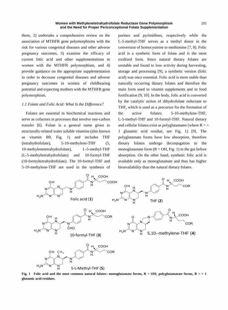
Folates are essential in biochemical reactions and
serve as cofactors in processes that involve one-carbon
transfer [6]. Folate is a general name given to
structurally-related water soluble vitamins (also known
as vitamin B9, Fig. 1) and includes THF
(tetrahydrofolate), 5-10-methylene-THF (5,
10-methylenetetrahydrofolate), L-5-methyl-THF
(L-5-methyltetrahydrofolate) and 10-formyl-THF
(10-formyltetrahydrofolate). The 10-formyl-THF and
5-10-methylene-THF are used in the synthesis of
purines and pyrimidines, respectively while the
L-5-methyl-THF serves as a methyl donor in the
conversion of homocysteine to methionine [7, 8]. Folic
acid is a synthetic form of folate and is the most
oxidized form. Since natural dietary folates are
unstable and found to lose activity during harvesting,
storage and processing [9], a synthetic version (folic
acid) was once essential. Folic acid is more stable than
naturally occurring dietary folates and therefore the
main form used in vitamin supplements and in food
fortification [9, 10]. In the body, folic acid is converted
by the catalytic action of dihydrofolate reductase to
THF, which is used as a precursor for the formation of
the active folates; 5-10-methylene-THF,
L-5-methyl-THF and 10-formyl-THF. Natural dietary
and cellular folates exist as polyglutamates (where R = >
1 glutamic acid residue, see Fig. 1) [9]. The
polyglutamate forms have low absorption, therefore
dietary folates undergo deconjugation to the
monoglutamate form (R = OH, Fig. 1) in the gut before
absorption. On the other hand, synthetic folic acid is
available only as monoglutamate and thus has higher
bioavailability than the natural dietary folates.
Fig. 1 Folic acid and the most common natural folates: monoglutamate forms, R = OH; polyglutamatate forms, R = > 1
glutamic acid residues.
5,10--methylene-THF (4)
Folic acid (1)
10-formyl-THF (3)
THF (2)
5-L-Methyl-THF (5)
Women with Methylenetetrahydrofolate Reductase Gene Polymorphism and the Need for Proper Periconceptional Folate Supplementation
206
1.2 Methylenetetrahydrofolate Reductase Gene
Polymorphisms and Its Effects on Folate Metabolism
and Folate-Based Biochemical Reactions
The MTHFR (methylenetetrahydrofolate reductase)
gene encodes MTHFR, an enzyme that catalyzes the
synthesis of L-5-methyl-THF from
5-10-methylene-THF [11]. The normal MTHFR allele
encodes a fully functional MTHFR enzyme. Many
genetic SNPs (single nucleotide polymorphisms) of the
MTHFR gene have been identified, but the MTHFR
C677T and A1298C variations are the most common
[12-15]. Each individual has two copies of the MTFHR
gene, one inherited from each parent. An individual can
either be heterozygous (SNP in one copy of the gene)
or homozygous (SNP in both copies of the genes) in the
MTHFR gene. It is also possible for some individuals
to present with a compounded heterozygous
polymorphism, which is one SNP located at two
different parts of the MTHFR gene (MTHFR 677CT
/1298AC) [16, 17].
The C677T polymorphism is a missense mutation on
exon 4. Cytosine which is normally found at position
677 is replaced with a thymine residue and this results
in valine being substituted for alanine [12, 18]. The
homozygous polymorphism (C677TT) reduces
enzymatic activity by 70%, while the heterozygous
polymorphism (C677CT) results in approximately 35%
reduction of enzymatic activity [7]. The A1298C
polymorphism results from a single nucleotide change
at exon 7 where adenosine is replaced by cytosine and
as a result glutamate is substituted for alanine. The
homozygous polymorphism (A1298CC) results in
about 40% reduction in enzymatic activity while the
compounded heterozygous polymorphism
(C677CT/A1298AC) has been shown to decrease
enzymatic activity by 40%-50% [4]. Hence, MTHFR
gene polymorphism leads to reduction in enzyme
activity, which results in lower levels of
L-5-methyl-THF.
Since L-5-methyl-THF serves as a methyl donor in
the conversion of homocysteine to methionine,
deficiencies in the enzymatic activity of MTHFR leads
to elevated homocysteine and low methionine levels.
This is important because further biochemical reactions
are disrupted as methionine is conjugated with
adenosine to form SAMe (S-adenosylmethionine).
SAMe is then used as a methyl donor in the
methylation of lipids, hormones, nucleic acids (DNA
and RNA) and myelin basic proteins. In summary,
MTHFR gene polymorphism can cause low
methionine levels, which may lead to low SAMe levels
and improper methylation of DNA and other
biomolecules [15, 19-22].
1.3 Populations Affected
Both C677T and A1298C MTHFR gene
polymorphisms have shown variation within different
ethnic groups. A HuGE review by Botto and Yang
published in 2000 reported on the MTHFR gene
polymorphisms by ethnicity and geographic area [23].
According to this review, Hispanics seem to have the
highest rate of C677T polymorphisms with up 21%
reportedly homozygous (677TT). European whites
were second with rates from 8-18%. Ireland and Britain
have had high rates of neural tube defects and their
reported rate of C677TT homozygous polymorphism
was 11-13%. Asians were next with reports of about 11%
frequency of C677TT homozygous polymorphism and
blacks were lowest at 1%-2%. The A1298C
polymorphism is not as well documented as the C677T
polymorphism. The A1298CC homozygous
polymorphism was found to be approximately 9%
based on studies in the Netherlands and Canada. The
compounded heterozygous populations
(C677T/A1298C) were estimated to be about 15 % in
Canada, 17% in US and 20% in Netherlands [23].
Esfahani et al studied the prevalence of MTHFR
polymorphism in different ethnic groups and also
found the 677TT to be highest in Mexican woman and
lowest in African American women. This was also the
finding for the compounded heterozygous genotype
Women with Methylenetetrahydrofolate Reductase Gene Polymorphism and the Need for Proper Periconceptional Folate Supplementation
207
with Mexican and white women having the highest
prevalence and Asian and African American women,
the lowest. The study also found the A1298CC
homozygous polymorphism to be highest in white
women [24].
1.4 Folate Status in Pregnancy
Maternal folate metabolism and levels plays an
important role in fetal development and having a
healthy baby [12, 25, 26].
Many studies have examined
the association of folate metabolism and levels with
fetal and maternal health. Pregnant women are at high
risk for folate deficiency due to the increase of folate
requirements for fetal growth [19]. Reduced MTHFR
enzymatic activity can cause low serum folate levels,
elevated homocysteine levels and decrease methylation
of DNA, proteins, lipids and hormones [16, 19, 27].
Homocysteine levels less than 13 µmol/L is normal,
levels between 13 to 60 µmol/L is moderate, and > 60
µmol/L is considered severely elevated [16]. High
homocysteine levels and folate deficiency could affect
development of ovarian follicle, fertilization, as well as
formation and development of the embryo [21]. Due to
the inability to properly process folate, pregnant
woman with MTHFR gene polymorphisms have an
increased risk for many deleterious defects, such as
having a baby with NTDs (neural tube defects),
congenital heart disease and cardiac septal effects,
down syndrome, cleft palate, omphalocele, attention
deficit hyperactivity disorder and low birth weight as
well as pregnancy loss, preeclampsia,
pregnancy-associated osteoporosis and susceptibility
to arsenic toxicity in pregnancy.
2. Association of MTHFR Gene
Polymorphism with Congenital Diseases and
Other Adverse Pregnancy Outcomes
2.1. Neural Tube Defects
NTDs (Neural tube defects) are a group of severe,
disabling and deadly congenital disorders of the
nervous system, arising from incomplete neural tube
closure during early stages of pregnancy [28-30].
Anencephaly, SB (spina bifida), encephalocele,
craniorachischisis and iniencephaly are the most
common [31-33]. There are several types of SB but
MM (myelomeningocele) commonly accounts for 90%
of cases [34]. Most infants with NTDs usually die
within days after birth or struggle with lifelong
disabilities [29, 32, 34, 35]. The incidence of NTDs
ranges from 1.0 to 10.0 per 1,000 births depending on
the country [30]. In the US (United States), the
prevalence of NTDs such as SB has decreased due to
FDA (food and drug administration) mandated
fortification of folic acid in enriched flours and grain
products [34, 35]. After fortification, SB in the US has
decreased by approximately 22.9% from 2.62 to 2.02
per 10,000 births [36]. Although this has been effective,
still 2,500 live births are affected by NTDs every year
[32].
NTDs occur more frequently in Irish and Mexican
populations compared to other Caucasians and Asians
[34, 37]. Other studies have also reported high
prevalence in China [27]. The pathogenesis of NTDs is
unknown and is caused by multiple factors such as
genetics, nutritional deficiencies, and environmental
determinants [29, 30]. Several studies have found
positive association between the common MTHFR
gene C677T polymorphism and risk of NTDs. The
MTHFR gene 677TT genotype has been associated
with a 4-fold increase risk of having a child with a NTD
[38]. Interestingly, to date there are few studies that
have extensively investigated the association between
A1298C polymorphism and NTDs [39]. MTHFR gene
C677T and A1298C polymorphisms result in
decreased MTHFR enzymatic activity, which increases
homocysteine levels and DNA hypomethylation [27,
40].
A case control study in Setif, Algeria found higher
homocysteine levels in MTHFR 677TT homozygous
mothers with spina bifida children than in mothers of
healthy children (P = 0.06) [38]. Although this is not
statistically significant the P-value is close to 0.05. The
Women with Methylenetetrahydrofolate Reductase Gene Polymorphism and the Need for Proper Periconceptional Folate Supplementation
208
same study also found an increased risk of NTDs in the
elevated homocysteine mothers (24%) versus control
(4.9%, P = 0.046) [38]. These observations therefore
support the hypothesis that elevated homocysteine
levels are commonly found in mothers with MTHFR
677TT genotype, which indicates an increased risk of
having a child with a NTD. A study conducted in
Mexico examining the risk of anencephaly in
association to folate, vitamin B12, homocysteine and
MTHFR gene polymorphism reported that mothers
with MTHFR 677TT genotype of anencephaly children
had significantly lower serum folate levels (8.2 ng/mL)
compared with mothers of healthy controls (14.1ng/mL,
P = 0.004) [37]. A review conducted by N. Greene et al.
also examined several studies that investigated the
association of MTHFR gene polymorphisms and NTDs
[41]. Pooled results from the studies demonstrated that
MTHFR 677TT genotype was strongly associated with
NTDs and there was no significant association between
the A1298C polymorphism and risk of NTDs. Thus,
mothers with MTHFR gene C677TT homozygous
polymorphisms seem to be more at risk of having a
child with NTDs compared to the other genotypes.
However, a study in a Turkish population showed that
MTHFR gene A1298C polymorphism might be
associated with development of NTDs [39].
Although the exact mechanism is not known,
research indicates that global DNA hypomethylation in
fetal brain is associated with the pathogenesis of NTDs.
DNA hypomethylation can alter normal repression or
expression of genes during fetal development [42].
Very few epidemiological studies have investigated the
association of DNA hypomethylation and risk of NTDs.
A case control study done in China, found positive
association between DNA hypomethylation in fetal
brain tissue and risk of NTD affected pregnancies [27].
This study reported that out of the 65 NTD affected
embryos (case), 8 cases resulted in craniorachischisis,
16 with anencephaly and open spina bifida, 14 with
encephalocele, 23 with open spina bifida and four
fetuses with spina bifida occulta. After stratification,
the study found that MTHFR 677CT heterozygous and
MTHFR 677 TT homozygous cases have lower DNA
methylation levels compared to healthy control fetuses
[27]. The study also found that the 677CC genotype
cases had similar DNA methylation levels to that of the
controls. Another case-control study conducted in two
populations (Dutch Caucasians and
Mexican-Americans in Texas) found significant
association between hypomethylation and the risk of
NTD in fetuses and very young children in the Dutch
Caucasian study group but not in the
Mexican-American study group [40]. The study found
an absolute decrease of MTHFR DNA methylation by
0.33% in the Dutch Caucasian children with SB
compared to controls (P = 0.001) [40]. Therefore, more
epidemiological studies need to be conducted to better
understand the association between impaired DNA
methylation and risk of NTDs in humans.
2.2 Congenital Heart Disease
CHD (congenital heart defects) are common birth
defects that often result from abnormal development of
the heart during the first 6 weeks of pregnancy [43, 44].
CHD is one of the major causes of infant death and
illnesses [45]. Heart diseases in infants mainly include
ASD (atrial septal defect), VSD (ventricular septal
defect), PDA (patent ductus arteriosus), and other types.
The majority of CHDs are affected by both genetic and
environmental factors.
In recent studies, plasma homocysteine level has
been shown to be elevated in CHD infants, which
might be a possible risk factor for CSD (cardiac septal
defects). A genetic variance in MTHFR is one possible
cause of elevated plasma homocysteine. The impaired
ability to process folate leads to the inability to convert
homocysteine to methionine. Several theories exist for
the mechanism by which elevated homocystiene levels
can lead to CHD. One study suggested that high
concentration of homocysteine was found to induce
electrical remodeling of ion channels in human atrial
cells that include potassium channels. The remodeling
Women with Methylenetetrahydrofolate Reductase Gene Polymorphism and the Need for Proper Periconceptional Folate Supplementation
209
of potassium currents has been hypothesized to induce
cardiac arrhythmias [46]. Other studies indicated that
hyperhomocystenimia causes interstitial and
perivascular fibrosis resulting in increased
myocardial stiffness, thereby affecting the pump
function of the myocardium, which then causes heart
failure [47].
Knowing that a genetic variance in the MTHFR gene
can lead to elevated homocysteine levels, several
studies have examined the direct association between
MTHFR gene C677T and A1298C polymorphism and
the development of CHD in infants and children.
Another case control study conducted in China that
investigated mothers of 57 children with congenital
heart disease of ASD, PDA or both versus 104 control
mothers, found that maternal MTHFR gene C677TT
homozygous polymorphism was associated with high
risk of PDA and ASD in offsprings [48]. A study
conducted in Canada looked at the relationship of
A1298CC homozygous polymorphism and congenital
heart diseases of ventricular septal defect and aortic
valve stenosis. It was demonstrated that maternal
MTHFR 1298AC heterozygous genotype was
associated with increased risk for aortic valve stenosis.
However, this study also reported mixed results on
negative association between 677TT genotype and
congenital heart defects [49].
CHD is caused by multiple factors including both
genetic and nongenetic factors. The pathophysiology
of CHD is complex and a single factor such as the
MTHFR gene polymorphism cannot explain the whole
cause. Although elevated homocysteinemia is observed
in populations with congenital heart diseases such as
cardiac septal defects, elevated homocysteine level is
not the only factor contributing to the pathophysiology.
Further studies need to be conducted in women with
MTHFR gene polymorphism and elevated
homocysteine to determine how proper folate
supplementation can affect maternal health and the
health of the offspring in terms of congenital heart
disease.
2.3 Down Syndrome
DS (down syndrome) also known as trisomy 21 is a
chromosomal disease that is associated with growth
and mental retardation [50, 51]. DS results from an
over-expression of three extra copies of the gene on
chromosome 21 causing failure of normal
chromosomal separation during meiosis [21, 51, 52].
DS affects approximately 1 in 1000 to 1100 live births
worldwide and 1 out of every 700 newborns in the US
[53, 54]. An important risk factor for DS is advanced
maternal age at the time of conception [51, 52].
Women who are 35 years or older are more at an
increased risk of having a child with DS, however DS
children have been born to younger mothers [55-58].
Studies have shown that mothers of children with DS
have abnormal folate metabolism, high homocysteine
and hypomethylation similar to NTDs, which result in
abnormal gene expression and chromosomal
segregation [20, 50, 53, 59].
Zampieri et al. investigated the influence of 12
genetic polymorphisms including the MTHR C677T
and A1298C on folate metabolism and risk of DS. The
MTHFR gene homozygous C677TT or heterozygous
C677CT polymorphisms was associated with the risk
of having a DS child (p value 0.0005). This study
confirmed low serum folate levels in DS mothers (12.2
ng/mL) compared to control group (14.6 ng/mL) (P =
0.028) as a maternal risk factor for a child’s
development of DS [58]. With respect to the impact of
MTHFR gene polymorphism and folate levels, folate
concentrations below the 25th percentile were
associated with presence of either MTHR gene
C677CT or C677TT polymorphism. This study
hypothesized that the increased maternal risk of DS
may be due to the reduction in enzymatic activity of
MTHFR [58]. Rai et al [56] conducted a study in
eastern state of India, which genotyped 312 mothers for
C677T and A1298C polymorphisms. The study
showed an association between both the C677T and
A1298C polymorphisms and maternal risk of having
DS children. The homozygous 677TT genotype was
Women with Methylenetetrahydrofolate Reductase Gene Polymorphism and the Need for Proper Periconceptional Folate Supplementation
210
7.6 times higher in the case study mothers compared to
controls. The distribution of C677T genotypes
according to age groups revealed that all 12
homozygous 677TT mothers were aged less than
31years among the total of 101 mothers aged less than
31 years. The frequency of homozygous A1298CC
polymorphism was higher in DS case mothers (22)
versus control (5) (P = 0.008). All of the young
mothers with homozygous 677TT genotype had either
a first or second child born with DS. The study revealed
the frequencies of the 1298C allele and homozygous
1298CC genotype to be higher in case mothers (46.6%,
24.1%) compared to controls (33.5%, 7.1%),
respectively. (p value 0.003, 0.008) [56]. Like the
previous study, Rai et al. concluded that
polymorphisms in MTHR gene results in
hypomethylation of DNA that could affect various
cellular functions, such as DNA repair, replication,
gene expression and chromatin conformation, leading
to disease conditions. Other epidemiological studies
have also found strong association between the
MTHFR 677T allele and risk of DS [17, 20, 52, 59-62].
Contrary to the above studies, others have shown that
there is no association between MTHR polymorphisms
and risk of DS, therefore further studies are necessary
[63-65].
2.4 Cleft Palate
CL/P (cleft lip with or without cleft palate) is birth
defect, which happens early during pregnancy where
baby’s lip or mouth does not form properly. During the
early pregnancy, a baby’s head forms by the joining of
body tissue from each side of the head in the center.
The lip is formed during the fourth and seventh week,
and the roof of the mouth between the sixth and ninth
week of pregnancy. If the lip tissues and/or palate
tissues do not join completely, it results in an opening,
which is cleft lip and/or cleft palate. According to CDC,
each year in the United States, about 2,650 babies are
born with a cleft palate and 4,440 babies are born with
a cleft lip with or without cleft palate [66]. Both genetic
and environmental factors are known to play a role in
the development of CL/P and many studies have been
conducted to verify the association between two
functional polymorphisms—C677T and A1298C—in
the MTHFR gene and an increased risk of CL/P.
Studies on the association of MTHFR gene
polymorphism with CL/P were performed in different
region of the world to find the prevalence among
subjects with CL/P. In many studies, it has been
proposed that low periconceptional folate intake
increases the risk of CL/P in offspring, and this risk is
even more pronounced in mothers with homozygous
MTHFR 677TT or 1298CC genotypes. In a study
performed in 153 patients with nonsyndromic oral
clefts and 205 control subjects genotyped for 176
different types of gene SNPs, MTHFR gene
polymorphism showed evidence of association with
CL/P [67]. There was a statistically significant
difference between genotype frequencies of MTHFR
C677T polymorphisms in non-syndromic CL/P cases
compared with controls [68]. These and similar studies
were performed in California, Brazil, Turkey, South
America, Germany, Poland, Paris, Korea, China, Brazil,
European and Northern Venezuela population samples.
MTHFR gene A1298C locus was analyzed and shown
positive and negative association with the cleft lip and
palate [15, 69-71]. Despite the association between
MTHFR C677T polymorphism and CL/P, there still
needs more studies to be performed to indicate if the
MTHFR gene polymorphism is the independent
marker for the CL/P [72-74].
2.5 Other Congenital Disease Sand Adverse Pregnancy
Outcomes
The MTHFR gene polymorphisms and mutation
have also been found to be associated with a number of
other congenital diseases and adverse pregnancy
outcomes. A recent case report from the New England
Journal of Medicine describes a boy with microcephaly
and episodic cyanosis. After a very long differential
diagnosis the boy was finally diagnosed at 10 months
Women with Methylenetetrahydrofolate Reductase Gene Polymorphism and the Need for Proper Periconceptional Folate Supplementation
211
with severe MTHFR deficiency. The boy was treated
with folinic acid (5-formyl-tetrahydrofolic acid) and
other active forms of the B vitamins, which resulted in
clinical and symptomatic improvement. The authors of
this case identified two novel mutations in the MTHFR
gene that put the patient at risk of developing
microcephaly and episodic cyanosis [75]. Other studies
have been conducted to extensively examine the
association of MTHFR polymorphisms and other
congenital diseases such as omphalocele, attention
deficit hyperactivity disorder, and microcephaly. A
study conducted on genetic information from all
newborn births in New York from 1998-1999, found
significant association between MTHFR 677T allele
and risk of omphalocele [76]. Other studies have also
shown strong association between risk of omphalocele
and MTHFR C677Tpolymorphism [77].
Homocysteine concentration has been reported to be
positively associated with the risk of pregnancy
complications such as low birth weight, increased
incidence of pregnancy loss, preeclampsia, intrauterine
growth retardation, and placental infarction [78]. A
prospective cohort study, which investigated the DNA
of pregnant women with complications (intrauterine
fetal death, preeclampsia, preterm delivery, and small
for gestational age infants) showed significantly higher
frequency of MTHFR gene C677T polymorphism
compared to control subjects. This study also showed
higher frequencies of homozygous 677TT allele than
the heterozygous 677CT allele. In a subgroup analysis,
statistically significant increase in MTHFR C677T
polymorphism was observed in women with small for
gestational age newborns [78]. In a meta-analysis study
performed in Caucasian and East Asian populations,
the MTHFR C677T polymorphism was associated with
a risk of preeclampsia [79]. In another study performed
in India, the data showed that MTHFR A1298C
heterozygous or homozygous genotypes significantly
increased the risk of recurrent pregnancy loss [80].
In a Mendelian randomization study conducted in
Seoul, Korea, birth weight significantly differed in
relation to MTHFR C677T genotype. The MTHFR
homozygous 677TT allele showed lower birth weights
than the normal 677CC allele. Birth weight also
decreased as maternal homocysteine level increased
[81]. According to another study conducted in Mexico
City, birth weight was 4 g higher for every 100 μg/day
increase in folate intake; however, neither maternal nor
infant MTHFR genotype was associated with
differences of infant weight at birth [82]. A similar
study conducted in the United Kingdom also showed
no significant relationship between MTHFR
polymorphism and low birth weight [83]. Different
studies demonstrated mixed results on association of
MTHFR C677T and/or A1298C polymorphism in
pregnancy outcomes [80, 84, 85]. Thus, it is suggested
that more studies with large samples be conducted to be
more conclusive.
3. Supplementation
3.1 Folic Acid
Adequate folate levels are paramount in pregnancy
for proper fetal development and maternal health.
There is a large body of evidence supporting the
protective effects of folic acid in preventing NTDs and
other adverse pregnancy outcomes such as
miscarriages and preeclampsia [86]. Folate deficiency
can result from poor dietary intake or failed
intracellular retention of folate and leads to elevated
homocysteine levels that are associated with various
congenital disorders such as NTDs, specifically spina
bifida and anencephaly [17]. Although the underlying
mechanism by which folic acid prevents NTDs is
widely unknown, many epidemiological studies have
shown that supplementation of folic acid during the
periconception period decreases the risk and
recurrence of NTD-affected pregnancies by
approximately 4 folds [13]. A four year
epidemiological study showed a reduction of 50%-70%
spina bifida cases and 25%-50% anencephaly cases
with supplementation of 5 mg/week folic acid [87]. It is
generally recommended that women of childbearing
Women with Methylenetetrahydrofolate Reductase Gene Polymorphism and the Need for Proper Periconceptional Folate Supplementation
212
age consume supplements containing at least 0.4-0.8
mg of folic acid daily to reduce the risk of NTDs.
However, most experts recommend higher doses of
folic acid supplements for women at intermediate to
high risk of having a child with a NTDs. Currently, the
US Preventive Services Task Force and American
Academy of Family Physicians recommend that all
women of childbearing age or women planning to be
pregnant take 0.4-0.8 mg/day of folic acid as a
supplement. This recommendation does not apply to
women who have had previous NTD affected
pregnancy and women on some antiseizure drugs [3].
Certain antiseizure drugs are known to cause folate
deficiency and higher dose of folic acid is
recommended for all women of childbearing age and
pregnant women [88]. Although there has not been any
study that determined the optimal dose of folic acid for
women on antiseizure drugs, it is usually recommended
that women of childbearing potential take 1mg/day of
folic acid and the dose be increased to 4 mg/day
following conception [88]. The American College of
Obstetricians and Gynecologists recommends a folic
acid supplement of 0.4 mg/day for women at low risk
of NTDs and 4 mg/day for women at high risk of NTDs
or who have had a previous pregnancy with an NTD
[89].
While research has indicated that MTHFR gene
polymorphism is associated with an increased risk for
neural tube defect, there is no guideline on folic acid
supplementation specific to women with MTHFR gene
polymorphism. However, the US Preventive Services
Task Force has listed mutations in folate-related
enzymes as one of the risk factors for NTDs and by
implication women with MTHFR gene polymorphism
may need to take higher doses of folic acid [3].
MTHFR catalyzes the conversion of 5,
10-methylene-THF to L-5-methyl-THF, one of the
active forms of folic acid that is used as a one-carbon
source in the synthesis of methionine from
homocysteine. MTHFR gene polymorphism reduces
levels of L-5-methyl-THF and increases homocysteine
levels [90]. Therefore, plasma homocysteine level is
used as a sensitive indicator of folate status and
MTHFR function. Plasma homocysteine levels can
also increase due to vitamin B6 or B12 deficiency.
Thus, B12 and B6 deficiency should be ruled out prior
to utilizing plasma homocysteine level as a biomarker
for folate status and MTHFR function.
The effects of folic acid (0.4 mg/day or 0.8 mg/day)
on homocysteine levels in Chinese hypertensive
patients with heterozygous C677CT or homozygous
C677TT polymorphism were assessed. This was done
in a multicenter, randomized, double-blind control
study. The results showed that homocysteine levels
remain high in patients with homozygous C677TT
polymorphism treated with either 0.4 mg/day or 0.8
mg/day of folic acid as well as in patients with
heterozygous C677CT polymorphism treated with 0.4
mg/day of folic acid [91]. In another large-scale
population-based double blind trial, the effects of six
months supplementation of different folic acid doses
(0.1 mg/day, 0.4 mg/day, 4 mg/day, 4 mg/week) on
plasma homocysteine levels, plasma folate levels and
red blood cell folate levels were evaluated [92]. The
study showed that folic acid dose of 0.1 mg/day and 4
mg/week given for 6 months did not reduce the high
homocysteine levels in those women with MTHFR
homozygous 677TT genotype. Even with the high dose
of 4 mg/day folic acid supplementation, women with
MTHFR 677TT genotype showed higher plasma
homocysteine levels as well as lower plasma and RBC
folate levels compared to women with the MTHFR
normal 677CC genotype [92]. In animal model of
severe MTHFR deficiency, folic acid failed to decrease
mortality while L-5-methyl-THF reduced mortality by
31%. Based on these and other studies, high dose of
folic acid alone may not be sufficient as a supplement
for women with MTHFR polymorphism and people
with severe MTHFR deficiency.
Although folic acid is generally considered safe,
there are several studies that raise concerns regarding
its safety. Folic acid is the most oxidized form of the
Women with Methylenetetrahydrofolate Reductase Gene Polymorphism and the Need for Proper Periconceptional Folate Supplementation
213
folates and is believed to be metabolized in the
intestine and the liver to tetrahydrofolic acid and
subsequently to 5-L-methyl-THF. However, a study
showed the presence of unmetabolized folic acid in the
blood and its level was dose-dependent and
accumulated with repeated exposure [93]. The
unmetabolized folic acid was associated with reduced
natural cell cytotoxicity and this effect was greater with
increased levels of unmetabolized folic acid [94]. High
folic acid intake was also found to be associated with
cognitive decline in older people [95]. In addition,
there are several reports on the association of folic acid
supplement with certain cancers such as prostate and
colorectal cancers [96-98]. High dose folic acid may
also mask Vitamin B12 deficiency by correcting
macrocytic anemia [99]. The effect of B12 deficiency
on RBC (macrocytic anemia) can be easily diagnosed
while the neurological effects may not be identified
until after the disease has progressed. Vitamin B12
mainly plays a role in the regeneration of THF and the
effect of B12 deficiency on RBC can be prevented by
administering higher dose of folic acid.
3.2 L-5-Methyltetrahydrofolate
L-5-methyl-THF (L-5-Methyltetrahydrofolate) is
the predominant active form of folic acid in the plasma
and in red blood cells. It is also naturally present in
dietary sources mostly as polyglutamates [9]. The
calcium salt of L-5-methyl-THF (L-5-methyl-THF
calcium), which is also known as levomefolate calcium
is available as a dietary supplement and is marketed
under the brand names Deplin [100] and Metafolin. It is
also available in combination with oral contraceptives
as Safyral and Beyaz. Each Safyral and Beyaz tablet
contains 0.451 mg of levomefolate calcium along with
3 mg of drospirenone. The difference between Safyral
and Beyaz is in the amount of ethylene estradiol. Both
Safyral and Beyaz were approved by the FDA in 2010
and the levomefolate calcium in the contraceptive pills
was intended to reduce the risk of NTD in a pregnancy
conceived while taking the contraceptive or soon after
discontinuation of the contraceptive. The European
Food Safety Authority Panel also evaluated
L-5-methyl-THF calcium in 2004 and concluded that
there is no safety concern when it is used as a source of
folate in foods and food supplements with a tolerable
upper level of 1mg/adult/day [101]. In 2013, the
European Food Safety Authority Panel similarly gave a
scientific opinion on the safety and bioavailability of
the glucosamine salt of L-5-methyl-THF
(L-5-methyl-THF glucosamine) in response to a
request from the European commission. Based on a
comparative crossover bioavailability study in human
volunteers, the panel concluded that L-5-methyl-THF
glucosamine has similar bioavailability to that of
L-5-methyl-THF calcium [102]. The panel also came
to the conclusion that there was no safety concern
regarding the genotoxicity of L-5-methyl-THF
glucosamine.
The approval of L-5-methyl-THF calcium as part of
a contraceptive by the US FDA and the scientific
comments by the European Food Safety Authority
ascertain the safety of L-5-methyl-THF. A number of
pharmacokinetic and pharmacodynamic studies have
indicated that L-5-methyl-THF has similar
bioavailability to that of folic acid and it is at least as
effective as folic acid in correcting folate status and
decreasing homocysteine levels [103-106]. An
epidemiological study compared the effects of a
prenatal supplement containing 1.13 mg of
L-5-methyl-THF plus folic acid 0.4 mg with standard
PNV (prenatal vitamins) containing 0.8 to 1 mg of folic
acid alone on decreasing the rate of anemia in pregnant
women. This study found that fewer women developed
anemia in the prenatal supplement containing
L-5-methyl-THF and folic acid compared with the
standard treatment of PNV [86]. In a randomized
double-blind study involving healthy women of
childbearing potential, steady-state plasma and RBC
folate levels were compared following administration
of oral contraceptives containing either 0.4 mg of folic
acid or 0.451 mg of levomefolate calcium for 24 weeks
Women with Methylenetetrahydrofolate Reductase Gene Polymorphism and the Need for Proper Periconceptional Folate Supplementation
214
[107]. The study results demonstrated that both plasma
and RBC folate levels were slightly higher in the
levomefolate group compared to the folic acid group
during the treatment phase as well as during the 20
weeks following cessation of treatment [107]. Another
randomized double-blind study that compared the
effects of equimolar amounts of L-5-methyl-THF and
folic acid on RBC folate levels demonstrated that
L-5-methyl-THF is more effective than folic acid in
correcting folate status [108]. In a randomized study
comparing the effects of 0.4 mg of folic acid and 0.412
mg of L-5-methyl-THF on the pharmacokinetics of
folate, Prinz-Langenohl et al showed that total plasma
folate levels and Cmax (the maximum folate
concentration) were higher and Tmax (the time to peak)
was shorter in women with homozygous 677TT
genotype supplemented with L-5-methyl-THF than
those supplemented with folic acid [109].
3.3 Betaine
Betaine is either obtained from diet or synthesized in
the body from choline [110]. The recommended
betaine intake is estimated at 0.5 to 3 grams per day
[111-113]. Homocysteine is methylated to methionine
with either betaine or L-5-methyl-THF as the methyl
donor. Thus, betaine and L-5-methyl-THF are
interchangeable sources of methyl group in the
conversion of homocysteine to methionine [110]. The
reaction that uses betaine as a methyl donor is
catalyzed by the enzyme BHMT
(betaine-homocysteine methyltransferase), which is
located only in the liver and kidney [112, 114, 115]. In
the liver, BHMT catalyzes up to 50% of homocysteine
metabolism [112]. Therefore, BHMT regulates
homocysteine levels in the liver. Epidemiological
studies have indicated that dietary supplementation
with betaine may reduce homocysteine levels in
healthy volunteers and individuals with
hyperhomocysteinemia [112, 114]. Studies have
shown that treatment with betaine may reduce the risk
of consequences of severe MTHFR deficiency [116]. A
meta-analysis of 15 case reports and case series
involving 35 patients investigated the effects of betaine
on survival and psychomotor development in patients
with severe MTHFR deficiency. The results indicated
that all five patients who were treated with betaine
early survived and had normal psychomotor
development. On the other hand, the 11 patients that
died were either not treated with betaine or their
treatment was delayed. None of the remaining 19
survivors whose treatment was delayed had normal
psychomotor development. A new born baby with
MTHFR deficiency was treated with increasing doses
of betaine for two years and the researchers reported
that 6 g/day of betaine administered in six divided
doses resulted in adequate control of homocysteine
levels [117]. Although physical development was
normal, language and motor developments were
delayed by 3-6 months. This study concluded that early
betaine treatment prevents mortality and allows normal
psychomotor development in patients with severe
MTHFR deficiency [116]. Holm and colleagues [118]
reported that plasma betaine levels are inversely
associated with plasma total homocysteine levels and
this is more pronounced in those with C677TT
homozygous polymorphism and low folate status,
implying that betaine is an important methyl donor
especially in those with low folate status and MTHFR
polymorphism. All the studies indicated that betaine is
an alternative methyl donor in those with MTHFR
polymorphism and deficiency. Therefore, betaine can
be used as add-on supplement for individuals with
MTHFR polymorphism.
4. Discussion
This article presents a comprehensive review on the
effects of MTHFR gene polymorphism on folate
metabolism and its association with an increased risk
for various congenital diseases and other adverse
pregnancy outcomes. It also examines the
pharmacodynamic effects and pharmacokinetic
properties of several supplements that are used to
Women with Methylenetetrahydrofolate Reductase Gene Polymorphism and the Need for Proper Periconceptional Folate Supplementation
215
prevent NTDs and other adverse outcomes related to
folate deficiency or inept folate metabolism. Based on
our assessment, we will provide a recommendation on
the supplementation to women of childbearing
potential with MTHFR gene polymorphism.
Folates are essential for proper fetal development
and maternal health. The demand for folates increases
during pregnancy, therefore folate supplementation is
necessary. Natural dietary folates are unstable and
degrade during harvesting, processing, and storage [9].
In addition, natural dietary folates exist as
polyglutamates, which are less absorbable forms.
Hence, natural dietary folates have to undergo
deconjugation to monoglutamate forms in the
gastrointestinal tract before absorption. As a result,
natural dietary folates have low bioavailability [10].
Because of the low bioavailability and stability issues,
the natural dietary source of folates may not be
adequate during pregnancy when the demand for
folates is higher. Folic acid, the synthetic and most
oxidized form of the folates, is used as supplement and
in food fortification due its stability and good
bioavailability [10]. Numerous studies have shown that
periconceptional folic acid supplementation decreases
the recurrence of fetal neural tube defects (NTDs) [119,
120]. Neural tube closes three to four weeks
postconception. Because the neural tube forms early in
the pregnancy, at a time when women are not even
aware of that they are pregnant, and half of the
pregnancies in the USA are unplanned [119], it is
recommended that all women of childbearing potential
take folic acid supplementation. In general, the current
folic acid recommendation is 0.4-0.8 mg/day for all
women of child bearing age who are low risk for
having a fetus with NTD [3]. Higher doses of folic acid
are recommended for women at higher risk for having
an NTD affected pregnancy.
MTHFR gene polymorphism, particularly C677T
and A1296C have been shown to be associated with an
increased risk for NTDs and other deleterious
pregnancy outcomes. Although mutations in folate
metabolizing enzymes have been recognized as one of
the risk factors for NTDs [3] and the WHO guideline
on folate levels [2] has documented that MTHFR
polymorphism is associated with low folate levels,
there are no recommendations on folate
supplementation specific to women with MTHFR
polymorphism. However, since higher doses of folic
acid is recommended for women at high risk for having
NTD affected pregnancy, by implication women with
MTHFR polymorphism may take high dose of folic
acid. Homozygous C677TT and A1298CC
polymorphism as well as compounded heterozygous
polymorphism (C677CT/A1298AC) have been shown
to decrease the enzymatic activity of the MTHFR by
40-70% [4, 5, 121]. MTHFR is a key enzyme that
catalyzes the synthesis of L-5-methyl-THF from folic
acid. L-5-methyl-THF is the major active form of
folates in the plasma and in red blood and serves as
methyl donor in the synthesis of methionine from
homocysteine.
Although current recommendations imply that
women with MTHFR polymorphism may take high
dose of folic acid, they may not benefit from high dose
of folic acid and may even have detrimental effect on
their health for a number of reasons. People with
MTHFR polymorphism cannot convert folic acid to
L-5-methyl-THF efficiently due to the reduced activity
of the MTHFR enzyme. Therefore, giving more folic
acid to process for an enzyme that is working at lower
capacity appears inconsistent with the scientific bases.
High dose of folic acid has also been found to mask
B-12 deficiency [99] and has been associated with
decreased immunity [94] as well as impaired cognition
[95].
Currently, the stable form of L-5-methyl-THF is
available as a calcium salt. L-5-methyl-THF calcium,
which is also known as levomefolate calcium is
commercially available under various proprietary
names. Pharmacokinetic and pharmacodynamic studies
have shown that the bioavailability of L-5-methyl-THF
is similar to that of folic acid and it is at least as
Women with Methylenetetrahydrofolate Reductase Gene Polymorphism and the Need for Proper Periconceptional Folate Supplementation
216
effective as folic acid in correcting folate status.
However, in people with MTHFR polymorphism,
L-5-methyl-THF has been shown to be more effective
in correcting folate status and reducing homocysteine
levels [109]. Our comprehensive literature study and
understanding of the biochemistry of folates and
folate-based biochemical reactions indicate that a
combination of L-5-methyl-THF and folic acid would
be a better choice for women of childbearing potential
with MTHFR polymorphism. However, a study is
needed to establish the optimal dose of
L-5-methyl-THF. L-5-methyl-THF calcium is
available in various strengths ranging from 0.4 mg to
15 mg of oral tablets or capsules. The FDA has
approved oral contraceptives containing 0.451 mg of
L-5-methyl-THF calcium. The European Food Safety
Authority Panel has also concluded that there is no
safety concern when L-5-methyl-THF calcium is used
as a supplement up to 1 mg/adult/day [101]. Therefore,
the safety of L-5-methyl-THF calcium at a dose greater
than 1 mg/day is unknown and requires studies. The
synthesis of methionine from homocysteine uses either
betaine or L-5-methyl-THF as a methyl donor.
Consequently, studies have shown that
supplementation with betaine reduces the risk of the
consequence of MTHFR gene polymorphism or severe
MTHFR deficiency. Therefore, betaine may be added
to the folic acid supplement in women with MTHFR
polymorphism. However, at this point we cannot
recommend betaine in pregnant women due to a lack of
safety studies.
5. Conclusion
MTHFR gene polymorphism is associated with an
increased risk for a number of birth defects including
NTDs as well as other adverse pregnancy outcomes.
Because of the reduced MTHFR enzymatic activity,
women with MTHFR gene polymorphism cannot get
the full benefit from high dose of folic acid
supplementation and even may have detrimental effect
on their health. Women with a homozygous or
compounded heterozygous polymorphism are at a
higher risk of adverse pregnancy outcome due to the
significant reduction in enzymatic activity. The stable
form of the end product of MTHFR, L-5-methyl-THF
is currently available. Pharmacokinetic studies have
shown that the bioavailability of L-5-methyl-THF is
similar to that of folic acid. Studies have also
demonstrated that there is no safety concern when
L-5-methyl-THF calcium is used up to 1mg/day and it
is more effective than folic acid at correcting folate
status in individuals with MTHFR gene polymorphism.
Therefore, L-5-methyl-THF calcium should be given
in combination with folic acid to women of
childbearing potential with MTHFR gene
polymorphism. However, study is needed to establish
the optimal dose. L-5-methyl-THF calcium is marketed
in various strengths ranging from 0.4 mg to 15 mg of
oral tablets or capsules. The safety of L-5-methyl-THF
at a dose greater than 1 mg/day/adult is unknown. Thus,
study is needed to determine the safety profile of
L-5-methyl-THF at higher doses. Betaine and
L-5-methyl-THF are interchangeable sources of
methyl group in the biochemical conversion of
homocysteine to methionine and alternatively betaine
may be added to folic acid supplement for women with
MTHFR polymorphism.
References
[1] Cordero, A. M., Crider, K. S., Rogers, L. M., Cannon, M.
J., and Berry, R. J. 2015. “Optimal Serum and Red Blood
Cell Folate Concentrations in Women of Reproductive age
for Prevention of Neural Tube Defects: World Health
Organization Guidelines.” MMWR Morb. Mortal. Wkly.
Rep. 64 (15): 421-3.
[2] WHO. 2015. “Guideline: Optimal Serum and Red Blood
Cell Folate Concentrations in Women of Reproductive
Age for Prevention of Neural Tube Defects.” Geneva,
Switzerland. Accessed: July 6, 2015.
http://www.who.int/nutrition/publications/guidelines/opti
malserum_rbc_womenrep_tubedefects/en/.
[3] U.S. Preventive Services Task Force. 2009. “Folic Acid
for the Prevention of Neural Tube Defects: U.S.
Preventive Services Task Force Recommendation
Statement.” Ann. Intern. Med. 150 (9): 626-31.
[4] Weisberg, I., Tran, P., Christensen, B., Sibani, S., and
Women with Methylenetetrahydrofolate Reductase Gene Polymorphism and the Need for Proper Periconceptional Folate Supplementation
217
Rozen, R. 1998. “A Second Genetic Polymorphism in
Methylenetetrahydrofolate reductase (MTHFR)
Associated with Decreased Enzyme Activity.” Mol. Genet.
Metab. 64 (3): 169-72.
[5] Yousefian, E., Kardi, M. T., and Allahveisi, A. 2014.
“Methylenetetrahydrofolate Reductase C677T and
A1298C Polymorphism in Iranian Women with Idiopathic
Recurrent Pregnancy Losses.” Iran Red. Crescent. Med. J.
16 (7): e16763.
[6] Arinze, I. J. 2005. “Facilitating Understanding of the
Purine Nucleotide Cycle and the One-Carbon Pool, Part II:
Metabolism of the One-Carbon Pool.” Biochem. Mol. Biol.
Educ. 33 (4): 255-9.
[7] Fukuda, N., Hamajima, N., Wakai, K., and Suzuki, K. A.
2014. “Cross-Sectional Study to Find out the Relationship
of Methylenetetrahydrofolate Reductase (MTHFR)
C677T Genotype with Plasma Levels of Folate and Total
homocysteine by daily folate intake in Japanese.” J. Nutr.
Sci. Vitaminol. 60 (4): 231-8.
[8] Barua, S., Kuizon, S., and Junaid, M. A. 2014. “Folic Acid
Supplementation in Pregnancy and Implications in Health
and Disease.” J. Biomed. Sci. 21 (1): 77.
[9] Report of a Joint FAO/WHO Expert Consultation. 2001.
“Human Vitamin and Mineral Requirements.” Thailand:
FAO/WHO, 53-64.
[10] Caudill, M. A. 2010. “Folate Bioavailability: Implications
for Establishing Dietary Reommendations and Optimizing
Status.” Am. J. Clin. Nutr. 91 (5): 1455S-60S.
[11] Ulvik, A., Ueland, P. M., Fredriksen, A., Meyer, K.,
Vollset, S. E., Hoff, G., and Schneede, J. 2007.
“Functional Inference of the Methylenetetrahydrofolate
Reductase 677C > T and 1298A > C Polymorphisms from
a Large-Scale Epidemiological Study.” Hum. Genet.121
(1): 57-64
[12] Yila, T. A., Sasaki, S., Miyashita, C., Braimoh, T. S.,
Kashino, I., Kobayashi, S., Okada, E., Baba, T., Yoshioka,
E., Minakami, H., Endo, T., Sengoku, K., and Kishi, R.
2012. “Effects of Maternal 5,
10-Methylenetetrahydrofolate Reductase C677T and
A1298C Polymorphisms and Tobacco Smoking on Infant
Birth Weight in a Japanese Population.” J. Epidemiol. 22
(2): 91-102.
[13] Obican, S. G., Finnell, R. H., Mills, J. L., Shaw, G. M., and
Scialli, A. R. 2010. “Folic Acid in Early Pregnancy: A
Public Health Success Story.” FASEB J. 24 (11): 4167-74.
[14] Pilsner, J. R., Hu, H., Wright, R. O., Kordas, K., Ettinger,
A. S., Sa, B. N., Cantonwine, D., Lazarus, A. L., Cantoral,
A., Schnaas, L., and Te, M. M. 2010. “Maternal MTHFR
Genotype and Haplotype Predict Deficits in Early
Cognitive Development in a Lead-Exposed Birth Cohort
in Mexico City.” Am. J. Clin. Nutr. 92 (1): 226-34.
[15] Bhaskar, L., Murthy, J., and Babu, G. 2011.
“Polymorphisms in Genes Involved in Folate. Metabolism
and Orofacial Clefts.” Arch. Oral. Biol. 56 (8): 723-37.
[16] Varga, E. A., Sturm, A. C., Misita, C. P., and Moll, S. 2005.
“Homocysteine and MTHFR Mutations: Relation to
Thrombosis and Coronary Artery Disease.” Circulation
111 (19): e289-e93.
[17] Guéant-Rodriguez, R. M., Guéant, J. L., Debard, R.,
Thirion, S., Hong, L. X., Bronowicki, J. P., Namour, F.,
Chabi, N. W., Sanni, A., Anello, G., Bosco, P., Romano,
C., Amouzou, E., Arrieta, H. R., Sánchez, B. E., Romano,
A., Herbeth, B., Guilland, J. C., and Mutchinick, O. M.
2006. “Prevalence of Methylenetetrahydrofolate
Reductase 677T and 1298 Cnalleles and Folate Status: A
Comparative Study in Mexican, West African, and
European Populations.” Am. J. Clin. Nutr. 83 (3): 701-7.
[18] Dalal, A., Pradhan, M., Tiwari, D., Behari, S., Singh, U.,
Mallik, G. K., Das, V., and Agarwal, S. 2007. “MTHFR
677C-->T and 1298A-->C Polymorphisms: Evaluation of
Maternal Genotypic Risk and Association with Level of
Neural Tube Defect.” Gynecol. Obstet. Invest. 63 (3):
146-50.
[19] FAO/WHO Expert Consultation. 2004. “Vitamin and
Mineral Requirements in Human Nutrition.” In
Consultation of 2nd ed World Heal Organ, Geneva,
289-341.
[20] Patterson, D. 2008. “Folate Metabolism and the Risk of
Down Syndrome.” Downs Syndr. Res. Pract. 12 (2): 93-7.
[21] Laanpere, M., Altmäe, S., Stavreus-Evers, A., Nilsson, T.
K., Yngve, A., and Salumets, A. 2010. “Folate-Mediated
One-Carbon Metabolism and Its Effect on Female Fertility
and Pregnancy Viability.” Nutr. Rev. 68 (2): 99-113.
[22] Reutter, H., Betz, R. C., Ludwig, M., and Boemers, T. M.
2006. “MTHFR 677 TT Genotype in a Mother and Her
Child with Down Syndrome, Atrioventricular Canal and
Exstrophy of the Bladder: Implications of a Mutual
Genetic Risk Factor?” Eur. J. Pediatr. 165 (8): 566-8.
[23] Botto, L., and Yang, Q. 2000. “5,
10-Methylenetetrahydrofolate Reductase Gene Variants
and Congenital Anomalies: A HuGE Review.” Am. J.
Epidemiol. 151 (9): 862-77.
[24] Esfahani, S. T., Cogger, E. A, and Caudill, M. A. 2003.
“Heterogeneity in the Prevalence of
Methylenetetrahydrofolate Reductase Gene
Polymorphisms in Women of Different Ethnic Groups.” J.
Am. Diet. Assoc. 103 (2): 200-7.
[25] Greenberg, J. A., Bell, S. J, Yong, G., and Yan-Hong, Y.
2011. “Folic Acid Supplementation and Pregnancy: More
than Just Neural Tube Defect Prevention.” Rev. Obstet.
Gynecol. 4 (2): 52-9.
[26] Antony, A. C. 2007. “In Utero Physiology: Role of Folic
Acid in Nutrient Delivery and Fetal Development.”Am. J.
Clin. Nutr. 85 (2): 598S-603S.
Women with Methylenetetrahydrofolate Reductase Gene Polymorphism and the Need for Proper Periconceptional Folate Supplementation
218
[27] Chen, X., Guo, J., Lei, Y., Lu, X., Bao, Y., Wu, L., Zheng,
X., Shen, Y., Wu, B.L., and Zhang, T. 2010. “Global DNA
Hypomethylation Is Associated with NTD-Affected
Pregnancy: A Case-Control Study.” Birth Defects Res.
Part A Clin. Mol. Teratol. 88 (7): 575-81.
[28] Cadenas-Benitez, N. M., Yanes-Sosa, F.,
Gonzalez-Meneses, A., Cerrillos, L., Acosta, D.,
Praena-Fernandez, J. M., Neth, O., Gomez de Terreros, I.,
and Ybot-Gonzalez, P. 2014. “Association of Neural Tube
Defects in Children of Mothers with MTHFR 677TT
Genotype and Abnormal Carbohydrate Metabolism Risk:
A Case-Control Study.” Genet. Mol. Res. 13 (1): 2200-7.
[29] Van der Linden, I. J. M., Afman, L. A., Heil, S. G., and
Blom, H. J. 2007. “Genetic Variation in Genes of Folate
Metabolism and Neural-Tube Defect Risk.” Proc. Nutr.
Soc. 65 (2): 204-15.
[30] Au, K. S., Ashley-Koch, A., and Northrup, H. 2011.
“Epidemiologic and Genetic Aspects of Spina Bifida and
Other Neural Tube Defects.” Dev. Disabil. Res. Rev. 16
(1): 6-15.
[31] Shin, M., Kucik, J. E., Siffel, C., Lu, C., Shaw, G. M.,
Canfield, M. A., and Correa, A. 2012. “Improved Survival
among Children with Spina Bifida in the United States.” J.
Pediatr. 161 (6): 1132-7.
[32] Yi, Y., Lindemann, M., Colligs, A., and Snowball, C. 2011.
“Economic Burden of Neural Tube Defects and Impact of
Prevention with Folic Acid: A Literature Review.” Eur. J.
Pediatr. 170 (11): 1391-400.
[33] Pangilinan, F., Molloy, A. M., Mills, J. L., Troendle, J. F.,
Parle-McDermott, A., Signore, C., O'Leary, V. B., Chines,
P., Seay, J. M., Geiler-Samerotte, K., Mitchell, A.,
Vander-Meer, J. E., Krebs, K. M., Sanchez, A.,
Cornman-Homonoff, J., Stone, N., Conely, M., Kirke, P.
N., Shane, B., Scott, J., and Brody, L. C. 2012.
“Evaluation of Common Genetic Variants in 82 Candidate
Genes as Risk Factors for Neural Tube Defects.” BMC
Med. Genet. 13: 62.
[34] Au, K. S. , Tran, P. X., Tsai, C. C., O'Byrne, M. R., Lin, J.
I., Morrison, A. C., Hampson, A. W., Cirino, P., Fletcher,
J. M., Ostermaier, K. K., Tyerman, G. H., Doebel, S., and
Northrup, H. 2008. “Characteristics of a Spina Bifida
Population Including North American Caucasian and
Hispanic Individuals.” Birth Defects Res. Part A Clin. Mol.
Teratol. 82 (10): 692-700.
[35] Heseker, H. 2011. “Folic Acid and Other Potential
Measures in the Prevention of Neural Tube Defects.” Ann.
Nutr. Metab. 59 (1): 41-5.
[36] CDC.2009. “Racial/Ethnic Differences in the Birth
Prevalence of Spina Bifida—United States, 1995-2005.”
MMWR. 57 (53): 1409-13.
[37] Lacasan, M., Blanco-mun, J., Borja-aburto, V. H.,
Gonzalez-alzaga, B., and Garcia-Cavazos, R. 2012.
“Effect on Risk of Anencephaly of Gene-Nutrient
Interactions between Methylenetetrahydrofolate
Reductase C677T Polymorphism and Maternal Folate,
Vitamin B 12 and Homocysteine Profile.” Public Health
Nutrition 15 (8): 1419-28.
[38] Houcher, B., Bourouba, R., Djabi, F., Yilmaz, E., Eğin, Y.,
and Akar, N. 2009. “Polymorphisms of 5,
10-Methylenetetrahydrofolate Reductase and
Cystathionine Beta-synthase Genes as a Risk Factor for
Neural Tube Defects in Sétif, Algeria.” Pediatr Neurosurg.
45 (6): 472-7.
[39] Boduroğlu, K., Alanay, Y., Alikaşifoğlu, M., Aktaş, D.,
and Tunçbilek, E. 2005. “Analysis of MTHFR 1298A>C
in Addition to MTHFR 677C>T Polymorphism as a Risk
Factor for Neural Tube Defects in the Turkish Population.”
Turk. J. Pediatr. 47 (4): 327-33.
[40] Stolk, L., Bouwland-Both, M. I., van Mil, N. H.,Verbiest,
M. M., Eilers, P. H., Zhu, H., Suarez, L., Uitterlinden, A.
G., and Steegers-Theunissen, R. P. 2013. “Epigenetic
Profiles in Children with a Neural Tube Defect; a
Case-Control Study in Two Populations.” PLoS One. 8
(11): e78462.
[41] Greene, N. D. E., Stanier, P. and Copp, A. J. 2009.
“Genetics of Human Neural Tube Defects.,” Hum. Mol.
Genet. 18 (R2): R113-R29
[42] Shookhoff, J. M, and Gallicano, G. I. 2010. “A New
Perspective on Neural Tube Defects: Folic Acid and
MicroRNA Misexpression.” Genesis 48 (5): 282-94.
[43] Youssef, O. I., and EL Sayed, G. M. 2012. “MTHFR 677
CT Polymorphism and the Risk of Cardiac Septal Defects:
A Pilot Study.” Life Sci. J. 9 (4): 4272-5.
[44] Van Beynum, I. M., den Heijer, M., Blom, H. J., and
Kapusta, L. 2007. “The MTHFR 677C->T Polymorphism
and the Risk of Congenital Heart Defects: A Literature
Review and Meta-Analysis.” QJM. 100 (12): 743-53.
[45] Huhta, J. C., and Hernandez-Robles, J. A. 2005.
“Homocysteine, Folate and Congential Heart Defects.”
Fetal Pediatr Pathol. 24 (2): 71-9.
[46] Cai, B. Z., Gong, D. M., Liu, Y., Pan, Z. W., Xu, C. Q., Bai,
Y. L., Qiao, G. F., Lu, Y. J., and Yang, B. F. 2007.
“HoMocysteine Inhibits Potassium Channels in Human
Atrial Myocytes.” Clin. Exp. Pharmacol Physiol. 34 (9):
851-5.
[47] Herrmann, M., Taban-Shomal, O., Hübner, U., Böhm, M.,
Herrmann, W. 2006. “A Review of Homocysteine and
Heart Failure.” Eur. J. Heart Fail 8 (6): 571-6.
[48] Zhu, W. L., Li, Y., Yan, L., Dao, J., and Li, S. 2006.
“Maternal and Offspring MTHFR Gene C677T
Polymorphism as Predictors of Congenital Atrial Septal
Defect and Patent Ductus Arteriosus.” Mol. Hum. Reprod.
12 (1): 51-4.
[49] Christensen, K. E., Zada, Y. F., Rohlicek, C. V.,
Women with Methylenetetrahydrofolate Reductase Gene Polymorphism and the Need for Proper Periconceptional Folate Supplementation
219
Andelfinger, G. U., Michaud, J. L., Bigras, J. L., Richter,
A., Dubé, M. P., and Rozen, R. 2013. “Risk of Congenital
Heart Defects Is Influenced by Genetic Variation in Folate
Metabolism.” Cardiol. Young. 23 (1): 89-98.
[50] Santos-Rebouças, C. B., Corrêa, J. C., Bonomo, A.,
Fintelman-Rodrigues, N., Moura, K. C., Rodrigues, C. S.,
Santos, J. M., and Pimentel, M. M. 2008. “The Impact of
Folate Pathway p Polymorphisms Combined to Nutritional
Deficiency as a Maternal Predisposition Factor for Down
Syndrome.” Dis. Markers 25 (3): 149-57.
[51] Costa-Lima, M. A., Amorim, M. R., and Orioli, I. M. 2013.
“Association of Methylenetetrahydrofolate Reductase
Gene 677C > T Polymorphism and Down Syndrome.” Mol.
Biol. Rep. 40 (3): 2115-25.
[52] Gilbody, S., Lewis, S., and Lightfoot, T. 2007.
“Methylenetetrahydrofolate Reductase (MTHFR) Genetic
Polymorphisms and Psychiatric Disorders: A HuGE
Review.” Am. J. Epidemiol. 165 (1): 1-13.
[53] Ragunath, P. K., and Abhinand, P. A. 2013. “Systems
Biological Approach to Investigate the Lack of Familial
Link Between Down’s Syndrome and Neural Tube
Disorders.” Bioinformation 9 (12): 610-6.
[54] Centers for Disease Control and Prevention. 2015. “Data
and Statistics Down Syndrom Birth Defects.” National
Center on Birth Defects and Developmental Disabilities.
Accessed June 15, 2015.
http://www.cdc.gov/ncbddd/birthdefects/downsyndrome/
data.html.
[55] Hao, L., Yang, Q., and Li, Z. 2008. “Folate Status and
Homocysteine Response to Folic Acid Doses and
Withdrawal among Young Chinese Women in a
Large-Scale Randomized Double-Blind Trial.” Am. J. Clin.
Nutr. 88 (2): 448-57.
[56] Rai, A. K., Singh, S., Mehta, S., Kumar, A., Pandey, L. K.,
and Raman, R. “MTHFR C677T and A1298C
Polymorphisms Are Risk Factors for Down ’s Syndrome
in Indian Mothers.” J. Hum. Genet. 51 (4): 278-83.
[57] Coppedè, F., Migheli, F., and Bargagna, S. 2009.
“Association of Maternal Polymorphisms in Folate
Metabolizing Genes with Chromosome Damage and Risk
of Down Syndrome Folate Metabolizing Genes with
Chromosome Damage and Risk of Down Syndrome
Offspring.” Neurosci Lett. 449 (1): 15-9.
[58] Zampieri, B. L., Biselli, J. M., Goloni-Bertollo, E. M.,
Vannucchi , H., Carvalho, V. M., Cordeiro, J. A., and
Pavarino, E. C. 2012. “Maternal Risk for Down Syndrome
Is Modulated by Genes Involved in Folate Metabolism.”
Dis. Markers 32 (2): 73-81.
[59] Yang, M., Gong, T., Lin, X., Qi, L., Guo, Y., Cao, Z.,
Shen, M., and Du, Y. 2013. “Maternal Gene
Polymorphisms Involved in Folate Metabolism and the
Risk of Having a Down Syndrome Offspring: A
Meta-Analysis.” Mutagenesis 28 (6): 661-71.
[60] Wu, X., Wang, X., Chan, Y., Jia, S., Luo, Y., and Tang, W.
2013. “Folate Metabolism Gene Polymorphisms MTHFR
C677T and A1298C and Risk for Down Syndrome
Offspring: A Meta-Analysis.” Eur. J. Obstet. Gynecol.
Reprod. Biol. 167 (2): 154-9.
[61] Coppedè, F., Grossi, E., Migheli, F., and Migliore, L. 2010.
“Polymorphisms in Folate-Metabolizing Genes,
Chromosome Damage, and Risk of Down Syndrome in
Italian Women: Identification of Key Factors Using
Artificial Neural Networks.” BMC Med. Genomics. 3: 42.
[62] Mohanty, P. K., Kapoor, S., Dubey, A. P., Pandey, S.,
Shah, R, Nayak, H. K., and Polipalli, S. K. 2012.
“Evaluation of C677T Polymorphism of the
Methylenetetrahydrofolate Reductase Gene and Its
Association with Levels of Serum Homocysteine, Folate,
and Vitamin B12 as Maternal Risk Factors for Down
Syndrome.” Indian J. Hum. Genet. 18 (3): 285-9.
[63] Božović, I. B., Vraneković, J., Cizmarević, N. S.,
Mahulja-Stamenković, V., Prpić, I., and Brajenović-Milić,
B. 2011. “MTHFR C677T and A1298C Polymorphisms as
a Risk Factor for Congenital Heart Defects in Down
Syndrome.” Pediatr Int. 53 (4): 546-50.
[64] Rai, V., Yadav, U., Kumar, P., and Yadav, S. K. 2014.
“Methylenetetrahydrofolate Reductase Polymorphism Is
Not Risk Factor for Down Syndrome in North India.”
Indian J. Hum. Genet. 20 (2): 142-7.
[65] Brandalize, A. P., Bandinelli, E., Dos Santos, P. A., and
Schüler-Faccini, L. 2010. “Maternal Gene Polymorphisms
Involved in Folate Metabolism as Risk Factors for Down
Syndrome Offspring in Southern Brazil.” Dis. Markers 29
(2): 95-101.
[66] Centers for Disease Control and Prevention. 2015. “Facts
about Cleft Lip and Cleft Palate—Birth Defects.” National
Center on Birth Defects and Developmental Disabilities.
Accessed June 15, 2015.
http://www.cdc.gov/ncbddd/birthdefects/cleftlip.html.
[67] Jagomägi, T., Nikopensius, T., Krjutskov, K., Tammekivi,
V., Viltrop, T., Saag, M., and Metspalu, A. 2010.
“MTHFR and MSX1 Contribute to the Risk of
Nonsyndromic Cleft Lip/Palate.” Eur. J. Oral. Sci. 118 (3):
213-20.
[68] Aşlar, D., Özdiler, E., Altuğ, A. T., and Taştan, H. 2013.
“Determination of Methylenetetrahydrofolate Reductase
(MTHFR) Gene Polymorphism in Turkish Patients with
Nonsyndromic Cleft Lip and Palate.” Int. J. Pediatr
Otorhinolaryngol. 77 (7): 1143-6.
[69] Semiç-jusufagiç, A., Bircan, R., Ç elebiler, Ö ., Erdim, M.,
Akarsu, N., and Elçioğlu, N. H. 2012. “Association
Between C677T and A1298C MTHFR Gene
Polymorphism and Nonsyndromic Orofacial Clefts in the
Turkish Population: A Case-Parent Study.” Turk J. Pediatr
Women with Methylenetetrahydrofolate Reductase Gene Polymorphism and the Need for Proper Periconceptional Folate Supplementation
220
54 (6): 617-25
[70] Han, Y., Pan, Y., Du, Y. L., and Wang, L. 2011.
“Methylenetetrahydrofolate Reductase C677T and
A1298C Polymorphisms and Nonsyndromic Orofacial
Clefts Susceptibility in a Southern Chinese Population.”
DNA Cell Biol. 30 (12): 1063-8.
[71] Sözen, M. A., Tolarova, M. M., and Spritz, R. A. 2009.
“The Common MTHFR C677T and A1298C Variants Are
Not Associated with the Risk of Non-syndromic Cleft
Lip/Palate in Northern Venezuela.” J. Genet. Genomics.
36 (5): 283-8.
[72] Johnson, C. Y., and Little, J. 2008. “Folate Intake, Markers
of Folate Status and Oral Clefts: Is the Evidence
Converging?” Int. J. Epidemiol 37 (5): 1041-58.
[73] Mills, J. L., Molloy, A. M., Parle-mcdermott, A., Troendle,
J. F., Brody, L. C., Conley, M. R., Cox, C., Pangilinan, F.,
Orr, D. J., Earley, M., McKiernan, E., Lynn, E. C., Doyle,
A., Scott, J. M., and Kirke, P. N. 2009. “Folate-Related
Gene Polymorphism as Risk Factors for Cleft Lip and
Cleft Palate.” Birth Defects Res. A. Clin. Mol. Teratol. 82
(9): 636-43.
[74] Blanton, S. H., Henry, R. R., Yuan, Q., Mulliken, J. B.,
Stal, S., Finnell, R. H., and Hecht, J. T. 2014. “The Folate
Pathway and Nonsyndromic Cleft Lip and Palate.” Birth
Defects Res. A. Clin. Mol. Teratol. 91 (1): 50-60.
[75] Sahai, I., Mochida, G. H., Grabowski, E. F., Caruso, P. A.
2014. “Case 27-2014 a 10-Month-Old Boy with
Microcephaly and Episodic Cyanosis.” New Engl. J. Med.
371 (9): 847-58.
[76] Mills, J. L., Druschel, C. M., Pangilinan, F., Pass, K., Cox,
C., Seltzer, R. R., Conley, M. R., and Brody, L. C. 2005.
“Folate-Related Genes and Omphalocele,” Am. J. Med.
Genet. 136 (1): 8-11.
[77] Mills, J. L., Carter, T. C., Kay , D. M., Browne, M. L.,
Brody, L. C., Liu, A., Romitti , P. A., Caggana, M., and
Druschel, C. M. 2013. “Folate and Vitamin B12-Related
Genes and Risk for Omphalocele.” Hum. Genet. 131 (5):
739-46.
[78] Stonek, F., Hafner, E., Philipp, K., Hefler, L. A., Bentz, E.,
and Tempfer, C. B. 2007. “Methylenetetrahydrofolate
Reductase C677T Polymorphism and Pregnancy
Complications.” Obstet Gynecol 110 (2): 363-8.
[79] Wang, X., Wu, H., and Qiu, X. 2013.
“Methylenetetrahydrofolate Reductase ( MTHFR ) Gene
C677T Polymorphism and Risk of Preeclampsia: An
Updated Meta-Analysis Based on 51 Studies.” Arch. Med.
Res. 44 (3): 159-68.
[80] Nair, R. R., Khanna, A., Singh, R., and Singh, K. 2013.
“Association of Maternal and Fetal MTHFR A1298C
Polymorphism with the Risk of Pregnancy Loss: A Study
of an Indian Population and a Meta-Analysis.” Fertil Steril.
99 (5): 1311-8.
[81] Lee, H. A., Park, E. A., Cho, S. J., Kim, H. S., Kim, Y. J.,
Lee, H., Gwak, H. S., Kim, K. N., Chang, N., Ha, E. H.,
and Park, H. 2013. “Mendelian Randomization Analysis
of the Effect of Maternal Homocysteine during Pregnancy,
As Represented by Maternal MTHFR C677T Genotype,
on Birth Weight.” J. Epidemiol. 23 (5): 371-5.
[82] Kordas, K., Ettinger, A. S., Lamadrid-Figueroa, H.,
Tellez-Rojo, M. M., Hérnandez-Avila, M., Hu, H., and
Wright, R. O. 2009. “Methylenetetrahydrofolate
Reductase (MTHFR) C677T, A1298C and G1793A
Genotypes, and the Relationship between Maternal Folate
Intake, Tibia Lead and Infant Size at Birth.” Br. J. Nutr.
102 (6): 907-14.
[83] Glanville, T., Yates, Z., Ovadia, L., Walker, J. J., Lucock,
M., and Simpson, N. A. 2016. “Fetal Folate C677T
Methylenetetrahydrofolate Reductase Gene
Polymorphism and Low Birth Weight.” J. Obstet.
Gynaecol. 26 (1): 11-4.
[84] Cao,Y., Xu, J., Zhang, Z., Huang, X., Zhang, A., Wang, J.,
Zheng, Q., Fu, L., and Du, J. 2013. “Association Study
between Methylenetetrahydrofolate Reductase
Polymorphisms and Unexplained Recurrent Pregnancy
Loss: A Meta-Analysis.” Gene 514 (2): 105-11.
[85] Mtiraoui, N., Zammiti, W., Ghazouani, L., Braham, N. J.,
Saidi, S., Finan, R. R., Almawi, W. Y., and Mahjoub, T.
2006. “Methylenetetrahydrofolate Reductase C677T and
A1298C Polymorphism and Changes in Homocysteine
Concentrations in Women with Idiopathic Recurrent
Pregnancy Losses.” Reproduction 131 (2): 395-401.
[86] Bentley, S., Hermes, A., Phillips, D., Daoud, Y. A., and
Hanna, S. 2011. “Comparative Effectiveness of a Prenatal
Medical Food to Prenatal Vitamins on Hemoglobin Levels
and Adverse Outcomes: A Retrospective Analysis.” Clin
Ther. 33 (2): 204-10.
[87] Martínez de Villarreal, L. E., Arredondo, P., Hernández,
R., and Villarreal, J. Z. 2006. “Weekly Administration of
Folic Acid and Epidemiology of Neural Tube Defects.”
Matern Child Health J. 10 (5): 397-401.
[88] Sethi, N. K., Wasterlain, A., and Harden, C. L. 2010.
“Pregnancy and Epilepsy—When You’re Managing Both.”
J. Fam. Pract. 59 (12): 675-9.
[89] Cheschier, N., and ACOG Committee on Practice
Bulletins-Obstetrics. 2003. “Neural Tube Defects.” Int. J.
Gynaecol. Obstet. 83 (1): 123-33.
[90] Golbahar, J., Fathi, Z., and Tamadon, M. 2005.
“Distribution of 5, 10-Methylenetetrahydrofolater
Reductase (C667T) Polymorphism and Its Association
with Red Blood Cell 5-Methyltetrahydrofolate in the
Healthy Iranians.” Clin. Nutr. 24 (1): 83-7.
[91] Qin, X., Li, J., Cui, Y., Liu, Z., Zhao, Z., Ge, J., Guan, D.,
Hu, J., Wang, Y., Zhang, F., Xu, X., Wang, X., Xu, X.,
Huo, Y. 2012. “MTHFR C677T and MTR A2756G
Women with Methylenetetrahydrofolate Reductase Gene Polymorphism and the Need for Proper Periconceptional Folate Supplementation
221
Polymorphisms and the Homocysteine Lowering Efficacy
of Different Doses of Folic Acid in Hypertensive Chinese
Adults.” Nutr. J. 11: 2.
[92] Crider, K. S., Zhu, J., Hao, L., Yang, Q. H., Yang, T. P.,
Gindler, J., Maneval, D. R., Quinlivan, E. P., Li, Z.,
Bailey, L. B., and Berry, R. J. 2011. “MTHFR 677C->T
Genotype Is Associated with Folate and Homocysteine
Concentrations in Large, Population-Based, Double-Blind
Trial of Folic Acid Supplementation.” Am J Clin Nutr. 93
(6): 1365-72.
[93] Powers, H. .J. 2007. “Folic Acid Under Scrutiny.” Br. J.
Nutr. 98 (4): 665-6.
[94] Troen, A. M., Mitchell, B., Sorensen, B., Wener, M. H.,
Johnston, A., Wood, B., Selhub, J., McTiernan, A., Yasui,
Y., Oral, E., Potter, J. D., and Ulrich, C. M. 2006.
“Unmetabolized Folic Acid in Plasma Is Associated with
Reduced Natural Killer Cell Cytotoxicity among
Postmenopausal Women.” J. Nutr. 136 (1): 189-94.
[95] Morris, M. C., Evans, D. A., Bienias, J. L.,Tangney, C. C.,
Hebert, L.E., Scherr, P. A., and Schneider, J. A. 2015.
“Dietary Folate and Vitamin B 12 Intake and Cognitive
Decline among Community-Dwelling Older Persons.”
Arch. Neurol. 62 (4): 641-5.
[96] Figueiredo, J. C., Grau, M. V., Haile, R. W., Sandler, R. S.,
Summers, R. W., Bresalier, R. S., Burke, C. A.,
McKeown-Eyssen, G. E., and Baron, J. A. 2009. “Folic
Acid and Risk of Prostate Cancer: Results from a
Randomized Clinical Trial.” J. Natl. Cancer Inst. 101 (6):
432-5.
[97] Kim, Y. 2007. “Folic Acid Fortification Folic Acid
Fortification and Supplementation—Good for Some but
Not So Good for Others.” Nutr. Rev. 65 (11): 504-11.
[98] Mason, J. B., Dickstein, A., Jacques, P. F., Haggarty, P.,
Selhub, J., Dallal, G., and Rosenberg, I. H. 2007. “A
Temporal Association between Folic Acid Fortification
and an Increase in Colorectal Cancer Rates May Be
Illuminating Important Biological Principles: A
Hypothesis.” Cancer Epidemiol Biomarkers Prev. 16 (7):
1325-9.
[99] Kernich, C. A. 2006. “Patient and Family Fact Sheet.
Vitamin B12 Deficiency and the Nervous System.”
Neurologist. 12 (3):169-70.
[100] Roman, M. W., and Bembry, F. H. 2011. “L-methylfolate
(Deplin® ): A New Medical Food Therapy as Adjunctive
Treatment for Depression.” Issues Ment. Health Nurs. 32
(2): 142-3.
[101] Written Procedure. 2004. “Opinion of the Scientific Panel
on Food Additives , Flavourings, Processing Aids and
Materials in Contact with Food on a Request from the
Commission Related to Calcium L-Methylfolate Question
N EFSA-Q-2004-007.” The EFSA Journal. 135: 1-20.
[102] EFSA Panel on Food Additives and Nutrient Sources
Added to Food (ANS). 2013. “Scientific Opinion on (6
S )-5-Methyltetrahydrofolic Acid , Glucosamine Aalt as a
Source of Folate Added for Nutritional Purposes to Food
Supplements.” EFSA Journal 11 (10): 1-20.
[103] Pietrzik, K., Bailey, L., and Shane, B. 2010. “Folic Acid
and L-5-Methyltetrahydrofolate Comparison of Clinical
Pharmacokinetics and Pharmacodynamics.” Clin.
Pharmacokinet 49 (8): 535-48.
[104] Pentieva, K., McNulty, H., Reichert, R., Ward, M., Strain,
J. J., McKillop, D. J., McPartlin, J. M., Connolly, E.,
Molloy, A., Krämer, K., and Scott, J. M. “The Short-Term
Bioavailabilities of [6S] -5-Methyltetrahydrofolate and
Folic Acid Are Equivalent in Men.” J. Nutr. 134 (3):
580-5.
[105] Houghton, L. A., Sherwood, K. L., Pawlosky, R., Ito, S.,
and O’Connor, D. L. 2006.
“[6S]-5-Methyltetrahydrofolate Is at Least as Effective as
Folic Acid in Preventing a Decline in Blood Folate
Concentrations During Lactation.” Am. J. Clin. Nutr. 83
(4): 842-50.
[106] Lamers, Y., Prinz-Langenohl, R., Brämswig, S., and
Pietrzik, K. 2006. “Red Blood Cell Folate Concentrations
Increase More after Supplementation with
[6S]-5-Methyltetrahydrofolate than with Folic Acid in
Women of Childbearing Age.” Am. J. Clin. Nutr. 84 (1):
156-61.
[107] Diefenbach, K., Trummer, D., Ebert, F., Lissy, M., Koch,
M., Rohde, B., and Blode H. 2013.
“EE-Drospirenone-Levomefolate Calcium Versus
EE-Drospirenone + Folic Acid: Folate Status during 24
Weeks of Treatment and over 20 Weeks Following
Treatment Cessation.” Int. J. Womens Health. 5: 149-63.
[108] Lamers, Y., Prinz-langenohl, R., and Bra, S. 2006. “Red
Blood Cell Folate Concentrations Increase More after
Supplementation with [6S]-5-Methyltetrahydrofolate than
with Folic Acid in Women of Childbearing Age.” Am. J.
Clin. Nutr. 84 (1): 156-61.
[109] Prinz-Langenohl, R., Brämswig, S., Tobolski, O.,
Smulders, Y. M., Smith, D. E., Finglas, P. M., and
Pietrzik K. 2009. “[6S]-5-Methyltetrahydrofolate
Increases Plasma Folate More Effectively than Folic Acid
in Women with the Homozygous or Wild-Type 677CT
Polymorphism of Methylenetetrahydrofolate Reductase.”
Br. J. Pharmacol. 158 (8): 2014-21.
[110] Ueland, P. M., Holm, P. I., and Hustad, S. 2005. “Betaine:
A Key Modulator of One-Carbon Metabolism and
Homocysteine Status.” Clin. Chem. Lab. Med. 43 (10):
1069-75.
[111] Cho, E., Zeisel, S. H., Jacques, P., Selhub, J., Dougherty,
L., Colditz, G. A., and Willett, W. C. 2008. “Dietary
Choline and Betaine Assessed by Food-Frequency
Questionnaire in Relation to Plasma Total Homocysteine
Women with Methylenetetrahydrofolate Reductase Gene Polymorphism and the Need for Proper Periconceptional Folate Supplementation
222
Concentration in the Framingham Offspring Study.” Am. J.
Clin. Nutr. 83 (4): 905-11
[112] McRae, M. P. 2013. “Betaine Supplementation Decreases
Plasma Homocysteine in Healthy Adult Participants: A
Meta-Analysis.” J. Chiropr. Med. 12 (1): 20-5.
[113] Chiuve, S. E., Giovannucci, E. L., Hankinson, S. E.,
Zeisel, S. H., Dougherty, L. W., Willett, W. C., and Rimm,
E. B. 2007. “The Association Between Betaine and
Choline Intakes and the Plasma Concentrations of
Homocysteine in Women.” Am. J. Clin. Nutr. (7):
1073-81.
[114] Olthof, M. R., and Verhoef, P. 2005. “Effects of Betaine
Intake on Plasma Homocysteine Concentrations and
Consequences for Health.” Curr. Drug Metab. 5 (1):
15-22.
[115] Lee, J. E., Jacques, P. F., Dougherty, L., Selhub, J.,
Giovannucci, E., Zeisel, S. H., and Cho, E. 2010. “Are
Dietary Choline and Betaine Intakes Determinants of
Total Homocysteine Concentration ?”Am. J. Clin. Nutr.
91 (5): 1303-10.
[116] Diekman, E. F., De Koning, T. J., Verhoeven-Duif, N. M.,
Rovers, M. M., and Van-Hasselt, P. M. “Survival and
Psychomotor Development with Early Betaine Treatment
in Patients with Severe Methylenetetrahydrofolate
Reductase Deficiency.” JAMA Neurol. 71 (2): 188-94.
[117] Ucar, S. K., Koroğlu, O. A., Berk, O., Yalaz, M.,
Kültürsay, N., Blom, H. J, and Coker, M. 2010. “Titration
of Betaine Therapy to Optimize Therapy in an Infant with
5,10-Methylenetetrahydrofolate Reductase Deficiency.”
Eur. J. Pediatr 169 (2): 241-3.
[118] Holm, P. I., Hustad, S., Ueland, P. M., Vollset, S. E.,
Grotmol, T., and Schneede J. 2007. “Modulation of the
Homocysteine-Betaine Relationship by
Methylenetetrahydrofolate Reductase 677 C->t Genotypes
and B-Vitamin Status in a Large-Scale Epidemiological
Study.” J. Clin. Endocrinol. Metab. 92 (4): 1535-41.
[119] Centers for Disease Control and Prevention (CDC). 2010.
“CDC Grand Rounds: Additional Opportunities to Prevent
Neural Tube Defects with Folic Acid Fortification.”
MMWR Morb. Mortal. Wkly. Rep. 59 (31): 980-4.
[120] Blencowe, H., Cousens, S., Modell, B., and Lawn, J. 2010.
“Folic Acid to Reduce Neonatal Mortality from Neural
Tube Disorders.” Int. J. Epidemiol. 39 (Suppl 1): i110-i21.
[121] Martin, D. N., Boersma, B. J., Howe, T. M., Goodman, J.
E., Mechanic, L. E., Chanock, S. J., and Ambs, S., 2006.
“Association of MTHFR Gene Polymorphisms with
Breast Cancer Survival.” BMC Cancer 6: 257.