2:1 bundle branch block∗: classification with special reference to the critical heart rate
TRANSCRIPT
Case Reports
2:l Bundle Branch Block*
Classification with Special Reference to the
Critical Heart Rate
HARRY VESEIJ., M.D. and JULES T.EVINE, M.D.
New York, New York
A LTHOUGH there had been previous reference to 2: 1 bundle branch block,‘-” the
first comprehensive and convincing report of this condition was made by Leinbach and White in 1928.+j It is extremely rare and its existence has even been questioned.” Only a few- cases were found in the literature by Boyer in 1939’ and by Sandberg et al. in 1951.8 A form of intermittent bundle branch block, its mechanism may be concerned with critical rate phenomena,Yp10 especially because of the 2: 1 pattern. Also, true cardiac alternans or alternation may t)e involved. It is our purpose to describe a case of 2: 1 bundle branch block, the thirteenth such case in the literature and the first with a twe1L.e lead electrocardiogram of this pattern, Ijricfly review the cases previously reported and discuss its mechanism, especiall) in regard to critical rate.
CASE REPORT
A. B., a sixty-two year old white man was first ad- mitted to Beth Israel Hospital, on May 10, 1957, be- cause of increasing congestive heart failure and pain in the chest. He had had a myocardial infarction two years previous and since that time had been experiencing increasing fatigue, dyspnea on effort, paroxysmal nocturnal dyspnea, orthopnea and edema of the ankles. One week prior to admission these symptoms grew worse and he began to have constricting pain in the chest. He had rarely con- sulted his physician and digitalis was taken only sporadically.
On physical examination he appeared acutely ill and short of breath. His pulse rate was 96 per minute,
respirations 30 per minute; temperature was 99’~. The veins in the neck were markedly distended when the patient was in the sitting position. There was dullness with absent breath sounds at both lung bases and moist rales up to the clavicles. The point of maximal cardiac impulse was at the left anteriol axillary line in the sixth intercostal space. The heart rate was regular with sounds of normal intensity. A, equalled P, and no murmurs were heard. The blood pressure was 124/94 mm. Hg. The liver was pal- pable 5 cm. below the costal margin and was moder- ately tender; the spleen was not palpable. There was 4 plus pitting edema of the extremities but no club- bing or cyanosis.
Laboratory Data: There was moderate leukocytosis of 15,800 but the sedimentation rate was normal. The serum transaminase (GOT) was 28 units. The venous pressure in the antecubital vein was 240 mm. saline, rising to 340 mm. on right upper quadrant pressure. The arm-to-tongue circulation time with Decholin”’ was prolonged to 29 seconds. Teleroent- genogram of the chest revealed a generally enlarged heart and the presence of increased bronchovasculal markings and bilateral pleural effusions. The rlrctro- cardiogram taken on admission showed changes typical of myocardial infarction of the posterior wall.
Coura: The treatment consisted of bed rest, a IOU sodium diet, Mercuhydrin and small doses of digoxin, 0.25 mg. daily. in view of the unknown state of dig;- talization. There was a weight loss of 8 pounds on the first hospital day. On the second day, a severe cougll developed with hemoptysis and the patient’s tcnl- perature rose to 102’~. Bilateral tenderness of thts calf was present at this time and a positive Homans’ sign. Because of suspected pulmonary embolism, the femoral veins were ligated. Anticoagulants wercb thought contraindicated because of a past history 01 “stomach ulcer” and ‘itarry” stools. Right thora-
* From the C:ardiographic Laboratory and Department of Medicine. Beth Israel Hospital, New York, N. >-.
NOVEMBER 1960 963
2: 1 Bundle Branch Block
ant aVL avF aVR aVL aVF
“4 “5 ‘6
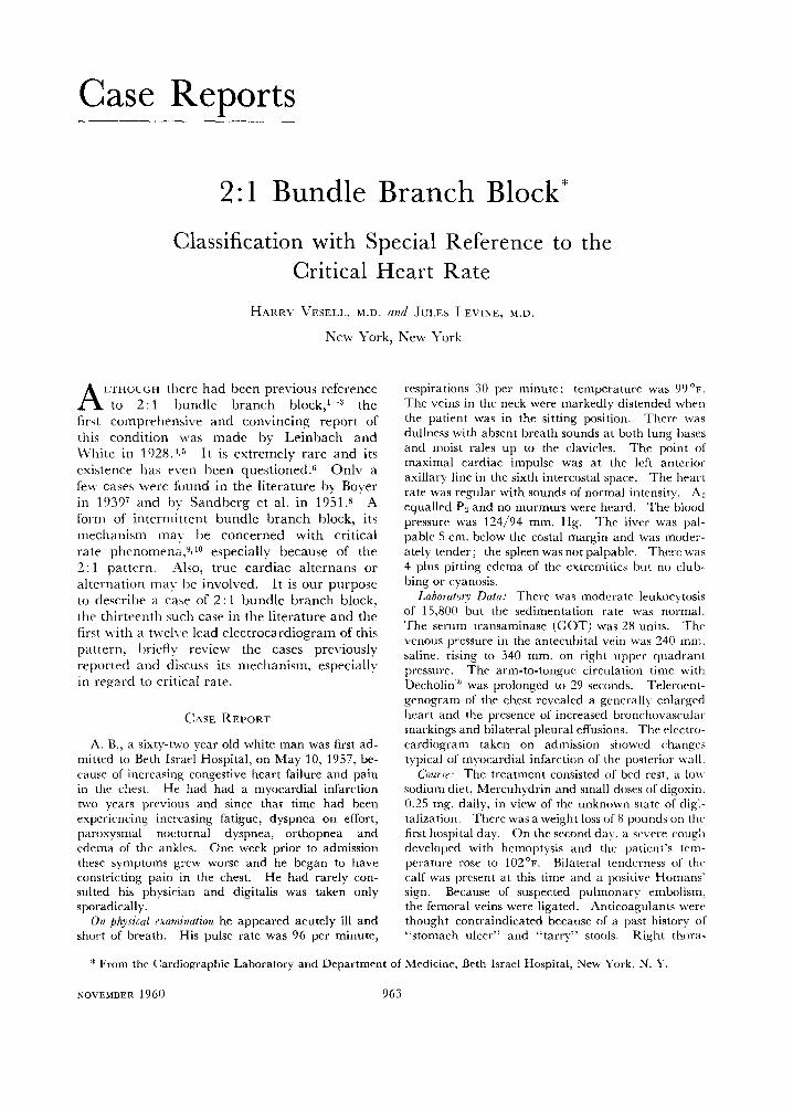
FIG. 1. Electrocardiogram, May 12, 1957, showing complete 1: 1 bundle branch block.
centesis was performed and 1,200 cc. of straw colored fluid was removed.
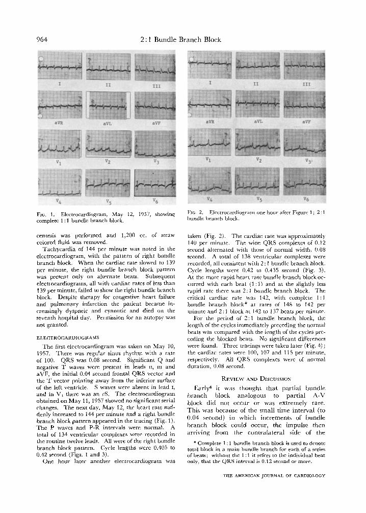
Tachycardia of 144 per minute was noted in the electrocardiogram, with the pattern of right bundle branch block. When the cardiac rate slowed to 139 per minute, the right bundle branch block pattern was present only on alternate beats. Subsequent electrocardiograms, all with cardiac rates of less than 139 per minute, failed to show the right bundle branch block. Despite therapy for congestive heart failure and pulmonary infarction the patient became in- creasingly dyspneic and cyanotic and died on the seventh hospital day. Permission for an autopsy was not granted.
ELECTROCARDIOGRAMS
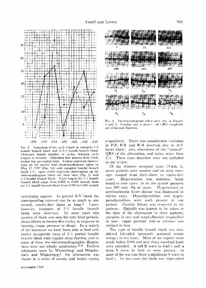
The first electrocardiogram was taken on May 10, 1957. There was regular sinus rhythm with a rate of 100. QRS was 0.08 second. Significant Q and negative T waves were present in leads II, III and aVF, the initial 0.04 second frontal QRS vector and the T vector pointing away from the inferior surface of the left ventricle. S waves were absent in lead I. and in VI there was an rS. The electrocardiogram obtained on May 11, 1957 showed no significant serial changes. The next day, May 12, the heart rate sud- denly increased to 144 per minute and a right bundle branch block pattern appeared in the tracing (Fig. 1). The P waves and P-R intervals were normal. A total of 134 ventricular complexes were recorded in the routine twelve leads. All were of the right bundle branch block pattern. Cycle lengths were 0.405 to 0.42 second (Figs. 1 and 3).
One hour later another electrocardiogram was
v4 v6
FIG. 2. Electrocardiogram one hour after Figure 1 ; 2 : 1 bundle branch block.
taken (Fig. 2). The cardiac rate was approximately 140 per minute. The wide QRS complexes of 0.12 second alternated with those of normal width, 0.08 second. A total of 138 ventricular complexes were recorded, all consistent with 2 : 1 bundle branch block. Cycle lengths were 0.42 to 0.435 second (Fig. 3). At the more rapid heart rate bundle branch block oc- curred with each beat (1 : 1) and at the slightly less rapid rate there was 2: 1 bundle branch block. The critical cardiac rate was 142: with complete 1 : 1 bundle branch block* at rates of 148 to 142 per minute and 2: 1 block at 142 to 137 beats per minute.
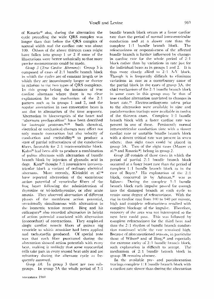
For the period of 2 : 1 bundle branch block, the length of the cycles immediately preceding the normal beats was compared with the length of the cycles pre- ceding the blocked beats. No significant differences were found. Three tracings were taken later (Fig. 4) ; the cardiac rates were 100, 107 and 115 per minute, respectively. All QRS complexes were of normal duration, 0.08 second.
REVIEW AND DISCUSSION
Early6 it was thought that partial bundle
branch block analogous to partial A-V
block did not occur or was extremely rare.
This was because of the small time interval (to
0.04 second) in which increments of bundle
branch block could occur, the impulse then
arriving from the contralateral side of the
* Complete 1 : 1 bundle branch block is used to denote total block in a main bundle branch for each of a series of beats; without the 1: 1 it refers to the individual beat only, that the QRS interval is 0.12 second or more.
THE AMERICAN JOURNAL OF CARDIOLOGY
Vesell and Levine 965
25
.405 .410 .415 .420 .425 .430 .435
FIG. 3. Variations of the cycle length in complete 1 :1 bundle branch block and in 2: 1 bundle branch block. Ordinates denotr number of cycles, abscissas cycle Itxngths in seconds. Unbroken line denotes limb leads, broken line precordial leads. Crosses represent observa- tions on the twelve lead electrocardiogram takrn on May 12, 1957 (Fig. 1A) with complete bundle branch block 1 : 1 ; open circles represent observations on the electrocardiogram taken one hour later (Fig. 2) with 2: 1 bundle branch block. Cycle lengths for 1 : 1 bundle branch block range from 0.405 to 0.420 second, those for 2: 1 bundle branch block from 0.420 to 0.435 srcond.
\.cntricular septum. In partial A-V block the corresponding interval can be as much as one second, twenty-five times as long.” Later, however, instances of 2: 1 bundle branch I)lock were observed. In most cases this pattern of block was seen for very brief periods, about fifteen to twenty-five cycles, in response to cxcrcise, vagus pressure or drugs. In a search of the literature we have been able to find only twelve acceptable cases of 2: 1 partial bundle I)ranch block with regular sinus rhythm, and in some of these the electrocardiographic illustra- tions were not wholly satisfactory.12,13 Earliest references were by Winterberg’ and Wencke- bath and Winterberg$ the alternation was shown in a series of twenty and twelve cycles,
aVL aW
Fro. 4. Electrocardiogram taken same dav as I“igurrs 2 and 3. Cardiac rate is slower: all (JR.4 complexes are of normal duration.
respectively. There was considerable \-ariation in P-P, P-R and R-R intervals due to A-1’ heart block; also, aberration of the “normal” QRS of the alternation and ratios other than 2 : 1. These cases therefore were not included in our review.
Of the thirteen accepted cases (‘l‘aljle I), seven patients were women and six \vere men; ages ranged from forty-three to eight).-five years. Hypertension was common. being noted in nine cases: in six the systolic pressure was 200 mm. Hg or more. H),pcrtensi\,e or arteriosclerotic heart disease was diagnosed in eleven cases. Hyperthyroidism and hyper- parathyroidism were each present in one patient. Cardiac failure was observed in six patients. Digitalis was known to t)e taken at the time of the alternation in three patients, atropine in one and corphyllamine (cuphyllin) in one: vagus pressure and “or esercisc was utilized in four.
The type of bundle branch block was con- sidered left-sided (presently accepted trrmi- nology) in ten cases. Most of the reports were made before 1944 and only three standard leads were recorded. A tall R wave in lead I and a deep S wave in lead III were present; in none of the ten was there a significant S wave in lead I. In two cases the block was right-sided
NOVEMBER 1960

966 2 : 1 Bundle Branch Block
TABLE I
Thirteen Reported Cases of 2: 1 Bundle Branch Block
B = QRS is wide.
Age (yr.) and Sex
Authors
Leinbach and 65,M Whit&
Stenstrom3 43,F
Diagnosis Cardiac
Bundle Branch
Failure
I P-R (set ) Rate
HCVD
HCVD
Block (per min.)
__----
0 Left 0.12-o. 13(N) 105 0.14-0.16(B)
0 Left 0.16(N) 97 0.17-0.18(B)
Kelly5 61, F
Donath’” 50, F
Master et a1.15 47, F
Boyer7 64,M
Rasario’” 66:M
Geill’? 85,M
HCVD
HCVD
HCVD AM1 Hyper-
thyroidism HCVD
HCVD
+ Left 0.12-0.16 140
-I- Left 0.14 136
O Left 0.16-0.18 103-111(N) 97-103(B)
? Left Normal 136-140
+ Left Normal 95.-100
+ Unknown 0.12(N) 100 0.14(B)
Kisch and 47:M. Grishman’s
Sandberg 57,M et al.R
AMI 0 Left 0.16 100
0 Left 0.16 107
Sandberr: et aLi
Shearn and Rytandlo
52,M
60,M
ASHD Hyperpara-
thyroidism HCVD
CVA HCVD
0 Right 0.14-0.16 139
+ Left 0.14-0.17 103-107
Vesell and 62,M ASHD + Right 0.13 137-142 Levine AM1
-
HCVD = hypertensive cardiovascular disease; ASHD = arteriosclerotic heart disease; N = QRS is normal;
and in one the QRS waves were concordant in the three limb leads.
The recorded duration of the 2: 1 bundle branch block was from six to 138 cycles, most often between fourteen and twenty-five. In one case the alternation was said to have continued for five minutes and in another for eight minutes. The cardiac rates during the period of 2: 1 bundle branch block were from 95 to 140 per minute. This was always a change from the rate during the period preceding or following the alternation when electrocardiograms were noted for these periods.
Electrocardiograms for the period preceding the alternation were reported in nine cases. In five of these complete 1 : 1 bundle branch block was present, one with a faster cardiac rate, three with a slower rate and in one the rate was not stated. In the other four of these
nine cases unstable bundle branch block was present with a critical heart rate for transitions between normal and prolonged intraventricular conduction; slower in two, faster in one and un- known in one.
Electrocardiograms for the period following the alternation were recorded in twelve of the thirteen cases. Complete 1 : 1 bundle branch block was present in two, both with slower cardiac rates. In seven the intraventricular condition time was normal, all with a cardiac rate slower than during the 2: 1 bundle branch block, in three there was unstable bundle branch block, all with a critical heart rate slower than the rate during the alternation.
During the period of 2 : 1 bundle branch block the alternate ventricular complexes with the normal QRS intervals frequently had an ab- normal configuration. In these marked left
THE AMERICAN JOURNAL OF CARDIOLOGY

Vesell and Levine 967
Duration (cycles)
Preceding Conduction
and Rate
30 and 27 Unknown
I.rss than 1 minute
Unstable bundle branch block critical rate 82-85
22 Unknown
“5 min.” 6 cycles recorded
1 6
Bundle branch block 1: 1
Bundle branch block 1: 1 95
.-8 min.”
14
26
21
20 and 27
14
9
138
Bundle branch block 1: 1 100-103 Unstable bundle branch block 97-100 Normal in 1926
Bundle branch block 1: 1
Unstable bundle branch block ; critical rate 78
Bundle branch block 1: 1 109 Unstable bundle branch
block
Bundle branch block 1: 1 142-l 48
Succeediq Conduction
and Rate -_ ~___
BBB 1:l 95 Unstable bundle branch
block slower
Bundle branch block 1: 1 105 Normal, slower
Normal 85 Normal, slower
Normal 90 Normal 80
Not given
Unstable bundle branch block ; critical rate 85
Normal 71 Unstable bundle branch
block ; critical rate 83-94
Normal 100-115
Remarks
T1. T, inverted with normal
QRS 2 : 1 bundle branch block after
digitalis, vagus prc’ssurr and exercise
Bundle branch block 1 : I after digitalis
2 : 1 bundle branch block after corphyllamine I-V
Bundle branch block for 4 weeks
2: 1 bundle branch block after atropine I-V
Bundle branch block other than 2: 1 with vagal pressure
Intermittent bundle branch block with csrrrisr or atro- pine
“True cardiac altrrnation”
Blood calcium 12-16 mg.
2: 1 bundle branch block fol- lowed 2 step test
3:l and 4:l bundle branch block also present
Critical rate for 2: 1 bundle branch block
AMI = acute myocardial infarction; CVA = cerebrovascular accident, probably hemorrhage
axis deviation of mean QRS was common. Q waves significant of myocardial infarction of the posterior wall were present in the electro- cardiogram with an aVF lead; T waves in leads I or II were frequently inverted, in some as secondary changes, part of a left ventricular hypertrophy and strain pattern, in others as primary RS-T changes due to digitalis or myocardial ischemia. Alternation of heart sounds with the 2: 1 bundle branch block was noted by Leinbach and White,4 and by Kelly5 \vho found no alternation in blood pressure.
(:LASSIFICATION OF P.4RTIAL BUNDLE BRANCH
BLOCK
Bundle branch block in general has been classified like A-V block into incomplete, partial and complete.*s3J Partial bundle branch Mock has been divided into type I (Wencke-
NOVEMBER 1960
bath) in which the QRS complexes gradually increase in width to that indicating complete blockage through one main bundle branch, and type II (Hay) with dropped “beats,” 3 : 1 : 3 : 1, 4: 1, etc. bundle branch block, without transi- tional QRS complexes. 2: 1 bundle branch block may be a form of partial bundle branch block, either type I (rarely if cvcr seen and proved) or type u.1g320
Group I (Alternation in Cardiac Ratta): The cases of 2: 1 bundle branch block reviewed in this report can be divided into three groups on the basis of specified changes in cardiac rate. Accordingly, group 1 consists of those cases in which, during the alternation, the cycle pre- ceding each QRS complex of normal width is longer than the cycle preceding the ad.jacent wide QRS. The conditions for the changes in conduction here are the same as in ordinar),

2: 1 Bundle Branch Block
intermittent bundle branch block, i.e., induced by fatigue, vagus or sympathetic effects, rate, chemical and nervous changes. The role of a critical heart rate has been described9mz1 and recently emphasized.‘0 An unstable state be- tween conduction and block is believed present in the His-Tawara system, usually due to disease. In this setting an increase or decrease in cardiac rate can trigger the bundle branch block. An efIective rate change has been found to be as little as one beat per minute or less than 0.01 second per individual beat at a cardiac rate of 100. The transition from conduc- tion to block or vice versa most often occurs within the interval of just one cycle, and it can take place many times within a minute. Aux- omerous condition has been suggested.21 Fried- man pointed out that heart rate is never absolutely constant even at rest and that in pulsus alternans careful measurement of the cycle length often showed prematurity of the weaker beats.22 This occurred in six or eight of his own cases and in many reported by others. He indicated that at a rapid heart rate slight alternation in cycle length may contribute to the development of mechanical alternation. Such alternation in cardiac rate may be responsible for alternation of conduction in group 1. By calculation this variation in rate should be at least 0.03 second or more when the QRS of bundle branch block is 0.12 second and the normal QRS 0.08 second.6r23 However, observations in ordinary intermittent as well as 2: 1 bundle branch block indicate it can be less than 0.03 second.7~s~‘g~23 Some or all of this variation in alternate cycle length is in duration of systole or QRS interval.
If in 2 : 1 bundle branch block only half the number of impulses traverse the fatigued bundle branch or its defective portion, this part should have a recovery period twice as long as for the contralateral bundle.24 This should be the basis for a stable and persistent 2: 1 bundle branch block. The alternation should be perpetuated even with a constant cardiac rate or with paradoxical rate change.24 It should be frequent as is 2: 1 A-V block. But 2: 1 bundle branch block is infrequent. Its great rarity, according to Wilson, is due to the fact that in bundle branch block the im- pulse is not prevented from reaching the farther side of the region of impaired conductivity. Arriving from the contra- lateral side within 0.04 second, he believed it would allow no improvement in the conductivity
of the region of block as a result of failure of the impulse to traverse it.6 However, the exact pathway and type of conduction beyond the site of block in a main bundle branch still does not appear to be definitely established. Recent pertinent and elaborate investigations by Scher25~26 and Sodi-Pallares27,2s and their col- leagues, and reasonings by Grant2g are in substantial disagreement concerning this path- way in specific conduction and in common myocardial tissue in bundle branch block. Elucidation of the definite mechanism of 2: 1 bundle branch block awaits a better solution of the problem of intraventricular conduction in common bundle branch block.
Group 1 is not true alternans. The 2:l arrangement is fortuitous based on small but adequate variations in rate of individual beats and critical rate phenomena. In the case of Boyerr fourteen cycles of 2: 1 bundle branch block are shown (his Fig. 3) in which by our measurements cycle lengths preceding the normal QRS were 0.44 to 0.45 second, and before the wide QRS 0.41 to 0.43 second. Each cycle before a normal QRS was 0.02 to 0.03 second longer than the preceding cycle before the wide QRS. This case and one reported by Slater of 3 : 1 and 4 : 1 bundle branch block’9 fit in the category of group 1.
Group 2 (Alternation Due to Supernormal Recovery Phase) : Group 2 consists of cases of 2 : 1 bundle branch block in which the cycle preceding the normal QRS complex is regularly shorter than the one preceding the adjacent wide QRS. The cardiac rate is about 100 per minute. The alternate normal ventricular con- duction (normal QRS) can be explained by the supernormal phase of recovery,11~30.31 the slightly faster rate of alternate beats being just suitable for this. The supernormal recovery phase has been calculated at about 0.40 to 0.64 second after the onset of QRS.32 Alternation due to supernormal recovery phase has been reported for A-V conduction in a small number of cases.31.33 One of the aforementioned cases’5 of 2: 1 bundle branch block had the character- istics of group 2. During the period of alterna- tion the cardiac rate was about 100 (103 to 111) for the cycles immediately preceding the normal QRS, and 97 to 103 before the bundle branch block complexes. Cycles were 0.02 to 0.08 second shorter before the normal complexes, so that at intervals of 0.54 to 0.58 second the QRS complexes were more within the supernormal recovery phase. In the case
THE AMERICAN JOURNAL OF CARDIOLOGY
Vesell and Levine 960
of RasarioL6 also, during the alternation the cycle preceding the wide QRS complex was longer than that before the QRS complex of normal width and the cardiac rate was about 100. Others of the above thirteen cases might Ilave fallen into group 1 or 2 if some of the illustrations were better technically so that more precise measurements could be made.
C&up .? (rrrl~e Cardiac Alternans) : Group 3 is composed of cases of 2 : 1 bundle branch block in which the cycles are of constant length or in \vhicll they are inconsistently longer or shorter in relation to the two types of QRS complexes. In this group belong the instances of true cardiac alternans where there is no clear explanation for the mechanism of the 2: 1 pattern such as in groups 1 and 2, and the regular alternation in two consecutive beats is not due to alternation of the time sequence. rilternation in bioenergetics of the heart and “alternans predispcsition” have been described for inotropic processes.3”r35 Such chemical, czlectrical or mechanical changes may affect not only muscle contraction but also velocity of conduction and excitability36 to produce a state of partial refractoriness of the conduction fibers, fa\,orable for 2 : 1 intraventricular block. Kah# had been able to produce a combination of pulsus alternans with alternation of bundle Ijranch block I)y injection of glyoxalic acid in doss. Katz”8 thought 2 : 1 intermittent intraven- tricular block a variant of ordinary electrical alternans. More recently, Kleinfeld et al.39 have reported alternation of the membrane action potential of ventricular fibers of the frog heart following the administration of thyroxine or tri-iodothyronine, or after acute anoxia. They observed alternation of different phases of the membrane action potential, occasionally simultaneous with alternation in thr isometric tension record. Bing and his colleagueslO also recorded alternation in height of action potential associated with alternation (concordant) of contractions. They employed single cardiac muscle fibers of guinea pig ventricle to which aconitine had been applied and tachycardia produced. Of special note was that each fiber punctured during the alternation showed action potentials with every I,eat, making it unlikely that some myocardial cells take part in every second beat only and are refractory during the alternate cycle as fre- quently assumed.
Chup .iiA: In group 3 there are two sub- groups. In group 3A the whole period of 2: 1
NOVEMBER 1360
bundle branch block occurs at a faster cardiac rate than the period of normal intraventricular conduction and at a slower rate than the complete 1 : 1 bundle branch block. The refractoriness or responsiveness of the affected bundle branch is further influenced by changes in cardiac rate for the whole period of 2: 1 block rather than by variations in rate just for the individual Ijeats as in groups 1 and 2. It is thus more closely allied to 2: 1 X-V block. Though it is frequently difficult to eliminate variations in rate as a contributory cause of the partial block in the cases of group 3A, the chief mechanism of the 2 : 1 bundle branch l)lock in some cases in this group may I)c that of true cardiac alternation unrelated to changes in heart rate.18 Electrocardiograms taken prior to the alternation were available in nine and postalternation tracings were a\railable in twelve of the thirteen cases. Complete 1 : 1 bundle branch block with a faster cardiac rate was present in one of these. Persistently normal intraventricular conduction timr with a ylowcr cardiac rate or unstable bundle t)ranch block with a slower critical rate was presrnt in seven others ; thus eight cases could IX placed in group 3A. Two of the eight cases (Master ct al.‘5 and Rasario’G) belong in ,group 2.
Grout, 3B consists of those cases in which the period of partial 2: 1 bundle branch block occurred at a faster heart rate than thr period of complete 1 : 1 bundle branch I)lock as in the case of Bo)-er.7 His explanation of the 2: 1 block, concurred in by Ashman,‘<” was as follows: During the complrtr 1 : 1 l)undle branch block each impulse passed far enough into the damaged branch at each cycle to retain some degree of refractoriness. ‘I\;‘ith the rise in cardiac rate from 100 to 140 prr minute, high and complete refractoriness resulted with complete blockage of the impulse. Therefore, recovery of the area was not intcrruptcd so the next beat could pass. This was followed by complete refractoriness for the third I)cat and thus the 2:l rhythm of bundle brancll conduc- tion continued while thr rate rcmaincd high. Because of aforetnentioned reasons, in particular those of Wilson6 and of Bin?,“0 and especially the extreme rarity of 2: 1 bundle branch block, such explanation is difficult to accept. ‘The mechanism of 2: 1 bundle branch I)lock in group 3B remains obscure.
In the available pre- and postalternation tracings complete 1: 1 bundle branch block with a cardiac rate slower than durin,? the alternation

2: 1 Bundle Branch Block
was present in four cases. One of these four (Boyer7) had been placed in group 1. Con- sistently normal intraventricular conduction time at a faster rate and unstable intraventricular conduction with a faster critical rate were not observed. There were thus three cases in group 3B.
Comment: The above findings suggest some correlation between 2: 1 bundle branch block and cardiac rate. At least three cases demon- strated the presence of alternation of block with alternation of rate for individual beats. Many showed a correlation between 2: 1 bundle branch block and changes in cardiac rate for the whole period of alternation compared to the rate when the intraventricular conduction was normal for each beat. The small number of cases available do not allow more general conclusions on the relationship between chrono- tropism (variations in rate) and dromotropism (variations in conduction) in 2: 1 bundle branch block.
SUMMARY
A case of 2 : 1 bundle branch block is described. All 138 consecutive ventricular complexes recorded in the routine twelve lead electro- cardiogram were of this pattern. Only twelve other acceptable cases of 2: 1 bundle branch block were found in a review of the literature. A synopsis of these is given. The mechanism of production of 2: 1 bundle branch block is dis- cussed, especially in regard to critical rate phenomena, theories of the site of delay in bundle branch block and especially in regard to the great rarity of 2: 1 bundle branch block. The cases of 2: 1 bundle branch block are classified into three groups on the basis of specified changes in cardiac rate.
REFERENCES
1. WINTERBERG, H. Beitrag zur Kenntnis der Sto- rungen in der Reizubertragung des menschlichen Herzens und der Anfalle bei Adams-Stokesschem Symptomenkomplex. Ztschr. ,p. exfier. Mfd., 8: 131, 1919.
2. WENCKEBACH, K. F. and WINTERBERG, H. Die Unregelmassige Herztatigkeit. Leipzig, 1927. Englemann.
3. STENSTROM, N. Further experiences in incomplete bundle branch block in man. Actn med. scandinau., 67: 353, 1927.
4. 11~~~~~~~, R. F. and WHITE, P. D. Two to one right bundle branch block. Am. Heart J., 3: 422, 1928.
5. KELLY, L. W. Two to one right bundle branch block. ,471~. Heart J., 6: 285, 1930.
6. WILSON, F. IN. and HERRMANN, G. .4n experimental study of incomplete bundle branch block and of the refractory period of the heart of the dog- Heart, 8 : 229, 1921.
7. BOYER, N. H. Paroxysmal bundle branch block converted into two to one bundle branch block by means of atropinr. Am. Heor/ J.. 18: 115, 1939.
8. SANDBERG, A. A., WENER, J., MASTER, A. M. and SCHERLIS, 1,. Intermittent and transient bundle branch block. .4 clinical and electrocardio- graphic study. r2nn. In/. Med., 35: 1085, 1951.
9. VESELL, H. Critical rates in ventricular conduction; unstable bundle branch block. .4m. ./. .\f. Jc., 202: 198, 1911.
10. SHEARN, M. A. and RYTAND, D. A. Inwr.nittent bundle branch block. Obser\-ations with special reference to the critical heart rate. ilrch. Int. Mtd., 91 : 418, 1953.
1. LEWIS, T. Mechanism and Graphic Reqistration of the Heart Beat, 3d ed. London. 1925. Shaw & Sons.
2. CAS~JJX, M. R. and RAMIREZ, R. I,. Alternancia electrica por bloquez de rama. RPL. nqvf. cardinl., 1: 55, 1931.
3. HANSEN, A. T. On intraventricular conduction disturbances in the specific system of the heart. Acta med. scqydinnu., 117: 104, 1944.
14. DONATH, F. Uber inkonstanten Schenkelblock und iiber die Wirkung van Corphyllamin auf das Herz. Ztschr. h&n. Med., 132: 802, 1937.
15. MASTFR, A. M., DACK, S. and JAFPE, H. L. Bundle branch and intraventricular block in acute coronary occlusion. Am. Hal-t J., 16: 283, 1938.
16. RASARIO, G. M. Sui blocchi di branca instabili. Cuorp c circolnz., 24: 139, 1940.
17. GEILL, T. Partieller schenkelblock two to one. A&. med. scnndinau., 109: 412, 1941.
18. KISCH, B. and GRISHMAN, A. Transient intraven- tricular conduction defect. !?‘apu. .Wfd. &T Sur,y., 2: 277, 1944.
19. SLATER, S. R. Partial bundle branch block. A case of three to one and four to one block. .h. Heart ~J., 5: 617, 1930.
20. HFRRMANN, G. and ASHMAN, R. A theoretical con- sideration of transient normal intrawntricular conduction in the presence of apparently com- plete bundlr branch block. rim. Hear/ J., 6: 375, 1931.
21. VESELL, H. and FRIEDYELD, L. Critical rates in ventricular conduction. I”. Am. Hal-t .J., 44: 830, 1952.
22. FRIEDMAN, B. Alternation of cycle length in pulsus alternans. Am. Ht-art ,I.: 51 : 701, 1956.
23. GARDBERG, M. and ROSEN, I. I.. Observations on conduction in a case of intermittent left bundle branch block. Am. Heart J.? 55: 677, 1958.
24. MYERS, G. The Interpretation of the Unipolar Electrocardiogram, p. 28. St. Louis, 1956. C. V. Mosby & Co.
25. ERICKSON, R. V., SCHER, A. M. and BECKER, R. A. Ventricular excitation in experimental bundle branch block. Circulation Res., 5 : 5, 1957.
26. BECKER, R. A., SCIIER, A. M. and ERICKSON, R. V. Ventricular excitation in experimental bundle branch block. Am. Heart J., 55: 547, 198.
27. RODRIGUEZ, M. I. and SODI-PALLARES, D. The
THE AMERICAN .JOURNAL OF CARDIOLOGY
Resell and Levine 071
mechanism of complete and incomplete bundle branch block. Am. Heart J., 44: 715, 1952.
28. MEDRANO, G. A., SODI-PALLARES, D., MARSICO, F. and BISTENI, A. The importance of septal ac- tivation in the electrogenesis of the unipolar mor- phologies in bundle branch block. Am. Hrnrt J., 57: 126. 1959.
19. GRAS r, R. P. CXnical Electrocardiography, p. 113, 126. New York, 1957. McGraw-Hill.
XI. SC~I~KF. D. and SCAARF, M. M. Supernormal phase of intravrntricular conduction. Am. Hmrt J., 36: 621. 1’148.
31. LAN~ENDOR~, K. nlternation of A-V conduction time-. .4m. If~nrl J., 55: 181, 1958.
32. NAI~M. I.. H. and HOFI:, H. E. The interpretation of the C-wave of the electrocardiogram. .4m. Hmr~ J.. 17: 585, 1939.
33. MACK. I., L.ANGENDORP,R. and KATZ, L.N. ‘rhe supwnormal phase of conduction in the human heart. .4m. Hmrt J., 34: 374, 1947.
34. Pou*ni\~~.~.ovx, M. Le Pouls Alternant; Etude
Uinique et Essai dv Pathogvniv. Paris. 1’130. Masson et Gie.
35. KIS~IX, B. Der Herzalternans. I-rgcbn. d. Kreis- laufforsch. Vol. 2, Dresden, 1932. ‘Theodor Steinkopff.
36. ASIJMAN, R. and HULL, 1;. I-swntials of E;lcrtro- cardiography, 2nd ed.. p. 111. NPW York. 1045. Macmillan C:o.
37. KAI~N, K. H. and ST.%RKENSTEIN, I<. 1X(- Storungcn drr HFrtztatikeit durch Glyoxylsalwr (Pulsus Altrrnans) in Electrokardiogrammc*. .,lrr,h. <TJ. I’hyriol., 133: 577, 1910.
38. KATZ, L. N. Electrocardiography, 2nd cd. Phila- delphia. 1947. Lea and Febigcr.
39. KIXINIXLD, M., STEIN, E. and h{ACtN. .I. 1,&x- trical alternans in single ventricular fibrrs of the frog heart. Am. ./. Phvriol., 187: 139, 1956.
40. HOGENCAMP, C:. E., K;ZKDESCH, M., DANFOR.I.H, W. H. and BING, R. J. Transmcmbrane clec- trical potentials in ventricular tarhycardia and fibrillation. :Im. Hmr/ J.. 57: 214. 1059.
NOVEMBER 1960