a galactose-functionalized dendritic sirna-nanovector to
TRANSCRIPT

Nanoscale
PAPER
Cite this: Nanoscale, 2015, 7, 16921
Received 4th May 2015,Accepted 14th August 2015
DOI: 10.1039/c5nr02898a
www.rsc.org/nanoscale
A galactose-functionalized dendriticsiRNA-nanovector to potentiate hepatitis Cinhibition in liver cells†
Abirami Lakshminarayanan,a,b B. Uma Reddy,b Nallani Raghav,c Vijay Kumar Ravi,c
Anuj Kumar,b Prabal K. Maiti,*c A. K. Sood,*c N. Jayaraman*a and Saumitra Das*b
A RNAi based antiviral strategy holds the promise to impede hepatitis C viral (HCV) infection overcoming
the problem of emergence of drug resistant variants, usually encountered in the interferon free direct-
acting antiviral therapy. Targeted delivery of siRNA helps minimize adverse ‘off-target’ effects and maxi-
mize the efficacy of therapeutic response. Herein, we report the delivery of siRNA against the conserved
5’-untranslated region (UTR) of HCV RNA using a liver-targeted dendritic nano-vector functionalized with
a galactopyranoside ligand (DG). Physico-chemical characterization revealed finer details of complexation
of DG with siRNA, whereas molecular dynamic simulations demonstrated sugar moieties projecting “out” in
the complex. Preferential delivery of siRNA to the liver was achieved through a highly specific ligand–recep-
tor interaction between dendritic galactose and the asialoglycoprotein receptor. The siRNA-DG complex
exhibited perinuclear localization in liver cells and co-localization with viral proteins. The histopathological
studies showed the systemic tolerance and biocompatibility of DG. Further, whole body imaging and
immunohistochemistry studies confirmed the preferential delivery of the nucleic acid to mice liver. Signifi-
cant decrease in HCV RNA levels (up to 75%) was achieved in HCV subgenomic replicon and full length
HCV-JFH1 infectious cell culture systems. The multidisciplinary approach provides the ‘proof of concept’ for
restricted delivery of therapeutic siRNAs using a target oriented dendritic nano-vector.
Introduction
Hepatitis C virus (HCV) is an important human pathogen pri-marily infecting the liver cells. The cascade of events caused bythe entry of HCV positive strand RNA to hepatocytes includesInternal Ribosome Entry Site (IRES) mediated translation toproduce an assortment of viral proteins, of which few areknown to be involved in the virion assembly along the lipiddroplets of ER.1,2 Virus entry into the cells is aided further byenvelope proteins E1 and E2 interacting with hepatocytes.1
Detailed investigations to understand important events of HCVlife cycle have allowed exploring various strategies against thisvirus. Several conventional approaches are reported to combatHCV infection which include (i) blocking the virus entry,3,4 (ii)
inhibiting the interaction of essential host factors, such as, La,miR 122 with HCV RNA by use of peptides,5,6 miravirsin;6,7 toinhibit 5′ IRES mediated translation, (iii) inhibiting replicationusing protease or polymerase inhibitors, such as boceprevir,telaprevir and sofosbuvir8 and small molecules like punicalin,punicalagin,9 (iv) inhibiting virion assembly and release.10,11
However, interferon based therapy combined with serine pro-tease inhibitors is prominent in current treatment againstHCV. Despite high potency of different antivirals, the emer-gence of drug resistant variants of HCV is a major concern.12
In order to overcome drug resistance, alternate approacheshave emerged in the past few years to inhibit viral translationand replication processes, by the use of antisense oligonucleo-tides,13,14 DNAzymes,15 and siRNAs16,17 and targeting the con-served 5′-untranslated region (UTR) of HCV.
The use of siRNA to inhibit HCV in JFH-1 infectious cellculture showed viral clearance rates up to 80%.16 Further, thedelivery of short hairpin RNA (shRNA), using Sendai viro-some, showed specific inhibition of HCV IRES mediated trans-lation in vivo. The membrane fusion glycoprotein of thevirosome, having exposed galactose residues specificallytargets hepatocytes and mediates localized delivery of theshRNA.14 A nanosome–siRNA formulation was also demon-
†Electronic supplementary information (ESI) available: Spectral data and experi-mental details. See DOI: 10.1039/c5nr02898a
aDepartment of Organic Chemistry, Indian Institute of Science, Bangalore 560012,
India. E-mail: [email protected] of Microbiology and Cell Biology, Indian Institute of Science, Bangalore
560012, India. E-mail: [email protected] of Physics, Indian Institute of Science, Bangalore 560012, India.
E-mail: [email protected], [email protected]
This journal is © The Royal Society of Chemistry 2015 Nanoscale, 2015, 7, 16921–16931 | 16921
Ope
n A
cces
s A
rtic
le. P
ublis
hed
on 2
8 Se
ptem
ber
2015
. Dow
nloa
ded
on 2
/18/
2022
2:2
4:27
PM
. T
his
artic
le is
lice
nsed
und
er a
Cre
ativ
e C
omm
ons
Attr
ibut
ion-
Non
Com
mer
cial
3.0
Unp
orte
d L
icen
ce.
View Article OnlineView Journal | View Issue

strated recently to inhibit the viral replication.17 These studiesexemplify the importance of siRNA mediated inhibition tocombat HCV infection and highlight the advantages of tar-geted delivery.
Target specific delivery of siRNA assumes greater impor-tance, especially in the in vivo systems, in order to enhanceinhibitory activities, minimize off-target effects and avoidadverse effects on healthy organs. Although the use of pseudo-viruses, virus-like particles, adenoviruses and virosomes wasstudied as delivery systems for antiviral agents inherentimmunogenicity and virulence of these vectors raise concernsin their therapeutic developments.19 Non-viral vectors haveemerged as efficient alternatives to deliver nucleic acids bothin vitro and in vivo.19 A recent entry to the repertoire of non-viral vectors is the dendritic macromolecule or dendrimers,characterized by the presence of a branches-upon-branchesstructural motif.20 We have demonstrated earlier the efficientnucleic acid delivery and non-toxic properties of novel syn-thetic dendritic vectors, namely, the poly(propyl ether imine)(PETIM) dendrimers.21–23 In an effort to deliver antiviral siRNAagainst HCV specifically to liver cells, we undertook a study ofthe liver target-specific PETIM dendrimer as a delivery system.The target-specific nature of the dendrimer delivery systemtook advantage of the ligand–receptor interaction between asugar ligand, namely, the galactoside ligand and its cognateasialoglycoprotein receptor (ASGPR), which is expressed abun-dantly in the liver cells.24 In the present study, a third gene-ration galactose-functionalized PETIM dendrimer (DG) wassynthesized and studied for its ability to interact with siRNAby biophysical techniques and molecular dynamic (MD) simu-lations. Immunofluorescence based confocal microscopyshowed that the delivered siRNA co-localized with the HCVviral proteins NS5B and NS3 which participate in HCV replica-tion. The study demonstrates, for the first time, that siRNAdelivered using DG is localized in the perinuclear regionwhich is the site for HCV replication. The target-specificPETIM dendrimer is found to deliver a chosen anti-HCV siRNAinto the liver cells preferentially and inhibit HCV proliferation.
MethodsSynthesis of galactose functionalized PETIM dendrimer
Benzobromogalactose25 (0.50 g, 0.76 mmol) in CH2Cl2 (30 mL)was added to a solution of dendritic alcohol21,26 (0.050 g,9.6 μmol), Ag2CO3 (0.20 g, 0.72 mmol) and AgClO4 (0.050 g,0.085 mmol), stirred for 30 h at room temperature, filtered, fil-trate concentrated in vacuo and purified by column chromato-graphy (Al2O3, CH2Cl2 : MeOH = 95 : 5). The resulting productin tetrahydrofuran : MeOH (1 : 1) (10 mL) was added withNaOMe in MeOH (1 M) (0.02 mL), stirred at room temperaturefor 16 h, neutralized with an Amberlite ion-exchange (H+)resin, filtered, concentrated in vacuo and the resulting solutionsubjected to dialysis (MW cut-off 3.5 kDa) to afford DG, as afoamy solid. Yield: 0.057 g (57%, after two steps); 1H NMR(D2O, 400 MHz) δ 1.87–1.92 (br, 132 H, CH2–CH2–CH2), 3.15
(br, 132 H, N–CH2–CH2 and CH2–CH2–N), 3.37 (t, 24 H, J = 8.4Hz,), 3.47–3.62 (br, 228 H), 3.62–3.80 (br, 72 H), 4.26 (d, 24 H,J = 4 Hz); 13C NMR (D2O, 100 MHz) δ 23.3, 31.3, 36.9, 60.9,68.6, 70.0, 70.7, 72.8, 75.1, 102.7.
UV-visible spectroscopy
siRNA (1 μM) in 1X siRNA buffer (Dharmacon) was titratedwith DG (25 mg mL−1) and the resultant solution equilibratedfor 20 min before the UV-visible spectrum was recorded. Titra-tions were carried out up to a weight ratio (R) of 100 in a10 mm stoppered quartz cuvette at a constant temperature of25 °C. UV-Vis absorption spectra were recorded on a Perkin-Elmer Lambda35 spectrophotometer.
Ethidium bromide displacement assay
A solution of siRNA (1 μM) in siRNA buffer was incubated for10 min with EB at a molar ratio of 6 : 1 siRNA : EB. The fluore-scence spectrum was recorded by exciting at 520 nm andrecording the emission in the 535–800 nm region using a10 mm quartz cuvette on a Varian Cary Eclipse Fluorimeter.The siRNA-EB solution was titrated with DG (25 mg mL−1) insiRNA buffer and the fluorescence spectrum recorded after20 min equilibration.
Zeta potential measurements
Aqueous solutions of siRNA–dendrimer complexes at R wereprepared by mixing siRNA (100 nM) with required amounts ofDG (10 mg mL−1) in siRNA buffer and equilibrated at 25 °C for45 min. Zeta potential and electrophoretic mobility measure-ments were carried out on a Brookhaven ZetaPals instrumentat a controlled temperature of 25 ± 0.5 °C. Each data was takenas an average of three runs with each run comprising tenmeasurements. The zeta potentials of siRNA alone (100 nM)and dendrimer alone (193 μM) were also measured.
Atomic force microscopy
DG (0.77 nM), siRNA (50 nM) and their complexes at R 1, 50and 100 were prepared for AFM imaging by drop-casting 10 μLof each sample on mica, drying under vacuum and imagingusing a VeecodiInnova AFM (Bruker, USA) under tappingmode in air. The resonance frequency of a tapping mode canti-lever was set at 300 kHz and the scanning rate was fixed at1 Hz. The z-height distributions were analyzed using Nano-scope Analysis software.
Molecular dynamic simulations
The PETIM dendrimer was built using a dendrimer buildertoolkit (DBT).27 The beta galactose structure was Gaussianoptimized with the HF 6-31 g basis set and was added to thehydroxy terminated PETIM dendrimer to derive the galactosefunctionalized PETIM dendrimer. Using the antechambermodule of Amber 12,28 the RESP charges are calculated andGAFF atom types are assigned for the dendrimer.29 ff99bsc030
with parmbsc031 correction was used to describe inter- andintra molecular interactions involving siRNA molecules. Thesolute (DG and siRNA) was solvated using the xleap module of
Paper Nanoscale
16922 | Nanoscale, 2015, 7, 16921–16931 This journal is © The Royal Society of Chemistry 2015
Ope
n A
cces
s A
rtic
le. P
ublis
hed
on 2
8 Se
ptem
ber
2015
. Dow
nloa
ded
on 2
/18/
2022
2:2
4:27
PM
. T
his
artic
le is
lice
nsed
und
er a
Cre
ativ
e C
omm
ons
Attr
ibut
ion-
Non
Com
mer
cial
3.0
Unp
orte
d L
icen
ce.
View Article Online

Amber maintaining a buffer of 15 Å on all sides. The TIP3Pwater model was used for solvation.32 Of the non-protonateddendrimer and siRNA, only siRNA required counter-ions tocharge neutralize the system. In non-protonated cases thesystem was charge neutralized with 44 Na+ ions. The systemwas then energy minimized for 1000 steps using the steepestdescent method and further 2000 steps by the conjugate gradi-ent method, while the solute was fixed using a harmonic con-straint of 500 kcal mol−1 Å−2, to eliminate bad contacts withthe solvent. Further 3000 steps of conjugate gradient minimiz-ation was carried out, reducing the harmonic constraint by5 kcal mol−1 Å−2 at every 600 steps from 20 kcal mol−1 Å−2 to0 kcal mol−1 Å−2. Subsequently, an equilibration run of 50 psin the NPT ensemble was performed before going into a pro-duction run of 100 ns in the NPT ensemble.33–35 All the simu-lations were carried out using the PMEMD module ofAmber12.36
Cell culture
Cell monolayers were maintained in Dulbecco’s modifiedEagle’s medium (DMEM) (Sigma Aldrich) supplemented with10% heat-inactivated foetal bovine serum (FBS) (Invitrogen) and1% penicillin–streptomycin at 37 °C in 5% CO2. Hygromycin B(25 mg/mL−1) was used as an additional supplement for stablemaintenance of replicon 2a harbouring Huh 7 cell line.
Transfection of siRNA–DG complexes in mammalian cells
1 × 10⁵ cells were grown on sterilized 18 mm cover slips in a 12well plate to 65% confluency. DG–siRNA complexes were pre-pared at various ratios using 50 nM siRNA and requiredamounts of dendrimers by incubating in Opti-MEM (SigmaAldrich, India) for 45 min. The cells were incubated with thecomplexes in Opti-MEM for 6 h. After transfection, cells werereplenished with complete growth medium. In the case ofexperiments where free sugars were used, a stock of 1 mMD-galactose or D-mannose was prepared by dissolving theappropriate amount of sugar in sterile double distilled waterand the solution was filtered through 0.2 μm pores before use.Cells were pre-incubated with the free sugars in Opti-MEM for20 min before the addition of the complexes for transfection.
Confocal microscopy and immunofluorescence
Post transfection, cells were washed with PBS, fixed with 4%paraformaldehyde solution, permeated with 0.1% Triton X andthe nuclei stained with DAPI. After blocking in 3% BSA (bovineserum albumin) solution for 40 min, cells were washed withPBS and mounted onto slides using glycerol. Fluorescentimages were obtained by using excitation wavelengths of350 nm, 488 nm and 657 nm (Zeiss Confocal microscope).Images were processed using ZEN10 software and quantifiedusing Image J. Immunofluorescence was carried out by incu-bating transfected cells, with NS5B or NS3 primary antibody(Thermo Fisher, India) diluted to 1 : 200 in 3% BSA for 2 h at4 °C. Cells were then washed with PBS and incubated at roomtemperature with secondary antibodies at 1 : 500 dilution
(Alexa 488 tagged antirabbit antibody for NS5B and Alexa 657tagged antimouse antibody for NS3) for 30 min.
Real time PCR to assess HCV RNA levels14
Huh 7 cells harbouring the HCV were transfected with siHCV–DG complexes as described previously. At the required time,cells were harvested in TRIzol reagent (Sigma Aldrich), andtotal cellular RNA was extracted and reverse transcribed withthe HCV forward primer and GAPDH reverse primer. Theresulting cDNA (1 : 10 diluted) was subjected to quantitativePCR using a SYBR green real-time assay mixture (ThermoScientific).
In vitro transcription of JFH1 RNA
The HCV-pJFH1 construct was linearized with the restrictionenzyme XbaI. The linear DNA was used as a template forin vitro transcription to synthesize JFH1 RNA using a RibomaxLarge scale RNA production T7 kit (Promega).
JFH1 replication inhibition assay37
Huh 7.5 cells were transfected with 25 nM and 50 nM ofsiHCV using DG. Four hours later, cells were transfected withHCV-JFH1 RNA using lipofectamine 2000 (Invitrogen). After48 h and 72 h, cells were harvested for total cellular RNA iso-lation. The relative levels of HCV RNA were quantified by realtime PCR.
Haemolysis assay38
500 μL blood was drawn by ocular bleeding into microfugetubes containing 50 μL of 6% EDTA as an anticoagulant. Thewhole blood was centrifuged at 10 000 rpm to separate plasmaand leukocytes. 1.78 × 10⁵ cells were suspended in 1 mL of PBSand required amounts of dendrimer (1 mg, 5 mg and 10 mg)solutions were added to the RBCs. Triton X (1% v/v) was addedin order to induce 100% haemolysis. The suspensions wereincubated in a 37 °C water bath. After incubation time, theRBC suspensions were centrifuged at 10 000 rpm for 5 min,the supernatant collected and the absorbance recorded at540 nm. Percentage haemolysis was calculated as follows:
% Haemolysis ¼ðAbsorbance of sample
� Absorbance of controlÞ=ðAbsorbance of Trition X
� Absorbance of controlÞ � 100
In vivo toxicity and histopathology
The mice were divided into six groups of three mice each.Group 1 served as the control; mice were administered with100 μL of sterile distilled water. Mice in groups 2 to 6 wereinjected with 100 μL DG at 50, 100, 200, 350 and 500 mg perkg body weight (b.w.). All mice were observed for morphologi-cal and behavioural responses, food–water intake, body weightchanges and mortality for 14 days. Subsequently, they weresacrificed, organs were excised and fixed in 10% NBF (neutralbuffered formalin) for 72 h. The tissues were then dehydrated
Nanoscale Paper
This journal is © The Royal Society of Chemistry 2015 Nanoscale, 2015, 7, 16921–16931 | 16923
Ope
n A
cces
s A
rtic
le. P
ublis
hed
on 2
8 Se
ptem
ber
2015
. Dow
nloa
ded
on 2
/18/
2022
2:2
4:27
PM
. T
his
artic
le is
lice
nsed
und
er a
Cre
ativ
e C
omm
ons
Attr
ibut
ion-
Non
Com
mer
cial
3.0
Unp
orte
d L
icen
ce.
View Article Online

in graded alcohol series of ethanol, mounted in paraffinblocks, sectioned to 5 μm thickness, deparaffinized, rehydratedand stained with haematoxylin–eosin, and examined using aZeiss microscope.
In vivo bioimaging and immunohistochemistry
Mice were injected with DG-HCVLucDNA or DG–HCVLuc–siRNA complexes via tail vein injection. Luciferase expressionin different organs of the mice was tracked by using a XenogenIn Vivo Imaging System (IVIS) by intraperitoneal administationof luciferin (150 mg per kg b.w.). The mice were sacrificed onthe 5th day post treatment, and tissue samples were isolatedand processed as described above. The presence of luciferaseproteins in tissues was detected using primary antibody(1 : 100 dilution, goat polyclonal antibody G7451, Promega)and horseradish peroxidase tagged secondary antibody (1 : 200dilution anti-goat IgG). The sections were treated with 3,3′-di-aminobenzidine stained with haematoxylin and imaged.
Ethical clearance
All animal experiments were performed in compliance withthe relevant laws and institutional guidelines and approved bythe ‘Institutional Animal Ethics Committee’ (IAEC) of IndianInstitute of Science. Healthy Balb/c mice (4–7 weeks old) wereselected from the inbred colony and maintained under a 16 : 8light : dark cycle at a temperature of 20 ± 1 °C. The animalswere fed with food pellets and water ad libitum.
ResultsFunctionalization of PETIM dendrimers with D-galactose
The PETIM dendrimer constitutes a tertiary amine as thebranching site, and an ether as the linker separating thebranching sites by n-propyl moieties.26 The third generationnitrogen-cored dendrimer presenting 24 hydroxyl group func-tionalities at its periphery (G3(OH)24) was used in thepresent study.23,26 Functionalization of the hydroxyl-groupterminated PETIM dendrimer was performed through theglycosylation using benzobromogalactose in the presence ofAg2CO3/AgClO4 (ESI Scheme S1†) (Fig. 1). The galactopyranoside-derivatized DG is soluble in water. The purity of DG was ascer-tained by 1H and 13C NMR spectroscopy analysis (ESI Fig. S2and S3†). A comparison of 1H NMR integrations of 4.26 ppm(H-1 of sugar) and 1.86 ppm (–CH2–CH2–CH2–) of the dendrimerstructure showed that ∼21–23 sugar moieties functionalize adendrimer molecule. A phenol-sulfuric acid assay further con-firmed the extent of galactose functionalization (ESI Fig. S4†).
Efficient complexation of siRNA with DG
The ability of DG to interact with siRNA and the nature of thisinteraction were assessed by gel retardation assay, zeta poten-tial measurements, UV-visible spectroscopy, ethidium bromideintercalation assay and visualization of the complex by atomicforce microscopy (AFM). UV-Visible spectroscopy analysis pro-vided an indication of siRNA-DG complexation, deduced from
the changes in siRNA absorbance at 260 nm occurring due tothe interaction of siHCV with DG (Fig. 2a). Displacement ofintercalated ethidium bromide (EB) from siRNA upon titrationwith DG, monitored by the decreasing fluorescence intensityof EB, also indicated formation of a stable complex betweensiHCV and DG (Fig. 2b). Complex formation between siHCVand DG was also apparent from the gel retardation assay,
Fig. 1 Molecular structure of DG. The molecular structure of galacto-pyranoside dendrimer (DG) with 24 peripheral galactose units.
Fig. 2 Characterization of siRNA–DG complexes. (a) UV-Visiblespectroscopy of siHCV and siHCV–DG complexes at different weightratios of siRNA–DG complexes, R: 10, 20, 30, 40, 50, 60, 70, 80, 90 and100. (b) Fluorescence emission profile of EB bound to siHCV, and, upontitration with DG at R 10, 30, 50, 70, 90 and 100. (c) Zeta potentialmeasurements of siRNA, DG and their complexes in 1× siRNA buffer.100 nM siRNA was used in each of the measurements. Complexes at R10, 20, 30, 40, 50, 60, 70, 80, 90, 100 as well as DG alone (193 μM) wereassessed for their zeta potentials and electroneutrality was found to beat R 50. (d) AFM height of siRNA-DG complexes at R 50. Scale bar =300 nm. Height profile of these complexes was 1.5–3.25 nm.
Paper Nanoscale
16924 | Nanoscale, 2015, 7, 16921–16931 This journal is © The Royal Society of Chemistry 2015
Ope
n A
cces
s A
rtic
le. P
ublis
hed
on 2
8 Se
ptem
ber
2015
. Dow
nloa
ded
on 2
/18/
2022
2:2
4:27
PM
. T
his
artic
le is
lice
nsed
und
er a
Cre
ativ
e C
omm
ons
Attr
ibut
ion-
Non
Com
mer
cial
3.0
Unp
orte
d L
icen
ce.
View Article Online

wherein, a gradual retardation and disappearance of the siRNAband, visualized on a 2% formaldehyde-agarose gel indicatedthe formation of siRNA–dendrimer complex (ESI Fig. S5†).
To study the charge neutralization upon complex for-mation, zeta potential measurements were performed. Thezeta potential of siRNA, DG and their complexes at varyingweight ratios (R) was determined (Fig. 2c). A charge of −28.15± 0.94 mV on siRNA and +15.13 ± 0.95 mV on DG wasobtained. Increasing concentration of DG in the complexresulted in an increase in the zeta potential up to +16.8 ±1.15 mV at R 100. The electroneutrality of the complexoccurred at R 50, which is in agreement with the gel retar-dation assay. The shape and size distribution of siRNA-DGcomplexes were characterized by atomic force microscopy(AFM), wherein, siRNA appeared globular with a z-height of∼1 nm, while DG was spherical with a height of 0.1–2 nm.DG–siRNA complexes at R 50 consisted of particles of variousshapes and height profiles ranging from 1.5 to 3.25 nm(Fig. 2d). Complexes formed at R 100 showed a z-height distri-bution of 3–8 nm and very few particles showed height profilesup to 25 nm (ESI Fig. S6†).
Structural analysis of siRNA–DG complexation by MDsimulation
In order to understand the complexation and to visualize thedendriplexes, a fully atomistic MD simulation of the siRNA–DG complexation was performed. Simulation studies revealedthat in case the dendrimer is not protonated, there is no com-plexation with siRNA. At physiological pH, tertiary amines atthe outer shells (penultimate shell) are protonated, as pKa
values are known to be in the range of 8–9. A lower pH isrequired generally for the tertiary amines at the interior andcore for their protonation. In the MD studies, tertiary aminesat the outer shells were protonated, as would be expected inexperimental studies conducted at pH 7.4.39,40 When dendri-mer amines are protonated, formation of a very tight complexwas observed, demonstrating the role of electrostatic inter-actions in the complexation process. Fig. 3a and b shows mole-cular level pictures of the siRNA–DG complex as a function ofsimulation time from 0 to 100 ns. Simulations also revealedthat the dendrimer preferentially binds to the major grooveduring complexation with siRNA. Instantaneous snapshots ofthe complex (ESI Fig. S7†) shown at few ns intervals reveal themajor groove binding nature of the dendrimer.
A graphical representation of the sizes of the complexes, aswell as sizes of the siRNA and DG in the complex as the com-plexation proceeds is shown in Fig. 3c. The radius of gyration(Rg) of the siRNA–DG complex was found to be ∼2 nm andthose of siRNA alone and dendrimer alone were 1.8 nm and1.3 nm, respectively. The Rg of siRNA remained almost con-stant in all the conditions studied. In order to verify thecomplex formation between the protonated dendrimer andsiRNA, the radial distribution of the dendrimer amines as wellas sugar groups and phosphates of the siRNA was studied (ESIFig. S8†). Further, a plot of the distance between the centre ofmass (COM) of a dendrimer and siRNA as a function of simu-
lation time showed that in the case of non-protonated dendri-mer, the dendrimer moves away from the siRNA oversimulation time. In contrast the protonated dendrimer bindstightly on the surface of siRNA, as seen by the very low COMdistance between the two (Fig. 3d).
Preferential perinuclear delivery of siRNA into liver cellsusing DG
Initial transfection studies were carried out by monitoring theinternalization of a green fluorescent tagged siRNA. Electro-neutral complexes formed at R 50 (siRNA : DG = 1 : 50) showedpreferential delivery of siRNA to liver derived Huh 7 cells overkidney derived BHK 21 cells (Fig. 4a). The siRNA deliveryefficiency was compared to other delivery agents, namely, lipo-fectamine 2000 and non-galactose functionalized dendrimerG3(OH)24. A higher transfection efficiency obtained using DGwhen compared to G3(OH)24 highlights the role of the ASGPR–galactose interaction in siRNA-DG internalization (Fig. 4b).
Importantly, the siRNA–DG complex was observed to bedelivered preferentially to the perinuclear region in liver cells.The fluorescence intensity around the perinuclear region ofthe cell was considerably higher for the delivery mediated bythe DG–siRNA complex, in comparison with the lipofecta-mine–siRNA complex (Fig. 4c). Further, siRNA internalizationoccurring in the perinuclear region was confirmed throughz-stack imaging. The higher fluorescence intensity at themiddle frame of the cell clearly indicated the internalization ofsiRNA occurring in the perinuclear region (Fig. 4d). The prefer-ential perinuclear localization can be due to ASGPR mediatedendocytosis, since ASGPR enters the cell during endocytosisand particularly delivers the bound ligand in the area sur-rounding the nucleus.41 In order to verify this hypothesis, theHuh 7 cells were pre-incubated with free D-galactose (a specificligand for ASGPR) or D-mannose (a non-specific control) atdifferent concentrations (45 μM, 90 μM, 180 μM and 360 μM)prior to the addition of siRNA–DG complexes. At 8 h postaddition of the complexes, the cells were fixed with parafor-maldehyde and imaged by CLSM. Internalization of siRNA–DGcomplexes was greatly reduced in cells pre-incubated withD-galactose due to saturation of the receptor sites with theligand which prevented the binding of DG. Cells pre-incubatedwith D-mannose did not show appreciable difference in siRNAinternalization when compared to control cells (Fig. 4e).Experiments were also carried out in BHK 21 cells, and noeffect on siRNA internalization was observed (ESI Fig. S9†).
Localization of DG–siRNA complex at the site of replicationand inhibition of viral RNA synthesis
Following successful transfection mediated by DG in culturedliver cells, the study was extended to a functional assay, todemonstrate the applicability of preferential liver delivery in adisease model involving hepatitis C virus (HCV). HCV repli-cates along the lipid rafts in the perinuclear region of livercells. The viral proteins NS5B and NS3 regulate the viral repli-cation.1 In order to identify the localization site of the deli-vered siRNA, Huh 7 cells harbouring a HCV monocistronic
Nanoscale Paper
This journal is © The Royal Society of Chemistry 2015 Nanoscale, 2015, 7, 16921–16931 | 16925
Ope
n A
cces
s A
rtic
le. P
ublis
hed
on 2
8 Se
ptem
ber
2015
. Dow
nloa
ded
on 2
/18/
2022
2:2
4:27
PM
. T
his
artic
le is
lice
nsed
und
er a
Cre
ativ
e C
omm
ons
Attr
ibut
ion-
Non
Com
mer
cial
3.0
Unp
orte
d L
icen
ce.
View Article Online

replicon were transfected with siRNA and an immunofluore-scence assay was performed. We also probed NS5B and NS3proteins so as to investigate co-localization of siRNA with theseproteins. Confocal microscopy images showed that labelledsiRNA co-localized with both the viral proteins (Fig. 5a, ESIFig. S10†), indicating that the siRNA delivered using DG wasprobably localized at the site of HCV RNA replication. Follow-ing this observation, the effect of DG-mediated delivery of apre-characterized therapeutic siRNA known to inhibit HCVRNA translation and replication18 was studied. Anti HCVsiRNA–DG complexes were prepared at R 50 using differentconcentrations of siRNA, ranging from 25 to 100 nM and trans-fected into Huh 7 cells harbouring the HCV replicon. About77% reduction in HCV RNA levels was noted up to 72 h, indi-cating the successful delivery of siRNA using DG (Fig. 4b).siHCV-G3(OH)24 and siNsp–DG complexes were used as the
non-targeting and non-specific siRNA (siNsp) controls,respectively. While transfection with the siHCV-G3(OH)24complex showed only about 25% inhibition of HCV RNA, thenon-specific siRNA did not show any effect on HCV RNA levels.Transfection with DG alone did not show any reduction in thelevels of HCV RNA indicating that the dendrimer alone doesnot interfere with the virus replication (ESI Fig. S11†).
The effectiveness of the siHCV–DG system was further vali-dated in an infectious JFH1 cell culture system.37 Huh 7.5 cellswere transfected with 25, 50, 100 and 200 nM siHCV using DG.Four hours later, the cells were transfected with JFH1 RNA andthe relative RNA levels were quantified at 48 h and 72 h posttransfection of siHCV. A dose dependent reduction in HCVRNA levels (up to 75%) was observed (Fig. 5c) which furtherconfirmed the successful delivery of siRNA to inhibit HCVreplication in the infectious cell culture system.
Fig. 3 MD simulation studies of siHCV–DG complexes. (a) Instantaneous snapshots of the siRNA–DG (protonated) complex at different time inter-vals. The red spheres are the hydroxyl oxygens of the galactose caps, and the blue spheres are the nitrogens in the dendrimer. (b) Instantaneoussnapshots of the complex of the siRNA–DG (non-protonanted) complex at different time intervals. (c) The radius of gyration (Rg) as a function of simu-lation time for the DG non-protonated complex (pink), DG protonated complex (red), siRNA alone in the non-protonated dendrimer complex (lightblue), siRNA alone in the protonated dendrimer complex (green), non-protonated dendrimer alone in the complex (grey) and protonated dendrimeralone in the complex (blue). (d) Time evolution of the distance between the centre of mass of dendrimer and siRNA for both the protonated and non-protonated cases.
Paper Nanoscale
16926 | Nanoscale, 2015, 7, 16921–16931 This journal is © The Royal Society of Chemistry 2015
Ope
n A
cces
s A
rtic
le. P
ublis
hed
on 2
8 Se
ptem
ber
2015
. Dow
nloa
ded
on 2
/18/
2022
2:2
4:27
PM
. T
his
artic
le is
lice
nsed
und
er a
Cre
ativ
e C
omm
ons
Attr
ibut
ion-
Non
Com
mer
cial
3.0
Unp
orte
d L
icen
ce.
View Article Online

Low toxicity and high tolerance of DG in animal cells
The toxicity of the siHCV–DG complex was evaluated in orderto extend the studies to in vivo systems. The cytotoxicity profileof DG was assayed by the MTT assay in four different celltypes, namely, Huh 7 (liver cells), BHK 21 (kidney cells) A549(lung carcinoma cells) and HeLa (cervical cancer cells). Thestudies showed that DG was completely non-toxic up to a con-centration of 225 μg mL−1 (ESI Fig. S12†). In order to ensurethat DG is not haemolytic, the release of haemoglobin uponincubation of RBCs with DG at different concentrations wasmonitored up to 24 h. Triton X, which is known to induce100% haemolysis, was used as a positive control. Cells nottreated with DG or Triton X were also assayed as controls for
comparison.38 The results indicate that DG induces only amarginal haemolysis of 2.4% at a concentration of 10 mgmL−1 after 24 h of incubation (Fig. 6a).
Toxicity studies in Balb/c mice were carried out by intra-venous tail vein injection of DG at doses of 100–500 mg per kgb.w., and monitoring their body weight, behavioural responses,food–water intake and mortality for 14 days. No statistically sig-nificant differences in the body weight were detected betweenmice injected with DG up to a dose of 350 mg per kg b.w. com-
Fig. 4 Transfection of siRNA-DG complexes in mammalian cellsmediated by the asialoglycoprotein receptor. Confocal microscopyimages of (a) Huh 7 and BHK 21 cells transfected with fluorescenttagged siRNA-DG complexes at R 50. siRNA transfected using G3(OH)24and lipofectamine 2000 and untransfected cells were used as controls.Scale bar 10 μm; (b) quantitative mean fluorescence intensities of thetransfections analysed by using Image J. (c) A comparison of the quanti-tative mean fluorescence intensities of siRNA in the perinuclear regionin transfections with DG and lipofectamine 2000. (d) z-Stack imaging ofa cell transfected with siRNA-DG complexes showing internalizationinside the cells. (e) A quantification of the mean fluorescence intensityof the internalized siRNA in Huh 7 cells in the presence or absence offree D-galactose or D-mannose at concentrations of 45, 90, 180 and360 μM. Cells with no added free sugar and a no transfection controlwere used for normalization. Cells incubated with D-galactose showed adose-dependent decrease of siRNA internalization while those incu-bated with D-mannose show no effect on siRNA internalization.
Fig. 5 Cellular localization of siRNA-DG complexes and their effect onHCV RNA levels. (a) Confocal imaging showing the co-localization ofred fluorescent tagged siRNA with NS5B protein (green). Maximum co-localization observed in the perinuclear region. Co-localization co-efficient ‘r’ in the perinuclear region was calculated to be 0.8. (b) Repli-con assay to determine HCV RNA levels. Huh7 cells harbouring the HCVsubgenomic replicon were transfected with the target siRNA or a non-specific siRNA and the HCV RNA levels were determined. The controlrepresents the percentage of RNA levels in the untransfected cells,taken as 100%. Total RNA isolated at 24 h, 48 h and 72 h was quantifiedfor the HCV RNA levels by real time PCR by normalizing with GAPDH. (c)Infectious JFH1 replication inhibition assay to authenticate the effect ofsiHCV. Huh 7.5 cells were transfected with 25, 50, 100 and 200 nM ofsiHCV using DG followed by JFH1 infectious RNA transfection. The HCVRNA levels at 48 h and 72 h post JFH1 transfection were determined byreal time PCR analysis and normalized with GAPDH.
Nanoscale Paper
This journal is © The Royal Society of Chemistry 2015 Nanoscale, 2015, 7, 16921–16931 | 16927
Ope
n A
cces
s A
rtic
le. P
ublis
hed
on 2
8 Se
ptem
ber
2015
. Dow
nloa
ded
on 2
/18/
2022
2:2
4:27
PM
. T
his
artic
le is
lice
nsed
und
er a
Cre
ativ
e C
omm
ons
Attr
ibut
ion-
Non
Com
mer
cial
3.0
Unp
orte
d L
icen
ce.
View Article Online
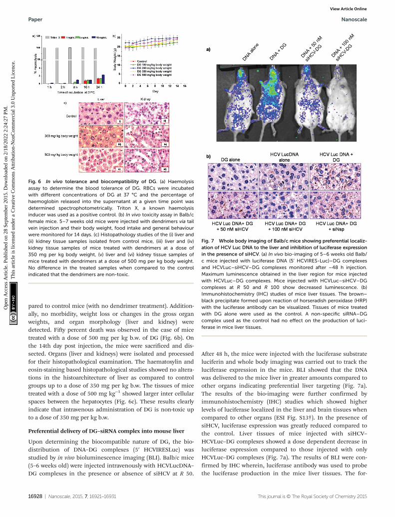
pared to control mice (with no dendrimer treatment). Addition-ally, no morbidity, weight loss or changes in the gross organweights, and organ morphology (liver and kidney) weredetected. Fifty percent death was observed in the case of micetreated with a dose of 500 mg per kg b.w. of DG (Fig. 6b). Onthe 14th day post injection, the mice were sacrificed and dis-sected. Organs (liver and kidneys) were isolated and processedfor their histopathological examination. The haematoxylin andeosin-staining based histopathological studies showed no altera-tions in the histoarchitecture of liver as compared to controlgroups up to a dose of 350 mg per kg b.w. The tissues of micetreated with a dose of 500 mg kg−1 showed larger inter cellularspaces between the hepatocytes (Fig. 6c). These results clearlyindicate that intravenous administration of DG is non-toxic upto a dose of 350 mg per kg b.w.
Preferential delivery of DG–siRNA complex into mouse liver
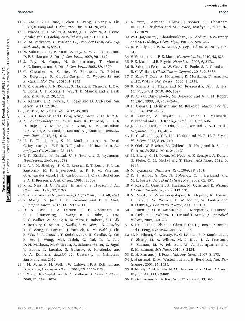
Upon determining the biocompatible nature of DG, the bio-distribution of DNA–DG complexes (5′ HCVIRESLuc) wasstudied by in vivo bioluminescence imaging (BLI). Balb/c mice(5–6 weeks old) were injected intravenously with HCVLucDNA–DG complexes in the presence or absence of siHCV at R 50.
After 48 h, the mice were injected with the luciferase substrateluciferin and whole body imaging was carried out to track theluciferase expression in the mice. BLI showed that the DNAwas delivered to the mice liver in greater amounts compared toother organs indicating preferential liver targeting (Fig. 7a).The results of the bio-imaging were further confirmed byimmunohistochemistry (IHC) studies which showed higherlevels of luciferase localized in the liver and brain tissues whencompared to other organs (ESI Fig. S13†). In the presence ofsiHCV, luciferase expression was greatly reduced compared tothe control. Liver tissues of mice injected with siHCV–HCVLuc–DG complexes showed a dose dependent decrease inluciferase expression compared to those injected with onlyHCVLuc–DG complexes (Fig. 7a). The results of BLI were con-firmed by IHC wherein, luciferase antibody was used to probethe luciferase production in the mice liver tissues. The for-
Fig. 6 In vivo tolerance and biocompatibility of DG. (a) Haemolysisassay to determine the blood tolerance of DG. RBCs were incubatedwith different concentrations of DG at 37 °C and the percentage ofhaemoglobin released into the supernatant at a given time point wasdetermined spectrophotometrically. Triton X, a known haemolysisinducer was used as a positive control. (b) In vivo toxicity assay in Balb/cfemale mice. 5–7 weeks old mice were injected with dendrimers via tailvein injection and their body weight, food intake and general behaviourwere monitored for 14 days. (c) Histopathology studies of the (i) liver and(ii) kidney tissue samples isolated from control mice, (iii) liver and (iv)kidney tissue samples of mice treated with dendrimers at a dose of350 mg per kg body weight, (v) liver and (vi) kidney tissue samples ofmice treated with dendrimers at a dose of 500 mg per kg body weight.No difference in the treated samples when compared to the controlindicated that the dendrimers are non-toxic.
Fig. 7 Whole body imaging of Balb/c mice showing preferential localiz-ation of HCV Luc DNA to the liver and inhibition of luciferase expressionin the presence of siHCV. (a) In vivo bio-imaging of 5–6 weeks old Balb/c mice injected with luciferase DNA (5’ HCVIRES-Luc)–DG complexesand HCVLuc–siHCV–DG complexes monitored after –48 h injection.Maximum luminescence obtained in the liver region for mice injectedwith HCVLuc–DG complexes. Mice injected with HCVLuc–siHCV–DGcomplexes at R 50 and R 100 show decreased luminescence. (b)Immunohistochemistry (IHC) studies of mice liver tissues. The brown–black precipitate formed upon reaction of horseradish peroxidase (HRP)with the luciferase antibody can be visualized. Tissues of mice treatedwith DG alone were used as the control. A non-specific siRNA–DGcomplex used as the control had no effect on the production of luci-ferase in mice liver tissues.
Paper Nanoscale
16928 | Nanoscale, 2015, 7, 16921–16931 This journal is © The Royal Society of Chemistry 2015
Ope
n A
cces
s A
rtic
le. P
ublis
hed
on 2
8 Se
ptem
ber
2015
. Dow
nloa
ded
on 2
/18/
2022
2:2
4:27
PM
. T
his
artic
le is
lice
nsed
und
er a
Cre
ativ
e C
omm
ons
Attr
ibut
ion-
Non
Com
mer
cial
3.0
Unp
orte
d L
icen
ce.
View Article Online

mation of a black–brown precipitate in tissues of mice treatedwith HCVLuc–DG complexes indicated successful delivery ofDNA to the liver (Fig. 7b). Tissues of mice treated with siHCV–HCVLuc–DG complexes showed a net decrease in luciferase pro-duction observed by the lower amounts of brown precipitatesformed. These results were consistent with that observed in thebioluminescence imaging assay. Further, the use of a non-specific siRNA in the DG–HCVLuc complex did not affect theluciferase production reiterating the specificity of siHCV to theHCV 5′ UTR. Mice injected with either DNA alone or DG alonewere used as controls to rule out background signals. Takentogether, the results clearly establish the potential use of DG fordelivery of therapeutic siRNA to inhibit HCV infection.
Discussion
Our study provides a ‘proof-of-concept’ approach for the use ofsiRNA technology for combating HCV infection based on anewly developed dendritic galactoside nanovector. Functionali-zation of the dendrimer with galactose units imparted theability of target-oriented delivery which is one of the key attri-butes to the successful action of the desired antiviral. Theformation of a stable complex with intact siRNA by the deliveryvector is essential to ensure crossing of cellular barriers andsuccessful delivery of siRNA at the target site to downregulategene expression.19 Gel retardation assay, zeta potentialmeasurements, ethidium bromide displacement assay andUV-Vis spectroscopy confirmed the formation of stable siRNA–DG complexes. Atomic force microscopy imaging showed thatmajority of DG particles had height profiles below 1 nm,similar to those observed in the case of PAMAM42 and PETIMdendrimers.21 The shape of siRNA and its non-aggregating be-haviour were consistent with the reports of siRNA AFMimaging.43,44 The morphologies of DG–siRNA complexes atdifferent weight ratios were visualized as dome shaped, spheri-cal and globular structures. The particle size distribution atR 50 was also monodispersed, possibly as a result of netneutral charge of the complex, as determined by the zetapotential measurements.
MD simulation showed that most of the galactose residuesare projected away from the siRNA whereas dendrimer nitro-gens (highlighted as blue spheres in Fig. 3a) are involved inelectrostatic complex formation with siRNA. The galactose resi-dues are free to interact with the asialoglycoprotein receptor(ASGPR) when these complexes are used for the transfection.The radius of gyration (Rg) values obtained from simulationstudies are in reasonable agreement with those obtained fromAFM imaging. For example, the z-height profile of the complexvaried between 1.5 and 3.25 nm which is in the same range asthe Rg value of the complex. The dendrimer structure was seento undergo a change becoming more compact and orientingitself such that it accommodates itself into the major groove.The constant value of Rg for siRNA indicates that siRNA main-tains its structural integrity in the complex. The analysis of theradial distribution function (RDF) of the dendrimer amines as
well as, galactose oxygens with respect to phosphate groups ofsiRNA showed complex formation between the protonated den-drimer and siRNA. This was further confirmed by the analysisof the change in the distance between the COM of dendrimerand siRNA with respect to simulation time.
Initial nucleic acid delivery experiments involved the trans-fection of fluorescent-tagged siRNA using DG at three ratios,R 25, R 50 and R 100. Comparison of these three ratios revealedthat the delivery at R 50 was more efficient. The use of low N/Pratios to mediate effective siRNA transfection was demon-strated recently45 and our observation was on similar lines tothat study. Due to the electroneutrality of the siRNA–DGcomplex at R 50, the entry inside the cell would occur preferen-tially by receptor-mediated endocytosis, involving the ASGPR–galactoside interaction. A comparison of transfection with anative hydroxyl functionalized dendrimer and lipofectamine,showed that DG preferentially localized in the perinuclearregion of liver cells with much higher fluorescence intensities.The z-stack imaging helped rule out surface adherence andascertain ASGPR mediated endocytosis in liver cells. ASGPRbelongs to the class of receptors that enter the cells duringendocytosis and deliver the bound ligand in the area sur-rounding the nucleus. Saunier and co-workers had earliershown co-localization of GFP-labelled ASGPR and red fluore-scence labelled galactoprotein which binds to ASGPR in theperinuclear region.41 These studies showed that ASGPR inter-nalized along with its bound ligand in the perinuclear regionsof the cell. The present study demonstrates that siRNA, deli-vered specifically to the liver cells by receptor mediated endo-cytosis, is localized in the perinuclear area.
The role of ASGPR in internalization was further validatedby the competition assay involving the pre-incubation of livercells with D-galactose or D-mannose. Ligand–receptor inter-actions are highly specific due to the ability of receptor torecognize subtle conformational differences of the ligandwhich alters the ligand-binding affinity.46 ASGPR specificallybinds to D-galactose over D-mannose by conformationalspecific hydrogen bonding interactions. Hence cells pre-incubated with D-galactose showed decreased siRNA uptakecompared to incubation with D-mannose.
Perinuclear localization of siRNA gains importance toinhibit HCV RNA replication, since it is known that the replica-tion occurs along the lipid droplets of the endoplasmic reticu-lum in the perinuclear region of hepatocytes.1,2 The viralproteins NS5B, the RNA dependent RNA polymerase (RdRp)and NS3 which exhibit RNA helicase activity are known to regu-late the viral replication.1 Both proteins are a part of the HCVreplication complex. We demonstrated here that siRNA taggedwith a fluorophore co-localizes with NS5B and NS3 proteins ofthe HCV virus replication complex. The proximity of siRNAlocalization at the site of viral RNA replication ensures theeffective action of siRNA, which was proven by deliveringsiHCV using DG in replicon harbouring cells and probing theviral RNA levels. The siRNA against HCV inhibits the replica-tion of viral mRNA through targeted binding to the 5′-UTR ofHCV RNA. The use of 5′ shDNA (short hairpin DNA), a precur-
Nanoscale Paper
This journal is © The Royal Society of Chemistry 2015 Nanoscale, 2015, 7, 16921–16931 | 16929
Ope
n A
cces
s A
rtic
le. P
ublis
hed
on 2
8 Se
ptem
ber
2015
. Dow
nloa
ded
on 2
/18/
2022
2:2
4:27
PM
. T
his
artic
le is
lice
nsed
und
er a
Cre
ativ
e C
omm
ons
Attr
ibut
ion-
Non
Com
mer
cial
3.0
Unp
orte
d L
icen
ce.
View Article Online

sor for the in situ generation of the siRNA to inhibit HCV IRESmediated translation and hence HCV RNA replication inhi-bition, was demonstrated earlier.14 In the present study, inhi-bition of HCV RNA up to ∼77% was obtained by the use ofsiRNA–DG delivery systems. The preferential perinuclear deliv-ery of siRNA in the vicinity of HCV virus replication site mightcontribute to the decrease in HCV RNA levels.
Dendrimer based delivery systems are injected intra-venously for in vivo studies, encountering blood in the firstinstance.38 In order to verify that intravenously injected com-pounds do not cause a haemolysis by rupturing the RBC mem-branes, a haemolytic assay was performed. The haemolysisinduced by DG was observed to be negligible when comparedto the dendritic vectors like PAMAM dendrimers, (60% haemo-lysis), PPI dendrimer (80% haemolysis) and polyethylenimine(PEI) based polymers (17–30% haemolysis).41,47–49 Theseresults showed that DG is safe to be injected intravenously.Further experiments in female Balb/c mice showed acute toler-ance dose for DG up to 350 mg per kg b.w. A comparison withreported studies on other dendritic systems47 shows DG to bemuch less toxic, since even a dose of 350 mg per kg b.w. is verywell tolerated by mice, which, to our knowledge, is perhaps themost non-toxic synthetic delivery vector in vivo reported so far.
In vivo whole body imaging showed a preferential biodistri-bution of the complexes to the liver. This observation was con-firmed further by immunohistochemistry (IHC) of the isolatedmice organs. Localization of large quantities of luciferaseenzymes in the liver when compared to other organs indicatedthe preferential targeting of DG to the mouse liver. Previousstudies using radiolabeled glycolipids and PAMAM dendrimersshowed that liver uptake of delivery vehicle 15 min to 1 h posti.v. injection was 60–90%. Minor or negligible uptake by otherorgans was also reported.49 The present study establishes thetargeted delivery and successful expression of a reporter geneusing a non-radiolabeled dendrimer, even after 84 h post injec-tion. Reports of siRNA being delivered to tumour cells in vivousing engineered PAMAM or PPI dendrimers showed only amodest success.50,51 Interestingly, luminescence signals wereobserved in the brain tissues which were further validated byIHC experiments. Crossing of the blood–brain barrier byhydroxyl-terminated PAMAM dendrimers was demonstratedrecently.52 Experiments are currently under progress to deci-pher the mechanism and specificity for delivery into neuronalcells. The use of siRNA in the complexes resulted in a netdecrease in the luciferase production probed by BLI, as well as,by IHC assays. The siRNA binds to the 5′-UTR and prevents theHCV IRES mediated translation of the subsequent luciferasegene, thereby reducing the luminescence signal, especially inthe liver. This resulted in the successful delivery of siHCV pre-ferentially to the liver cells by using DG.
Conclusion
RNAi technology and dendrimer based delivery are being usedas popular strategies to combat diseases, such as, cancer and
HIV.53–55 Significant progress has been made to implementthis technology to overcome HCV infection.54,56 Our resultsestablish that siRNA–DG is a biocompatible system for prefer-ential liver targeted delivery. The ability of DG to deliver siRNAin the perinuclear region near the HCV replication complexand observation of a significant decrease in JFH1 RNA levelsauthenticates the siRNA–DG complex as an efficient deliverysystem. Our study shows that DG as a targeted delivery vectorcan be used for the effective delivery of antivirals against HCV.The novel siRNA–DG nano-system appears to be a promisingoption for HCV related therapeutics.
Acknowledgements
Authors would like to thank Ralf Bartenschlager, CharlesM. Rice and Takaji Wakita for sharing plasmid constructs andcell lines. We thank the Central Animal Facility, Confocalimaging facility, Department of Microbiology and Cell Biologyand Centre for Nanoscience and Engineering (CeNSE), IISc.Current work was supported by grants from the Department ofBiotechnology, India to SD, NJ, AKS, PKM; and from the Scienceand Engineering Research Board, DST, India to BUR and NJ.AL and AK thank Council of Scientific and Industrial Research(CSIR), India for a research fellowship. SD also acknowledges aJ. C Bose fellowship from DST, India.
References
1 D. Moradpur, F. Penin and C. M. Rice, Nat. Rev. Microbiol.,2007, 5, 453.
2 Y. Ma, M. Anantpadma, J. M. Timpe, S. Shanmugam,S. M. Singh, S. M. Lemon and M. Yi, J. Virol., 2011, 85, 86.
3 S. Das, R. K. Shetty, A. Kumar, R. N. Shridharan,R. Tatineni, G. Chi, A. Mukherjee, S. Das, S. M. Subbaraoand A. A. Karande, PLoS One, 2013, 8, e53619.
4 C. Ji, Y. Liu, C. Pamulapati, S. Bohini, G. Fertig,M. Schraeml, W. Rubas, M. Brandt, S. Ries, H. Ma andK. Klumpp, Hepatology, 2015, 61, 1136.
5 A. K. Manna, A. Kumar, U. Ray, S. Das, G. Basu and S. Roy,Antiviral Res., 2013, 97, 223.
6 T. J. Liang and M. G. Ghany, N. Engl. J. Med., 2013, 368,1907.
7 R. E. Lanford, E. S. Hildebrandt-Eriksen, A. Petri,R. Persson, M. Lindow, M. E. Munk, S. Kauppinen andH. Ørum, Science, 2010, 327, 198.
8 P. J. Pockros and J. S. Au, Clin. Pharmacol. Ther., 2014, 95,78.
9 B. U. Reddy, R. Mullick, A. Kumar, G. Sudha, N. Srinivasanand S. Das, Sci. Rep., 2014, 24, 5411.
10 T. L. Foster, G. S. Thompson, A. P. Kalverda, J. Kankanala,M. Bentham, L. F. Wetherill, J. Thompson, A. M. Barker,D. Clarke, M. Noerenberg, A. R. Pearson, D. J. Rowlands,S. W. Homans, M. Harris, R. Foster and S. Griffin, Hepato-logy, 2014, 59, 408.
Paper Nanoscale
16930 | Nanoscale, 2015, 7, 16921–16931 This journal is © The Royal Society of Chemistry 2015
Ope
n A
cces
s A
rtic
le. P
ublis
hed
on 2
8 Se
ptem
ber
2015
. Dow
nloa
ded
on 2
/18/
2022
2:2
4:27
PM
. T
his
artic
le is
lice
nsed
und
er a
Cre
ativ
e C
omm
ons
Attr
ibut
ion-
Non
Com
mer
cial
3.0
Unp
orte
d L
icen
ce.
View Article Online
11 Y. Gao, X. Yu, B. Xue, F. Zhou, X. Wang, D. Yang, N. Liu,L. Xu, X. Fang and H. Zhu, PLoS One, 2014, 28, e90333.
12 E. Poveda, D. L. Wyles, A. Mena, J. D. Pedreira, A. Castro-Iglesias and E. Cachay, Antiviral Res., 2014, 108, 181.
13 M. M. Verstegen, Q. Pan and L. J. van der Laan, Adv. Exp.Med. Biol., 2015, 848, 1.
14 N. Subramanian, P. Mani, S. Roy, S. V. Gnanasundram,D. P. Sarkar and S. Das, J. Gen. Virol., 2009, 90, 1812.
15 S. Roy, N. Gupta, N. Subramanian, T. Mondal,A. C. Banerjea and S. Das, J. Gen. Virol., 2008, 89, 1579.
16 C. Chevalier, A. Saunier, Y. Benureau, D. Fléchet,D. Delgrange, F. Colbère-Garapin, C. Wychowski andA. Martin, Mol. Ther., 2013, 2, 1452.
17 P. K. Chandra, A. K. Kundu, S. Hazari, S. Chandra, L. Bao,T. Ooms, G. F. Morris, T. Wu, T. K. Mandal and S. Dash,Mol. Ther., 2012, 20, 1724.
18 R. Kanasty, J. R. Dorkin, A. Vegas and D. Anderson, Nat.Mater., 2013, 12, 967.
19 J.-P. Behr, Acc. Chem. Res., 2012, 45, 980.20 X. Liu, P. Rocchic and L. Peng, New J. Chem., 2012, 36, 256.21 A. Lakshminarayanan, V. K. Ravi, R. Tatineni, Y. B. R.
D. Rajesh, V. Maingi, K. S. Vasu, N. Madhusudhan,P. K. Maiti, A. K. Sood, S. Das and N. Jayaraman, Bioconju-gate Chem., 2013, 24, 1612.
22 U. P. Thankappan, S. N. Madhusudhana, A. Desai,G. Jayamurugan, Y. B. R. D. Rajesh and N. Jayaraman, Bio-conjugate Chem., 2011, 22, 115.
23 T. R. Krishna, M. Belwal, U. S. Tatu and N. Jayaraman,Tetrahedron, 2005, 61, 4281.
24 L. A. J. M. Sliedregt, P. C. N. Rensen, E. T. Rump, P. J. vanSantbrink, M. K. Bijsterbosch, A. R. P. M. Valentijn,G. A. van der Marel, J. H. van Boom, T. J. C. van Berkel andE. A. L. Biessen, J. Med. Chem., 1999, 42, 609.
25 R. K. Ness, H. G. Fletcher Jr. and C. S. Hudson, J. Am.Chem. Soc., 1950, 72, 2200.
26 T. R. Krishna and N. Jayaraman, J. Org. Chem., 2003, 68, 9694.27 V. Maingi, V. Jain, P. V. Bharatam and P. K. Maiti,
J. Comput. Chem., 2012, 33, 1997–2011.28 D. A. Case, T. A. Darden, T. E. Cheatham III,
C. L. Simmerling, J. Wang, R. E. Duke, R. Luo,R. C. Walker, W. Zhang, K. M. Merz, B. Roberts, S. Hayik,A. Roitberg, G. Seabra, J. Swails, A. W. Götz, I. Kolossváry,K. F. Wong, F. Paesani, J. Vanicek, R. M. Wolf, J. Liu,X. Wu, S. R. Brozell, T. Steinbrecher, H. Gohlke, Q. Cai,X. Ye, J. Wang, M.-J. Hsieh, G. Cui, D. R. Roe,D. H. Mathews, M. G. Seetin, R. Salomon-Ferrer, C. Sagui,V. Babin, T. Luchko, S. Gusarov, A. Kovalenko andP. A. Kollman, AMBER 12, University of California,San Francisco, 2012.
29 J. M. Wang, R. M. Wolf, J. W. Caldwell, P. A. Kollman andD. A. Case, J. Comput. Chem., 2004, 25, 1157–1174.
30 J. Wang, P. Cieplak and P. A. Kollman, J. Comput. Chem.,2000, 21, 1049–1074.
31 A. Perez, I. Marchan, D. Svozil, J. Sponer, T. E. CheathamIII, C. A. Laughton and M. Orozco, Biophys. J., 2007, 92,3817–3829.
32 W. L. Jorgensen, J. Chandrasekhar, J. D. Madura, R. W. Impeyand M. L. Klein, J. Chem. Phys., 1983, 79, 926–935.
33 B. Nandy and P. K. Maiti, J. Phys. Chem. B, 2011, 115,217.
34 V. Vasumati and P. K. Maiti, Macromolecules, 2010, 43, 8264.35 P. K. Maiti and B. Bagchi, Nano Lett., 2006, 6, 2478.36 R. Salomon-Ferrer, A. W. Goetz, D. Poole, S. L. Grand and
R. C. Walker, J. Chem. Theory Comput., 2013, 9, 3878.37 T. Kato, T. Date, A. Murayama, K. Morikawa, D. Akazawa
and T. Wakita, Nat. Protoc., 2006, 1, 2334.38 B. Klajnert, S. Pikala and M. Bryszewska, Proc. R. Soc.
London, Ser. A, 2010, 466, 1527.39 R. C. van Duijvenbode, M. Borkovec and G. J. M. Koper,
Polymer, 1998, 39, 2657–2664.40 D. Cakara, J. Kleimann and M. Borkovec, Macromolecules,
2003, 36, 4201–4207.41 B. Saunier, M. Triyatni, L. Ulianich, P. Maruvada,
P. Yenand and L. D. Kohn, J. Virol., 2003, 77, 546.42 J. Li, L. T. Piehler, D. Qin, J. R. Baker and D. A. Tomalia,
Langmuir, 2000, 16, 5613.43 H. G. Abdelhady, Y.-L. Lin, H. Sun and M. E. H. El-Sayed,
PLoS One, 2013, 8, e61710.44 P. Ofek, W. Fischer, M. Calderón, R. Haag and R. Satchi-
Fainaro, FASEB J., 2010, 24, 3122.45 M. Zheng, G. M. Pavan, M. Neeb, A. K. Schaper, A. Danai,
G. Klebe, O. M. Merkel and T. Kissel, ACS Nano, 2012, 6,9447.
46 N. Jayaraman, Chem. Soc. Rev., 2009, 38, 3463.47 K. L. Allion, Y. Xie, N. El-Gendy, C. J. Berkland and
M. L. Forrest, Adv. Drug Delivery Rev., 2009, 61, 457.48 V. Russ, M. Gunther, A. Halama, M. Ogris and E. Wnage,
J. Controlled Release, 2008, 132, 131.49 N. Malik, R. Wiwattanapatapee, R. Klopsch, K. Lorenz,
H. Frey, J. W. Weener, E. W. Meijer, W. Paulus andR. Duncan, J. Controlled Release, 2000, 65, 133.
50 O. Taratula, O. B. Garbuzenko, P. Kirkpatrick, I. Pandya,R. Savla, V. P. Pozharov, H. He and T. Minko, J. ControlledRelease, 2009, 140, 284.
51 X. Liu, C. Liu, J. Zhou, C. Chen, F. Qu, J. J. Rossi, P. Rocchiand L. Peng, Nanoscale, 2015, 7, 3867.
52 M. K. Mishra, C. A. Beaty, W. G. Lesniak, S. P. Kambhapati,F. Zhang, M. A. Wilson, M. E. Blue, J. C. Troncoso,S. Kannan, M. V. Johnston, W. A. Baumgartner andR. M. Kannan, ACS Nano, 2014, 8, 2134.
53 D. H. Kim and J. J. Rossi, Nat. Rev. Genet., 2007, 8, 173.54 J. Haasnoot, E. M. Westerhout and B. Berkhout, Nat. Bio-
technol., 2007, 25, 1435.55 B. Nandy, D. H. Bindu, N. M. Dixit and P. K. Maiti, J. Chem.
Phys., 2013, 139, 024905.56 D. Grimm and M. A. Kay, Gene Ther., 2006, 13, 563.
Nanoscale Paper
This journal is © The Royal Society of Chemistry 2015 Nanoscale, 2015, 7, 16921–16931 | 16931
Ope
n A
cces
s A
rtic
le. P
ublis
hed
on 2
8 Se
ptem
ber
2015
. Dow
nloa
ded
on 2
/18/
2022
2:2
4:27
PM
. T
his
artic
le is
lice
nsed
und
er a
Cre
ativ
e C
omm
ons
Attr
ibut
ion-
Non
Com
mer
cial
3.0
Unp
orte
d L
icen
ce.
View Article Online