a neurologist and ataxia: using eye movements to learn
TRANSCRIPT

REVIEW Open Access
A neurologist and ataxia: using eyemovements to learn about the cerebellumDavid S. Zee
Abstract
The cerebellum, its normal functions and its diseases, and especially its relation to the control of eye movements, hasbeen at the heart of my academic career. Here I review how this came about, with an emphasis on epiphanies,“tipping points” and the influences of mentors, colleagues and trainees. I set a path for young academicians, bothclinicians and basic scientists, with some guidelines for developing a productive and rewarding career in neuroscience.
Keywords: Cerebellum, Ataxia, Eye movements
BackgroundThe normal functions of the cerebellum and its diseaseshas been at the heart of my academic career of morethan 45 years – both in clinical care of patients, and inclinical and experimental research. More than 85 of mypublications have the word cerebellum in the title, or thecerebellum is central to the problems discussed in thepublication (see Additional files 1 and 2). Most of thesepublications emphasize some aspect of the relationshipof the cerebellum to the control of eye movements, in-cluding all its subtypes, vestibular, saccade, pursuit andvergence. The visual symptoms from the disturbed ocu-lar motor control in cerebellar patients are often ex-tremely disabling and life altering, for example, doublevision due to ocular misalignment, and oscillopsia dueto nystagmus or other unwanted ocular oscillations. Thishas been one reason for my sustained focus, over somany years, on this relatively small, but vital part of thebrain. My interests in the cerebellum followed upon aseries of epiphanies, based on people – patients, physi-cians and scientists – with whom I came in contact; onthe times; and on chance and good fortune. At everystep of the way, I reached a “tipping point” that pushedme in a new direction or to a particular person who be-came an influential mentor, colleague, or trainee. Here Irecapitulate some of this story and based on my experi-ence suggest some “tips” for success (Table 1), which I
hope will help those early in their careers as they makedecisions about how their academic lives are to unfold.
Why I chose neuroscience“Keep an eye out for something new and exciting tostudy”. In 1965, I began medical school classwork atJohns Hopkins in neuroanatomy and immediately be-came enthralled with the brain, marveling at its exquisiteconnectivity. Later in the same year, I was able to watchProfessor Vernon Mountcastle, chair of physiology andan eminent neurophysiologist especially for his discoveryof the columnar architecture of somatosensory cerebralcortex, perform experiments in his laboratory. He wasrecording the activity in individual nerve fibers ofexperimental animals in response to different sensorystimuli. The ability to “see” how neural activity in thebrain encodes experiences from the outside world wasan epiphany for me and further piqued my interest in acareer in neuroscience. In 1966, after my first year ofmedical school, I opted for a summer elective with thechair of the anatomy department, Professor DavidBodian, well known for his seminal studies on the patho-genesis of poliomyelitis, which made possible the devel-opment of the polio vaccine. He also developed the“Bodian” silver stain for identifying nerve fibers andnerve endings in neuroanatomical sections. That sum-mer we spent many hours together at the microscope,examining the upper cervical spinal cord trying to de-cipher propriospinal pathways. Nowadays how oftendoes a departmental chair have even a small amount oftime, let alone almost daily sessions, to spend with a first
Correspondence: [email protected] of Neurology, Ophthalamology, Otolaryngology-Head and NeckSurgery, and Neuroscience, The Johns Hopkins University School ofMedicine, The Johns Hopkins Hospital, Path 2-210, Baltimore, MD 21287, USA
© The Author(s). 2018 Open Access This article is distributed under the terms of the Creative Commons Attribution 4.0International License (http://creativecommons.org/licenses/by/4.0/), which permits unrestricted use, distribution, andreproduction in any medium, provided you give appropriate credit to the original author(s) and the source, provide a link tothe Creative Commons license, and indicate if changes were made. The Creative Commons Public Domain Dedication waiver(http://creativecommons.org/publicdomain/zero/1.0/) applies to the data made available in this article, unless otherwise stated.
Zee Cerebellum & Ataxias (2018) 5:2 DOI 10.1186/s40673-018-0081-2
year medical student on an elective in the laboratory?My fascination with the anatomical and physiologicalorganization of the brain continued throughout medicalschool so that on our own time, a classmate, TomWoolsey, who was in a similar state of anatomical“ecstasy” and I dissected a gross brain specimen. Wewere trying to envision in three dimensions the compli-cated relationships among the fluid spaces and fissuresof the brain. Tom ultimately achieved considerable famefor his discovery, while still a medical student, of the“barrel” organization of the projections of the whiskers(vibrissa) in the cerebral cortex of the rat.
Why I chose neurologyWhen it came time to choose a clinical specialty,neurology was the natural choice. Again, an experience(another summer elective, this time at the Mayo Clinicin neurology in 1968), and an exposure to some of thegiants of clinical neurology there (Dr. Frank Howard ofmyasthenia gravis fame, and Drs. Thomas Kearns andRobert Hollenhorst of neuroophthalmolgy fame) madeneurology an inevitable decision. My interests in thecerebellum were also stirred at the Mayo Clinic whenone of the patients assigned to me was being evaluatedfor a chronic cerebellar ataxia. I was told to look up aclassic paper on cerebellar degeneration in alcoholics byMaurice Victor and colleagues entitled “A restrictedform of cerebellar cortical degeneration occurring inalcoholic patients”, which was 109 pages long [1]. I
confess I did not read this paper from start to finish butthe ability to correlate function and anatomy using theclinical examination and subsequent pathology was a“tipping point” that pushed me towards neurology andeventually the cerebellum. This experience also empha-sized to me the importance of reading and knowing themedical literature. “Know, but not necessarily accept, whathas been said, written and accomplished in the past.”
Why I chose neuroophthalmologyAll medical students visiting the Mayo Clinic for thesummer elective program were obliged to take a week ofneuroophthalmology. At that time, I came across theclassic textbook, “The Neurology of the Ocular Muscles”by David Cogan, the eminent neuroophthalmologist andChair of Ophthalmology at the Harvard Medical School.About 6 years later, in 1974-75, while I was serving inthe Public Health Service at the National Institutes ofHealth in Bethesda, by chance my small cubicle was nextdoor to the office of Dr. Cogan. He had relocated to theNational Eye Institute in Bethesda after retiring fromHarvard. Dr. Cogan took me under his wing and sent meto my first international conference (in Stockholm in1975) simply as an observer because he thought it wouldbe “good for me”. The other major individual who stirredmy interest in neuroophthalmology was Dr. Frank Walshat Johns Hopkins. As a neurology resident at Hopkins(1970-1973), I attended Dr. Walsh’s Saturday morningneuroophthalmology conferences and he, like Dr. Cogan,took considerable interest in my career. He sent me to aninternational colloquium on the pupil in Detroit so that Imight gain more exposure in the field. Dr. Walsh told methat someone (even a lowly neurology resident) shouldrepresent the Wilmer Eye Institute. I have never forgottenthe generosity and interest in my early career of these twogiants. One important caveat. Take your mentor’s sugges-tions seriously. Dr. Cogan and I were evaluating a patientwith slow saccades and he suggested ocular electromyog-raphy might help. He asked if I would be the controlsubject. I thought he was joking but about 45 min later, Iwas lying on a table with a huge needle in my lateralrectus (in those days ocular electromyographic needleswere large and foreboding). A functional MRI would haveshown my entire brain, in some sort of limbic seizure,lighting up as I watched Dr. Cogan approach my eye withneedle in hand. I can at least report that the experiencewas more frightening than painful.
Why I chose eye movementsAlmost every neurology resident at some time duringhis or her training becomes infatuated with neu-roophthalmolgy. Examining the eyes is perhaps the mostfascinating part of the neurological evaluation, makingthe performance of the brain easily accessible to simple
Table 1 Ten Tips For Academic Success
• Keep an eye out for something new, exciting, and important to study.
• Interact and collaborate with colleagues and trainees who have skillsyou do not or see or do things differently than you. Look foranalogies to see how problems have been solved in other fields.
• Listen, more than talk.
• Pick a mentor who, at any level of career, is looking to the futureand striving to be at the forefront of the field. Joining a newenterprise at its inception under a young, inspiring leader, is often asgood or even better option than joining a large, establishedenterprise under an older but busy entrenched leader.
• Know, but not necessarily accept, what has been said, written andaccomplished in the past.
• Persevere but be willing to change course when you should changecourse; be focused but flexible.
• Make your research quantitative and hypothesis driven, and whenthings look like they fit, try to prove your hypotheses wrong!
• Learn how to teach effectively and how to write concisely. Getfeedback from mentors and students.
• Broaden your horizons. Meet colleagues and students from othercountries and cultures. You gain much from collaborating, teachingand learning with them, and establishing enduring friendships.Take sabbaticals.
• If you are a clinician, learn from your patients, take physiology andanatomy to the bedside, figure out how the brain works and writepapers, or even a book to educate your colleagues.
Zee Cerebellum & Ataxias (2018) 5:2 Page 2 of 9

visual inspection using only a penlight, ophthalmoscopeand a target for the patient to fix upon or track. Thefindings on the neuroophthalmologic examination arecommonly the key to localizing lesions in many parts ofthe brain and especially in the brainstem and cerebel-lum. As a second year resident, I attended an introduc-tory lecture for neurology residents on eye movementsgiven by David A. Robinson, a bioengineer and ocularmotor physiologist, working in the Wilmer Eye Institute.His topic was the pathophysiology of internuclear oph-thalmoplegia (INO), a common ocular motor disorder ofthe brainstem in which the medial longitudinal fascic-ulus (MLF), which conveys information to the oculo-motor nuclei, is interrupted. He used a simple controlsystems approach to the signal processing needed togenerate normal eye movements, and then derived whathappens when there is an interruption in the flow ofinformation in the MLF. This remarkable exposition ledto an immediate epiphany. Applying simple mathematicsto understanding a complex pattern of pathological eyemovements, and being able to pinpoint the location ofthe defect in the processing of information by the brain,tipped me forever into normal and pathological ocularmotor control.After the lecture, I asked Dave Robinson if I could
work with him during my elective time in the last yearof my residency. He immediately accepted, saying, “Ihave been waiting for years for a neurologist to come byand work with me”. Asking Dave Robinson to be myscientific mentor was a key point in my career as he hadrealized early on how much we could learn about thefunctioning of the normal brain from examining patientswho suffered the unfortunate accidents and diseases ofnature. “Pick a mentor who, at any level of career, islooking to the future and striving to be at the forefrontof the field”. After I joined his laboratory, we beganweekly hospital rounds in which Dave and his graduatestudents and post-doctoral fellows, and our clinicalgroup, including residents and medical students, wouldgo to the bedside of a patient who had a challengingocular motor problem. We examined the patient to-gether, and afterwards discussed the mechanism, whatnew questions to ask, and what experiments could an-swer them. Publications often grew from these bedsideconversations, usually with us challenging Dave to makea model [2]. This experience emphasized for me theimportance of interacting with people who came fromdifferent fields, with different scientifc and clinical back-grounds and expertise. “Interact and collaborate withcolleagues and trainees who have skills you do not orsee or do things differently than you”.When I joined the laboratory in 1972 Dave’s first job
was teach me control systems using eye movements asthe model. We met several times each week, for an hour
or so, one on one. These sessions often involved home-work problems for me. Dave and I also sat down at theanalog computer together to test our ideas (Fig. 1). Thisteaching tutorial began with an analysis of signal pro-cessing in the vestibulo-ocular reflex (VOR). When thehead moves the brain must program an eye movementthat is exactly compensatory for us to see clearly whenwe walk or turn our head. In another epiphany, I real-ized that understanding the vestibular system – beingthe fundamental evolutionary scaffolding upon which allthe subtypes of eye movements developed – was key forme to becoming an ocular motor clinician-scientist.The most important projects in Dave’s laboratory at
that time related to the function of the cerebellum in thecontrol of the VOR. He was studying how the brainmaintains the correct timing (phase) of the VOR, bothadaptively in the long-term and in its immediate onlinecontrol. These experiments led to the idea of a cerebellarocular motor “repair shop”, compensating when the ocu-lar motor control system goes awry [3]. Another key con-cept from these experiments emerged that became afundamental building block in ocular motor physiology –the idea of an ocular motor integrator, not only to insurethat the phase of the VOR was correct, but also to hold
Fig. 1 Analog computer in which our first simulations of downbeatnystagmus were made in 1973. Differentiators, integrators and pulsegenerators were simulated with capacitors, resisters, amplifiers andone-shot multivibrators
Zee Cerebellum & Ataxias (2018) 5:2 Page 3 of 9

the eyes still after the eyes finished moving [4]. Gaze-evoked nystagmus, a common sign of cerebellar dysfunc-tion, could then be interpreted as a disorder in a neuralnetwork that mathematically integrates a velocity (move)command into a position (holding) command. More re-cently, this concept of mathematical neural integrators hasbeen applied to the control of the head and other parts ofthe body by my colleagues Aasef Shaikh and Reza Shad-mehr and their collaborators [5, 6]. “Look for analogies tosee how problems have been solved in other fields”.This exciting research in Dave Robinson’s laboratory kin-
dled my interest in both the vestibular system and the cere-bellum. Shortly after I started working in the laboratory, thechief of my department, Dr. Guy Mckhann, referred to meseveral patients with a persistent spontaneous downbeatingnystagmus as part of a clinical cerebellar syndrome. GuyMcKhann was the new and young chair of a just establisheddepartment of Neurology at Johns Hopkins. He alwayslooked out for and referred patients with clinical problemsthat his young trainees might profitably investigate. Guyand I also began a therapeutic drug trial in a group of ataxiapatients which was perhaps one of the earliest such trials incerebellar patients. Unfortunately, the medication was nothelpful. Two key paths for my research followed from theinvestigation of these patients: 1) using control systemsmodels to interpret abnormal eye movements, and 2) devel-oping an animal model in monkeys of the effects of experi-mental lesions of different parts of the cerebellum on eyemovements. First, with Dave Robinson, using the analogcomputer (Fig. 1) we made a control systems model ofdownbeat nystagmus. This was one of the first neurologicaldisorders studied and interpreted in this way [7]. This led tomy first scientific presentation, at the Association forResearch in Vision and Ophthalmology (ARVO) meeting in1973. Moreover, once we started modeling the disorder werealized we had to know more about the function of thevertical VOR. I realized we could engage and measure thevertical VOR simply by rotating a subject in a vestibularchair around an earth vertical axis with the head tilted 90degrees to one side to stimulate the vertical semicircular ca-nals. Not a great scientific discovery to be sure, but probablyhad never been performed on a patient before. The messagehere, of course, is that mathematical models allow you totest your hypotheses rigorously and suggest new quantita-tive experiments to challenge your hypotheses. “Make yourresearch quantitative and hypothesis driven, and whenthings look like they fit, try to prove your hypotheseswrong!” This same approach led to seminal models of thecontrol of saccades and the pathogenesis of various formsof oscillations and nystagmus, which we will discuss later.
Why I chose the cerebellumOur studies with downbeat nystagmus pointed to a largegap in knowledge about how the cerebellum works and
how cerebellar disease is manifest. The intricate connec-tions of the cerebellum to the brain stem (and now thethalamus and even cerebral cortex) always hung over thequestion of what is a cerebellar eye sign. “Keep an eyeout for something new, exciting, and important tostudy”. We needed an animal model to study the effectsof lesions in the cerebellum on eye movements. Withthe advent of Robinson’s search coil technique allowingthe accurate recording of eye movements, and usingmonkeys that I could train to fix upon and follow tar-gets, I hoped to make progress toward delineating acerebellar ocular motor syndrome. Over the next quartercentury, we recorded and analyzed eye movements inmonkeys before and after focal cerebellar lesions includ-ing the flocculus and paraflocculus (tonsil), the dorsalvermis, and the nodulus [8–15]. My long-standing col-leagues at Johns Hopkins, Mark Walker, Richard Lewisand Rafael Tamargo played key roles in these experi-ments. These studies enhanced our clinical diagnosticacumen and our ability to infer what might be the func-tions of different parts of the cerebellum. At the sametime, we carefully quantified eye movements in patientswho had naturally occurring dysfunction of the cerebel-lum and compared their findings with our experimentalresults (e.g., [16]). We used a version of the search-coiltechnique for humans to measure eye movements(Fig. 2), and control systems techniques to analyze thedata. In a true model of translational research, we wentback and forth, iteratively, between studies in patientsand in experimental animals, to learn what the cerebel-lum does and how we might better localize and diagnose
Fig. 2 The magnetic field search coil technique applied to humansubjects. David A Robinson (right) inserting a small scleral annulusthat was developed by Han Collewijn from The Netherlands toprecisely measure eye movements, with David Zee (middle) lookingon. Circa 1980s
Zee Cerebellum & Ataxias (2018) 5:2 Page 4 of 9

lesions of the cerebellum in our patients. We constantlykept in mind the Robinsonian approach; careful meas-urement, quantitative analysis, hypothesis testing andanalytical modelling but always with the patient at theback of our minds, both to improve their lot and to dis-cover what they can teach us about how the brainworks.An exemplar of this approach was developing a model
for the premotor circuits that generate saccade com-mands. We based our ideas on a single patient whomade slow saccades as part of a spinocerebellar degener-ation [17]. Her saccades were slow because of degener-ation in the premotor saccade “burst” neurons withinthe pontine paramedian reticular formation. Her sloweye movements provided us with the opportunity to seeif saccades were preprogrammed and ballistic, as was theconventional wisdom in the 1970s. I reasoned that byjumping the target while a slow saccade was in flight, wecould test the idea of preprogramming by seeing if ourpatient could change the course or direction of her sac-cades in midflight. Indeed, when the target jumped backto the starting position after she began the saccade, hereyes turned around without stopping and returned tothe starting position. If the target jumped onwards inmidflight as her eyes began slowing down, her eyeswould again pick up speed in response to the new targetlocation and eventually reach the target in just onemovement. These results suggested her saccades wereunder a type of internal feedback control. This “localfeedback model” with only slight modifications has stoodthe test of time for how the brain generates normal sac-cades. Moreover, this model has been an impetus tomany current ideas about how the cerebellum and otherstructures optimize control of movements, for both im-mediate online adjustments of motor performance andlong-term adaptive motor learning. Furthermore, thismodel can simulate certain saccadic oscillations such asocular flutter – intrusive, uncalled for, and often dra-matic, back-to-back saccades [18].Another example of the control systems approach to
ocular motor disorders was a study by John Leigh, DaveRobinson, and me of a patient with a cerebellar lesioncausing periodic alternating nystagmus (PAN), a disorderin which a spontaneous nystagmus alternates directionevery 2 min [19]. It was early on a Saturday morning inthe basement of the Wilmer Eye Institute, that Dave,John and I were recording the eye movements of thispatient. The idea was to test the then current model ofprocessing of information in the VOR to see how PANmight arise. A key test of the model was how one mightstop the nystagmus and John and Dave had come up withsome predictions. Accordingly, we measured the patient’snystagmus that morning and then Dave—working furi-ously with a paper and pencil— came up with an
amplitude and a duration of a rotational vestibular stimu-lus that if delivered in the right part of the patient’s nystag-mus cycle would —the model predicted—stop thenystagmus. We tried it—it worked—and the patient wasecstatic. Her visual blurring from the nystagmus was re-lieved, albeit for only about 10 min, for the first time inmany years! Experiments in animals a few years latershowed that a loss of function of Purkinje cells in the cere-bellar nodulus was the cause of PAN because of disinhib-ition and consequent instability of a central “velocity-storage” mechanism within the vestibular nuclei [20].Fortunately, shortly after we saw our patient, and
somewhat serendipitously after a casual discussion withcolleagues from the United Kingdom at an ARVO meet-ing, we reported that baclofen, a GABA-like drug, couldpermanently stop her nystagmus [21]. Baclofen was asurrogate for the missing GABA-mediated inhibitionfrom the nodulus on the vestibular nuclei. This was thefirst example of a medication that could completely stopa persistent pathological nystagmus! This successful out-come arising from a fortuitous interaction at a scientificmeeting emphasizes the importance of “broadening yourhorizons” by interacting with colleagues from afar. Thiscase also illustrates the power of the control systems ap-proach to clinical problems and, in this day of high tech-nology, the importance of imaginative thinking with thehelp of just a paper and a pencil, especially when theyare in the hands of someone like David Robinson.There are many other examples of how studying the
cerebellum and cerebellar patients has revealed muchabout how the brain works, and how we can better diag-nose and treat patients with cerebellar diseases. The firstdescriptions of an unstable neural integrator came fromstudies of animals with experimental lesions in the floc-culus, and in a patient with a paraneoplastic cerebellardegeneration [8, 22]. Recent studies in patients withacute strokes who had lesions isolated to the flocculusor to the paraflocculus (tonsil) have allowed us to pin-point a role for these particular structures in the finegrain control of eye movements and the VOR [23–25].These studies have led my close colleagues, DavidNewman-Toker, Jorge Katah and Ji-Soo Kim, and theircollaborators, to develop better and critically neededalgorithms for diagnosing patients with strokes in thebrainstem and cerebellum [23–26]. Quantification of theVOR can be an important biomarker of progression ofsome forms of cerebellar disease and potentially amarker of response to treatment [27, 28]. Correlations ofocular motor behavior with findings on functional andstructural imaging of the cerebellum have been a boonto our knowledge of the behaviors in which the cerebel-lum is involved [29]. Studies of patients with a curiousneurological disorder (ocular-palatal tremor syndrome)associated with hypertrophy and degeneration of the
Zee Cerebellum & Ataxias (2018) 5:2 Page 5 of 9
inferior olive, have given insights into what happenswhen the cerebellum attempts to compensate for motordysfunction with feedback about motor performance thatis inaccurate. With the cerebellum playing a centralrole in the adaptive responses of the brain to diseaseand trauma, a knowledge of how the cerebellum pro-motes compensation for lesions elsewhere in the brainbecomes a key pillar for developing better physicaltherapy programs for rehabilitation of brain-damagedpatients [30–32].With an almost daily increase in the knowledge of the
genetics of cerebellar disease, ocular motor function isoften the cornerstone of phenotypic classification anddifferential diagnosis (e.g., [33]). Particularly satisfyinghas been the identification of the genetic defect in twogroups of patients that we studied in the 1970s. First, alarge pedigree of patients with a late-onset, isolated cere-bellar degeneration eventually turned out to have spino-cerebellar ataxia type 6 (SCA6) with an abnormality inthe calcium channel on chromosome 21 [16, 34]. I havefollowed four generations in one family with this syn-drome. Secondly, the patients with slow saccades whowere the basis of our local feedback model of control ofsaccades, turned out to have spinocerebellar ataxia type2 (SCA2) with an abnormality on chromosome 12(ATXN2 gene). In the last decade, my interests in thecerebellum led me to be a co-founding member of themultidisciplinary Johns Hopkins ataxia clinic generouslysupported by the Macklin Foundation. Patients come fora complete evaluation and management of their ataxia; aneurologist, geneticist, physical and occupational thera-pists, social worker, etc., all see the patient in the clinicon the same day to deliver expert, comprehensive andefficient clinical care.
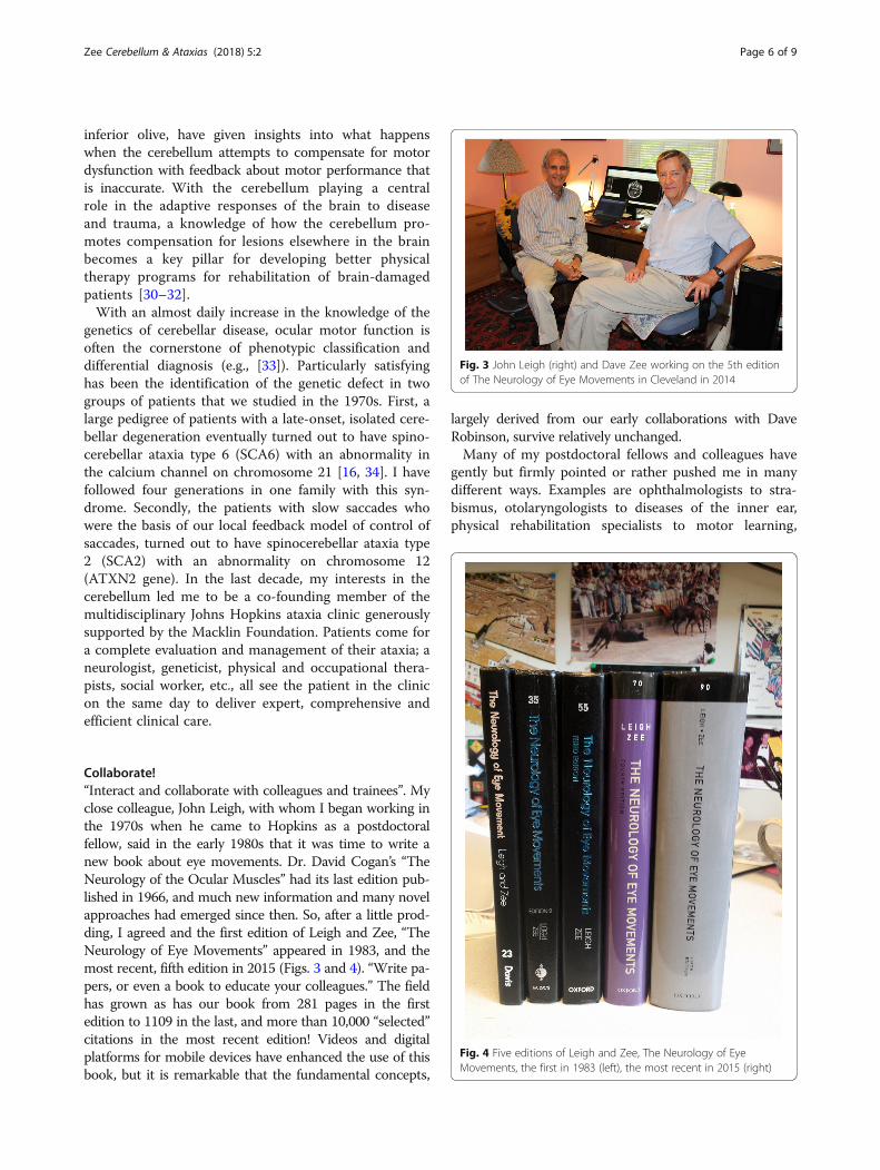
Collaborate!“Interact and collaborate with colleagues and trainees”. Myclose colleague, John Leigh, with whom I began working inthe 1970s when he came to Hopkins as a postdoctoralfellow, said in the early 1980s that it was time to write anew book about eye movements. Dr. David Cogan’s “TheNeurology of the Ocular Muscles” had its last edition pub-lished in 1966, and much new information and many novelapproaches had emerged since then. So, after a little prod-ding, I agreed and the first edition of Leigh and Zee, “TheNeurology of Eye Movements” appeared in 1983, and themost recent, fifth edition in 2015 (Figs. 3 and 4). “Write pa-pers, or even a book to educate your colleagues.” The fieldhas grown as has our book from 281 pages in the firstedition to 1109 in the last, and more than 10,000 “selected”citations in the most recent edition! Videos and digitalplatforms for mobile devices have enhanced the use of thisbook, but it is remarkable that the fundamental concepts,
largely derived from our early collaborations with DaveRobinson, survive relatively unchanged.Many of my postdoctoral fellows and colleagues have
gently but firmly pointed or rather pushed me in manydifferent ways. Examples are ophthalmologists to stra-bismus, otolaryngologists to diseases of the inner ear,physical rehabilitation specialists to motor learning,
Fig. 3 John Leigh (right) and Dave Zee working on the 5th editionof The Neurology of Eye Movements in Cleveland in 2014
Fig. 4 Five editions of Leigh and Zee, The Neurology of EyeMovements, the first in 1983 (left), the most recent in 2015 (right)
Zee Cerebellum & Ataxias (2018) 5:2 Page 6 of 9

adaptation and compensation, movement disorder neu-rologists to dystonia and tremor, bioengineers to modelsof nystagmus and other oscillations, and brilliant stu-dents who just brought their native intelligence and curi-osity to the lab and clinic and raised important andoften discomforting questions. Collaboration, free ex-change of information, and coming out of your silo tosee what else is around is at the core of scientific pro-gress (e.g., [21]). Three individual sabbatical years at theNational Eye Institute in Bethesda, working with LanceOptican, Ed Fitzgibbon, Christian Quaia and other col-leagues in the laboratory of sensorimotor research (LSR),led to fruitful publications and changes in my researchpriorities [35–42]. Several different summers in which Ispent a month at the University of Zurich in the laboratoryof Dominik Straumann to revitalize my thinking, were vitalto me for receiving a continuous RO1 individual researchgrant for 36 years. “Take sabbaticals” and “Persevere but bewilling to change course when you should change course”.My own current research focus is on how magnetic fields
stimulate the labyrinth and produce nystagmus, and whatthis tells us about how the brain adapts to vestibular disor-ders [43, 44]. This new area of research for me grew froma casual conversation with an Italian neuro-otologist,Vincenzo Marcelli, on one of many visits to the Universityof Siena where I have had a long-standing collaborationwith Professor Daniele Nuti (Fig. 5). “Broaden your hori-zons. Meet colleagues and students from other countriesand cultures”. One of the most rewarding aspects of mycareer has been collaborations, teaching and visits withclose colleagues in countries all over the world. I have also
been fortunate to collaborate for years with wonderfulscientists and clinicians at Johns Hopkins: in biomedicalengineering, ophthalmology, otolaryngology, neurologyand neuroscience. Collegiality is a core tenet of theHopkins experience.
Why teach?“Learn how to teach effectively and how to write concisely.Get feedback from mentors and students”. Working withstudents and trainees is also a pillar of scientific progressand personal satisfaction. As Sir William Osler emphasized,conveying new knowledge and stimulating people to learnabout or jump into one’s field is perhaps the most funda-mental and gratifying contribution that most of us can makein our academic lives. Students have forced me to learnabout something new or to do something differently, oropened up a new way of thinking about a problem. Youknow you have succeeded as a teacher when you are learn-ing more from your trainees than they from you. Teachingprods us to examine our frequently superficial understand-ing of key issues and concepts. When things are murky,teaching pushes us back to the drawing board. Every timewe teach, we learn is not a hackneyed phrase but a genuineacknowledgement of a mainstay of academic life. Teachingallows us to disseminate knowledge to many scientists orphysicians at once, and in the case of clinical audiences toaffect the medical care immediately of hundreds or eventhousands of patients. Perhaps one of the most importantrecent applications of our studies of the effects of cerebellarlesions on eye movements has been the development of al-gorithms to distinguish stroke in the cerebellum or brain-stem from benign afflictions of the peripheral labyrinth inacutely vertiginous patients [26]. Teaching and stimulatingstudents allows us to bring new blood into one’s field byattracting the smartest and most imaginative to follow ourexample. Teaching allows us to meet students and col-leagues from all over the world, from different cultures withdifferent approaches to medicine, to science and to life.Learning to write concisely, too, is a core skill of an
effective teacher. “Get feedback!” Dave Robinson’s no-torious red pen with which he butchered my first draftsof a paper, and the stinging critiques from (usually)thoughtful reviewers (“if the reviewer did not understandwhat you wrote, it was your problem, not the reviewers”)have been painful but essential experiences in learninghow to disseminate scientific knowledge effectively.
ConclusionsSome of these personal recommendations for choosingand developing one’s scientific path are summarized inTable 1. In many ways, these suggestions follow on thosefrom Francis Bacon, which Gordon Holmes offered asthe best advice one could give, “Desire to seek, patienceto doubt, fondness to meditate, slowness to assert,
Fig. 5 Long-term collaborators. David Zee and Daniele Nuti (right),Professor of Otolaryngology at the University of Siena in Italy havebeen meeting in Siena annually for more than 25 years. Dave joinedTartuca, Daniele’s contrada. There are 17 contradas (local districts) inSiena. They compete fierecely twice each year during the summer ina famous horse race (the Palio) three times around the town square.Here David Zee has just been “baptized” into the Contrada withabout 25 others who were mostly under the age of 2 years
Zee Cerebellum & Ataxias (2018) 5:2 Page 7 of 9

readiness to reconsider, carefulness to dispose and set inorder” [45, 46].
Additional files
Additional file 1: Cerebellar related publications. (DOCX 35 kb)
Additional file 2: Curriculum vitae, David S Zee. (DOCX 126 kb)
AbbreviationsARVO: Association for Research in Vision and Ophthalmology; PAN: periodicalternating nystagmus; VOR: vestibulo-ocular reflex
AcknowledgementsFor many years, the National Institutes of Health, Fight-for-Sight, Research toPrevent Blindness, the Leon Levy Foundation, the Gordon and MarilynMacklin Foundation, the Weiss Family Foundation, Linda Trippe and theArnold-Chiari Foundation, the Johns Hopkins Brain-Science Institute, WarrenSchwerin, Charles and Diane Lott, Betty and Paul Cinquegrana, and manyother generous donors and philanthropic organizations have supported us. Ido not have room to acknowledge all the partners in my research but Iespecially thank Corena Bridges, Phyllis Butler, Adrian Lasker, VendettaMatthews, Dale Roberts and Rebecca Scholz as their decades of service hasbeen the sine qua non of our research group.
FundingNot applicable
Availability of data and materialsNot applicable
Author’s contributionsSole author
Ethics approval and consent to participateNot applicable
Consent for publicationNot applicable
Competing interestsThe author declares that he has no competing interests.
Publisher’s NoteSpringer Nature remains neutral with regard to jurisdictional claims inpublished maps and institutional affiliations.
Received: 19 December 2017 Accepted: 25 January 2018
References1. Victor M, Adams RD, Mancall EL. A restricted form of cerebellar cortical
degeneration occurring in alcoholic patients. Arch Neurol. 1959;1:579–688.2. Robinson DA. Bielschowsky head-tilt test: II quantitative mechanics of the
Bielschowsky head-tilt test. Vis Res. 1985;25:1983–8.3. Robinson DA. Adaptive gain control of the vestibuloocular reflex by the
cerebellum. J Neurophysiol. 1976;39:954–69.4. Robinson DA. The effect of cerebellectomy on the cat's vestibulo-ocular
integrator. Brain Res. 1974;71:195–207.5. Shadmehr R. Distinct neural circuits for control of movement vs. holding
still. J. Neurphysiol. 2017;117:1431–60.6. Shaikh AG, Zee DS, Crawford JD, Jinnah HA. Cervical dystonia: disorder of a
neural integrator. Brain. 2016;139:2590–9.7. Zee DS, Friendlich A, Robinson DA. The mechanism of downbeat
nystagmus. Arch Neurol. 1974;30:227–37.8. Zee DS, Yamazaki A, Butler PH, Gücer G. Effects of ablation of the flocculus and
paraflocculus on eye movements in primate. J Neurophysiol. 1984;6:878–99.9. Takagi M, Zee DS, Tamargo R. Effects of lesions of the oculomotor vermis
on eye movements in primate: saccades. J Neurophysiol. 1998;80:1911–31.
10. Takagi M, Zee DS, Tamargo R. Effects of lesions of the oculomotor cerebellarvermis on eye movements in primate: smooth pursuit. J Neurophysiol.2000;83:2047–62.
11. Takagi M, Tamargo R, Zee DS. Effects of lesions of the cerebellar oculomotorvermis on eye movements in primate: binocular control. In, neural control ofspace coding and action production eds. Prablanc, C., Pélisson, D., Rossetti, Y.Prog Brain Res. 2003;142:19–33.
12. Walker M, Tian J, Shan X, Tamargo R, Ying H, Zee DS. Lesions of thecerebellar nodulus and uvula impair downward pursuit. J Neurophysiol.2008;100:1813–23.
13. Walker M, Tian J, Shan X, Tamargo R, Ying H, Zee DS. Lesions of thecerebellar nodulus and uvula in monkeys: effect on otolith-ocular reflexes.Progress Brain Res. 2008;171:167–72.
14. Walker M, Tian J, Shan X, Tamargo R, Ying H, Zee DS. Enhancement of thebias component of downbeat nystagmus after lesions of the nodulus anduvula. Ann N Y Acad Sci. 2009;1164:482–5.
15. Walker, M, Tian, J, Shan, X, Tamargo, R, Ying, H, Zee, DS, The cerebellarnodulus/uvula integrates otolith signals for the translational vestibulo-ocularreflex, PLoS One 2011;5: e13981. doi:10.1371/.
16. Zee DS, Yee RD, Cogan DG, Robinson DA, Engel WK. Ocular motorabnormalities in hereditary cerebellar ataxia. Brain. 1976;99:207–34. 1976
17. Zee DS, Optican L, Cook JD, Robinson DA, Engel WK. Slow saccades inspinocerebellar degeneration. Arch Neurol. 1976;33:243–51.
18. Zee DS, Robinson DA. A hypothetical explanation of saccadic oscillations.Ann Neurol. 1979;5:405–14.
19. Leigh RJ, Robinson DA, Zee DS. A hypothetical explanation of periodicalternating nystagmus: instability in the optokinetic-vestibular system.Ann N Y Acad Sci. 1981;374:619—35.
20. Waespe W, Cohen B, Raphan T. Dynamic modification of the vestibulo-ocularreflex by the nodulus and uvula. Science. 1985;228:199–202.
21. Halmagyi G, Rudge P, Gresty M, Leigh RJ, Zee DS. Treatment of periodicalternating nystagmus. Ann Neurol. 1980;8:609–11.
22. Zee DS, Leigh RJ, Mathieu-Millaire F. Cerebellar control of ocular gazestability. Ann Neurol. 1980;7:37–40.
23. Park H-K, Kim J-S, Strupp M, Zee DS. Isolated floccular infarction:impaired vestibular responses to horizontal head impulse. J Neurology.2013;260:1576–82.
24. Lee S-H, Park S-O, Kim J-S, Kim H-J, Yunusov F, Zee DS. Isolated unilateralinfarction of the cerebellar tonsil: ocular motor findings. Ann Neurol.2014;75:429–34.
25. Yacovino, DA, Akly, MP, Leonel, L, Zee, DS, The floccular syndrome: dynamicchanges in eye movements and vestibulo-ocular reflex in isolated infarctionof the cerebellar flocculus, The Cerebellum, 2017published on line,.
26. Tehrani AS, Kattah J, Kerber K, Gold D, Zee DS, Urrutia V, Newman-Toker D.Diagnosing Stroke in Acute Dizziness and Vertigo: Pitfalls and Pearls, Stroke,2017 in press.
27. Huh YE, Kim J-S, Kim H-J, Park S-H, Jeon BS, Kim J-M, Chu JW, Zee DS.Vestibular performance during high-acceleration stimuli correlates withclinical decline in SCA6. Cerebellum. 2015;14:284–9.
28. Ying S, Choi S, Perlman S, Baloh R, Zee DS, Toga A. Pontine and cerebellaratrophy correlate with clinical disability in SCA2. Neurology. 2006;66:424–6.
29. Lee SU, Choi JY, Kim HJ, Park JJ, Zee DS, Kim JS. Impaired tilt suppression ofpost-rotatory nystagmus and cross-coupled head-shaking nystagmus incerebellar lesions: image mapping study. Cerebellum. 2017;16:95–102.
30. Xu-Wilson M, Chen-Harris H, Zee DS, Shadmehr R. Cerebellar contributions toadaptive control of saccades in humans. J Neuroscience. 2009;29:12930–9.
31. Shaikh AG, Hong S, Liao K, Tian J, Solomon D, Zee DS, Leigh RJ, Optican LM.Oculopalatal tremor explained by model with inferior olivary hypertrophyand cerebellar plasticity. Brain. 2010;133:923–40.
32. Shaikh AG, Wong AL, Optican LM, Zee DS. Impaired motor learning in adisorder of the inferior olive: is the cerebellum confused? Cerebellum.2017;16:158–67.
33. Schiess N, Zee DS, Siddiqui KA, Szolics M, El Hattab AW. Novel PNKP mutationin siblings with ataxia-Oculomotor Apraxia type 4. J Neurogenetics.2017;31:23–5.
34. Gomez CM, Thompson RM, Gammack JT, Perlman SL, Dobyns WB,Truwit CL, Zee DS, Clark HB, Anderson JH. Spinocerebellar ataxia type 6:gaze-evoked and vertical nystagmus, Purkinje cell degeneration, andvariable age of onset. Ann Neurol. 1997;42:933–50.
35. Optican LM, Zee DS. A hypothetical explanation of congenital nystagmus.Biol Cybern. 1984;50:119–34.
Zee Cerebellum & Ataxias (2018) 5:2 Page 8 of 9

36. Optican LM, Zee DS, Chu FC. Adaptive response to ocular muscleweakness in human pursuit and saccadic eye movements. J Neurophysiol.1985;54:110–22.
37. Zee DS, FitzGibbon E, Optican LM. Saccade-vergence interactions in humanbeings. J Neurophysiol. 1992;68:1624–41.
38. Shan X, Tian J, Ying H, Quaia C, Optican L, Walker M, Tamargo R, Zee DS.Acute superior oblique palsy in monkeys: I. Changes in static eye alignment.Invest Ophthalmol Vis Sci. 2007;48:2602–11.
39. Shan X, Tian J, Ying H, Quaia C, Optican L, Walker M, Tamargo R, Zee DS.Acute superior oblique palsy in monkeys: II. Changes in dynamic propertiesduring vertical saccades. Invest Ophthalmol Vis Sci. 2007;48:2612–20.
40. Tian J, Shan X, Zee DS, Ying H, Quaia C, Optican L, Tamargo R, Walker M.Acute superior oblique palsy in monkeys: III. Changes in Listing’s lawbehavior. Invest Ophthalmol Vis Sci. 2007;48:2621–5.
41. Quaia C, Shan X, Tian J, Ying H, Optican LM, Walker M, Tamargo R, Zee DS.Acute superior oblique palsy in the monkey: effects of viewing conditionson ocular alignment and modeling of the ocular motor plant. ProgressBrain Res. 2008;171:47–52.
42. Shaikh AG, Miura K, Optican LM, Ramat S, Leigh RJ, Zee DS. A new familialdisease of saccadic oscillations and limb tremor provides clues tomechanisms of common tremor disorders. Brain. 2007;130:3020–31.
43. Roberts DC, Marcelli V, Gillen JS, Carey JP, DellaSantina CC, Zee DS.MRI magnetic field stimulates rotational sensors of the brain. Curr Biol.2011;21:1635–40.
44. Zee DS, Jareonsettasin P, Leigh RJ. Ocular stability and set-point adaptation.Phil Trans R Soc B. 2017;372:1718.
45. Penfield W. Obituaries. Sir Gordon Morgan Holmes (1876-1965). J Neurol Sci.1967;5:185–92.
46. Sak JJ, Grzybowski, A, Baj, J, Sir Gordon Morgan Holmes (1876–1965): one ofthe founders of modern neurology, Neurol Sci, 2017 published online.
• We accept pre-submission inquiries
• Our selector tool helps you to find the most relevant journal
• We provide round the clock customer support
• Convenient online submission
• Thorough peer review
• Inclusion in PubMed and all major indexing services
• Maximum visibility for your research
Submit your manuscript atwww.biomedcentral.com/submit
Submit your next manuscript to BioMed Central and we will help you at every step:
Zee Cerebellum & Ataxias (2018) 5:2 Page 9 of 9