activated mhc-mismatched t helper-1 lymphocyte infusion
TRANSCRIPT

Activated MHC-Mismatched T Helper-1 LymphocyteInfusion Enhances Graft-Versus-Leukemia
with Limited Graft-Versus-Host Disease
Item Type text; Electronic Thesis
Authors Stokes, Jessica Lynnette
Citation Stokes, Jessica Lynnette. (2013). Activated MHC-Mismatched THelper-1 Lymphocyte Infusion Enhances Graft-Versus-Leukemiawith Limited Graft-Versus-Host Disease (Bachelor's thesis,University of Arizona, Tucson, USA).
Publisher The University of Arizona.
Rights Copyright © is held by the author. Digital access to this materialis made possible by the University Libraries, University of Arizona.Further transmission, reproduction or presentation (such aspublic display or performance) of protected items is prohibitedexcept with permission of the author.
Download date 25/02/2022 05:38:52
Item License http://rightsstatements.org/vocab/InC/1.0/
Link to Item http://hdl.handle.net/10150/311807

1
ABSTRACT
Donor lymphocyte infusion (DLI) is traditionally used to provide graft-versus-leukemia
(GvL) effects when given to patients relapsing post-hematopoietic stem cell
transplantation (HSCT), but is often associated with significant graft-versus-host
disease (GvHD). Therefore, novel cellular therapies are needed to improve the outcome
of high-risk or relapsed leukemia patients following HSCT. These cell therapies need to
be optimized to induce GvL with limited GvHD. Activated T helper-1 (aTh-1)
lymphocytes are CD4+ CD25+ CD40L+ CD62Llo effector memory cells that produce
large amounts of IFN-γ and TNF-α. We have previously shown that aTh-1 cells, when
combined with tumor vaccination, augment long-lasting protection against murine
leukemia. In the current study, we demonstrate that post-transplant adoptive aTh-1 cell
therapy enhances GvL with limited GvHD in a major histocompatibility complex (MHC)-
mismatched murine bone marrow transplantation (BMT) model. aTh-1 infusions result in
superior leukemia-free survival when compared to unstimulated splenocytes (SC),
purified CD4+ T-cells, and T-cell enriched SC. aTh-1 cells display cytotoxicity against
A20 leukemia cells in vitro and persist in vivo for at least two months following adoptive
transfer. Furthermore, contrasting unstimulated SC infusion, aTh-1 cell therapy is
associated with only transient, mild suppression of donor-derived hematopoiesis. aTh-1
cell therapy may provide an effective alternative to DLI.
2
INTRODUCTION
Allogeneic hematopoietic stem cell transplantation (HSCT) results in the cure of a
significant number of patients with relapsed, refractory leukemia, especially when
patients are in remission. Patients with residual disease undergoing HSCT may achieve
short-lived remissions, but the majority will eventually relapse. Donor lymphocyte
infusion (DLI) may provide graft-versus-leukemia (GvL) effects when given to patients
relapsing post-HSCT, but is often associated with significant graft-versus-host disease
(GvHD). As such, various approaches have been utilized to prevent or alleviate GvHD.
In vivo donor T-cell depletion using antithymocyte globulin or alemtuzumab, although
reducing the severity of GvHD, leads to delayed immune reconstitution, higher
incidence of infection, higher relapse rates, and a lower chance of disease-free survival
in patients (1, 2). It has been shown that alloreactive T-cells, identified by the activation
marker CD25, can be effectively depleted through the use of anti-CD25 immunotoxin,
magnetic beads that selectively bind to CD25, and by photodynamic purging,
accelerating post-transplant T-cell reconstitution and reducing infection-related
mortality, without causing GvHD (3-5) and while retaining some anti-tumoral effects (6).
However, none of these strategies provide enough of the GvL effect that is crucial for
the cure of hematological malignancies following allogeneic HSCT. Therefore, novel
cellular therapies are a potential strategy to improve the outcome of high-risk patients
with hematologic malignancies receiving HSCT. Such cell therapies should be
optimized to induce GvL with minimal GvHD. In the current study, we investigate
activated allogeneic T helper-1 (aTh-1) lymphocytes as a potential post-HSCT cell
therapy.
3
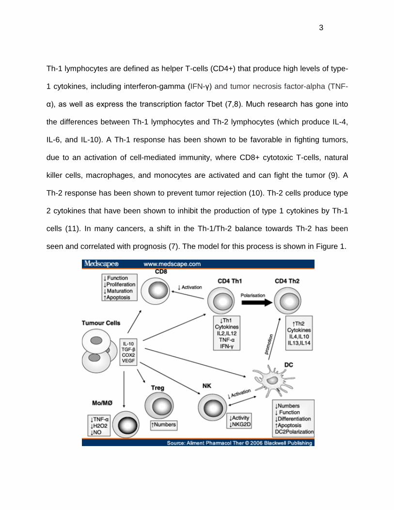
Th-1 lymphocytes are defined as helper T-cells (CD4+) that produce high levels of type-
1 cytokines, including interferon-gamma (IFN-γ) and tumor necrosis factor-alpha (TNF-
α), as well as express the transcription factor Tbet (7,8). Much research has gone into
the differences between Th-1 lymphocytes and Th-2 lymphocytes (which produce IL-4,
IL-6, and IL-10). A Th-1 response has been shown to be favorable in fighting tumors,
due to an activation of cell-mediated immunity, where CD8+ cytotoxic T-cells, natural
killer cells, macrophages, and monocytes are activated and can fight the tumor (9). A
Th-2 response has been shown to prevent tumor rejection (10). Th-2 cells produce type
2 cytokines that have been shown to inhibit the production of type 1 cytokines by Th-1
cells (11). In many cancers, a shift in the Th-1/Th-2 balance towards Th-2 has been
seen and correlated with prognosis (7). The model for this process is shown in Figure 1.

4
Figure 1. The effect of tumor on the Th1/Th2 balance. Tumor cells release factors including IL-10, COX2, VEGF, and TGF-β, that increase the number of Tregs, decrease the activity of natural killer cells, suppress Th-1 cytokine production, and increase apoptosis of CD8+ T-cells. This cascade of effects leads to the polarization of the Th-1/Th-2 balance towards Th-2 (7).
Due to this model, as well as our previous findings, aTh-1 cells were interesting as a
potential post-HSCT cell therapy. aTh-1 cells generated in vitro were previously
phenotyped, showing that they are CD4+ CD25+ CD40L+ CD62Llo, which is consistent
with an effector memory T-cell phenotype (13). We have previously demonstrated that
aTh-1 lymphocytes, when combined with tumor vaccination, result in improved survival
and long-lasting protection in an aggressive (non-HSCT) leukemia model (12). The
release of IFN-γ was shown to contribute to this effect. We found that in addition to
polarizing type-1 anti-tumor immune responses, aTh-1 cells also impair
immunosuppressive T regulatory lymphocyte (Treg) function (12). As shown in the
model in Figure 1, tumor cells are thought to increase the number of Treg cells,
contributing to the tumor’s immune system evasion.
The purpose of the current study was to test, in an allogeneic murine bone marrow
transplantation (BMT) setting, the potential of aTh-1 cells to induce GvL, while
evaluating GvHD effects. We aim to provide pre-clinical data and proof of principle that
aTh-1 cellular therapy may be superior to DLI, the currently widely used strategy, in
improving the outcome of patients following allogeneic HSCT. Though not used
clinically, in order to evaluate the full GvHD potential of aTh-1 cells, we used a major
histocompatibility complex (MHC)-mismatched murine BMT model. Unstimulated
splenocyte (SC) infusion was used to mimic DLI in patients because the CD4 to CD8 T
5
lymphocyte ratio in unstimulated splenocytes is approximately 2:1, which is comparable
to that in the peripheral blood of mice, mimicking the composition of DLI in humans
(data not shown). Herein, we demonstrate that aTh-1 cell therapy enhances GvL with
minimal GvHD in mice with aggressive lymphoblastic leukemia, resulting in superior
leukemia-free survival when compared to unstimulated splenocyte infusion.
MATERIALS AND METHODS
Mice
Age-matched 4-10 week old female BALB/c (CD45.2+, H-2Kd), C57BL/6 (CD45.2+, H-
2Kb), and BoyJ (CD45.1+, H-2Kb) mice were obtained from the National Cancer
Institute’s Frederick National Laboratory for Cancer Research (Frederick, MD). Mice
were housed in specific pathogen-free conditions and cared for according to the
guidelines of the University of Arizona Institutional Animal Care and Use Committee.
Cell culture
Unless otherwise specified, cells were cultured in complete RPMI medium, which is
RPMI 1640 Media with L-glutamine (Thermo-Scientific, Hudson, NH), supplemented
with 10% heat-inactivated fetal bovine serum (FBS; Thermo-Scientific), 1% 100 mM
sodium pyruvate (Thermo-Scientific), 1% MEM Nonessential Amino Acids (Mediatech,
Manassas, VA), and 1% penicillin-streptomycin (Gibco, Grand Island, NY) at 37°C and
5% CO2.
6
aTh-1 cell generation
aTh-1 cells were generated using a protocol adapted from Har-noy et al (14). CD4+
cells were isolated from splenocytes by positive selection using CD4 (L3T4) MicroBeads
(Miltenyi Biotec, Auburn, CA). The purity of the CD4+ cells was consistently over 90%,
as determined by flow cytometry (Figure 2B). CD4+ cells were cultured with anti-CD3
and anti-CD28 paramagnetic activation beads (Dynabeads Mouse T-activator
CD3/CD28 for T-cell Expansion and Activation; Gibco) at a ratio of 3 beads:1 cell in the
presence of 10 ng/mL of recombinant murine IL-12, 20 ng/mL of recombinant murine IL-
7, 20 IU/mL of recombinant murine IL-2 (Peprotech, Rocky Hill, NJ), and 5 μg/mL of
mouse anti-IL-4 antibody (eBioscience, San Diego, CA). Cells were placed in culture at
a concentration of 0.5*106 cells/mL. Cells were culture in RPMI complete media, with
the addition of 0.1% 55mM beta-mercaptoethanol (Gibco). On day 3 of culture, cells
were harvested, and activation beads were removed using a magnet. Cells were then
placed in culture for 4 hours with activation beads at a ratio of 1 bead:1 cell in the
presence of 20 IU/mL of IL-2. After 4 hours, activated cells were used for in vivo and in
vitro assays, with the activation beads intact. For in vitro phenotyping of aTh-1 cells,
cells were cultured for five days. For these experiments, on day 3 of culture, cells were
harvested and diluted with fresh media to a concentration of 0.5x10⁶ cells/mL.
Additional beads were added at a ratio of 1 bead:2 new cells (determined by the
increase in cell number from initial plating). IL-12, IL-7, IL-2, and anti-IL-4 were added at
the same concentrations as above. All cells were cultured at 37°C and 5% CO2 in 24-
well plates with 1 mL volume/well.
7
Preparation of CD4+ T-cells and T-cell enriched splenocytes
CD4+ T-cells were isolated from splenocytes by negative selection using mouse CD4+
T-Cell Isolation Kit II (Miltenyi Biotec). The purity of CD4+ T-cells was greater than 90%,
as determined by flow cytometry. T-cell enriched splenocytes were obtained by running
splenocytes on nylon wool fiber columns for one hour (Polysciences, Inc., Warrington,
PA). This cell population consisted of over 65% CD8a+ and CD4+ T-cells, as
determined by flow cytometry.
Flow cytometry
Approximately 106 cells per sample were washed in 1X phosphate buffered saline
(PBS, Thermo Scientific) with 0.5% heat-inactivated FBS. Cells were then incubated
with anti-mouse CD16/CD32 (BD Biosciences, Franklin Lakes, NJ) for 15 minutes to
block Fc-receptors, followed by incubation with fluorochrome-conjugated antibodies for
30 minutes. For intracellular staining, cells were permeabilized and fixed (Foxp3
Transcription Factor Fixation/Permeabilization; eBioscience) and incubated with
fluorochrome-conjugated antibodies for 30 minutes. Cells were washed with PBS and
analyzed using an LSR Fortessa cell analyzer (BD Biosciences). Data were analyzed
using FlowJo 2 (Tree Star, Inc., Ashland, OR). Antibodies used were anti-mouse IFN-γ
PE, CD40L APC, CD69 PE, Tbet PE, CD44 PE-Cy7, CD25 APC, CD62L FITC,
CD45RB FITC, CD4 APC, CD45.1 APC, CD45.2 APC-eFluor780, CD8a PE-Cy7, Ki-67
eFluor450, Gr-1 FITC, CD11b eFluor450, FoxP3 APC (eBioscience), B220 BV510, CD4
PE-Cy5 (BioLegend, San Diego, CA), and CD3ε PE-CF594 (BD Biosciences). Isotype
controls used were rat IgG1 PE, mouse IgG1 κ PE, rat IgG2a κ APC, rat IgG2a κ FITC,
8
rat IgG1 κ APC, Armenian hamster IgG PE, Armenian hamster IgG APC, rat IgG2b κ
PE-Cy7, and rat IgG2a eFluor450 (eBioscience).
ELISA
The concentrations of interferon gamma (IFN-γ), tumor necrosis factor alpha (TNF-α),
granulocyte macrophage colony stimulating factor (GM-CSF), and interleukin-17 (IL-17)
in aTh-1 cell culture after 4 hour stimulation with activation beads and IL-2 was
determined using ELISA kits following manufacturer’s protocol (eBioscience).
Tumor cell line
The A20 cell line, a B-cell lymphoblastic leukemia cell line of BALB/c origin, was
obtained from the American Type Culture Collection (ATCC, Manassas, VA) and
cultured in complete RPMI medium for five days prior to use. A20 cells are not
radiosensitive and have been previously used in murine bone marrow transplantation
studies (15).
MHC-mismatched bone marrow transplantation experiments
On day -5, recipient BALB/c mice were injected intravenously (i.v.) with 106 A20 cells.
On day -1, these mice received a lethal dose of 750 cGy total body irradiation (TBI)
using a Cesium 137 Irradiator at the University of Arizona. On day 0, 107 T-cell-depleted
(TCD) C57BL/6 bone marrow (BM) cells were injected i.v. into the recipient mice. T-cells
were depleted from the BM cells using a CD3ε MicroBead Kit (Miltenyi Biotec) and an
autoMACS Separator (Miltenyi Biotec). On days 12 and 17, BALB/c mice were injected
9
i.v. with one of the following cell populations: 3*106 aTh-1 cells, 3*106 CD4+ cells, 3*106
T-cell enriched splenocytes, or 107 total splenocytes. Control groups received i.v.
injections of PBS. Starting day 0, mice were weighed every three to four days, and
moribund mice were euthanized. Mice were considered cured after day 100 if no tumor
was found during necropsy. Lack of tumor was confirmed by histology (data not shown).
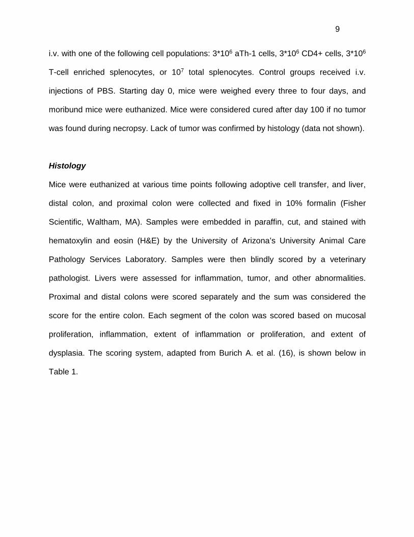
Histology
Mice were euthanized at various time points following adoptive cell transfer, and liver,
distal colon, and proximal colon were collected and fixed in 10% formalin (Fisher
Scientific, Waltham, MA). Samples were embedded in paraffin, cut, and stained with
hematoxylin and eosin (H&E) by the University of Arizona’s University Animal Care
Pathology Services Laboratory. Samples were then blindly scored by a veterinary
pathologist. Livers were assessed for inflammation, tumor, and other abnormalities.
Proximal and distal colons were scored separately and the sum was considered the
score for the entire colon. Each segment of the colon was scored based on mucosal
proliferation, inflammation, extent of inflammation or proliferation, and extent of
dysplasia. The scoring system, adapted from Burich A. et al. (16), is shown below in
Table 1.
10
Table 1.
In vivo tracking of adoptively transferred lymphocytes
BALB/c mice received 750 cGy TBI on day -1 and C57BL/6 TCD BM cells on day 0 as
described above. On day 12, mice received 3*106 allogeneic aTh-1 cells or 3*106 SC
generated from CD45.1+ BoyJ mice. On days 3, 7, 10, 17, 24, 31, and 66 after adoptive
cell transfer, peripheral blood was collected from recipient BALB/c mice by tail vein
bleeding. Complete blood counts (CBC) were obtained using a Coulter AcT counter
(Beckman Coulter, Brea, CA). After red blood cell lysis, the presence and phenotype of
adoptively transferred aTh-1 cells and splenocytes, as well as lineage differentiation of
engrafted donor BM cells, were determined by flow cytometry. Ki-67 staining was done
separately on days 4, 8, 11, 18, and 25. Gates were determined using fluorescence
minus ones (FMOs) for all markers except Ki-67, for which an isotype control was used.
11
Cytotoxicity assay
Chromium-release (51Cr) assays were performed as previously described (17). Briefly,
target A20 tumor cells were incubated with 500 µCi Na51CrO4 (5 mCi/mL, ICN
Pharmaceuticals, Irvine, CA) for 1 hour at 37°C. Target cells were then washed three
times and co-cultured with effector cells in triplicate in 96-well V-bottomed microtiter
plates (100 µL, 5,000 targets/well) at 37 °C for 4 hours. Spontaneous-release wells
contained only tissue culture medium and 51Cr-labeled target cells; maximal-release
wells contained 5% Triton X-114 (Sigma-Aldrich, St. Louis, MO) and 51Cr-labeled target
cells. After the four hour incubation, the plates were then centrifuged at 200g for 10
minutes, after which 100 µL aliquots of supernatant were harvested and added to 3 mL
scintillation fluid. The radioactivity (cpm) was measured on a beta counter (Packard,
Palo Alto, CA).
Cytotoxicity was determined by the following formula:
cytotoxicity (%) = 100*(mean experimental release – mean spontaneous release)/(mean
maximal release – mean spontaneous release).
Statistics
Kaplan-Meier curves were generated and analyzed by the log-rank test to determine
survival percentages and differences between treatment groups. In other experiments,
Student’s t-tests were used to determine significant differences (p<0.05) between
groups.
12
RESULTS
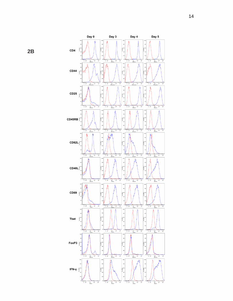
aTh-1 cells express surface markers consistent with effector memory T-cells and
secrete high levels of type-1 cytokines
We have previously reported the in vivo effects of aTh-1 cells that had been cultured for
6 days (12). We were, however, concerned that with prolonged culture, aTh-1 may
undergo activation-induced cell death shortly after their infusion, thus limiting their in
vivo activity. We, therefore, evaluated cytokine production kinetics and surface marker
expression of cultured aTh-1 cells serially over 5 days. On each day, cells were
harvested, beads were removed, and cells were stimulated for four hours as described
above. We found that the production of IFN-γ and TNF-α peaks on day 4 of culture,
while GM-CSF was highest on day 5 (Figure 2A). IL-17 production is low throughout,
indicating these cells do not possess the characteristics of Th-17 cells. Expression of
CD44, CD45RB, CD69, CD40L, and Tbet is consistent with activated T-helper 1 cells
that evolve into an effector memory phenotype (CD62Llo CD45RBhi CD44hi Tbet+).
Furthermore, these cells do not express markers for regulatory T-cells such as FoxP3.
This phenotype already exists by day 3 of culture (Figure 2B). Collectively, these data
suggest that infusion of day 3 aTh-1 cells, right before cytokine production peaks on day
4, may have the advantage of producing more cytokines in vivo. In addition, reducing
the in vitro culture time by 50% makes these cells more appealing for a potential clinical
application.

13
2A
Figure 2. aTh-1 cells express surface markers consistent with effector memory T-cells and secrete high levels of type-1 cytokines. aTh-1 cells were cultured in vitro for indicated duration of time. On each day, cells were harvested, de-beaded, and stimulated for 4 hours with IL-2 and anti-CD3/CD28 activation beads as described. (A) Supernatant was collected, and the amount of IFN-γ, TNF-α, GM-CSF, and IL-17 was determined by ELISA. Each value represents the average of at least three independent experiments. Day 3 vs. Day 5: p<0.05 for all cytokines measured, except IL-17. (B) Expression of surface markers CD4, CD44, CD25, CD45RB, CD62L, CD40L, and CD69, as well as transcription factors Tbet and FoxP3 and intracellular IFN-γ, was determined by flow cytometry. Representative histograms from at least three independent experiments are shown.
14
2B
15
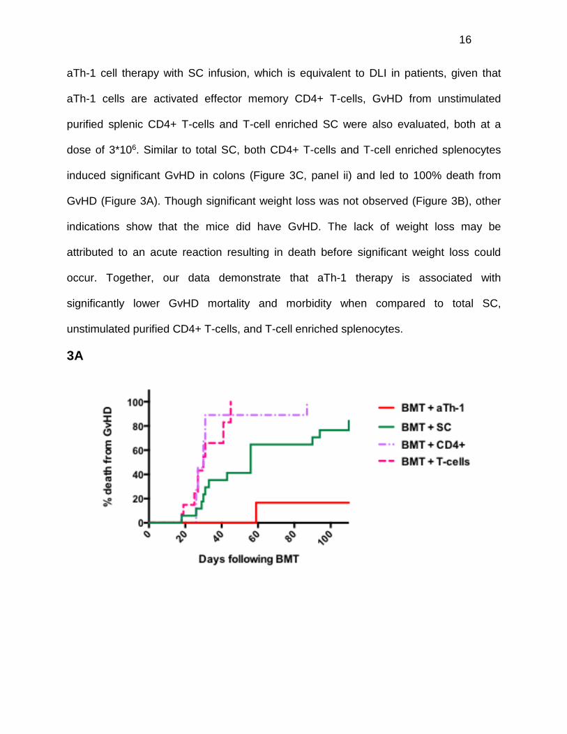
aTh-1 cell infusion is associated with significantly reduced GvHD compared to
unstimulated splenocytes or T-cells
To evaluate whether aTh-1 cell therapy induces GvHD, a fully MHC-mismatched T-cell-
depleted (TCD) murine BMT model was utilized. Following 750 cGy of TBI on day -1
and TCD BMT on day 0, recipient BALB/c mice were infused with C57BL/6 unstimulated
SC or aTh-1 cells on days 12 and 17. Mice were injected with 107 SC, which contain
approximately 3*106 T-cells (data not shown), while other groups received 3*106 aTh-1
cells. As expected, greater than 90% of SC-treated mice died of GvHD. In contrast,
GvHD was the cause of death in less than 20% of the mice receiving aTh-1 cells (Figure
3A). Moreover, mice treated with SC exhibited significant weight loss when compared to
control mice, while mice receiving aTh-1 cells did not (Figure 3B). GvHD was further
confirmed histologically as previously described (16). The degree and extent of
inflammation, mucosal proliferation, and dysplasia of the colons were evaluated and
scored on day 5 following adoptive cell transfer. Colons from mice treated with SC
showed subacute multifocal coalescing colitis with crypt hyperplasia and mixed
lymphocytic and neutrophilic infiltrates, consistent with GvHD (Figure 3C, panel i). Colon
GvHD scores of mice receiving aTh-1 cells were comparable to those of mice receiving
BMT alone (Figure 3C, panel ii). Livers were also evaluated for GvHD. On day 5
following adoptive cell transfer, mice receiving aTh-1 showed no significant abnormality
in their livers. In comparison, mice treated with PBS showed leukemic infiltration of their
livers, indicating inadequate GvL effects provided by BMT alone. Mice receiving SC,
while showing no leukemic infiltration, developed multifocal chronic active cholangitis,
indicating liver GvHD (Figure 3D). Although the focus of the current study is to compare
16
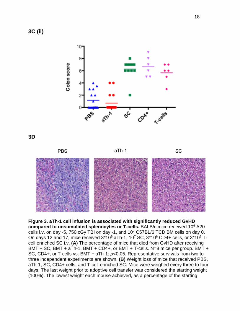
aTh-1 cell therapy with SC infusion, which is equivalent to DLI in patients, given that
aTh-1 cells are activated effector memory CD4+ T-cells, GvHD from unstimulated
purified splenic CD4+ T-cells and T-cell enriched SC were also evaluated, both at a
dose of 3*106. Similar to total SC, both CD4+ T-cells and T-cell enriched splenocytes
induced significant GvHD in colons (Figure 3C, panel ii) and led to 100% death from
GvHD (Figure 3A). Though significant weight loss was not observed (Figure 3B), other
indications show that the mice did have GvHD. The lack of weight loss may be
attributed to an acute reaction resulting in death before significant weight loss could
occur. Together, our data demonstrate that aTh-1 therapy is associated with
significantly lower GvHD mortality and morbidity when compared to total SC,
unstimulated purified CD4+ T-cells, and T-cell enriched splenocytes.
3A

17
3B
3C (i)
18
3C (ii)
3D
Figure 3. aTh-1 cell infusion is associated with significantly reduced GvHD compared to unstimulated splenocytes or T-cells. BALB/c mice received 106 A20 cells i.v. on day -5, 750 cGy TBI on day -1, and 107 C57BL/6 TCD BM cells on day 0. On days 12 and 17, mice received 3*106 aTh-1, 107 SC, 3*106 CD4+ cells, or 3*106 T-cell enriched SC i.v. (A) The percentage of mice that died from GvHD after receiving BMT + SC, BMT + aTh-1, BMT + CD4+, or BMT + T-cells. N=8 mice per group. BMT + SC, CD4+, or T-cells vs. BMT + aTh-1: p<0.05. Representative survivals from two to three independent experiments are shown. (B) Weight loss of mice that received PBS, aTh-1, SC, CD4+ cells, and T-cell enriched SC. Mice were weighed every three to four days. The last weight prior to adoptive cell transfer was considered the starting weight (100%). The lowest weight each mouse achieved, as a percentage of the starting
19
weight, was determined. This illustrates the mean lowest weight achieved by each group of mice. SC vs. aTh-1: p<0.05; SC, CD4+, or T-cells vs. BMT: p<0.05; aTh-1 vs. BMT, CD4+, or T-cells: not significant. (C) Representative H&E staining of the colon of mice five days after receiving PBS, aTh-1, or SC at 50X magnification. N=3 mice per group for that time point (panel i). Mean colon GvHD scores of mice receiving PBS were compared to scores of mice receiving 3*106 aTh-1 cells, 107 SC, 3*106 total CD4+ cells, and 3*106 T-cell enriched SC (panel ii). aTh-1 vs. PBS: not significant; SC, CD4+ cells, or T-cells vs. PBS or aTh-1: p<0.05. N=6-17 mice per group. (D) Representative H&E staining of the livers of mice five days after receiving PBS, aTh-1, or SC at 50X magnification. N=3 mice per group per time point.
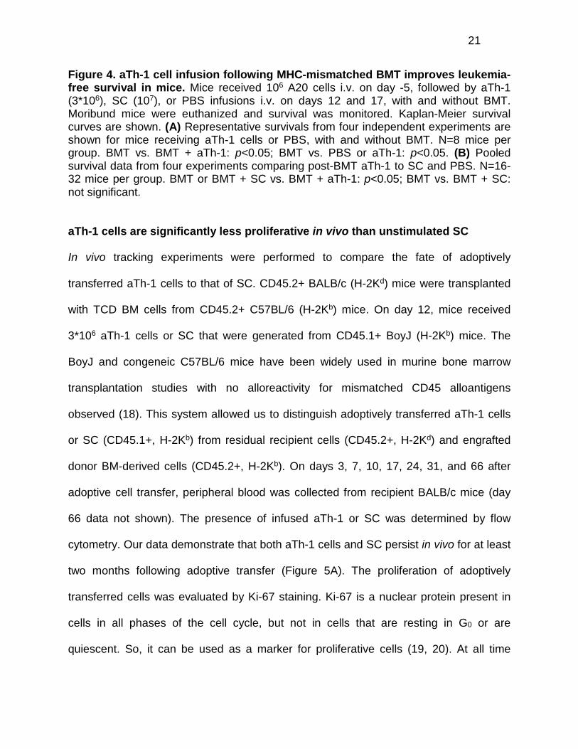
aTh-1 cell infusion following MHC-mismatched BMT improves leukemia-free
survival in mice
MHC-mismatched allogeneic aTh-1 cells have been previously reported to induce anti-
leukemic effects without GvHD toxicity in non-transplant murine settings (13, 14). Given
that aTh-1 cells induced significantly less GvHD than SC, we used the same model to
evaluate whether aTh-1 cell therapy was capable of promoting GvL effects. Recipient
BALB/c mice were injected i.v. with 106 A20 B-cell leukemia cells on day -5. Without
BMT, 100% of the mice died from leukemia. Additionally, without BMT, the aTh-1 cells
showed no effect on survival (Figure 4A). TBI, followed by BMT, although significantly
prolonging survival, was also not curative (Figure 4A, 4B). While SC effectively abated
the leukemia, as evidenced by a lack of tumor seen upon necropsy and confirmed by
histological analysis (data not shown), survival rate was not improved as over 90% of
mice died of GvHD (Figure 3A, 4B). Infusion of aTh-1 cells post-BMT led to leukemia-
free survival in 50% of the mice (Figure 4A, 4B). The absence of leukemia in the
surviving mice 100 days post-BMT was confirmed by histological analysis (data not
shown). In separate experiments, mice were treated with lower number of aTh1 cells
(1*106). We found no difference in GvHD mortality rate or leukemia-free survival with

20
this reduction in dose (data not shown). In summary, aTh-1 cells, while demonstrating
minimal GvHD in a fully MHC-mismatched BMT model, retain significant anti-leukemic
properties.
4A
4B
21
Figure 4. aTh-1 cell infusion following MHC-mismatched BMT improves leukemia-free survival in mice. Mice received 106 A20 cells i.v. on day -5, followed by aTh-1 (3*106), SC (107), or PBS infusions i.v. on days 12 and 17, with and without BMT. Moribund mice were euthanized and survival was monitored. Kaplan-Meier survival curves are shown. (A) Representative survivals from four independent experiments are shown for mice receiving aTh-1 cells or PBS, with and without BMT. N=8 mice per group. BMT vs. BMT + aTh-1: p<0.05; BMT vs. PBS or aTh-1: p<0.05. (B) Pooled survival data from four experiments comparing post-BMT aTh-1 to SC and PBS. N=16-32 mice per group. BMT or BMT + SC vs. BMT + aTh-1: p<0.05; BMT vs. BMT + SC: not significant.
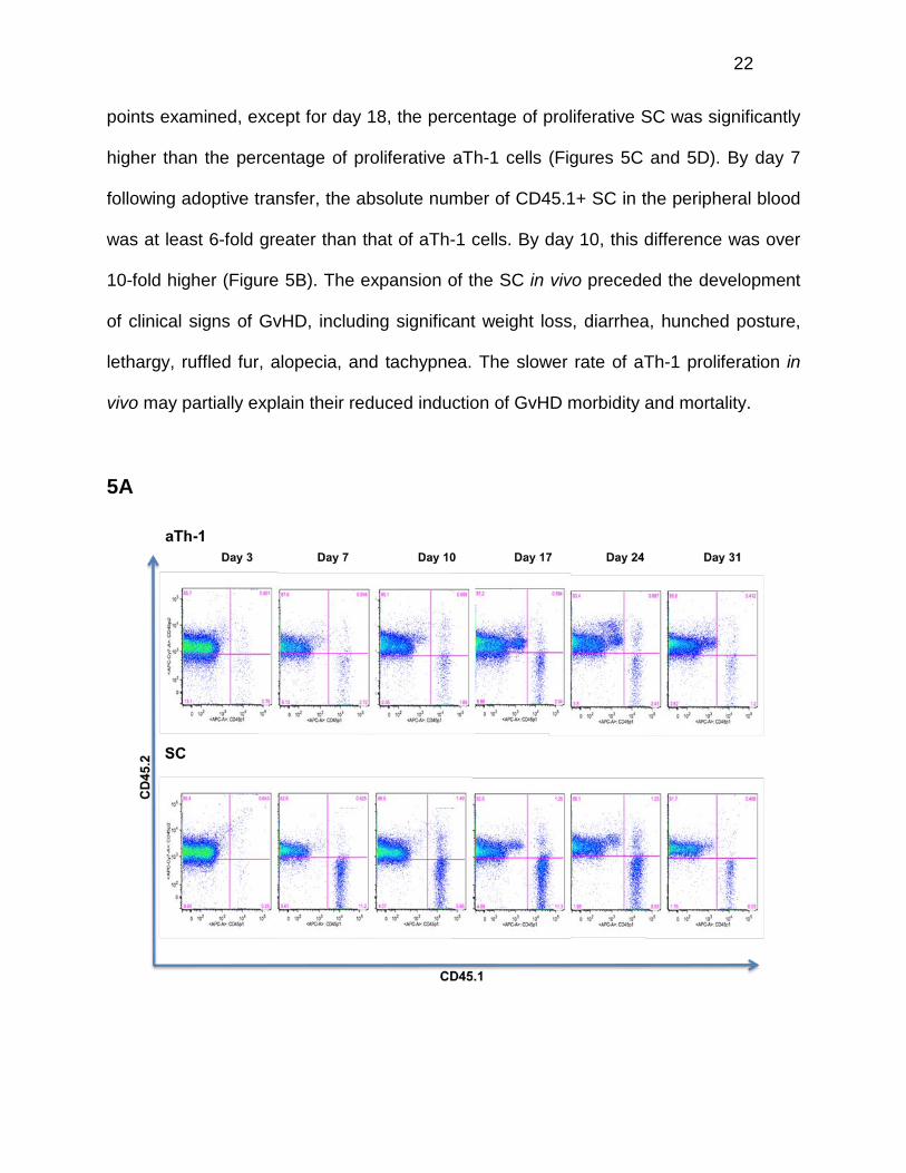
aTh-1 cells are significantly less proliferative in vivo than unstimulated SC
In vivo tracking experiments were performed to compare the fate of adoptively
transferred aTh-1 cells to that of SC. CD45.2+ BALB/c (H-2Kd) mice were transplanted
with TCD BM cells from CD45.2+ C57BL/6 (H-2Kb) mice. On day 12, mice received
3*106 aTh-1 cells or SC that were generated from CD45.1+ BoyJ (H-2Kb) mice. The
BoyJ and congeneic C57BL/6 mice have been widely used in murine bone marrow
transplantation studies with no alloreactivity for mismatched CD45 alloantigens
observed (18). This system allowed us to distinguish adoptively transferred aTh-1 cells
or SC (CD45.1+, H-2Kb) from residual recipient cells (CD45.2+, H-2Kd) and engrafted
donor BM-derived cells (CD45.2+, H-2Kb). On days 3, 7, 10, 17, 24, 31, and 66 after
adoptive cell transfer, peripheral blood was collected from recipient BALB/c mice (day
66 data not shown). The presence of infused aTh-1 or SC was determined by flow
cytometry. Our data demonstrate that both aTh-1 cells and SC persist in vivo for at least
two months following adoptive transfer (Figure 5A). The proliferation of adoptively
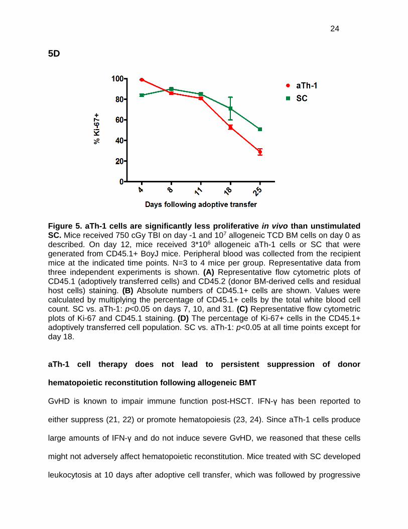
transferred cells was evaluated by Ki-67 staining. Ki-67 is a nuclear protein present in
cells in all phases of the cell cycle, but not in cells that are resting in G0 or are
quiescent. So, it can be used as a marker for proliferative cells (19, 20). At all time
22
points examined, except for day 18, the percentage of proliferative SC was significantly
higher than the percentage of proliferative aTh-1 cells (Figures 5C and 5D). By day 7
following adoptive transfer, the absolute number of CD45.1+ SC in the peripheral blood
was at least 6-fold greater than that of aTh-1 cells. By day 10, this difference was over
10-fold higher (Figure 5B). The expansion of the SC in vivo preceded the development
of clinical signs of GvHD, including significant weight loss, diarrhea, hunched posture,
lethargy, ruffled fur, alopecia, and tachypnea. The slower rate of aTh-1 proliferation in
vivo may partially explain their reduced induction of GvHD morbidity and mortality.
5A

23
5B
5C
24
5D
Figure 5. aTh-1 cells are significantly less proliferative in vivo than unstimulated SC. Mice received 750 cGy TBI on day -1 and 107 allogeneic TCD BM cells on day 0 as described. On day 12, mice received 3*106 allogeneic aTh-1 cells or SC that were generated from CD45.1+ BoyJ mice. Peripheral blood was collected from the recipient mice at the indicated time points. N=3 to 4 mice per group. Representative data from three independent experiments is shown. (A) Representative flow cytometric plots of CD45.1 (adoptively transferred cells) and CD45.2 (donor BM-derived cells and residual host cells) staining. (B) Absolute numbers of CD45.1+ cells are shown. Values were calculated by multiplying the percentage of CD45.1+ cells by the total white blood cell count. SC vs. aTh-1: p<0.05 on days 7, 10, and 31. (C) Representative flow cytometric plots of Ki-67 and CD45.1 staining. (D) The percentage of Ki-67+ cells in the CD45.1+ adoptively transferred cell population. SC vs. aTh-1: p<0.05 at all time points except for day 18.
aTh-1 cell therapy does not lead to persistent suppression of donor
hematopoietic reconstitution following allogeneic BMT
GvHD is known to impair immune function post-HSCT. IFN-γ has been reported to
either suppress (21, 22) or promote hematopoiesis (23, 24). Since aTh-1 cells produce
large amounts of IFN-γ and do not induce severe GvHD, we reasoned that these cells
might not adversely affect hematopoietic reconstitution. Mice treated with SC developed
leukocytosis at 10 days after adoptive cell transfer, which was followed by progressive

25
leukopenia, correlating with the clinical course of GvHD (Figure 6A). In comparison,
aTh-1 cell therapy caused only mild leukopenia (Figure 6A). The effect of aTh-1 cells on
hematopoiesis was further evaluated by assessing the absolute number of donor BM-
derived CD45.2+ cells in the peripheral blood (Figure 6B). SC infusion led to transient,
rapid expansion of BM-derived CD45.2+ cells at 10 days after adoptive cell transfer,
followed by a rapid decline in cell numbers. By day 31 post-SC infusion, the absolute
number of donor BM-derived CD45.2+ cells was only a mean of 61% of the absolute
number of donor BM-derived CD45.2+ cells in mice receiving PBS. This observation
indicates persistent suppression of donor hematopoietic reconstitution by SC. In
contrast, aTh-1 cell infusion resulted in transient, mild suppression of donor
hematopoiesis that recovered over time (Figure 6B). Furthermore, SC infusion resulted
in skewed immune reconstitution, including an increased percentage of myeloid cells,
particularly polymorphonuclear neutrophils (PMNs), a decreased percentage of B-cells
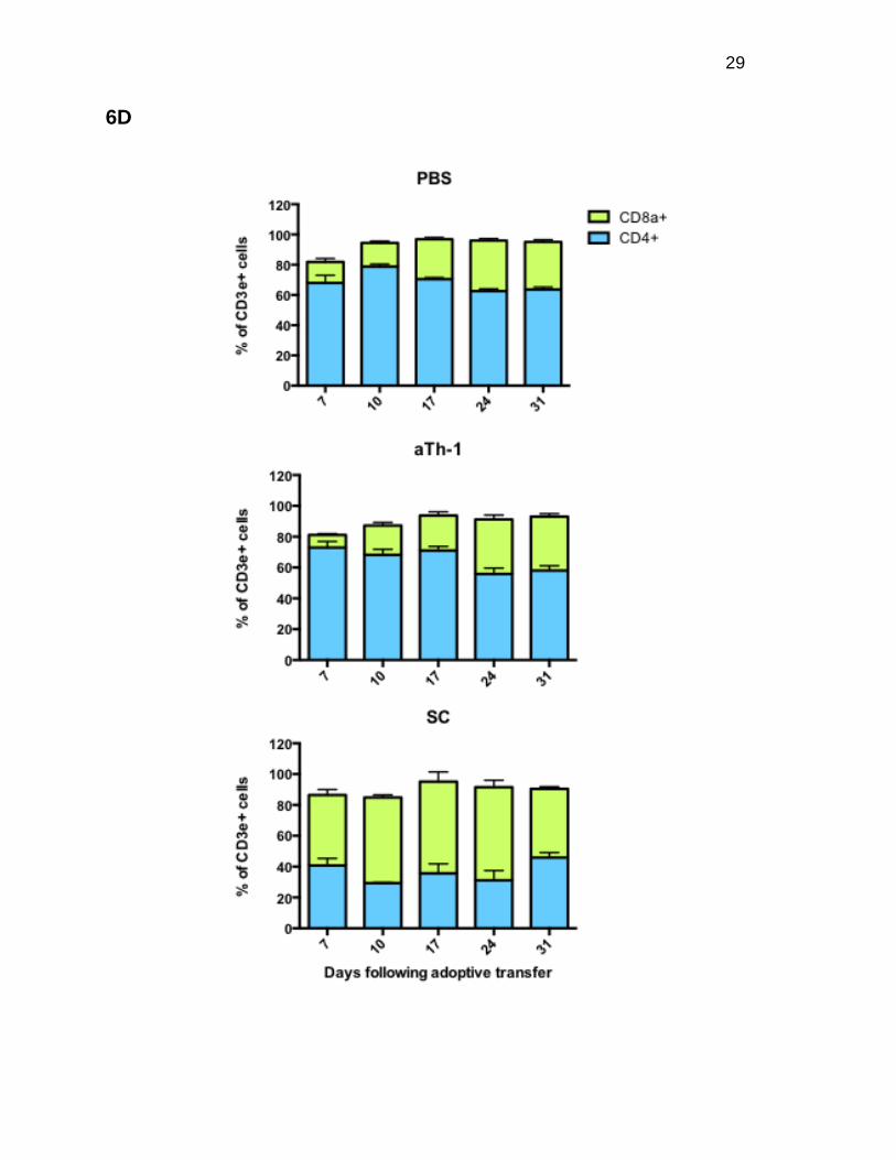
(Figure 6C), and a decreased or inverted CD4 to CD8 T-cell ratio (Figure 6D). When
compared to control mice, the increase in monocytes was lower and transient in mice
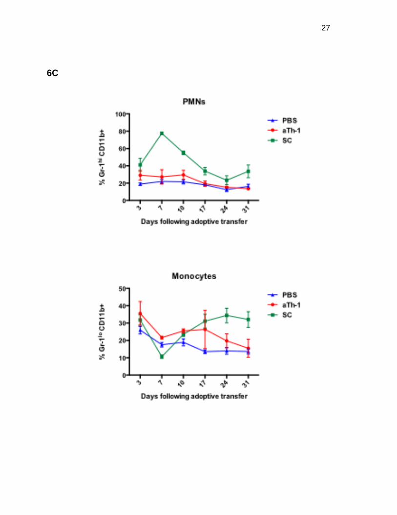
treated with aTh-1 cells (Figure 6C), with no significant differences in the percentages of
PMNs, B-cells, and T-cells and in the CD4 to CD8 ratio (Figures 6C and 6D).

26
6A
6B
27
6C

28
29
6D
30
Figure 6. aTh-1 cell therapy does not lead to persistent suppression of donor hematopoietic reconstitution following allogeneic BMT. Peripheral blood from mice from the experiment described in Figure 5 was used to examine hematopoietic reconstitution and lineage differentiation. (A) Total white blood cell (WBC) counts. SC vs. PBS: p<0.05 on days 10 and 31; aTh-1 vs. PBS: p<0.05 on day 24. (B) The actual number of donor BM-derived CD45.2+ H-2Kb+ cells was calculated by multiplying the percentage of these cells by the total WBC count. SC vs. PBS: p<0.05 on days 10, 24, and 31; aTh-1 vs. PBS: p<0.05 on day 24. (C) The percentage of PMNs (CD3e- B220-Gr-1hi CD11b+), monocytes (CD3e- B220- Gr-1lo CD11b+), B-cells (B220+ CD3e-), and T-cells (B220- CD3e+) in the donor BM-derived CD45.2+ H-2Kb+ population. For B-cells, SC vs. PBS: p<0.05 at all time points. For PMNs, SC vs. PBS: p<0.05 on days 3, 7, 10, and 17. For monocytes, SC vs. PBS: p<0.05 on days 7, 17, 24, and 31. For T-cells, SC vs. PBS: not significant at any time point. For monocytes, aTh-1 vs. PBS: p<0.05 on days 7 and 10. aTh-1 vs. PBS: not significant at any time point for any other cell types. (D) The percentage of CD4+ and CD8a+ cells in the donor BM-derived CD45.2+ H-2Kb+ CD3e+ T-cell population. SC vs. PBS: p<0.05 at all time points. aTh-1 vs. PBS: not significant at any time point. N=3 to 4 mice per group. Representative data from three independent experiments is shown.
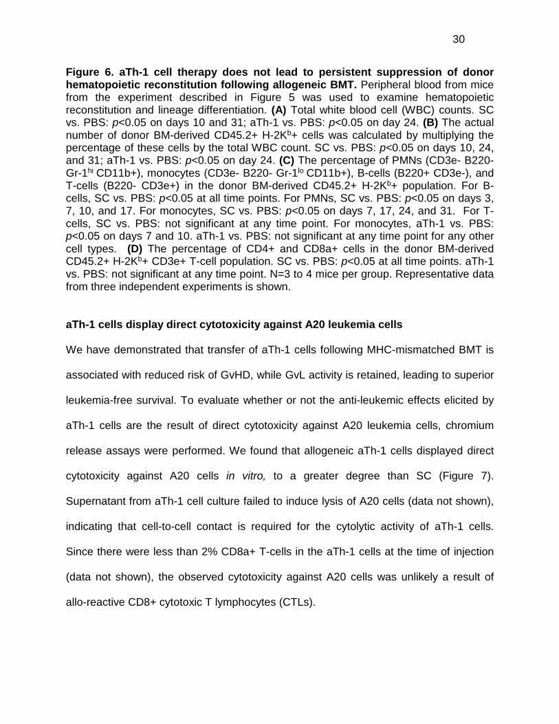
aTh-1 cells display direct cytotoxicity against A20 leukemia cells
We have demonstrated that transfer of aTh-1 cells following MHC-mismatched BMT is
associated with reduced risk of GvHD, while GvL activity is retained, leading to superior
leukemia-free survival. To evaluate whether or not the anti-leukemic effects elicited by
aTh-1 cells are the result of direct cytotoxicity against A20 leukemia cells, chromium
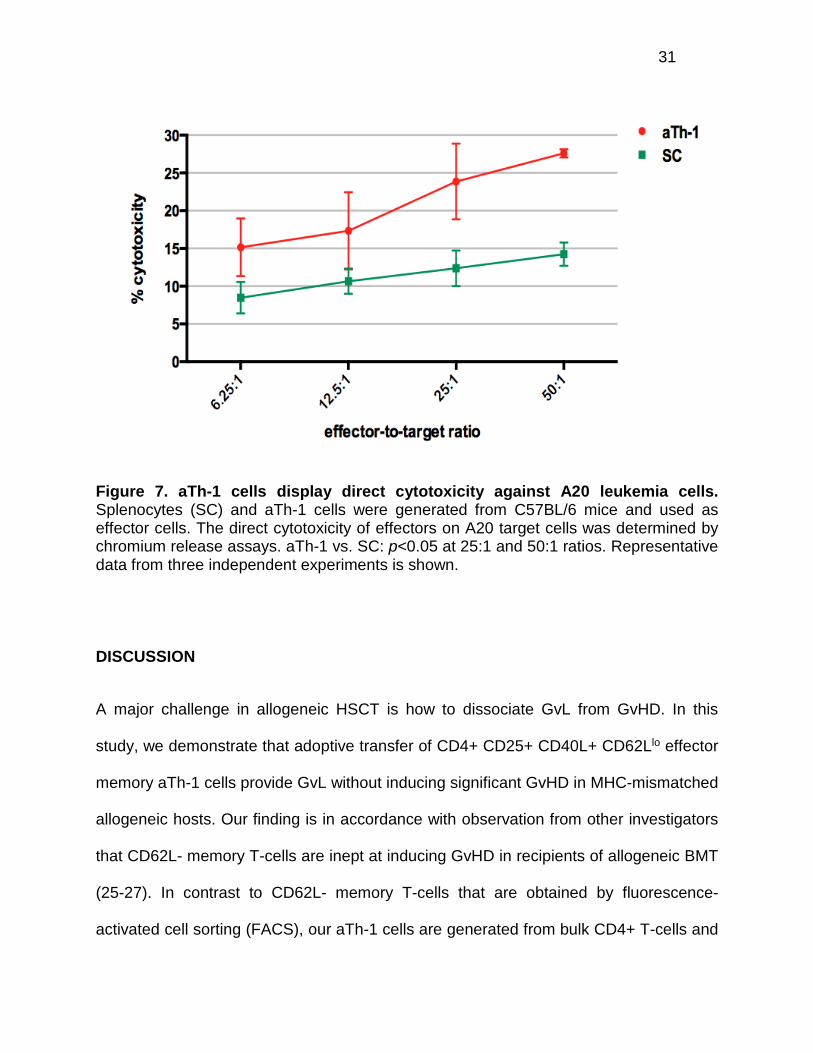
release assays were performed. We found that allogeneic aTh-1 cells displayed direct
cytotoxicity against A20 cells in vitro, to a greater degree than SC (Figure 7).
Supernatant from aTh-1 cell culture failed to induce lysis of A20 cells (data not shown),
indicating that cell-to-cell contact is required for the cytolytic activity of aTh-1 cells.
Since there were less than 2% CD8a+ T-cells in the aTh-1 cells at the time of injection
(data not shown), the observed cytotoxicity against A20 cells was unlikely a result of
allo-reactive CD8+ cytotoxic T lymphocytes (CTLs).
31
Figure 7. aTh-1 cells display direct cytotoxicity against A20 leukemia cells. Splenocytes (SC) and aTh-1 cells were generated from C57BL/6 mice and used as effector cells. The direct cytotoxicity of effectors on A20 target cells was determined by chromium release assays. aTh-1 vs. SC: p<0.05 at 25:1 and 50:1 ratios. Representative data from three independent experiments is shown.
DISCUSSION
A major challenge in allogeneic HSCT is how to dissociate GvL from GvHD. In this
study, we demonstrate that adoptive transfer of CD4+ CD25+ CD40L+ CD62Llo effector
memory aTh-1 cells provide GvL without inducing significant GvHD in MHC-mismatched
allogeneic hosts. Our finding is in accordance with observation from other investigators
that CD62L- memory T-cells are inept at inducing GvHD in recipients of allogeneic BMT
(25-27). In contrast to CD62L- memory T-cells that are obtained by fluorescence-
activated cell sorting (FACS), our aTh-1 cells are generated from bulk CD4+ T-cells and
32
are used following three days of in vitro culture, resulting in a potentially clinically useful
higher cell yield of at least three times the starting cell number (data not shown).
A key question is why aTh-1 cells provide GvL without significant GvHD. aTh-1 cells do
not express markers for regulatory T-cells such as FoxP3 (Figure 2B), suggesting that
the lack of significant GvHD in vivo is not due to immunosuppressive activity of
regulatory T-cells in the adoptively transferred population. Following adoptive transfer,
aTh-1 cells proliferated in vivo and persisted for at least two months, indicating that
activation-induced cell death did not take place post-infusion. The magnitude of T-cell
response has been suggested to be essential for T-cell-infiltrative GvHD (20). At the
onset of GvHD, a threshold number of effector T-cells may need to infiltrate a given
target tissue to initiate sufficient tissue damage to establish local changes that promote
recruitment of additional activated T-cells, thus leading to the clinical manifestation of
GvHD (28-30). We have shown that adoptively transferred SC underwent rapid
expansion shortly after infusion at a much higher rate than aTh-1 cells, suggesting that
GvHD may require a sustained highly proliferative allogeneic T-cell response that SC,
but not aTh-1 cells, can generate. Because the absolute number of aTh-1 cells was
much lower, the threshold number of effector T-cells may not have been reached.
aTh-1 cells differ from effector memory cells reported by others in that they are
activated and secrete large amounts of type-1 cytokines and are therefore possibly
endowed with an enhanced GvL capacity. Previous murine studies demonstrated that
CD4+ effector memory T-cells mediate GvL via direct cytotoxicity against leukemia cells
(27). On the other hand, Zorn et al. has reported that CD4+ DLI infusion induces
expansion of CD8+ donor CTLs with cytolytic activity directed against leukemia cells
33
and residual recipient hematopoietic cells. It is noteworthy that the methodology utilized
by Zorn et al. did not allow one to distinguish between CD8+ T-cells derived from donor
BM cells and those derived from CD8+ T-cells that remained in the CD4+ DLI
preparation (31). In our study, allogeneic aTh-1 cells exhibited direct cytotoxicity against
A20 leukemia cells in vitro, consistent with the findings of Zheng et al. (27). Our data
also showed that the ratio of CD8 to CD4 T-cells in the donor-derived engrafted BM was
unchanged by our aTh-1 cell infusion, indicating that an expansion of CD8+ CTLs was
likely not the source of the anti-leukemic effect, as it was in the Zorn et al. experiments.
Syngeneic aTh-1 cells also displayed some cytotoxicity against A20 leukemia cells,
although at a much lower level than allogeneic aTh-1 cells (data not shown), indicating
that allo-antigen-mediated T-cell response contributed to the overall cytotoxicity, but
was not the only factor. Furthermore, given that aTh-1 cells provide adjuvant effects to
tumor vaccination (12), it is also possible that other effector cells, such as natural killer
cells and macrophages, are activated by aTh-1 cells via type-1 cytokines and mediate
GvL in vivo. Additionally, it has been shown that aTh-1 cells reduce the suppressive
function of FoxP3+ Treg (12). Thus, aTh-1 may also enhance GvL by modulating
FoxP3+ Treg, thus contributing to breaking tumor tolerance.
Hematopoietic stem cell engraftment and reconstitution following HSCT are affected by
a variety of factors, including graft source, GvHD, and host BM microenvironment. The
effect of mature donor T-cells on donor hematopoietic stem cell engraftment and
immune recovery has been controversial. Early attempts to reduce GvHD by T-cell
depletion were associated with increased risk of graft failure and suppressed immune
recovery (32-35), indicating that donor T-cells may facilitate engraftment and
34
hematopoietic reconstitution. However, later clinical trials suggested that these results
could have been due to insufficient hematopoietic stem/progenitor content in the
manipulated BM, rather than the lack of T-cells (36-38). Moreover, murine studies
provide evidence that allogeneic T-cells impair hematopoietic reconstitution after stem
cell transplantation (39, 40). On the other hand, CD62L- effector memory T-cells have
been shown to promote donor BM stem/progenitor-derived T-cell reconstitution (19). In
our study, aTh-1 cell therapy induced mild transient suppression of donor
hematopoiesis. In other studies (19), the cells are unactivated. The activation of our
cells and the release of such large amounts of type 1 cytokines could explain the mild
suppression of hematopoiesis. In contrast, SC resulted in severe, persistent
suppression of donor-derived hematopoiesis, consistent with previous reports that
allogeneic T-cells suppress hematopoietic reconstitution. Adoptive transfer of SC led to
skewed immune reconstitution characterized by severe B lymphopenia, inverted CD4 to
CD8 T-cell ratio, and predominance of the myeloid fraction in the blood. Our finding is
consistent with prior reports that GvHD is associated with lymphoid hypoplasia and
impairs immune function post-HSCT [22, 23, 30-33]. Lack of significant GvHD caused
by aTh-1 cell infusion may partially explain why the cells do not have detrimental effects
on hematopoiesis following allogeneic BMT.
In summary, utilizing a MHC-mismatched murine BMT model, we demonstrate that aTh-
1 cells enhance GvL without inducing significant GvHD following allogeneic HSCT.
Furthermore, in contrast to SC, aTh-1 cell infusion does not cause severe, persistent
suppression of donor-derived hematopoiesis. aTh-1 cell therapy, therefore, may provide
an effective alternative to DLI. Considering that GvHD is more severe in an MHC-

35
mismatched HSCT than in minor antigen-mismatched HSCT (MHC-matched HSCT),
our finding that aTh-1 cellular therapy is associated with significantly less GvHD in an
MHC-mismatched BMT model makes this approach even more attractive for potential
clinical application. Future potential applications include moving this cellular therapy into
a haploidentical model, making it very clinically relevant and solving most issues in
locating BM donors.
36
ACKNOWLEDGEMENTS
I would like to thank Dr. Emmanuel Katsanis for the amazing opportunities he has given
me in allowing me to join his lab, for having confidence my abilities and pushing me to
work harder, and for all of his guidance and support as a research mentor. I would like
to thank Dr. Yi Zeng for her support, guidance, collaboration, and valuable insight.
Thanks to Emely Hoffman for countless hours of help and support in planning, data
collection, analysis, writing, and everything else. Thanks to Min Hahn for his valuable
contributions to data collection and analysis. Thanks to Alexis Lorenz for taking me
under her wing and training me when I joined the lab, giving me the skill set that has
allowed me to be successful here. I would like to thank Martin Asimis for his support and
for always being willing to pitch in when needed. Thanks to Dr. Darya Alizadeh for
sharing her flow cytometry expertise and to Dr. Neale Hanke for always being willing to
help. Thanks to Vanessa Frisinger and Kim Carpenter for their invaluable administrative
support. I would also like to thank our sources of funding: the National Institutes of
Health grant R01 CA104926, Hyundai Hope on Wheels, and Tee Up for Tots.
37
REFERENCES
1. Soiffer RJ, Lerademacher J, Ho V, et al. Impact of immune modulation with anti-T-cell antibodies on the outcome of reduced-intensity allogeneic hematopoietic stem cell transplantation for hematologic malignancies. Blood 2011;117:6963-6970. 2. D'Sa S, Peggs K, Pizzey A, et al. T- and B-cell immune reconstitution and clinical outcome in patients with multiple myeloma receiving T-cell-depleted, reduced-intensity allogeneic stem cell transplantation with an alemtuzumab-containing conditioning regimen followed by escalated donor lymphocyte infusions. British journal of haematology 2003;123:309-322. 3. Bastien JP, Roy J, Roy DC. Selective T-cell depletion for haplotype-mismatched allogeneic stem cell transplantation. Seminars in oncology 2012;39:674-682. 4. Perruccio K, Topini F, Tosti A, et al. Optimizing a photoallodepletion protocol for adoptive immunotherapy after haploidentical SCT. Bone marrow transplantation 2012;47:1196-1200. 5. Amrolia PJ, Muccioli-Casadei G, Huls H, et al. Adoptive immunotherapy with allodepleted donor T-cells improves immune reconstitution after haploidentical stem cell transplantation. Blood 2006;108:1797-1808. 6. Chen B, Cui X, Liu C, Chao N. Prevention of graft-versus-host disease while preserving graft-versus-leukemia effect after selective depletion of host-reactive T cells by photodynamic cell purging process. Blood 2002;99:3083-3088. 7. Evans C, Dalgleish G, Kumar D. Immune Suppression and Colorectal Cancer. Aliment Pharmacol Ther. 2006;24(8):1163-1177. 8. Gattinoni L, Klebanoff C, Restifo N. Paths to stemness: building the ultimate antitumour T cell. Nature Reviews Cancer. 2012;12:671-684. 9. Kidd P. Th1/Th2 balance: the hypothesis, its limitations, and implications for health and disease. Alternative Medicine Review 2003;8(3):223-246. 10. Romagnani S. The Th1/Th2 paradigm. Immunology Today 1997;18(6):263-266. 11. Sharma R, Dalgleish A, Steward W, O'Byrne K. Angiogenesis and the immune response as targets for the prevention and treatment of colorectal cancer (review). Oncology Reports 2003;10(5):1625-1631. 12. Janikashvili N, LaCasse CJ, Larmonier C, et al. Allogeneic effector/memory Th-1 cells impair FoxP3+ regulatory T lymphocytes and synergize with chaperone-rich cell lysate vaccine to treat leukemia. Blood 2011;117:1555-1564. 13. Har-Noy M, Zeira M, Weiss L, Fingerut E, Or R, Slavin S. Allogeneic CD3/CD28 cross-linked Th1 memory cells provide potent adjuvant effects for active immunotherapy of leukemia/lymphoma. Leuk Res 2009;33:525-538. 14. Har-Noy M, Zeira M, Weiss L, Slavin S. Completely mismatched allogeneic CD3/CD28 cross-linked Th1 memory cells elicit anti-leukemia effects in unconditioned hosts without GVHD toxicity. Leuk Res 2008;32:1903-1913. 15. Zeis M, Uharek L, Glass B, et al. Eradication of residual disease by administration of leukemia-specific T cells after experimental allogeneic bone marrow transplantation. Experimental hematology 1998;26:1068-1073. 16. Burich A, Hershberg R, Waggie K, et al. Helicobacter-induced inflammatory bowel disease in IL-10- and T cell-deficient mice. Am J Physiol Gastrointest Liver Physiol 2001;281:G764-778.
38
17. He L, Feng H, Raymond A, et al. Dendritic-cell-peptide immunization provides immunoprotection against bcr-abl-positive leukemia in mice. Cancer immunology, immunotherapy : CII 2001;50:31-40. 18. Sykes M, Chester CH, Sundt TM, Romick ML, Hoyles KA, Sachs DH. Effects of T cell depletion in radiation bone marrow chimeras. III. Characterization of allogeneic bone marrow cell populations that increase allogeneic chimerism independently of graft-vs-host disease in mixed marrow recipients. Journal of immunology 1989;143:3503-3511. 19. Scholzen T, Gerdes J. The Ki- 67 protein: from the known and the unknown. Cell Physiology 2000;182:311-322. 20. Brown DC, Gatter KC. Ki67 protein: the immaculate deception? Histopathology 2002;40:2-11. 21. Selleri C, Sato T, Anderson S, Young NS, Maciejewski JP. Interferon-gamma and tumor necrosis factor-alpha suppress both early and late stages of hematopoiesis and induce programmed cell death. J Cell Physiol 1995;165:538-546. 22. de Bruin AM, Demirel O, Hooibrink B, Brandts CH, Nolte MA. Interferon-gamma impairs proliferation of hematopoietic stem cells in mice. Blood 2013;121:3578-3585. 23. Caux C, Moreau I, Saeland S, Banchereau J. Interferon-gamma enhances factor-dependent myeloid proliferation of human CD34+ hematopoietic progenitor cells. Blood 1992;79:2628-2635. 24. Kawano Y, Takaue Y, Hirao A, et al. Synergistic effect of recombinant interferon-gamma and interleukin-3 on the growth of immature human hematopoietic progenitors. Blood 1991;77:2118-2121. 25. Anderson BE, McNiff J, Yan J, et al. Memory CD4+ T cells do not induce graft-versus-host disease. J Clin Invest 2003;112:101-108. 26. Chen BJ, Cui X, Sempowski GD, Liu C, Chao NJ. Transfer of allogeneic CD62L- memory T cells without graft-versus-host disease. Blood 2004;103:1534-1541. 27. Zheng H, Matte-Martone C, Li H, et al. Effector memory CD4+ T cells mediate graft-versus-leukemia without inducing graft-versus-host disease. Blood 2008;111:2476-2484. 28. Wysocki CA, Panoskaltsis-Mortari A, Blazar BR, Serody JS. Leukocyte migration and graft-versus-host disease. Blood 2005;105:4191-4199. 29. Chakraverty R, Cote D, Buchli J, et al. An inflammatory checkpoint regulates recruitment of graft-versus-host reactive T cells to peripheral tissues. J Exp Med 2006;203:2021-2031. 30. Mapara MY, Leng C, Kim YM, et al. Expression of chemokines in GVHD target organs is influenced by conditioning and genetic factors and amplified by GVHR. Biol Blood Marrow Transplant 2006;12:623-634. 31. Zorn E, Wang KS, Hochberg EP, et al. Infusion of CD4+ donor lymphocytes induces the expansion of CD8+ donor T cells with cytolytic activity directed against recipient hematopoietic cells. Clin Cancer Res 2002;8:2052-2060. 32. Martin PJ, Hansen JA, Torok-Storb B, et al. Effects of treating marrow with a CD3-specific immunotoxin for prevention of acute graft-versus-host disease. Bone marrow transplantation 1988;3:437-444. 33. Marmont AM, Horowitz MM, Gale RP, et al. T-cell depletion of HLA-identical transplants in leukemia. Blood 1991;78:2120-2130.

39
34. Kernan NA, Bordignon C, Heller G, et al. Graft failure after T-cell-depleted human leukocyte antigen identical marrow transplants for leukemia: I. Analysis of risk factors and results of secondary transplants. Blood 1989;74:2227-2236. 35. Schmeiser T, Wiesneth M, Bunjes D, et al. Infectious complications after allogeneic bone marrow transplantation with and without T-cell depletion of donor marrow. Infection 1989;17:124-130. 36. Soiffer RJ, Murray C, Mauch P, et al. Prevention of graft-versus-host disease by selective depletion of CD6-positive T lymphocytes from donor bone marrow. J Clin Oncol 1992;10:1191-1200. 37. Wagner JE, Thompson JS, Carter SL, Kernan NA. Effect of graft-versus-host disease prophylaxis on 3-year disease-free survival in recipients of unrelated donor bone marrow (T-cell Depletion Trial): a multi-centre, randomised phase II-III trial. Lancet 2005;366:733-741. 38. Jakubowski AA, Small TN, Young JW, et al. T cell depleted stem-cell transplantation for adults with hematologic malignancies: sustained engraftment of HLA-matched related donor grafts without the use of antithymocyte globulin. Blood 2007;110:4552-4559. 39. Tsao GJ, Allen JA, Logronio KA, Lazzeroni LC, Shizuru JA. Purified hematopoietic stem cell allografts reconstitute immunity superior to bone marrow. Proc Natl Acad Sci U S A 2009;106:3288-3293. 40. Muller AM, Linderman JA, Florek M, Miklos D, Shizuru JA. Allogeneic T cells impair engraftment and hematopoiesis after stem cell transplantation. Proc Natl Acad Sci U S A 2010;107:14721-14726.