activation-dependent recognition by hematopoietic cells of the
TRANSCRIPT

Activation-dependent Recognition by Hematopoietic Cellsof the LDV Sequence in the V Region ofFibronectinE. A. Wayner* and N. L. Kovach** University ofMinnesota, Department of Laboratory Medicine and Pathology, Minneapolis, Minnesota 55455 ; and$University ofWashington School ofMedicine, Division of Hematology, Seattle, Washington 98195
Abstract . It has been shown that the a4ß1 integrin isthe lymphocyte receptor for the carboxy terminalcell-binding domain of fibronectin which comprisesadhesion sites in Hep 2 and a high affinity site, CS-1,in the type III connecting segment or V (for variable)region . In the present studies, using a series of pep-tides derived from CS-1, we identify the tripeptide leu-asp-val (LDV), as the minimal peptide capable of sup-porting stable lymphocyte or melanoma cell adhesion .However, only cells which expressed an active form ofthe a401 complex were capable of attaching to andspreading on LDV peptide. On a molar basis, LDVminimal peptides were either not active or 10-20times less active than intact CS-1 in promoting theadhesion of lymphocytes expressing the resting formof the receptor. In cells which express the high avidityform of the receptor, LDV and CS-1 were equallyeffective in promoting cell adhesion and spreading.
V4,Rlt)US cell populations can interact with adhesionsequences in the carboxy terminal cell-binding do-main (CTCBD) , of fibronectin (McCarthy et al .,
1986 ; Bernardi et al ., 1987; Humphries et al ., 1986, 1987;Liao et al ., 1989 ; Mould et al ., 1990; Wayner et al ., 1989 ;Garcia-Pardo et al ., 1990 ; Guan and Hynes, 1990), in addi-tion to the arg-gly-asp-set (RGDS) adhesion sequence lo-cated in the central cell binding domain (CCBD) offibronec-tin (Pierschbacher and Ruoslahti, 1983) . Lymphocytes, inparticular, bind with high affinity to a 38-kD tryptic frag-ment of fibronectin (Garcia-Pardo et al ., 1987 ; Garcia-Pardoet al ., 1990 ; Wayner et al ., 1989) which contains the Hepa-rin 2 (Hep 2) domain and 67 (out of 120) amino acid residuesofthe alternatively spliced type III connecting segment or V(for variable) region (Schwarzbauer et al ., 1983, 1985 ;Kornblihtt et al ., 1985 ; Paul et al ., 1986) . The 67 aminoacids ofthis fragment (Garcia-Pardo et al ., 1987) which are
Requests for the 84,2 mAb should be directed to Dr. N. Kovach.
1 . Abbreviations used in this paper: CCBD, central cell binding domain ;CTCBD, carboxy terminal cell-binding domain; Hep2, Heparin 2; LDV,leu-asp-val ; RSA, rabbit serum albumin.
O The Rockefeller University Press, 0021-9525/92/01/489/9 $2.00The Journalof Cell Biology, Volume 116, Number 2, January 1992489-497
The avidity of the a4ß l complex could be altered withmAbs to ßl which specifically activate ßl dependentfunction . The high avidity form of the a4ß1 complexcould be induced on U937 cells, T, and B lympho-blastoid cell lines, or PHAstimulated T cell blasts.Resting PBL could not be induced to bind LDV pep-tide conjugates by activating antibodies to ßl implyingthat two signals are required for LDV recognition by Tcells . In conclusion, these data show clearly that theminimal peptide for the a4ß1 complex in CS-1 is theLDV sequence. Although numerous cell populationscan interact with intact CS-1 only cells which expressan active a4ß1 complex can bind the LDV sequence .This implies that cell interaction with the carboxy ter-minal cell-binding domain of fibronectin can be regu-lated at several levels : (a) a4ß1 expression ; (b) activa-tion of the a4ß1 complex ; and (c) alternate splicing ofCS-1 into V+ isoforms of fibronectin .
derived from alternative splicing ofthe fibronectin gene spanthe CS-1, CS-2, and CS-3 regions defined by Humphries etal . (1986, 1987) . Our previous findings indicated that it wasthe CS-1 sequence which contributed to the high affinitybinding site for lymphocytes on this fragment (Wayner et al .,1989 ; Garcia-Pardo et al ., 1990) . Interestingly, in humanfibronectin two splice sites exist in V120 cDNA (Kornblihttet al ., 1985 ; Paul et al ., 1986), one at the COOH-terminalend of CS-1 and another just NH2-terminal to the CS-5 re-gion . Therefore, the entire CS-1 region can either be presentor absent in V+ fibronectins . The importance of this withregard to lymphocyte function is, at present, unknown .mAb inhibition studies showed that it was the a4ßl inte-
grin receptor which mediated lymphocyte binding to CS-1(Wayner et al ., 1989 ; Garcia-Pardo et al ., 1990) . This find-ing was later confirmed by affinity chromatographic isola-tion of the a4ß1 integrin from hematopoietic and melanomacell populations on CS-1 sepharose (Mould et al ., 1990;Guan and Hynes, 1990) . Further, it has been reported thatmelanoma cells can recognize and bind the LDV sequencelocated in the COOH-terminal portion of CS-1 strongly sug-gesting that LDV is a peptide ligand for a4ß1 (Mould et al .,1991) . However, the minimal sequence in CS-1 capable of
489
promoting stable hematopoietic cell adhesion has not yet beenidentified although two recent studies have shown that hema-topoietic cells can interact with LDVcontaining peptides de-rived from the COOH-terminal portion of CS-1 (Garcia-Pardo et al ., 1990 ; Guan and Hynes, 1990) .In the present paper we have identified the minimal peptide
ligand in CS-1 that is capable of supporting stable hemato-poietic cell adhesion via a4ß1 . Surprisingly, this varied ac-cording to the cell population examined . Although the mini-mal peptide for melanoma cell adhesion was leu-asp-val (orLDV), many hematopoietic cell lines required larger por-tions of the COOH-terminal end of CS-1, while still otherpopulations could be identified that required the entire lengthof CS-1 to form stable attachments . This suggested that theLDV sequence might be recognized by some cell populationsonly in the context of intact CS-1 and that this recognitionmay be regulated in a cell type-specific manner. Furtherstudies revealed that LDV recognition was in fact determinedby the avidity of the a4ß1 complex expressed by an in-dividual cell population . The low avidity receptor, expressedon Jurkat, Ramos, U937, or PHA-activated T cells, could notbind LDV outside the context of CS-1 while the high avidityreceptor, expressed by HUT 78 or A375 melanoma cells,could directly interact with LDV peptide-coated surfaces .The avidity of the a4ß1 complex could be altered by a mAbto ßl, 8A2, which up-regulates a4ß1-dependent function(Kovach et al ., 1992) but not surface expression (this manu-script) . Therefore, in the presence of mAb 8A2 hematopoi-etic cells could be induced to form stable attachments toLDVcoated surfaces . This suggests that recognition of theLDV sequence in CS-1 requires activation of the a4ß1 com-plex . Finally, PHAstimulated but not resting T cells couldbe induced by mAb 8A2 to bind LDV suggesting that restingT cells require an additional signal(s) for LDV recognition .Together, these data strongly suggest that hematopoietic
cell interaction with the CTCBD of fibronectin is regulatedat several levels : (a) a4ß1 expression ; (b) activation of a4ß1;(c) presence of the CS-1 sequence in V+ fibronectin iso-forms . Furthermore, these data also suggest that multiplesignals, one of which is transduced through ßl, are requiredfor T lymphocyte activation and LDV recognition .
Materials andMethods
MaterialsFibronectin was purified from human plasma as previously described(Wayner and Carter, 1987) . Fragments of fibronectin were the same as pre-viously described (Wayner et al ., 1989 ; Garcia-Pardo et al., 1987) . 51Cr-
sodium chromate was from New England Nuclear (Boston, MA) . Rabbitserum albumin (RSA), BSA and protein A-agarose were from Sigma Chem-ical Co . (St . Louis, MO) . Protein G-agarose was a generous gift from Dr.Kurt Gehlsen (Pharmacia, La Jolla, CA) .
Peptides and Peptide ConjugatesA series of peptides spanning the entire CS-1 region were synthesized byDrs . Jim Blake and Wes Cosand (Oncogen, Seattle, WA) using an AppliedBiosystems 430A peptide synthesizer and were a generous gift from theBristol Myers-Squibb Pharmaceutical Research Institute, Oncogen Divi-sion (Seattle, WA) . Synthetic peptides were HPLC purified and tested fordirect toxicity and growth-inhibitory activity. Some peptides were synthe-sized with an NH2-terminal cysteine at the end of a gly-gly-gly tail andwere chemically conjugated to SMCC-derivatized RSA for use in cellularadhesion assays . None of the peptides, either inhibitory or noninhibitory,or any of the peptide conjugates were toxic or growth inhibitory.
The Journal of Cell Biology, Volume 116, 1992
Cells and Cell CultureThe A375 (human melanoma) cell line was obtained from Dr. Diane Horn(Oncogen, Seattle, WA) and the Jurkat (human T lymphoblastoid) cell linewas obtained from Dr. Dave Urdal (Immunex Corp, Seattle, WA) . TheHT1080, RD, Ramos, and HUT 78 cells were obtained from the AmericanType Culture Collection (Rockville, MD) . All cell culture conditions wereas previously described (Wayner and Carter, 1987) . PHA-stimulated T cellblasts were prepared exactly as described from normal fresh human blood(Wayner et al ., 1989) .
Monoclonal AntibodiesAll mAbs to adhesion receptors were produced and characterized as de-scribed (Wayner and Carter, 1987; Wayner et al ., 1989 ; Kovach et al .,1992) . The 8A2 mAb has been shown to recognize an epitope on the inte-grin ßl subunit using the previously described anti-ßl, P400 (Kovach etal ., 1992) . The anti-a4 mAb P4C2 has been previously described (Wayneret al ., 1989) . In every case, control antibody consisted of protein G-purifiednonimmune mouse IgG .
Activation ofßl with Mab 842The ßl subunit was activated with mAb 8A2 several ways . In some experi-ments, cell adhesion assays were carried out in the presence of 8A2 . In otherexperiments, cells were pretreated with 8A2 mAb for 30 min, washed, andthen used in cell adhesion assays. The effects of 8A2 mAb could be mea-sured within 10 min and as little as 0.1 ug/ml 8À2 could stimulate 01function .
Inhibition of Cell Adhesion to IntactFFbronectin and CS-1 Peptide-RSA Conjugateswith MonoclonalAntibodiesAntibodies that would alter cell adhesion to fibronectin orCS-1 peptide RSAconjugates were identified as previously described (Wayner and Carter,1987; Wayneret al ., 1989) . Briefly, 48-well virgin styrene plates (No. 3547 ;Costar, Cambridge, MA) were coated with 5,ug/ml plasma fibronectin, orpeptide conjugate (with the final concentration of peptide being 5 wg/ml) .Na251Cr04-labeled cells were incubated with mAbs to adhesion receptorsfor 15 min at room temperature and were then allowed to attach to thecoated substrates in the presence of the antibodies for 30-60 min at 37°C .In some cases antibodies to adhesive ligands were pre-incubated with thesubstrates for 15 min before the cells were added . At the end of the incuba-tion, nonadherent cells were removed by washing withPBSandthe adherentcells were dissolved in 0.1 N NaOH/0.25% SDS and hound 51 Cr cpm werequantitated in a gamma counter.
Inhibition ofCellAdhesion to FIbronectin with CSIDerived PeptidesFor peptide inhibition studies, 51 Cr-labeled cells were pre-incubated withCS-1-derived peptides at various concentrations for 15 min at room temper-ature. The cells were then allowed to attach to fibronectin-coated surfacesin the presence of exogenous peptides for 30-60 min. The assay thenproceeded as described above .
Results
a4ß1 Is the Receptorfor the CSI SequenceLocated in the Carboxy Terminal Cell-binding DomainofFYbronectinWe and others have previously reported that antibodiesspecific for epitopes on the a4 subunit inhibit T or B lym-phocyte adhesion to plasma fibronectin, fragments of fibro-nectin containing adhesion sites in the Hep 2 domain, andthe first 25 amino acids (CS-1) of the V (for variable) region(Wayner et al ., 1989 ; Garcia-Pardo et al ., 1990 ; Guan andHynes, 1990) . Other workers have shown that the conjugateof a4 with ßl can be affinity purified on immobilized CS-1
490

Figure 1. Adhesion of Jurkat T lymphoblastoid cells to plasmafibronectin, fragments of plasma fibronectin, or CS-1-rsa pep-tide-coated surfaces in the presence of inhibitory anti-integrinmAbs . 5 'Cr-labeled cells were incubated in the presence of the in-dicated mAbs (10,ug/ml-purified antibody) for 10 min at ambienttemperature and allowed to attach to the protein or peptidecoated-surfaces for 15-30 min in the presence of the inhibitorymAbs . Adhesion is expressed as percent of control (protein G-pu-rified non-immune mouse IgG) . The plasma fibronectin and theplasma fibronectin fragments are identical to those used and de-scribed previously (Wayner et al ., 1989) .
(Mould et al ., 1990 ; Guan and Hynes, 1990) indicating thatthe cellular receptor for CS-1 is a4ß1 . However, the contri-bution of ßl in mediating adhesion to fragments of fibro-nectin containing Hep 2 (58 kD derived from the B chain ofplasma fibronectin) or the high affinity CTCBD (38 kD de-rived from the A chain offibronectin and contains Hep 2 andCS-1) has not yet been elucidated . Therefore, we examinedthe adhesion of T (Jurkat ; Fig . 1) or B (not shown) lympho-blastoid cell lines to surfaces coated with various fragmentsoffibronectin in the presence ofmAbs to inhibitory epitopeson a4 or ßl . The data in Fig . 1 show clearly that Jurkat celladhesion to plasma fibronectin, CS-1, or fragments of fibro-nectin containing Hep 2 (58 kD) and the entire CTCBD (38kD) can be inhibited by mAbs to a4 (P4C2) or ßl (P4C10) .This confirms the role of 01 in mediating adhesion of cellsto the Hep 2 site as well as to CS-1 . As we have alreadyreported, adhesion to the CCBD (80-kD fragment) whichcontains the RGDS sequence is inhibited by mAbs directedto a5 (PID6) or ßl (P4C10) .
Identification ofthe Minimal Peptide Ligandforthe Lymphocyte a4ß1 Receptor in CS-1The first step taken to define a minimal peptide ligand inCS-1 for the a4ß1 integrin receptor was to divide CS-1 intotwo smaller peptides, an N112-terminal (A13) and a 000H-terminal (B12) peptide. The ability of these and smaller pep-tides to inhibit Jurkat cell adhesion to substrates coated withintact fibronectin was examined (Table I) . As we and others(Humphries et al ., 1986, 1987 ; Garcia-Pardo et al ., 1990 ;Guan and Hynes, 1990) have reported CS-1 was a potent in-hibitor of cell adhesion to fibronectin (Table I) . Interestingly,only the COOH-terminal B12 peptide was effective in in-hibiting T cell adhesion to intact fibronectin (Table I) . Thisis in agreement with our previously published findings with
Wayner and Kovach Lymphocyte 81 Activation and LDVAdhesion
Table I. Molar Concentration ofCS-1 PeptidesRequiredfor 50% Inhibition ofJurkat CellAdhesionto Plasma Fibronectin
"Cr-labeled Jurkat cells (105/well) were incubated in the presence of thevarying concentrations (starting at 2 mg/ml) of the indicated peptides for 15min . The Jurkat cells were then allowed to adhere toplasma fibronectin-coatedsurfaces (5 ug/ml) in the presence of the peptides for 30 min . Adhesion tofibronectin was evaluated as cpm bound to the fibronectin surface (Y axis),plotted as a function ofmolar peptide concentration (X axis) and the 5056 inhi-bition point was determined .
human B lymphocytes (Garcia-Pardo et al ., 1990) and alsocorroborates another report identifying the GPEILDVPSTas the active peptide for murine hematopoietic cell adhesion(Guar and Hynes, 1990) . Next, a series of smaller peptidesderived from B12 were tested for their ability to inhibit Jur-kat cell adhesion to intact fibronectin (Table 1) . The resultsshow clearly that deletion of the N112-terminal LHGP orCOOH-terminal PST residues had little effect on the abilityof a particular B12 derived peptide to inhibit Jurkat adhesionto fibronectin . In fact, several of these truncated B12 pep-tides (Table I) were similar in their ability to inhibit T cellfibronectin interaction as long as the minimal EILDV se-quence was conserved (Table I) . Interestingly, although LDVhas been reported to be the minimal peptide for the mela-noma a4ß l receptor (Mould et al ., 1991 ; Komoriya et al .,1991), LDV was not capable of inhibiting Jurkat-fibronectinadhesion . The data in Table I suggest that the minimal pep-tide ligand in CS-1 for the T lymphocyte a4ß1 receptoris glu-iso-leu-asp-val or EILDV. Identical results were ob-tained with a B lymphoblastoid cell line, Ramos (not shown)and an identical pattern was observed for peptide inhibitionof Jurkat or Ramos cell adhesion to CS-1-coated surfaces ;
Table II. Molar Concentration ofCS-1 Peptide ConjugatesRequiredfor 50% Adhesion ofJurkat CellsPeptide or fragment
Molar concentration
rsa-CS-1rsa-B12rsa-EILDVPSTrsa-EILDVrsa-LDVrsa-A13
49 1
115 .0492 .0
1,007 .01,129 .0
>2,000 .0No adhesion
"Cr-labeled Jurkat cells (105/well) were incubated on surfaces coated withvarying concentrations of peptide-rsa conjugates starting at 50 ug/ml (wt/vol)fragment or peptide (based on peptide not peptide-conjugate weight) . Adhesionwas evaluated as cpm bound to the surfaces (Y axis), plotted as a function offragment or peptide concentration (X axis) and the molar concentration of apeptide required to support 5056 (oftotal input cpm) adhesion was calculated .No adhesion, no measurable adhesion above negative controls .
Peptide Sequence Molar concentration(MM)
CS-1 DELPQLVTLPHPNLHGPEILDVPST 0.18B12 LHGPEILDVPST 0.40
GPEILDVPST 0.45EILDVPST 0.55LDVPST >1.60
VPST >2.50EILDV 0.66LDV >2.70
A13 DELPQLVTLPHPN >1 .00
The Journal of Cell Biology, Volume 116, 1992
492
MI
X
aVvZONW2DQJJWU
CELL LINE
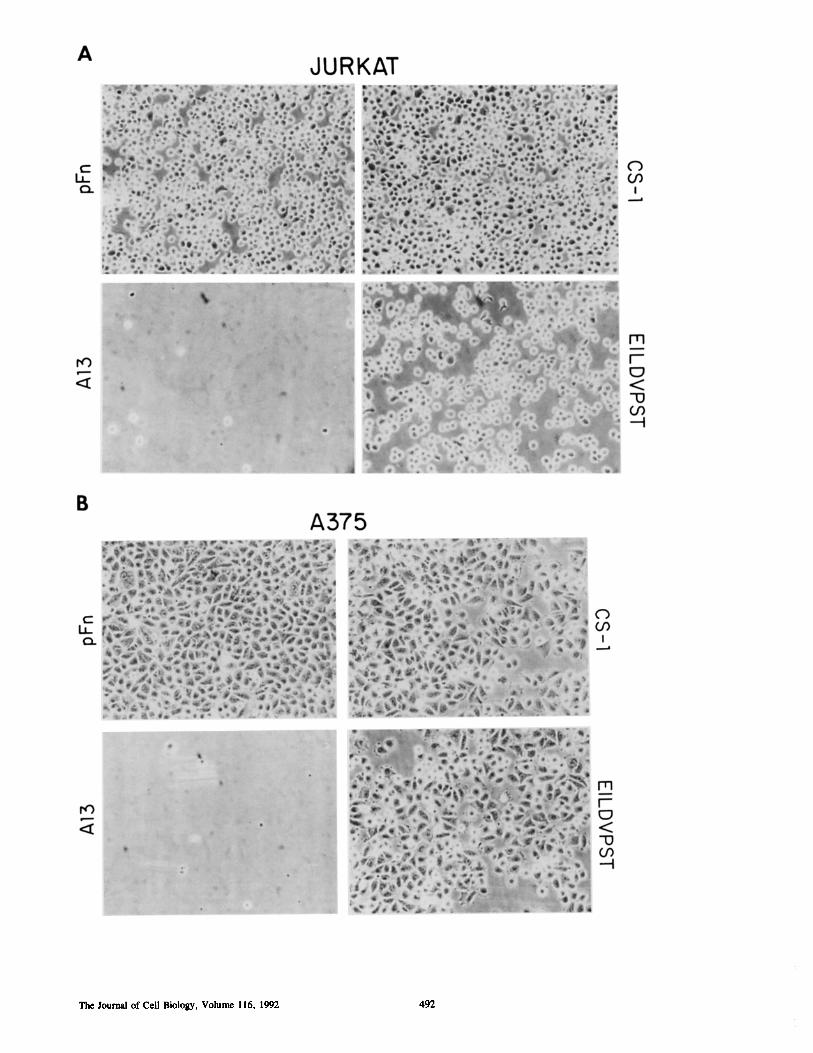
Figure 3. Adhesion of various hematopoetic cell lines to CS-1(o) or LDV (®) -coated surfaces . "Cr-labeled Jurkat (T lympho-blastoid), A375 (melanoma), U937 (monocytic), Ramos (B lym-phoblastoid), and ST-1 (EBV transformed B lymphoblastoid) cellswere allowed to adhere to CS-1-rsa or LDVPSTrsa (51g/ml pep-tide)-coated surfaces for 30 min at 37°C. At the end of this timethe nonadherent cells were washed offand the adherent cells weresolubilized inNaOH/SDS and quantitated in a gamma counter. Theresults are expressed as bound counts per minute.
deletion of the NHZ-terminal glutamic acid and isoleucineresidues resulted in a peptide with no inhibitory activity (notshown) .
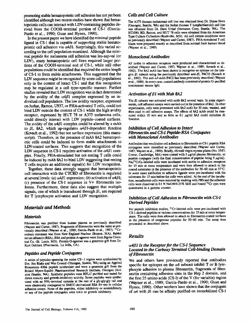
Minimal CS-1 FL-ptides Support Stable CellAdhesionand SpreadingIt was of interest to determine if CS-1 or derivative peptideswere capable of inducing stable hematopoietic cell adhesionand spreading. Therefore, RSA-peptide conjugates were pre-pared and their ability to support cell adhesion was examinedand compared to intact fibronectin and fragments of fibro-nectin containing the CTCBD (Table II and Figs. 2 and 3) .As expected CS-1 and a peptide containing EILDVwere ableto support the adhesion of Jurkat and A375 melanoma cells(Fig . 2, A and B). Interestingly, peptide conjugates contain-ing EILDV (Fig. 2 B) or LDV (not shown) also supportedmelanoma cell spreading. However, on a molar basis therewere significant differences in the ability of CS-1 versus CS-1derivative peptides to promote Jurkat cell adhesion (TableII) . In general, truncated CS-1 peptides were inefficientmediators of hematopoietic cell adhesion and none of thetruncated CS-1 peptide conjugates could support the adhe-sion of PHA activated T cell blasts (not shown) . Further-more, although the minimal peptide sequence required tosupport melanoma cell adhesion has been reported to beLDV (Mould et al ., 1991 ; Komoriya et al ., 1991) and, in ourhands, A375 melanoma cells did adhere to LDVPSTcoatedsurfaces (Fig . 3), of the hematopoietic cell lines we exam-
MI0
-X
aY v
c0
a
Wayner and Kovach Lymphocyte 81 Activation and LDVAdhesion
493
20
15
10
5
0
ADHESION SURFACE W
Figure 4. Adhesion of Jurkat cells to surfaces coated with pFN-,CS-1-, Al 3-, or B12-derived peptide-rsa conjugates in the presenceof mAb 8A2. (A) Adhesion in the presence ofpurified nonimmunemouse IgG (5 ug/ml) . (B) Adhesion in the presence of mAb 8A2(5 Ag/ml) . 48-well plates were coated with 5 ug/ml (based on pep-tide weight) peptide-rsa conjugates overnight in PBS at 4°C. s'Cr-labeled Jurkat cells were allowed to adhere to the peptide-coatedsurfaces in the presence of IgG or 8A2 for 30 min at 37°C. The restof the adhesion assay proceeded as for Fig. 3. Results are expressedas bound counts per minute .
ined only Jurkat cells adhered slightly to surfaces coatedwith the LDVPSTrsa conjugate (Fig . 3) . Furthermore, someof the hematopoietic cell lines we examined, such as U937or ST-1 cells did not adhere to surfaces coated with intactCS-1 (Fig . 3) . Thereason for the apparent inability of somecell populations to adhere to CS-1 or LDVPSTcoated sur-faces was not immediately obvious. Flow cytometry analysisrevealed that cell surface expression of a4 or X31 could notaccount for the functional differences we observed in theability of a particular cell population to adhere to CS-1 orLDVcoated surfaces . As we have previously reported (Way-ner et al ., 1989) U937 (monocytic) and ST-1 (B lympho-blastoid) cells express high levels of cell surface a4 and 01
Figure 2. Adhesion of Jurkat (A) or A375 melanoma cells (B) to plasma fibronectin (pFN), CS-1, A13, or EILDVPST-coated surfaces.Plasma fibronectin (pFN) CS-1, A13, or EILDVPSTrsa conjugates were coated on virgin styrene surfaces (5 ug/ml) . The A375 or Jurkatcells were allowed to adhere for 1 h. Nonadherent cells were washed off and the resulting monolayers were photographed with an invertingmicroscope and phase contrast . These data show that A375 cells spread on surfaces coated with LDVcontaining peptides . Evidence ofJurkat spreading can also be seen with CS-1 (A, top right) .
MOX
ôU VnvM COMmL
Q
MOX
ÔU nnMN C
OâtVQ
The Journal of Cell Biology, Volume 116, 1992
Activation ofthe a4ß1 ComplexEnhances Hematopoietic Cell RecognitionofCS-1 Peptides
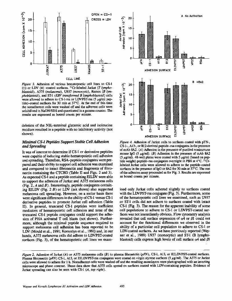
Figure 5. Adhesion of U937 cells to surfaces coated with pFN-,CS-1-, Al3-, or B12-derived peptide-rsa conjugates in the presenceof mAb SA2 . (A) Adhesion in the presence of purified nonimmunemouse IgG (5 pg/ml) . (B) Adhesion in the presence of mAb 8A2(5 jig/ml) . 48-well plates were coated with 5 pg/ml (based on pep-tide weight) peptide-rsa conjugates overnight in PBS at 4°C. "Cr-labeled U937 cells were allowed to adhere to the peptide-coatedsurfaces in the presence ofIgG or 8A2 for 30 min at 37°C . The restof the adhesion assay proceeded as for Fig . 3 . Results are expressedas bound counts per minute . U937 cells do not adhere to B12 pep-tides without activation .
which are equivalent to the levels expressed by Jurkat andA375 melanoma cells (Wayner et al ., 1989) . Together, thedata in Table II and Fig . 3 suggested that CS-1 or LDVpeptide recognition and binding might be regulated in a celltype specific manner with melanoma cells possessing thehighest affinity receptor.
The results of the preceeding experiments suggested thatadhesion of cells to CS-1 might involve the LDV sequenceand that interaction of a4ß1 with this sequence outside thecontext of intact CS-1 may be regulated in a cell-specificmanner. Several reports have suggested that the interactionof cells with ECM proteins may require activation (Neuge-bauer and Reichardt, 1991 ; Shimizu et al ., 1990) . Further-more, we have recently described a mAb to ßl, SA2, that up-regulates a4ß1-dependent lymphocyte adhesion to VCAM-1
494
ADHESION SURFACE W
ADHESION SURFACE W
Figure 6. Adhesion of HUT 78 cells to surfaces coated with pFN-,CS-1-, A13, or B12-derived peptide-rsa conjugates in the presenceofmAb 8A2 . (A) Adhesion in the presence ofpurified nonimmunemouse IgG (5 /ig/ml) . (B) Adhesion in the presence of mAb 8A2(5 ug/ml) . 48-well plates were coated with 5 vg/ml (based on pep-tide weight) peptide-rsa conjugates overnight in PBS at 4°C . 5 'Cr-labeled HUT 78 cells were allowed to adhere to the peptide-coatedsurfaces in the presence of IgG or SA2 for 30 min at 37°C . The restofthe adhesion assay proceeded as for Fig . 3. Results are expressedas bound counts per minute . Unlike U937 cells, HUT 78 cells ad-here to LDVcoated surfaces without activation.
(Kovach et al ., 1991) . Therefore, we examined the effects of8,A2 on hematopoietic cell adhesion to CS-1 and derivativepeptides . These results are shown in Figs . 4 (Jurkat), 5(U937 cells), and 6 (HUT 78) . As can be seen from thesedata Jurkat or U937 cells are capable ofmaximal interactionwith CS-1 and LDVcontaining derivative peptides only afteractivation with the 8A2 mAb . U937 cells, in fact, do not ad-here to any of the B12-derived peptides without activation .Since pretreatment ofcells with 8A2 does not up-regulate ex-pression of either 01 or a4 on U937 cells (not shown), thesedata strongly suggest that recognition of the LDV sequenceby a4ß1 requires an activation signal which can be trans-duced through01 . As we have previously shown (Tàbles I andII), VPSTrsa conjugates are inactive at inducing stable celladhesion (Figs . 4, 5, and 6) . These data strongly suggest thatthe minimal essential adhesion sequence in CS-1 for acti-vated hematopoietic cells is LDV. Interestingly, HUT 78cells appeared to possess an active a4ß1 complex ; restingHUT 78 cells adhered to LDV and this adhesion was notsignificantly up-regulated by 8A2 (Fig . 6) .
MONOCLONAL ANTIBODY
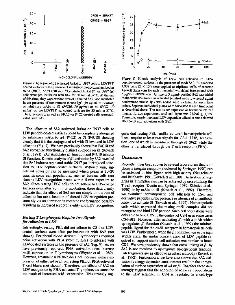
Figure 7. Adhesionof01 activated Jurkat or U937 cells to LDVPSTcoated surfaces in the presence ofinhibitory monoclonal antibodiesto a4 (P4C2) or 01 (P4C10). 5'Cr-labeled Jurkat (o) or U937 (®)cells were pre-incubated with 84,2 for 30 min at 37°C . At the endofthis time, they were washed free ofunbound 8A2, and incubatedin the presence of nonimmune mouse IgG (]0 Pg/ml = Control)or inhibitory mAbs to 01 (P4C10, 10 lzg/ml) or a4 (P4C2, 10ttg/ml) on the LDVPSTrsa-coated surfaces for 30 min at 37°C .Thus, the control as well as P4C10- or P4C2-treated cells were acti-vated with 8A2 .
The adhesion of 8A2 activated Jurkat or U937 cells toLDV peptide-coated surfaces could be completely abrogatedby inhibitory mAbs to a4 (P4C2) or ßl (P4C10) showingclearly that it is the conjugate of a4 with ßl involved in LDVadhesion (Fig. 7) . We have previously shown that P400 and8A2 recognize functionally distinct epitopes on ßl (Kovachet al ., 1991) : 8A2 stimulates ßl function and P4C10 inhibitsßl function . Kinetic analysis ofßl activation by 8A2 revealedthat 8A2 induces rapid and stable U937 (or Jurkat) cell adhe-sion to LDV peptide-coated surfaces . Within 5 min, sig-nificant adhesion can be measured which peaks at 10-20min . In some cell populations, such as Jurakat cells (notshown) LDV recognition peaks within 5-min exposure to8A2 . Since resting U937 cells do not adhere to LDVcoatedsurfaces even after 90 min of incubation, these data clearlyindicate that the effects of 8A2 are not simply on the rate ofadhesion but are a function of altered LDV recognition pre-sumably via an alteration in receptor conformation possiblyresulting in increased receptor avidity and LDV recognition .
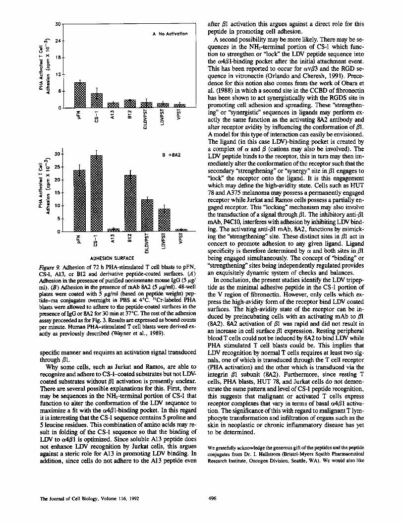
Resting TLymphocytes Require Two Signalsfor Adhesion to LDVInterestingly, resting PBL did not adhere to CS-1 or LDVcoated surfaces even after pre-incubation with 8A2 (notshown) . Peripheral blood-derived T lymphocytes requiredprior activation with PHA (72-h culture) to interact withLDVcoated surfaces in the presence of 8A2 (Fig. 9) . As wehave previously reported, PHA activation does increasea4ß1 expression on T lymphocytes (Wayner et al ., 1989) .However, treatment with 8A2 does not increase surface ex-pression of either a4 or ßl on resting PBL or PHAactivatedT cell blasts (not shown) . Therefore, the effects of 8A2 onLDV recognition by PHA-activated T lymphocytes cannot bethe result of increased a4ß1 expression . This strongly sug-
Wayner and Kovach Lymphocyte 61 Activation and LDVAdhesion
Figure 8. Kinetic analysis of U937 cell adhesion to LDVpeptide-coated surfaces in the presence of mAb 84,2 . "Cr-labeledU937 cells (2 x 10⁵) were applied to triplicate wells of separate48-well plates (one for each timepoint) which had been coated with5 ug/ml LDVPST-rsa . At time 0, 5 ug/ml-purified 8A2 was addedto the wells designated as activated (control wells to which 51g/mlnommmune mouse IgG was added were included for each timepoint) . Separate individual plates were harvested at each time pointas described above . The results are expressed as bound counts perminute. In this experiment total cell input was 28,796 t 1,389.Therefore, nearly maximal LDV-dependent adhesion was achievedafter 5-10 min activation with 8A2 .
gests that resting PBL, unlike cultured hematopoietic celllines, require at least two signals for CS-1 (LDV) recogni-tion, one of which is transduced through ßl (8A2) while theother is transduced through the T cell receptor (PHA) .
DiscussionRecently, it has been shown by several laboratories that lym-phocyte integrin receptors (reviewed by Springer, 1990) canbe activated to bind ligand with high avidity (Neugebauerand Reichardt, 1991 ; Kovach et al ., 1991) . Activation ofinte-grins in T lymphocytes can be achieved by cross-linking theT cell receptor (Dustin and Springer, 1989; Shimizu et al .,1990) or by mAbs to 01 (Kovach et al ., 1992) . Therefore,we examined hematopoietic cell adhesion to CS-1 andderivative peptides in the presence or absence of an antibodyknown to activate 01 (Kovach et al ., 1992) . Hematopoieticcells which expressed the resting a4ß1 complex did notrecognize and bind LDV peptide . Such cell populations wereonly able to bind LDV in thecontext ofCS-1 orin some casesCS-1-B12 . However, after activating ßl with a mAb whichup-regulates ßl function (Kovach et al ., 1992) the minimalpeptide ligand for the a4ß1 receptor in hematopoietic cellswas LDV. Furthermore, when the ßl complex was in the highavidity state, the molar concentration of LDV peptide re-quired to support stable cell adhesion was similar to intactCS-1 . We have previously shown that cross-linking of ßl by8A2 is not required to up-regulate ßl-dependent function ;Fab fragments are as effective as intact antibody (Kovack etal ., 1992) . Furthermore, we have also shown that 8A2 acti-vation is energy dependent and does not result in the upregu-lation of surface expression of a4 or ßl . Together these datastrongly suggest that the adhesion of some cell populationsto the LDV sequence in CS-1 is regulated in a cell-type
495
25~ MOPEN = JURKAT 30-M CROSS = U937
0
1c 20- X
â25- r +8A2
-~"-1_X 1Z 15- 2 20-0W 0 ô
t 152O 10 v
QaJ m 10
UWU 5 "0
nM "
5
0 r116-6 r-Ô~O---- -~0 Control
N O Ô N O 00 20 40 60 80 100
0â â Û
âUâ
Time (min)
M-Im o
MI
U O
ô E
30
24
18
12
6
0
ADHESION SURFACE
The Journal of Cell Biology, Volume 116, 1992
Figure 9. Adhesion of 72 h PHA-stimulated T cell blasts to pFN,CS4, A13, or B12 and derivative peptide-coated surfaces. (A)Adhesion inthe presence ofpurified nonimmune mouse IgG (5 lg/ml) . (B) Adhesion in the presence ofmAb SA2 (5 ug/ml) . 48-wellplates were coated with 5 pg/ml (based on peptide weight) pep-tide-rsa conjugates overnight in PBS at 4°C. 5'Cr-labeled PHAblasts were allowed to adhere to the peptide-coated surfaces in thepresence ofIgG or8A2 for 30 minat 37°C . The rest ofthe adhesionassayproceeded as forFig. 3 . Results are expressed as bound countsper minute. Human PHA-stimulated T cell blasts were derived ex-actly as previously described (Wayner et al ., 1989) .
specific manner and requires an activation signal transducedthrough ßl .Why some cells, such as Jurkat and Ramos, are able to
recognize and adhere to CS-1-coated substrates but not LDVcoated substrates without ßl activation is presently unclear.There are several possible explanations for this . First, theremay be sequences in the NH2-terminal portion of CS-1 thatfunction to alter the conformation of the LDV sequence tomaximize a fit with the a4ß1-binding pocket . In this regardit is interesting that the CS-1 sequence contains 5 proline and5 leucine residues . This combination ofamino acids may re-sult in folding of the CS-1 sequence so that the binding ofLDV to a4ß1 is optimized . Since soluble Al3 peptide doesnot enhance LDV recognition by Jurkat cells, this arguesagainst a steric role for A13 in promoting LDV binding . Inaddition, since cells do not adhere to the A13 peptide even
after ßl activation this argues against a direct role for thispeptide in promoting cell adhesion .A second possibility may be more likely. There may be se-
quences in the N112-terminal portion of CS-1 which func-tion to strengthen or "lock" the LDV peptide sequence intothe a4ß1-binding pocket after the initial attachment event.This has been reported to occur for avß3 and the RGD se-quence in vitronectin (Orlando and Cheresh, 1991) . Prece-dence for this notion also comes from the work of Obara etal . (1988) in which a second site in the CCBD of fibronectinhas been shown to act synergistically with the RGDS site inpromoting cell adhesion and spreading . These "strengthen-ing" or "synergistic sequences in ligands may perform ex-actly the same function as the activating 8A2 antibody andalter receptor avidity by influencing the conformation of ßl .A model for this type ofinteraction can easily be envisioned .The ligand (in this case LDV)-binding pocket is created bya complex of a and ß (cations may also be involved) . TheLDV peptide binds to the receptor, this in turn may then im-mediately alter the conformation ofthe receptor such that thesecondary "strengthening" or "synergy" site in ßl engages to"lock" the receptor onto the ligand . It is this engagementwhich may define the high-avidity state. Cells such as HUT78 and A375 melanoma may possess a permanently engagedreceptor while Jurkat and Ramos cells possess a partially en-gaged receptor. This "locking" mechanism may also involvethe transduction of a signal through ßl . The inhibitory anti-ßlmAb, P4C10, interferes with adhesion by inhibiting LDVbind-ing . The activating anti-ßl mAb, 8A2, functions by mimick-ing the "strengthening" site . These distinct sites in ßl act inconcert to promote adhesion to any given ligand . Ligandspecificity is therefore determined by a and both sites in ßlbeing engaged simultaneously. The concept of "binding" or"strengthening" sites being independently regulated providesan exquisitely dynamic system of checks and balances .
In conclusion, the present studies identify the LDV tripep-tide as the minimal adhesive peptide in the CS-1 portion ofthe V region of fibronectin . However, only cells which ex-press the high-avidity form of the receptor bind LDV coatedsurfaces. The high-avidity state of the receptor can be in-duced by preincubating cells with an activating mAb to ßl(8A2) . 8A2 activation of ßl was rapid and did not result inan increase in cell surface ßl expression . Resting peripheralblood T cells could not be induced by 8A2 to bind LDV whilePHA stimulated T cell blasts could be. This implies thatLDV recognition by normal T cells requires at least two sig-nals, one of which is transduced through the T cell receptor(PHA activation) and the other which is transduced via theintegrin ,ßl subunit (8A2) . Furthermore, since resting Tcells, PHA blasts, HUT 78, and Jurkat cells do not demon-strate the same pattern and level ofCS-1 peptide recognition,this suggests that malignant or activated T cells expressreceptor complexes that vary in terms of basal a4ß1 activa-tion . The significance ofthis with regard to malignant T lym-phocyte transformation and infiltration of organs such as theskin in neoplastic or chronic inflammatory disease has yetto be determined.
We gratefully acknowledge the generous gift of the peptides andthe peptideconjugates from Dr. I. Hellstrom (Bristol-Myers Squibb PharmaceuticalResearch Institute, Oncogen Division, Seattle, WA) . We would also like
496
to thankDr . David Cheresh (La Jolla, CA) and Dr . William Carter (Seattle,WA) for their valuable assistance and critical comments on the manuscript .
Dr . N . Kovach is supported by U.S . Public Health Service grants HL-and HL-07093-16 .
Received for publication 7 August 1991 and in revised form 20 September1991 .
References
Bernardi, P ., V. P . Patel, and H . F. Lodish . 1987 . Lymphoi d precursor cellsadhere to two different sites on fibronectin . J. Cell Biol. 105 :489-498 .
Dustin, M . L ., and T . A . Springer . 1989 . T-cell receptor cross-linking tran-siently stimulates adhesiveness through LFA-1 . Nature (Land.). 341 :619-624 .
Garcia-Pardo, A ., A . Rostagno, and B . Frangione . 1987 . Primary structure ofhuman plasma fibronectin . Characterization of a 38 kDa domain containingthe C-terminal heparin-binding site (Hep III site) and a region of molecularheterogeneity . Biochem . J . 241 :923-928 .
Garcia-Pardo, A ., E . A. Wayner, W . G . Carter, and O . C. Ferreira . 1990. Hu-man B lymphocytes define an alternative mechanism of adhesion to fibronec-tin . The interaction of the a4ß1 integrin with the LHGPEILDVPST sequenceof the type III connecting segment is sufficient to promote cell attachment .J. Immunol. 144 :3361-3366.
Guan, J . L ., and R . O . Hynes . 1990. Lymphoid cells recognize an alternativelyspliced segment of fibronectin via the integrin receptor a4ß1 . Cell. 60 :53-61 .
Humphries, M . J ., S. K . Akiyama, A . Komoriya, K . Olden, and K . M . Yamada .1986. Identification of an alternatively spliced site in human plasma fibro-nectin that mediates cell type-specific adhesion . J. Cell Biol. 103 :2637-2647 .
Humphries, M . J ., A . Komoriya, S . K. Akiyama, K. Olden, and K . M . Yamada .1987 . Identificationoftwo distinct regions ofthe type III connecting segmentof human plasma fibronectin that promote cell type-specific adhesion . J.Biol. Chem. 262 :6886-6892 .
Komoriya, A ., L . J . Green, M . Mervic, S . Yamada, K . M . Yamada, and M . J .Humphries. 1991 . The minimal essential sequence for a major cell type spe-cific adhesion site (CS-1) within the alternatively spliced type III connectingsegment domain of fibronectin is leucine-aspartic acid-valine . J. Biol . Chem .266 :15075-15079 .
Komblihtt, A . R ., K. Umezawa, K . Vibe-Pedersen, and F . Baralle . 1985. Pri-mary structure of human plasma fibronectin: differential splicing may gener-ate at least 10 polypeptides from a single gene . EMBO (Eur. Mol. Biol. Or-gan .) J . 4 :1755-1759 .
Kovach, N . L ., T . M . Carlos, E . Yee, and J . M . Harlan . 1992 . A monoclonalantibody to 01 integrin (CD29) stimulates VLA-4-dependent adherence ofleukocytes to vascular cell adhesion molecule-1 (VCAM-1) . J. Cell Biol.116 :499-509 .
Liao, N . S ., J . St . John, Z . J . Du, H . T . Cheung . 1987 . Adhesion of lymphoidcell lines to fibronectin-coated substratum : biochemical and physiologicalcharacterization and the identification of a 140-kDa fibronectin receptor.
Wayner and Kovach Lymphocyte 81 Activation and LDV Adhesion
Exp. Cell Res. 171 :306-320 .McCarthy, J . B ., S . T . Hagen, and L . T . Furcht . 1986 . Human fibronectin con-
tains distinct adhesion and motility promoting domains for metastatic mela-noma cells . J. Cell Biol . 102 :179-188 .
Merrifield, R. B . 1963 . Solid phase peptide synthesis . I . The synthesis of atetrapeptide . J. Am. Chem. Soc. 85 :2149-2154 .
Mould, A . P ., L. A . Wheldon, A. Komoriya, E . A . Wayner, K . M . Yamada,and M . J . Humphries . 1990 . Affinity chromatographic isolation of the mela-nomaadhesion receptor for the IIICS region of fibronectin and its identifica-tion as the integrin a4ß1 . J. Biol. Chem . 265:4020--4024 .
Mould, P . A ., A . Komoriya, K . Yamada, and M . J . Humphries . 1991 . Th eCS5 peptide is a second site in the IIICS region of fibronectin recognizedby the integrin a4ß1 . J. Biol. Chem . 266 :3579-3585 .
Orlando, R . A ., andD. A . Cheresh . 1991 . Arg-Gly-Asp-binding leading to mo-lecular stabilization between integrin av03 and its ligand. J. Biol. Chem . Inpress .
Neugebauer, K. M ., and L . F . Reichardt . 1991 . Cell-surface regulation of 01-integrin activity on developing retinal neurons . Nature (Land.).350:68-71 .
Obara, M ., M.S . Kang, and K . M . Yamada . 1988 . Site-directed mutagenesisof the cell-binding domain ofhuman fibronectin : separable, synergistic sitesmediate adhesive function . Cell. 53 :649-657 .
Paul, J . I ., J . E. Schwarzbauer, J . W . Tamkun, and R . O . Hynes. 1986 . Cell-type-specific fibronectin subunits generated by alternative splicing . J. Biol.Chem. 261 :12258-12265 .
Pierschbacher, M . D ., and E . Ruoslahti . 1984 . Cell attachment activity offibronectin can be duplicated by small synthetic fragments of the molecule.Nature (Land.). 309 :30-33 .
Pytela, R ., M.D . Pierschbacher, and E . Ruoslahti . 1985 . Identification and iso-lation of a 140 kd cell surface glycoprotein with properties expected of afibronectin receptor . Cell. 40:191-198 .
Schwarzbauer, J . E ., J . I . Paul, and R. O . Hynes . 1985 . O n the origin of speciesof fibronectin . Proc. Natl . Acad. Sci. USA. 82 :1424-1428 .
Schwarzbauer, J . E ., J . W . Tamkun, I. R . Lemischka, and R. O . Hynes. 1983 .Thre e different fibronectin mRNAs arise by alternative splicing within thecoding region . Cell. 35 :421-431 .
Shimizu, S ., G . A . Van Seventer, K . J . Morgan, and S . Shaw . 1990 . Regulatedexpression and binding of three VLA (/31) integrin receptors on T cells . Na-ture (Land.) . 345 :250-253 .
Springer, T . A. 1990 . Adhesion receptors of the immune system . Nature(Land.). 346:425-434 .
Wayner, E . A ., andW . G . Carter. 1987 . Identification of multiple cell adhesionreceptors for type VI collagen and fibronectin in human fibrosarcoma cellspossessing unique a and common 0 subunits . J. Cell Biol. 105 :1873-1884 .
Wayner, E . A ., W . G . Carter, R . Piotrowicz, and T . J . Kunicki. 1988 . Thefunction of multiple extracellular matrix receptors (ECMRs) in mediatingcell adhesion to ECM : preparation ofmonoclonal antibodies to the fibronec-tin receptor that specifically inhibit cell adhesion to fibronectin and react withplatelet glycoproteins Ic/IIa . J. Cell Biol. 107 :1881-1891 .
Wayner, E . A ., A . Garcia-Pardo, M . J . Humphries, J . A . McDonald, andW . G. Carter . 1989 . Identificationand characterization of the T lymphocyteadhesion receptor for an alternative cell attachment domain (CS-1) in plasmafibronectin . J. Cell Biol . 109 :1321-1330 .
497