an advanced in vitro liver tissue model by combination of on-site
TRANSCRIPT

659© 2008, Japanese Society for Alternatives to Animal Experiments
An advanced in vitro liver tissue model by combination of on-site oxygenation and double-layer coculture with fibroblasts
Masaki Nishikawa1, Nobuhiko Kojima1, Takatoki Yamamoto1, Teruo Fujii1 and Yasuyuki Sakai1, 2
1Institute of Industrial Science, University of Tokyo2Center for Disease Biology and Integrative Medicine, Graduate School of Medicine, University of Tokyo
Corresponding author: Masaki NishikawaInstitute of Industrial Science, University of Tokyo4-6-1 Komaba, Meguro-ku, Tokyo 153-8505, Japan
Phone: +(81)-3-5452-6349, Fax: +(81)-3-5452-6348, [email protected]
AbstractIn in vivo liver tissue, each hepatocyte has intimate interactions not only with adjacent hepatocytes but also with nonparenchymal cells in a three-dimensional (3D) manner. We recently reported that hepatic function is highly maintained on collagen covalently-immobilized poly-dimethylsiloxane (PDMS) membranes through which oxygen is supplied directly to the cells. In this study, to further enhance performances of hepatocytes culture, we investigated cocultivation of rat hepatocytes with a mouse fibroblast, NIH/3T3 (3T3) in the same PDMS membranes. Various functions of hepatocytes were better maintained on the membrane at remarkably higher levels, particularly albumin secretion on such coculture was about 20 times higher than that in conventional coculture on tissue-culture-treated polystyrene (TCPS) surfaces. The remarkable functional enhancements are likely to be explained by the net growth of hepatocytes and very intimate contact between hepatocytes and 3T3 cells in almost continuous double-layered structures under the adequate oxygen supply. The results demonstrate that simultaneous realization of different requirements toward mimicking in vivo liver tissue microstructure is effective in improving performance of hepatocytes culture system.
Keywords: rat hepatocytes, coculture, oxygenation, poly-dimethylsiloxane, liver tissue model
1. IntroductionImprovement of primary culture of hepatocyte
is definitely important for in vitro toxicity tests for various industrial and clinical chemicals. However, it is still difficult to maintain in vivo-like high liver-specific functions in in vitro culture. In in vivo liver tissue, each hepatocyte has intimate interactions not only with adjacent hepatocytes but also with nonparenchymal cells in a three-dimensional (3D) manner. In in vitro, however, it was difficult to realize such 3D interactions (Harimoto 2002). Apparent shortage of oxygen supply to the hepatocytes was reported previously, because the hepatocytes consume oxygen at a 10 times higher rate than other cell types (Smith et al. 1996). Then, on-site oxygenation using highly-oxygen-permeable materials has been shown to improve hepatocyte functions (De Bartolo et al. 2006; Tilles et al. 2001). However, there were few reports that investigated high-cell-density coculture under such direct oxygenation. In this report, the combined effect of the coculture and the on-site oxygenation on primary rat hepatocytes was investigated.
2. Materials and methods2.1 Cell Culture Plates
Collagen covalently immobilized PDMS plates and collagen coated tissue culture polystyrene (TCPS-CN) plates (IWAKI, Japan) were used to culture cells. The PDMS plates were prepared according to a procedure described previously (Nishikawa et al. 2007a; Nishikawa et al. 2007b). Briefly, a PDMS membrane (thickness: 1.5 mm) was stuck and clamped between a 24-well poly-carbonate (PC) frame and a stainless-steel board possessing 24 holes not to inhibit oxygen permeation through the PDMS membranes (PDMS plate). Subsequently, PDMS plates were treated by oxygen plasma and coupled with aminosilane (Shinetsu Silicone, KBE-903; Japan). The introduced amino groups were reacted with a photo-reactive cross linker, SAND (Pierce, P-C21549; USA), by exposing to UV light. Using this cross linker, collagen Type 1-P (Nitta gelatin, Japan) was covalently immobilized on the PDMS surface.
AATEX 14, Special Issue, 659-663Proc. 6th World Congress on Alternatives & Animal Use in the Life SciencesAugust 21-25, 2007, Tokyo, Japan
660
Masaki Nishikawa, et. al.
2.2 NIH/3T3 and hepatocytes preparationsFor NIH/3T3 (3T3) culture, Dulbeccos modified
Eagle medium (DMEM) (Kojin Bio; Japan) was used as a base and supplemented with 10% fetal bovine serum (FBS) (Gemini Bio-Product; USA), 20 mM HEPES (Dojindo; Japan), 100 units-penicillin/ml, 100 µg streptomycin/ml, and 1 µg amphotericin/ml (Wako; Japan).
For the culture of rat primary hepatocytes, serum free (SF) DMEM and 5% FBS DMEM were prepared as follows. For SF DMEM, the medium for 3T3 was additionally supplemented with non-essential amino acid (Gibco; USA), 10 ng/ml mouse epidermal growth factor (EGF), 0.1 µM insulin (Takara; Japan), 0.1 µM dexamethasone, 0.8 µM copper sulfate (CuSO4, 5H2O), 2 nM selenious acid (H2SeO3), 2.6 µM zinc sulfate (ZnSO4, 7H2O), 0.3 µM manganese sulfate (MnSO4, 5H2O) (Wako; Japan). For 5% FBS DMEM, fi rstly 10% FBS DMEM was prepared by supplying with 10% FBS, HEPES, antibiotics, EGF, insulin, and dexamethasone at the same concentrations as SF DMEM, and was mixed with SF DMEM at a ratio of 1:1.
A m a l e Wi s t a r r a t a g e d 5 – 8 w e e k s w a s anaesthetized and its hepatocytes were isolated by the collagenase perfusion method. Viability was determined by the trypan blue exclusion method and hepatocytes with over 85% viability were used for the culture.
2.3 Homoculture and Coculture of Hepatocytes and NIH/3T3 cells
For the homoculture, isolated hepatocytes were inoculated and cultured at a cell density of 1.1 × 10⁵ viable-cells/cm2 in TCPS-CN plates and surface-modifi ed PDMS plates described in 2.1.
Simultaneously, two types of double-layered coculture were investigated. In the first type coculture, hepatocytes were cultured on the NIH/3T3 (3T3) monolayer (Hep/3T3 coculture). The 3T3 cells were inoculated at a cell density of 1 × 10⁵ viable-cells/cm2 in TCPS-CN plates and surface-modified PDMS plates and cultured for eight hours. Then, isolated hepatocytes were inoculated in the same way as the homoculture. In the second type coculture, 3T3 cells were cultured on the hepatocytes monolayer (3T3/Hep coculture) at 24 hour after the hepatocytes inoculation in reverse order of the Hep/3T3 coculture.
The experiments were performed during 13 days in an incubator at 37 °C, 5% CO2, with 100 % humidity. The culture medium, 5% FBS DMEM, was changed at 24 hour after the inoculation and then after everyday. Morphologies of cells were routinely observed with phase-contrast microscopy (Olympus, ITM-2; Japan).
2.4 Measurement of various cellular activitiesWe measured albumin secretion, ammonium
removal, urea synthesis, and numbers of survival cells. Secreted albumin in the culture medium was measured by the sandwich-type enzyme-linked immuno-sorbent assay (ELISA). Anti-rat albumin goat antibody (MP Biomedicals-Cappel products, Irvine, CA) was used as the primary antibody, and horseradish peroxidase-conjugated anti-rat albumin sheep antibody (MP Biomedicals-Cappel products) was used as the secondary antibody. Once every two days the culture mediums were analyzed using ELISA.
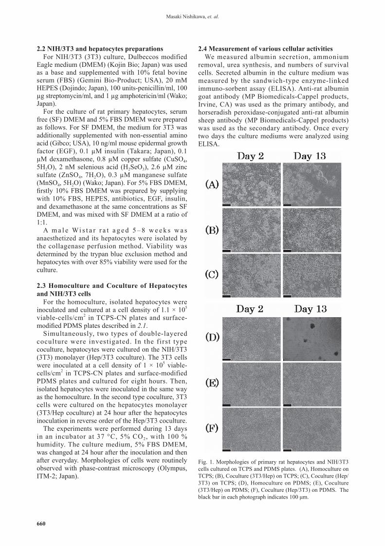
Fig. 1. Morphologies of primary rat hepatocytes and NIH/3T3 cells cultured on TCPS and PDMS plates. (A), Homoculture on TCPS; (B), Coculture (3T3/Hep) on TCPS; (C), Coculture (Hep/3T3) on TCPS; (D), Homoculture on PDMS; (E), Coculture (3T3/Hep) on PDMS; (F), Coculture (Hep/3T3) on PDMS. The black bar in each photograph indicates 100 μm.

661
On Day 7 and Day 13, to measure urea synthesis, the culture medium was changed to SF DMEM con ta in ing 2mM ammonium ch lo r ide fo r 2 hours. Urea concentration was measured by the diacecylmonooxium methods.
2.5 Histological staining of cocultured cells on PDMS
For both the cocutlures on PDMS, that is, 3T3/Hep and Hep/3T3, remaining cells were fixed on Day 13 in formalin solution, and embedded in paraffin. Seven-micron thick vertical sections against the cell layers were obtained from the center region of each specimen, and stained with hematoxylin and eosin (HE).
3. Results3.1 Morphologies of rat hepatocytes
In the homoculture on usual TCPS, a rapid decrease in the number of hepatocytes and a slight growth of fi broblast like cells that might be initially contained in the hepatocyte fraction were observed (Fig. 1 (A)). In the both two types of coculture on usual TCPS,
3T3/Hep and Hep/3T3, hepatocytes monolayers were maintained until around Day 7, and then they were detached gradually. However, hepatocytes remained better in these cocultures than in the homoculture on TCPS (Fig. 1 (B), (C)). On PDMS, even on Day 2, the surface densities of hepatocytes were clearly higher than those on TCPS (Fig. 1), suggesting that hepatocytes grew actively during the initial two days. Thereafter, in the homoculture on PDMS, detachment of hepatocytes and a slight growth of fi broblast-like cells were also observed, but hepatocytes monolayer gradually changed into stably attached spheroids (Fig. 1 (D)). In contrast, in the two types of coculture on PDMS, hepatocytes monolayers were completely maintained until the end of the culture (Fig. 1 (E), (F)).
Fig. 2. Photographs of hematoxylin and eosin staining of vertical thin sections of two types of coculture on PDMS on Day 13. (A), Coculture (3T3/Hep) on PDMS; (B), Coculture (Hep/3T3) on PDMS. The black bar in each photograph indicates 20 μm.
Fig. 3. Time course changes in albumin production of the six culture groups. Homoculture and cocultures on TCPS (A) and on PDMS (B). Point and error bar represent the mean +/- S.D. of six wells from two independent experiments.

662
Masaki Nishikawa, et. al.
3.2 Histological stain of cocultured cells on PDMSAs shown in Fig. 1, usual observation using
a phase-inverted microscope did not give clear information about the proximity between hepatocytes and 3T3 cells cultured on PDMS surfaces. We therefore prepared thin sections vertical to the cell layers and did histological staining on Day 13 as shown in Fig. 2. These photographs show that in both types of coculture almost complete double-layered structure of hepatocytes and 3T3 cell monolayers were formed and maintained until the end of the culture in the same stacking order in the inoculation.
3.3 Measurement of cellular albumin secretion Both on TCPS and on PDMS, albumin secretion
remarkably increased and was maintained by coculture (Fig. 3 (A), (B)) when compared with those in homocultures. Namely in these cocultures, albumin secretion gradually increased, peaked during Day 7 to Day 9, and was maintained at high levels until the end of culture. In homoculture, albumin secretion on PDMS was better maintained than that on TCPS even after the initial decrease until Day 7. This result was consistent with the morphological observation as shown in Fig. 1. In addition, there were marked differences in the secretion levels between cocultures on TCPS and PDMS; the secretion on PDMS was about 20 times higher than that on TCPS, irrespective of the stacking order of cell-layers.
3.4 Urea synthesisIn urea synthesis rates , cul tures on PDMS
exhibited much higher rates than those on TCPS. Both on PDMS and TCPS, coculture with 3T3 cells enhanced the rates, particularly those on Day 13 (Fig. 4), irrespective of the stacking order of the two cell populations. These results agree well with the morphological changes shown in Fig. 1.
4. DiscussionIn this report, we observed dramatic enhancement
of the culture performances by the combination of predicted adequate oxygen supply and the coculture with 3T3 fibroblast cells using surface-modified PDMS. In the coculture on PDMS, various functions of hepatocytes were better maintained at a remarkably higher level (Fig. 3, 5), particularly albumin secretion on such coculture on PDMS was about 20 times higher than even that in coculture on TCPS (Fig. 3). Such remarkable enhancements are likely to be explained by the remarkably higher number of retained hepatocytes in culture and very close contact between hepatocytes and 3T3 cells in almost continuous double-layered structures which were realized under predicted sufficient oxygen supply, as revealed in Fig. 2.
By using PDMS, the oxygen requirement of hepatocytes can be satisfied adequately (Nishikawa et al. 2007a; Nishikawa et al. 2007b). This is mainly because of the higher oxygen diffusion coefficient in PDMS (4.1 × 10⁻⁵ cm2/sec) compared with culture medium (2 × 10⁻⁵ cm2/sec) and the nine-times higher solubility of oxygen in PDMS (0.18 cm3 (STP)/cm3 atm: 10.6 nmol/ml/mmHg) compared with that in medium (1.19 nmol/ml/mmHg). Consequently, the maximum oxygen flux through PDMS is calculated 0.41 nmol/sec/cm2, and can satisfy the oxygen consumption of hepatocytes, 0.05 – 0.1 nmol/sec/cm2. In equilibrated state, the oxygen concentration in the vicinity of hepatocytes is estimated at about 150 nmol/ml, which is close to the concentration in the medium equilibrated to the gaseous phase (170 nm/ml).
The coculture performed in this study enables hepatocytes to make close contact at an individual cell level not only with adjacent hepatocytes but also with a 3T3 cell monolayer over or beneath the hepatocytes monolayer. The effects of coculture are known to be quite limited only in hepatocytes directly contacting nonparenchymal cells. Harimoto et al. demonstrated the overlay of pre-incubated endothelial cell sheet onto hepatocytes monolayer (Harimoto 2002). However, in the coculture, the oxygen shortage limits the hepatic functions and the flexibility of cocultures. In contrast, this report demonstrated that a high oxygen permeable material, PDMS, realized the almost continuous double-layered coculture by just direct overlaying of the second cells suspension onto the first cells monolayer.
PDMS has an advantage of the applicability to various microdevices over other oxygen permeable materials. However, one big problem of PDMS in cell culture is the detachment of cells (Lee et al. 2004). We showed that collagen conjugated PDMS inhibited the detachment of both rat primary hepatocytes and 3T3 cells from PDMS (Nishikawa et al. 2007a; Nishikawa et al. 2007b). Using such
Fig. 4. Urea synthesis rate of the six culture groups on Day 7 and Day 13. Column and error bar represent the mean +/- S.D. of six wells from two independent experiments.

663
surface-modified PDMS, various complicated culture systems, such as cell culture insert-based separated double-layered coculture, can be investigated with better flexibility (Choi et al. 2004). In addition, the encouraging results of this study also shows a promise in better performances in PDMS-based liver tissue microdevices (Leclerc et al. 2004; Ostrovidov et al. 2004).
In conclusion, the double-layered coculture system of hepatocytes and 3T3 cells on surface-modified PDMS with direct oxygenation remarkably improved performance of rat hepatocytes.
Acknowledgments This work was supported in part by both the Global
COE Program for Chemistry Innovation and a NEDO (New Energy and Industrial Technology Development Organization) project, Development of Simple and Highly Functional Hazardous Assessment Methods.
ReferencesChoi SH, Nishikawa M, Sakoda A, Sakai Y. (2004) Feasibility
of a simple double-layered coculture system incorporating metabolic processes of the intestine and liver tissue: application to the analysis of benzo[a]pyrene toxicity. Toxicol In Vitro 18:393-402.
De Bartolo L, Salerno S, Morelli S, Giorno L, Rende M, Memoli B, Procino A, Andreucci VE, Bader A, Drioli E. (2006) Long-term maintenance of human hepatocytes in oxygen-permeable membrane bioreactor. Biomaterials 27(27):4794-803.
Harimoto MY, M. Hirose, C. Takahashi, Y. Isoi, A. Kikuchi, T. Okano. (2002) Novel approach for achieving double-layered cell sheets co-culture: overlaying endothelial cell sheets onto monolayer hepatocytes utilizing temperature-responsive culture dishes. Journal of Biomedical Materials Research 62(3):464-470.
Leclerc E, Sakai Y, Fujii T. (2004) Microfluidic PDMS (polydimethylsiloxane) bioreactor for large-scale culture of hepatocytes. Biotechnol Prog 20(3):750-5.
Lee JN , J i ang X , Ryan D , Whi t e s ides GM. (2004) Compatibil i ty of mammalian cells on surfaces of poly(dimethylsiloxane). Langmuir 20(26):11684-91.
Nishikawa M, Nobuhiko K, Komori K, Yamamoto T, Fujii T, Sakai Y. (2007a) Enhanced maintenance and functions of rat hepatocytes induced by combination of on-site oxygenation and coculture with fibroblasts. J Biotechnol 133(2):253-260.
Nishikawa M, Yamamoto T, Nobuhiko K, Komori K, Fujii T, Sakai Y. (2007b) Stable immobilization of rat hepatocytes as hemispheroids onto collagen-conjugated poly-dimethylsiloxane (PDMS) surfaces: Importance of direct oxygenation through PDMS for both formation and function. Biotechnol Bioeng accepted.
Ostrovidov S, Jiang J, Sakai Y, Fujii T. (2004) Membrane-based PDMS microbioreactor for perfused 3D primary rat hepatocyte cultures. Biomed Microdevices 6(4):279-87.
Smith MD, Smirthwaite AD, Cairns DE, Cousins RB, Gaylor JD. (1996) Techniques for measurement of oxygen consumption rates of hepatocytes during attachment and post-attachment. Int J Artif Organs 19(1):36-44.
Tilles AW, Baskaran H, Roy P, Yarmush ML, Toner M. (2001) Effects of oxygenation and flow on the viability and function of rat hepatocytes cocultured in a microchannel flat-plate bioreactor. Biotechnol Bioeng 73(5):379-389.
