recent advances in the development of in vitro liver
TRANSCRIPT

Vol.:(0123456789)1 3
Bio-Design and Manufacturing (2021) 4:717–734 https://doi.org/10.1007/s42242-021-00142-7
REVIEW
Recent advances in the development of in vitro liver models for hepatotoxicity testing
Siming Lu1 · Jingwei Zhang5 · Sha Lin1 · Danfeng Zheng1 · Yan Shen1 · Jiale Qin4 · Yangyang Li4 · Shuqi Wang2,3
Received: 15 November 2020 / Accepted: 4 June 2021 / Published online: 5 August 2021 © Zhejiang University Press 2021
AbstractLiver injury is a common cause of drug approval withdrawal during drug development, pre-clinical research, and clinical treatment. If not properly treated, patients with severe liver injury can suffer from acute liver failure or even death. Thus, utilization of the convenient in vitro hepatotoxicity assessment model for early detection of drug-induced hepatotoxicity is vital for drug development and safe personalized medication. Biomaterials (e.g., hydrogels, nanofibers, decellularized liver matrix) and bioengineering technologies (e.g., microarrays, micropatterns, 3D printing, and microfluidics) have been applied for in vitro hepatotoxicity assessment models. This review summarizes the structure and functions of the liver as well as the components of in vitro hepatotoxicity assessment models. In addition, it highlights the latest advances in developing hepatotoxicity models with the ultimate goal of further clinical translation.
Graphic abstract
Keywords Liver injury · Drug toxicity · In vitro model · 3D culture
Extended author information available on the last page of the article

718 Bio-Design and Manufacturing (2021) 4:717–734
1 3
Introduction
Drug-induced liver injury (DILI) remains a common cause of drug withdrawal in drug development, pre-clinical research, and clinical treatment [1]. Patients with severe liver injury may suffer from acute liver failure or even death. The annual incidence of DILI is reported to be between 1 in 10,000 and 1 in 1,000,000 people worldwide [1]. In addition, Chinese medicine and dietary supple-ments are believed to be important factors causing DILI [2–6]. Rarely, medications can induce hepatotoxicity in individuals with the toxicity remaining undetected until the late clinical stage [7]. Moreover, the response of differ-ent individuals to the same dose of the same drug and the final progress of the disease are different [8, 9]. Therefore, there is an urgent need to develop an effective and reliable method to investigate liver toxicity in the drug develop-ment stage, before clinical usage.
The importance of in vitro hepatotoxicity assessment models is widely recognized, since they can help prevent hepatotoxicity and are vital for drug development and safe personalized medication [10, 11]. Current methods for drug screening in liver disease mainly depend on in vitro cell culture and in vivo animal models [10, 12]. Among these methods, human primary hepatocyte culture is considered the gold standard for in vitro hepatotoxic-ity assessment models. However, human primary hepato-cytes de-differentiate and rapidly lose liver-specific func-tions when maintained in conventional two-dimensional (2D) monolayer culture [10]. As such, conventional 2D human primary hepatocyte cultures for long-term stud-ies of liver biology and assays that require liver-specific functions. Although animal models can mimic the human in vivo environment, they cannot always predict toxicity in human due to fundamental biological differences [13, 14]. Moreover, these animal models have a high cost, a long experimental period, and doubtful ethics and thus are not suitable for rapid hepatotoxicity testing and drug discov-ery. More recently, the shortcomings of traditional in vitro drug testing models have been overcome with the advent of the three-dimensional (3D) in vitro cell culture models. Likewise, the 3D liver drug testing models provide rapid and simple detection of drug hepatotoxicity, offering great advantages over 2D cell models and animal models for drug development and safe personalized medication [11, 15]. In vitro liver models, such as liver spheroids [16], scaffold-based 3D tissue construction [17, 18], 3D printing [19, 20], and livers-on-a-chip [21–24], have been widely explored for the detection of drug hepatotoxicity. The 3D cell culture system can establish a 3D growth state simi-lar to tissues and organs in vivo, accurately simulating the in vivo microenvironment [25–29]. Drug screening
models based on 3D in vitro cell culture more accurately demonstrate toxic responses and can also be used in high-throughput screening and individualized assays.
The in vitro drug hepatotoxicity model is a promising and rapidly emerging branch of liver tissue engineering. In this review, we provide an overview of 3D hepatotoxicity testing models. First, we give a detailed introduction to liver structure and liver function. Second, we summarize the com-ponents of the liver model and review the latest advances in in vitro hepatotoxicity assessment models. Finally, the com-mon obstacles and future prospects for developing in vitro hepatotoxicity assessment models are discussed.
Liver structure and function
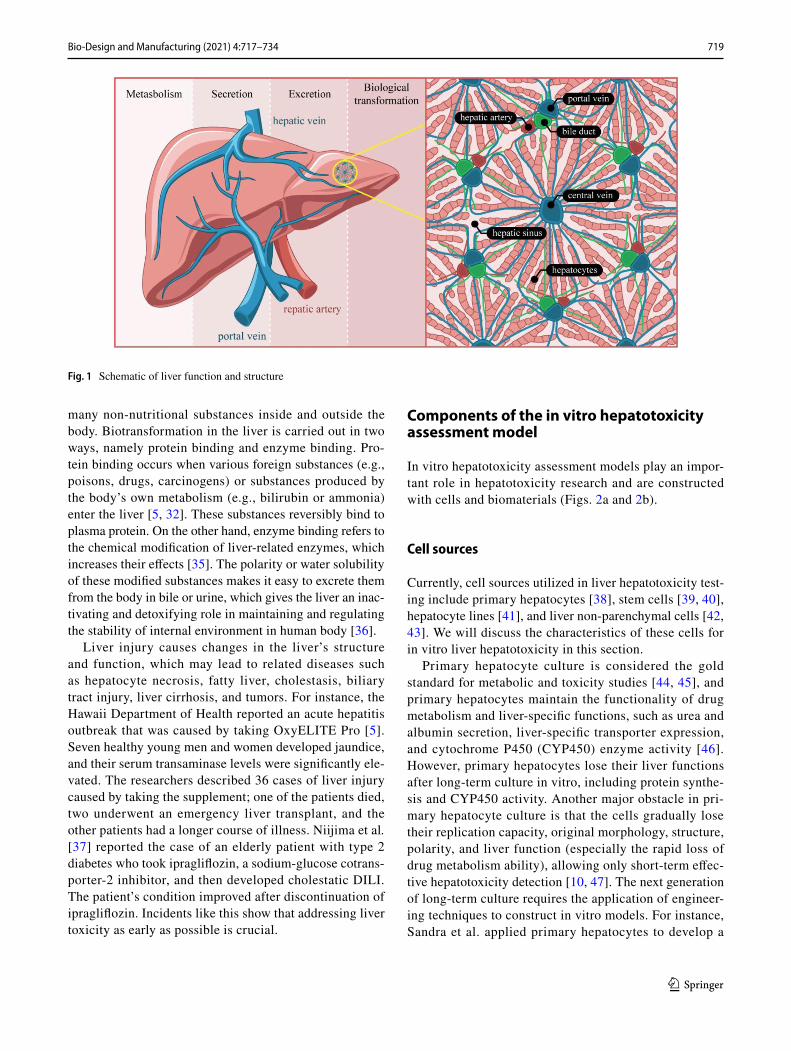
The liver is the largest solid organ of the human body, with more than 1500 functions such as metabolism, secretion, excretion, and biological transformation [30, 31]. Liver damage causes corresponding functional abnormalities and metabolic disorders in the human body, and sometimes even death. The liver tissue is composed of numerous hepatic lobules with the same structure, and similar size and shape, and its main functions are detoxification and drug metabo-lism (Fig. 1).
Hepatic lobule structure
The hepatic lobule is a hexagonal structure composed of a central vein, a liver plate, a hepatic sinus, and a capillary bile duct [32]. The liver plate and hepatic sinus are distrib-uted radially around the central vein, and the capillaries are distributed among hepatocytes. The irregular cavity formed between the liver plates is called the hepatic sinusoid and allows for blood flow between the liver plates. The hepatic sinus consists of a sinus wall and a sinus cavity, which par-ticipate in the immune response [30]. Capillary bile ducts constitute the hepatobiliary system, which participates in material metabolism and drug conversion.
The microenvironment (extracellular matrix (ECM)) is a diverse collection of proteins and carbohydrates around the hepatocytes and plays an important role in cell growth [33, 34]. In summary, hepatocytes, endothelial cells, liver macrophages, and their extracellular microenvironment form the basic unit of hepatic lobules, and they work together to make the liver function.
Liver function
Liver tissue is the most important detoxifying and drug-metabolizing organ of the human body, and it can transform
719Bio-Design and Manufacturing (2021) 4:717–734
1 3
many non-nutritional substances inside and outside the body. Biotransformation in the liver is carried out in two ways, namely protein binding and enzyme binding. Pro-tein binding occurs when various foreign substances (e.g., poisons, drugs, carcinogens) or substances produced by the body’s own metabolism (e.g., bilirubin or ammonia) enter the liver [5, 32]. These substances reversibly bind to plasma protein. On the other hand, enzyme binding refers to the chemical modification of liver-related enzymes, which increases their effects [35]. The polarity or water solubility of these modified substances makes it easy to excrete them from the body in bile or urine, which gives the liver an inac-tivating and detoxifying role in maintaining and regulating the stability of internal environment in human body [36].
Liver injury causes changes in the liver’s structure and function, which may lead to related diseases such as hepatocyte necrosis, fatty liver, cholestasis, biliary tract injury, liver cirrhosis, and tumors. For instance, the Hawaii Department of Health reported an acute hepatitis outbreak that was caused by taking OxyELITE Pro [5]. Seven healthy young men and women developed jaundice, and their serum transaminase levels were significantly ele-vated. The researchers described 36 cases of liver injury caused by taking the supplement; one of the patients died, two underwent an emergency liver transplant, and the other patients had a longer course of illness. Niijima et al. [37] reported the case of an elderly patient with type 2 diabetes who took ipragliflozin, a sodium-glucose cotrans-porter-2 inhibitor, and then developed cholestatic DILI. The patient’s condition improved after discontinuation of ipragliflozin. Incidents like this show that addressing liver toxicity as early as possible is crucial.
Components of the in vitro hepatotoxicity assessment model
In vitro hepatotoxicity assessment models play an impor-tant role in hepatotoxicity research and are constructed with cells and biomaterials (Figs. 2a and 2b).
Cell sources
Currently, cell sources utilized in liver hepatotoxicity test-ing include primary hepatocytes [38], stem cells [39, 40], hepatocyte lines [41], and liver non-parenchymal cells [42, 43]. We will discuss the characteristics of these cells for in vitro liver hepatotoxicity in this section.
Primary hepatocyte culture is considered the gold standard for metabolic and toxicity studies [44, 45], and primary hepatocytes maintain the functionality of drug metabolism and liver-specific functions, such as urea and albumin secretion, liver-specific transporter expression, and cytochrome P450 (CYP450) enzyme activity [46]. However, primary hepatocytes lose their liver functions after long-term culture in vitro, including protein synthe-sis and CYP450 activity. Another major obstacle in pri-mary hepatocyte culture is that the cells gradually lose their replication capacity, original morphology, structure, polarity, and liver function (especially the rapid loss of drug metabolism ability), allowing only short-term effec-tive hepatotoxicity detection [10, 47]. The next generation of long-term culture requires the application of engineer-ing techniques to construct in vitro models. For instance, Sandra et al. applied primary hepatocytes to develop a
Fig. 1 Schematic of liver function and structure

720 Bio-Design and Manufacturing (2021) 4:717–734
1 3
micropatterned co-culture (MPCC) for liver disease research [38]. They found that hepatocytes in the MPCC model maintained high levels and stable liver function and polarity during several weeks of culture. In addition, they found that this use of hepatocytes could be extended to multiple liver-related infectious disease models and showed potential for drug development. Another study utilized MPCC to develop a micropatterned tri-culture (MPTC) platform for drug development [48]. Here, MPTC was composed of hepatic stellate cells (HSCs) around MPCC. This study found that the secretion of albumin and urea in MPTC and MPCC was similar, indicating that primary human hepatocytes (PHHs) differentiate well. However, the increased number of HSCs down-regulated the activity of hepatic CYP450 and transporters in MPTC and caused steatosis in two weeks. In addition, inhibi-tion of farnesol X receptor (FXR) and NADPH oxidase (NOX) activation with drugs can reduce liver dysfunction in MPTC. Despite the advances in culture conditions and technologies, there are still considerable technical obsta-cles to the isolation and culture of primary hepatocytes in liver tissue engineering.
More recently, hepatocytes derived from stem cells have provided an opportunity for patient-specific hepatotoxicity testing, since stem cells (e.g., embryonic stem cells (ESCs),
Fig. 2 Model system for hepatotoxicity assessment. a The cells used in the in vitro hepatotoxicity assessment test model are stem cells, primary hepatocytes, hepatocyte lines, and liver NPCs. b The scaf-fold materials used in the in vitro liver hepatotoxicity detection model are nanofibers, hydrogels, and decellularized LEM. c Liver spheroids and organoids provide a unique model for hepatotoxicity assessment,
which, through cellular interactions, simulates the internal envi-ronment of liver tissues and spontaneously forms blood vessel-like structures. d Four kinds of in vitro hepatotoxicity assessment models achieved toxicity testing in vitro and provide new platforms for clini-cal diagnosis and drug development. NPC: non-parenchymal cell; LEM: liver extracellular matrix
mesenchymal stem cells (MSCs), and induced pluripotent stem cells (iPSCs)) have a pluripotent nature and the ability to proliferate [39, 40, 49, 50]. For instance, human-induced pluripotent stem cell (hiPSC) derived hepatic progenitor cells (hiPSC-HPCs) with adipose-derived stem cells and endothelial cells were embedded in a triculture to form an in vitro liver model [51]. After several weeks of culture, it was found that hiPSC-HPCs expressed higher levels of liver-specific genes and enhanced CYP450 and metabolites which could be used for drug screening. These studies dem-onstrated the feasibility of differentiating hepatocytes from stem cells. Nevertheless, differentiated hepatocytes still have lower enzyme activity and gene expression profiles com-pared with cells in vivo.
To surmount the limitations of hepatocytes and stem cells in the most common applications, cell lines derived from hepatocellular carcinoma are widely used [52]. A few com-monly used cell lines are HepG2, C3A, HepaRG, and Huh7. These cell lines can proliferate indefinitely, have a relatively stable phenotype, are easy to manipulate, and overcome the problem of inter-individual variation. In addition, transcrip-tion factor (nuclear factor erythroid 2-related factor 2 [Nrf2]) is still expressed in these cells, which is essential for drug metabolism and toxicity [41]. For example, encapsulating HepG2 cells on alginate provided an in vitro 3D liver model

721Bio-Design and Manufacturing (2021) 4:717–734
1 3
for drug hepatotoxicity testing [53]. In this study, 10 mM 7-ethoxy-4-trifluoromethyl coumarin (EFC) stock solution mixed with HepG2 medium demonstrated the effectiveness of drug metabolism in the liver chamber. The drug metabo-lism conversion rate increased by 26%, which reproduced liver function in vivo. Thus, this model can be used to sup-port drug toxicity screening. In another study, Deng et al. used four cell lines (HepG2, LX-2, EAhy926, and U937) to fabricate a microfluidic liver model for hepatotoxicity detec-tion [54]. The 3D cell cultures maintained liver function (e.g., albumin and urea secretion). In addition, compared with the gold standard primary hepatocyte plate model, the response of this platform to acetaminophen (APAP) treat-ment was similar. However, the main disadvantage is the lack of expression of key metabolic enzymes and the low metabolic capacity compared with other cell types.
In order to further circumvent the challenges of stem cell-delineated cultures, hepatocytes can be co-cultured with non-parenchymal cells (NPCs) (e.g., Kupffer cells and HSCs) to preserve certain cellular mechanisms and meta-bolic pathways in vitro for drug toxicity testing. NPCs make up roughly 20% of liver mass and exhibit mediated immune/pro-inflammatory responses and functional 3D tissue struc-ture [10, 46]. As liver cytotoxicity involves cascading pro-inflammatory pathways and complex interactions between NPCs and ECM, the presence of NPCs is largely considered to be a necessary condition for accurate modeling of toxicity assays in vivo [10, 42, 43, 46, 55]. Hence, the presence of NPCs not only helps to prolong the survival and function of hepatocytes in culture, but also increases their sensitiv-ity to DILI detection involving inflammatory mediators and hepatocyte NPC “communication.” Therefore, NPCs and ECM components, which can be also co-cultured with 3D liver buds to further improve physiological relevance, are important to develop accurate in vitro liver models for meta-bolic and hepatotoxicity testing.
Scaffolds for in vitro liver models
A 3D bioscaffold is an important element in the construc-tion of 3D liver tissue structures and drug testing models. Scaffold materials generally should have the following char-acteristics: (1) a 3D porous structure that can facilitate cell growth and timely supply of nutrients and then discharge metabolic waste; (2) good biocompatibility that can promote cells to grow, proliferate, and adhere, and easily degrade in the body; (3) plasticity; (4) the ability to bind to the ECM naturally and better reshape the body [56]. For liver tissue construction, nanofibers [57–59], hydrogels [60–62], and a decellularized liver matrix [63–66] have been used as suit-able ECMs for hepatocyte culture, maintenance of hepato-cyte function, or promotion of liver differentiation of stem cells for drug hepatotoxicity testing (Fig. 3).
Nanofiber scaffold materials
In recent years, nanomaterials have been developed rapidly in the field of liver tissue engineering. Nanofibrous scaffolds in particular not only provide structures similar to natural tis-sues, but have mechanical properties and high surface area/volume which promote cell proliferation in vitro. Nanofib-ers for in vitro liver models also have the potential to pre-dict drug toxicity, since this nanomaterial can enhance liver function and upgrade critical drug-metabolizing enzyme activity (CYP) [57, 58, 67, 68]. For example, Brophy et al. demonstrated that hepatocytes cultured on nanofibers main-tain high CYP450 activity [58]. In addition, the amount of albumin produced by hepatocytes on nanofibers was twice that of hepatocytes produced on laminin. In another study, a 3D scaffold of multi-walled carbon nanotubes (MWCNTs) was fabricated for liver tissue engineering and drug discov-ery (Fig. 3a). Primary rat hepatocytes were cultured using aligned nanogrooved MWCNT sheets, MWCNT yarns, or 2D standard culture. The cell viability between these three culture conditions was comparable, but in cells cultured on MWCNTs, the production of albumin, a hepatocyte-specific marker, increased. In addition, the activity of CYP1A2 was enhanced on MWCNTs. These results showed that car-bon nanotubes can be used in in vitro liver models with enhanced liver-specific function. To further promote the adhesion properties of nanomaterials for hepatocyte cul-ture, researchers used chitosan to prepare polycaprolactone (PCL)/chitosan nanofibers using electrospinning technology [57]. The pores of the scaffolds were suitable for infiltration of mouse hepatocytes. In addition, the in vitro cell culture results confirmed the biocompatibility of the scaffold for later construction of an in vitro liver model. The increased liver function of this model showed potential for hepatotox-icity detection. Thus, the biocompatibility of nanomaterials and their retention of affinity and enzyme activity in hepato-cyte culture make them useful in both liver tissue construc-tion and hepatotoxicity testing.
Hydrogel scaffold materials
To construct liver extracellular matrix in vitro, biomimetic properties and soft materials are required, and thus hydro-gels are a good option [60, 61, 69]. Moreover, the cells in hydrogels have a 3D environment that is similar to natu-ral tissues. Utilizing hydrogels to fabricate an in vitro liver model reproduces the true state of the liver in the body, and the model can be used for hepatotoxicity drug detection.
Hydrogels are of two types, natural and synthetic. Synthetic hydrogels, such as polyethylene glycol (PEG) [60, 69] and polypropylene amine (PAAm) [61], are arti-ficial hydrogels with a defined composition and a strong mechanical structure which are widely used. For example,
722 Bio-Design and Manufacturing (2021) 4:717–734
1 3
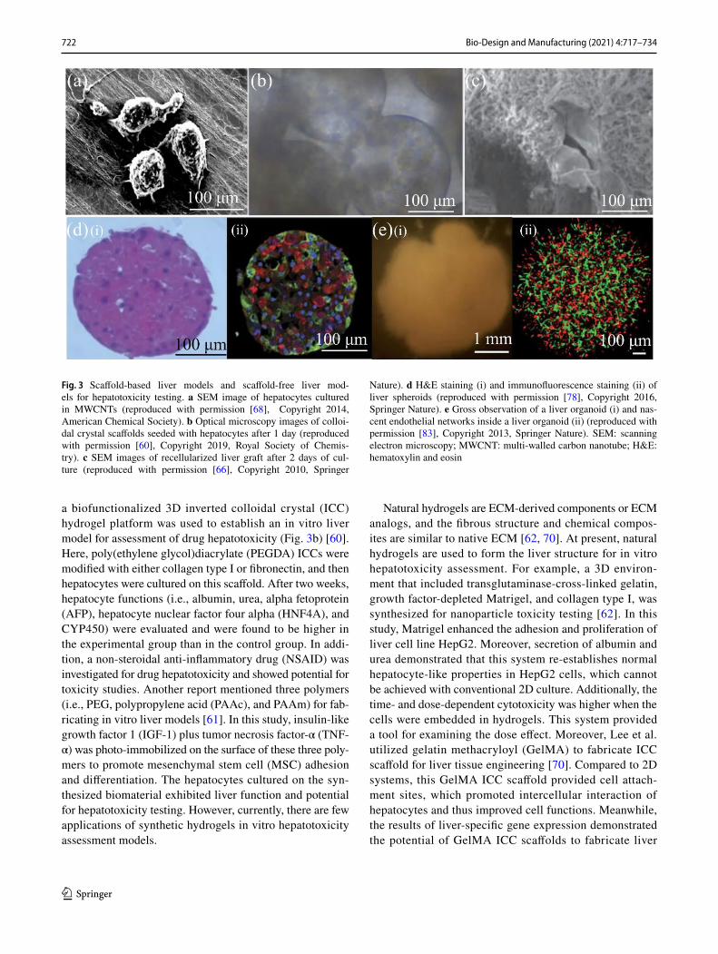
a biofunctionalized 3D inverted colloidal crystal (ICC) hydrogel platform was used to establish an in vitro liver model for assessment of drug hepatotoxicity (Fig. 3b) [60]. Here, poly(ethylene glycol)diacrylate (PEGDA) ICCs were modified with either collagen type I or fibronectin, and then hepatocytes were cultured on this scaffold. After two weeks, hepatocyte functions (i.e., albumin, urea, alpha fetoprotein (AFP), hepatocyte nuclear factor four alpha (HNF4A), and CYP450) were evaluated and were found to be higher in the experimental group than in the control group. In addi-tion, a non-steroidal anti-inflammatory drug (NSAID) was investigated for drug hepatotoxicity and showed potential for toxicity studies. Another report mentioned three polymers (i.e., PEG, polypropylene acid (PAAc), and PAAm) for fab-ricating in vitro liver models [61]. In this study, insulin-like growth factor 1 (IGF-1) plus tumor necrosis factor-α (TNF-α) was photo-immobilized on the surface of these three poly-mers to promote mesenchymal stem cell (MSC) adhesion and differentiation. The hepatocytes cultured on the syn-thesized biomaterial exhibited liver function and potential for hepatotoxicity testing. However, currently, there are few applications of synthetic hydrogels in vitro hepatotoxicity assessment models.
Natural hydrogels are ECM-derived components or ECM analogs, and the fibrous structure and chemical compos-ites are similar to native ECM [62, 70]. At present, natural hydrogels are used to form the liver structure for in vitro hepatotoxicity assessment. For example, a 3D environ-ment that included transglutaminase-cross-linked gelatin, growth factor-depleted Matrigel, and collagen type I, was synthesized for nanoparticle toxicity testing [62]. In this study, Matrigel enhanced the adhesion and proliferation of liver cell line HepG2. Moreover, secretion of albumin and urea demonstrated that this system re-establishes normal hepatocyte-like properties in HepG2 cells, which cannot be achieved with conventional 2D culture. Additionally, the time- and dose-dependent cytotoxicity was higher when the cells were embedded in hydrogels. This system provided a tool for examining the dose effect. Moreover, Lee et al. utilized gelatin methacryloyl (GelMA) to fabricate ICC scaffold for liver tissue engineering [70]. Compared to 2D systems, this GelMA ICC scaffold provided cell attach-ment sites, which promoted intercellular interaction of hepatocytes and thus improved cell functions. Meanwhile, the results of liver-specific gene expression demonstrated the potential of GelMA ICC scaffolds to fabricate liver
Fig. 3 Scaffold-based liver models and scaffold-free liver mod-els for hepatotoxicity testing. a SEM image of hepatocytes cultured in MWCNTs (reproduced with permission [68], Copyright 2014, American Chemical Society). b Optical microscopy images of colloi-dal crystal scaffolds seeded with hepatocytes after 1 day (reproduced with permission [60], Copyright 2019, Royal Society of Chemis-try). c SEM images of recellularized liver graft after 2 days of cul-ture (reproduced with permission [66], Copyright 2010, Springer
Nature). d H&E staining (i) and immunofluorescence staining (ii) of liver spheroids (reproduced with permission [78], Copyright 2016, Springer Nature). e Gross observation of a liver organoid (i) and nas-cent endothelial networks inside a liver organoid (ii) (reproduced with permission [83], Copyright 2013, Springer Nature). SEM: scanning electron microscopy; MWCNT: multi-walled carbon nanotube; H&E: hematoxylin and eosin

723Bio-Design and Manufacturing (2021) 4:717–734
1 3
engineering platforms for drug toxicity testing and regen-erative medicine.
Decellularized liver matrix
Decellularization refers to the removal of all cells from the original tissues/organs by physical and chemical methods to obtain cell-free, non-immunogenic/low immunogenic tissue scaffolds. Decellularized scaffolds have a unique value in the field of tissue engineering [71, 72]. Decellularization retains the ECM components of the original tissues and organs and some unique growth factors. Therefore, recellularization using a decellularized scaffold can closely conform to the characteristics of the original tissue for organ transplanta-tion, tissue repair, drug testing, and other applications.
Although decellularized scaffolds present challenges for in situ colonization, they are rich in natural ECM compo-nents and essential growth factors for cells, which allows accurate construction of drug screening models [66, 72–74]. A classic study showed that an acellular liver matrix can be used for liver tissue engineering via characterization of its components (Fig. 3c) [66]. In this study, decellulariza-tion retained the structural and functional characteristics of the natural microvascular network, allowing hepatocytes to effectively recellularize the liver matrix. The recellularized grafts supported liver-specific functions such as albumin secretion, urea synthesis, and CYP450 expression, which were similar to in vivo conditions. Hussein et al. utilized a decellularized rat liver scaffold to create a hepatocellular carcinoma model for pharmacological studies [72]. In this work, a simplified closed-perfusion system was developed as a bioreactor and HepG2 cells were infused into the decel-lularized liver scaffold, which maintained their function and oncogenicity. Through anti-cancer drug testing, it was dem-onstrated that this in vitro system could provide a precious 3D cell culture environment and thus evaluate the efficacy of new antitumor drugs. Another team developed a decellular-ized liver matrix solution for 3D cell culture [73]. Here, 3D printing was used to create the in vitro 3D structure, and the decellularized liver matrix bioink improved the functionality of printed structures.
It has also been found that combining acellular matrix with a hybrid scaffold not only preserves its unique biologi-cal factors but also increases the performance of the hybrid scaffold. For example, Lee et al. enzymatically decomposed decellularized liver scaffold and combined it with type I col-lagen to form a composite hydrogel; they then cultured pri-mary hepatocytes using the composite hydrogel [74]. Mass spectrometry revealed a variety of ECM components and characteristic growth factors in the acellular matrix. Acel-lular matrix composite hydrogels had improved biophysical and mechanical properties compared to single hydrogels. They also promoted cell viability and liver function, and the
animal experiments have confirmed the superiority of com-posite hydrogels [74]. In summary, acellular matrix compos-ite hydrogels are a promising biological material and play a unique role in the in vitro culture of hepatocytes for drug hepatotoxicity evaluation.
In vitro models for hepatotoxicity testing
The different components of in vitro hepatotoxicity assess-ment models, including cell sources, scaffold materials, and liver spheroids, were described in the previous sec-tions. Now, we will introduce the technology behind these models. At present, the foremost models are liver spheroids, organoids, microarrays, micropatterns, livers-on-a-chip, and 3D-printed livers (Figs. 2c and 2d).
Scaffold‑free construction of liver models
Liver spheroids
Liver spheroids are a rising model for 3D culture of hepato-cytes. These spheroids can mimic the ECM and improve hepatic function in vitro [11, 16, 75]. The principle of forma-tion of hepatic spheroids is that monodispersed cells are able to change the 3D configuration by self-repolymerizing, with the unavailability of a suitable biomaterial for attachment causing cell–cell attachment into 3D spheroid structures. The production of spheres increases proliferative activity and metabolic function, leading to enhanced cell viability, stable morphology, and polarization [76]. Thus, compared to 2D cultures, liver spheroids have more powerful functions and provide a valuable tool for drug development and pre-clinical hepatotoxicity testing.
For instance, liver spheroids produced by HepaRG2 cells showed the morphological characteristics of hepatocyte-like cells and the formation of bile duct-like structures [77]. In this study, researchers used a rotating bioreactor to construct spheroids with diameters of < 200 μm. This system main-tained stable albumin secretion, CYP3A4 induction, and uri-dine diphosphate glucuronic acid transferase (UGT) activity for seven weeks, and immunofluorescence staining revealed the distribution of bile duct-like structures and transporters. In addition, computer modeling analysis of acetaminophen (APAP) for hepatotoxicity demonstrated the utility of this system in assessing the biological activation of APAP and its associated cytotoxicity. In another study, Bell et al. used ultra-low attachment plates to produce monoculture PHH spheroids (Fig. 3d) [78]. During the 35-day culture period, a bile duct-like structure was formed in the spheroids, indi-cating stable functional polarization of hepatocytes. Sub-sequently, the spheroids were dosed every two days with several drugs (i.e., amiodarone, fialuridine, diclofenac,

724 Bio-Design and Manufacturing (2021) 4:717–734
1 3
bosentan, and tolcapone) and the viability was determined to investigate hepatotoxicity. Hepatotoxicity was not identi-fied after short-term (48 h) exposure but was clearly detected after long-term (8 and 28 days) exposure that approached clinically relevant concentrations. Another team pre-laid 1.5% agarose to construct a low surface adsorption environ-ment to culture C3A cell spheres [79]. The C3A spheres maintained culture for 32 days and showed similar structural features and liver-specific functions to the in vivo environ-ment, such as the ability to synthesize and secrete urea and albumin, and functional tubule transporter and CYP2E1 expression. Additionally, analysis of four hepatotoxins (APAP, diclofenac, trovafloxacin, and floxuridine) indicated that the spheroid model was able to predict hepatotoxicity with higher sensitivity than conventional 2D monolayer cultures. Therefore, liver spheroids have great application potential for in vitro hepatotoxicity assessment.
Liver organoids
Generally, current spheroid model systems exhibit a large gap between the cellular level and organ level. Stem cell-derived organoids are being used to develop 3D self-organ-izing tissue models and provide a compelling new biologi-cal model to act as both tissue and organ agents [80–82]. Organoids recapitulate a large number of biological param-eters, including the spatial organization of heterogeneous tissue-specific cells, intercellular interaction, cell–matrix interactions, and certain physiological functions produced by tissue-specific cells in the organoid [83]. By controlling self-renewal and differentiation, specific tissues and organs can be formed. Liver organoids bridge a gap in existing model systems via providing a stable and scalable system to extend cultivation and manipulation, which better repre-sents the in vivo physiology. Therefore, construction of liver organoids can not only be used in regeneration engineering, but also has irreplaceable value in drug development and toxicity evaluation.
A major investigation into liver organoids was previously reported, mainly using the self-assembly of three types of cells (i.e., endothelial cells, human iPSC-HEs, and MSCs) (Fig. 3e) [83]. In this work, gene expression and immu-nostaining analyses revealed sufficient similarity between in vitro and in vivo liver buds to permit liver regenerative therapy and drug toxicity testing. For drug metabolism investigation, liver organoids from stem cells have been used [84]. In this study, the CYP subfamilies (CYPs) in liver organoids were expressed. Then, the activity of doc-etaxel in a mouse liver organoid and human pancreatic carcinoid co-culture platform was measured. The results indicated that the cell survival rates of pancreatic tumors co-cultured with undifferentiated, differentiated, and CYPs-induced differentiated organoids treated with docetaxel were
(66.05 ± 2.14)%, (89.20 ± 2.67)%, and (101.90 ± 0.94)%, respectively. Thus, the organoids performed drug metabo-lism assessment and gene expression modification. In vivo-like in vitro investigation on drug toxicity may potentially be done with organoids as a stepping-stone to the clinical trial stage. It seems that although liver organoids have bet-ter function than liver spheroids, in practice, they are more commonly used in liver regeneration studies and are rarely used for hepatotoxicity testing.
Liver cell microarrays
In the early phase of drug development, it is necessary to screen the metabolism of thousands of compounds. As the main detoxification organ, the liver is critical for clear-ing drugs. Assessing the ability of the liver to clear drugs requires a high-throughput artificial liver platform that pro-vides operational data rapidly at a relatively low cost and that can be miniaturized. Next, we describe a state-of-the-art microarray platform for drug metabolism studies, which offers support for drug hepatotoxicity research.
Cell microarrays are used to seed cells on materials or biomolecules for high-throughput studies of microenvi-ronmental signals on cells [85–87]. Kwon et al. reported a Transfected Enzyme and Metabolism Chip (TeamChip) that was developed for high-throughput investigation of drug metabolism toxicity screening in the human liver (Fig. 4a) [86]. This TeamChip was prepared by using a recombinant adenovirus in a complementary microwell chip to transfer genes into a miniature 3D cell microarray on a micro-pillar chip. This platform was able to transduce viral genes such as CYP3A4, CYP2C9, CYP2D6, CYP2E1, CYP1A2, and UGT1A4 for high-throughput screening in 3D cell culture microarrays. To verify the hepatotoxicity of this TeamChip platform, the effects of ketoconazole (a CYP3A4 inhibi-tor) and buthionine sulfoximine (BSO) were investigated in the TeamChip. The enhanced toxicity of APAP on the CYP3A4-expressing chip exposed to BSO was due to the lack of sufficient glutathione to detoxify the reactive inter-mediate. Accordingly, this TeamChip platform provided critical information for high-throughput assessment of metabolism-induced toxicity. In another work, a miniatur-ized 3D cell culture array (DataChip 2.0) and a metaboliz-ing enzyme microarray (MetaChip 2.0) were developed by Yu et al. to predict metabolism-induced hepatotoxicity with high throughput [87]. This DataChip supported the Hep3B cells in a 3D microarray format, and then 22 compounds were distributed into the MetaChip and sandwiched with the DataChip. The drug toxicity testing results demon-strated that the platform had 100% sensitivity, 86% speci-ficity, and 93% overall predictivity at optimum cutoffs of median inhibitory concentration (IC50) and Cmax values. As a multi-well platform, the DataChip/MetaChip was useful for

725Bio-Design and Manufacturing (2021) 4:717–734
1 3
high-throughput drug screening. The researchers concluded that cell microarrays could be utilized as high-throughput platforms and rapid, inexpensive, and microscale alterna-tive assessment of in vitro toxicity at early phases of drug development and clinical treatment.
Micropatterned hepatocytes
Micropatterns, another kind of high-throughput platform, use accurate techniques such as lithography to construct an in vitro liver model in a size range of a few microns to a few nanometers [88–91]. Micropatterning was first reported in 1994 [88], when an elastomeric stamp was used to imprint a gold surface and create islands of a defined shape. With this technique, cells were placed in predetermined locations and arrays separated by a defined distance, assigned their shape, and then provided control over cell growth and pro-tein secretion. Subsequently, Bhatia et al. used lithography to design the micropatterned co-cultures (MPCCs) platform, which was widely used in various in vitro models in subse-quent studies. For this particular platform, hepatocytes and fibroblasts were seeded in a co-culture system by micropat-terning [89]. This co-culture technique allowed control of the level of homotypic interaction in cultures of a single cell type and of the degree of heterotypic contact in co-cultures over a wide range, which allowed manipulation of the initial cellular microenvironment without variation of cell num-ber. Later, Lin et al. utilized MPCCs to predict clearance of 26 drugs and drug–drug interactions (Fig. 4b) [90]. In this study, the long-term activity of cytochrome P450 enzymes produced by unpooled cryopreserved PHH donors was char-acterized in a 96-well plate and then MPCCs were used to predict metabolism of 26 drugs with a wide range of in vivo turnover rates. Meanwhile, the team detected the effects of drug-mediated P450 modulation on drug clearance rates to find out whether they could mimic in vivo drug–drug inter-action (DDI) situations. More recently, this MPCC model was also employed to assess the safety and efficacy of RNA interference (RNAi)-based therapeutics (Fig. 4c) [91]. Here, the endogenous RNAi pathway was utilized to post-tran-scriptionally silence central drug metabolism genes and the team evaluated the impact of these changes on a natural sub-strate, as well as on DILI assessment of known hepatotoxins. This new capability could be utilized to open the door for structure–activity relationship detection of compounds in the setting of both high- and low-metabolizing genotypes. Therefore, this methodology has great potential for drug development and hepatotoxicity testing.
Livers‑on‑a‑chip
At present, livers-on-a-chip have become a research hot spot. They can simulate the structure of the liver in vitro, which
provides a flow environment to simulate blood flow. Livers-on-a-chip create a long-term culture environment that main-tains the cell phenotype and function to fine-tune physical, chemical, and biological signals [92–96].
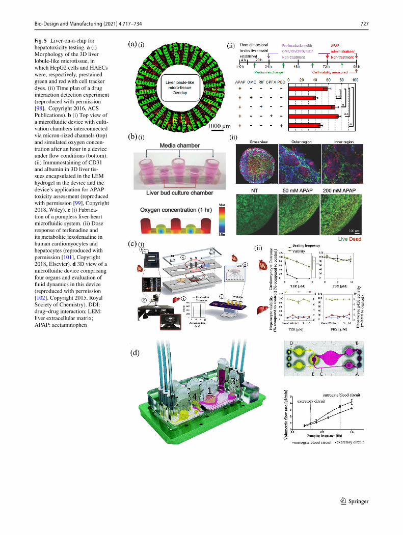
Since perfusion-based 3D culture mimics the growth of hepatocytes in vivo, hepatocytes on a chip can be used for drug detection [97–99]. For instance, a digital drug hepa-totoxicity screening platform based on microfluidic tech-nology was developed for detection of the dose effect of APAP on hepatocyte apoptosis [97]. In the microfluidic device, HepG2 cells and NIH-3T3 fibroblasts were encap-sulated in a hydrogel to form a liver-like organ. Compared to a 2D culture system, hepatocyte function was enhanced. A 5 mM APAP dose did not have a significant toxic effect, while 10 and 20 mM of APAP demonstrated dose-dependent toxicity in the liver compared to non-treated controls. In another study, drug interaction testing was performed on a liver-on-a-chip platform with hepatic lobular micro-tis-sues (Fig. 5a) [98]. Drug hepatotoxicity testing showed that omeprazole or probenecid treatment for 48 h before APAP treatment aggravates APAP toxicity, while rifampicin or ciprofloxacin pretreatment relieves APAP toxicity. This work supports livers-on-a-chip as a potential drug devel-opment platform. To construct complex liver organs, iHep cells were cultured in a decellularized liver extracellular matrix (LEM)-coated microfluidic system to form a 3D vascularized liver organ (Fig. 5b) [99]. iHep cells, which transfected liver transcription factors, were co-cultured with endothelial cells in 3D LEM hydrogels. In the physiologi-cally relevant culture microenvironment, the resulting 3D vascularized liver-like organs exhibited improved liver func-tion. Finally, the feasibility of iHep-based 3D liver organs as a high-throughput drug screening platform was validated and applied to a multi-organ model consisting of multiple internal organoids. One recent work presented a microflu-idic multi-tissue platform named metaEST that seamlessly integrates liver metabolism into embryonic stem cell tests (ESTs) [22]. In this “metaEST” platform, embryoid bodies (EBs) were formed on a chip using hanging-drop network technology as a micro-physiological system [39] and were co-cultured with the main 3D human liver micro-tissues (hLiMT). A metaEST assay was performed in a single microfluidic device, which was operated by gravity-driven flow to ensure constant intertissue communication and rapid and efficient exchange of metabolites. A test with the prod-rug cyclophosphamide revealed a fourfold reduction in the median infective dose (ID50) after biotransformation, which demonstrated the potential adverse effects of metabolites in embryotoxicity.
Livers-on-a-chip can be applied as a stand-alone unit or combined with other organ chips (e.g., heart, kidney, intes-tine) for investigating drug toxicity [100–102]. Because most drug failures are associated with liver or heart toxicity

726 Bio-Design and Manufacturing (2021) 4:717–734
1 3
or dysfunction, an integrated liver chip was developed for drug screening and biophysical study of physiologically rel-evant microenvironments [100]. In this study, a dynamic fluid transport system was constructed by establishing an endothelial-like barrier that mimicked the continuous nutri-ent exchange and intercellular interaction between ven-tricular myocardium and hepatic sinusoid cells. Moreover,
using microfluidic channels and control of the fluid rate, the team imitated blood flow between the liver and heart. More importantly, the effect of different parameters, such as flow rate, was investigated via real-time detection of the morphology and metabolism of cultured cells. In another work, a heart-liver system was fabricated to better predict human drug metabolism (Fig. 5c) [101]. The heart-liver
Fig. 4 Microarrays and micropatterns for hepatotoxicity testing. a Schematic of micropillar/microwell chip components (i); scanned image of a combination of multiple drug-metabolizing enzymes expressed by THLE-2 cells. The chip was exposed to 200 µM tamox-ifen for 48 h (top) and normalized THLE-2 cell viability at different drug-metabolizing enzyme expression levels (bottom) (ii) (repro-duced with permission [86], Copyright 2014, Springer Nature). b
Morphology and functional (albumin and urea secretion) characteri-zation of MPCCs (reproduced with permission [90], Copyright 2016, ASPET). c Fabrication of gene-silenced MPCC to evaluate safety and efficacy of RNAi-based therapeutics (reproduced with permission [91], Copyright 2019, Elsevier). MPCC: micropatterned co-culture; RNAi: RNA interference
727Bio-Design and Manufacturing (2021) 4:717–734
1 3
Fig. 5 Liver-on-a-chip for hepatotoxicity testing. a (i) Morphology of the 3D liver lobule-like microtissue, in which HepG2 cells and HAECs were, respectively, prestained green and red with cell tracker dyes. (ii) Time plan of a drug interaction detection experiment (reproduced with permission [98], Copyright 2016, ACS Publications). b (i) Top view of a microfluidic device with culti-vation chambers interconnected via micron-sized channels (top) and simulated oxygen concen-tration after an hour in a device under flow conditions (bottom). (ii) Immunostaining of CD31 and albumin in 3D liver tis-sues encapsulated in the LEM hydrogel in the device and the device’s application for APAP toxicity assessment (reproduced with permission [99], Copyright 2018, Wiley). c (i) Fabrica-tion of a pumpless liver-heart microfluidic system. (ii) Dose response of terfenadine and its metabolite fexofenadine in human cardiomyocytes and hepatocytes (reproduced with permission [101], Copyright 2018, Elsevier). d 3D view of a microfluidic device comprising four organs and evaluation of fluid dynamics in this device (reproduced with permission [102], Copyright 2015, Royal Society of Chemistry). DDI: drug–drug interaction; LEM: liver extracellular matrix; APAP: acetaminophen

728 Bio-Design and Manufacturing (2021) 4:717–734
1 3
microfluidic system was integrated with a computational model to predict terfenadine and fexofenadine kinetics. This model recapitulated the fate of terfenadine, which improved the prediction of xenobiotic toxicity. To establish a drug absorption, distribution, metabolism, and excretion (ADME) map, and to perform a systemic toxicity test, the researchers proposed a four-organ chip (Fig. 5d) [102]. This four-organ chip system ensured a ratio of physiological fluid close to that of in vivo tissue. Intensive metabolic and genetic analysis indicated that the establishment of homeo-stasis in co-cultures for at least 28 days was independent of individual human cell lines or tissue/body background for each organ equivalent. The future direction of organ-on-a-chip research is likely to be aimed toward connecting these authentic functionalities to mimic a whole-body response for drug hepatotoxicity testing.
3D‑printed livers
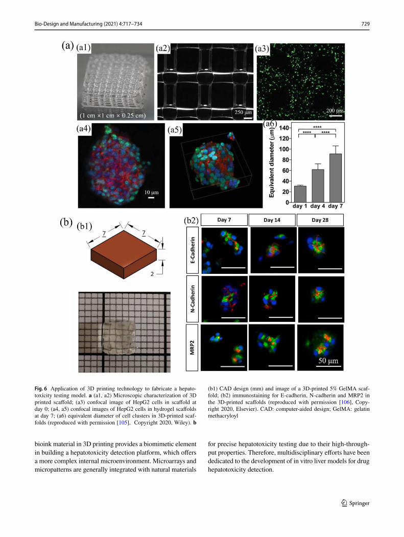
Recently, bioprinting approaches have facilitated auto-mated and high-throughput fabrication of precisely con-trolled 3D liver architectures, which promotes accurate prediction of drug hepatotoxicity [103]. For example, Organovo’s exVive3D liver is an already commercialized bioprinted human tissue model for drug hepatotoxicity assessment [104]. Hematoxylin and eosin staining have shown that exVive3D liver tissues have similar intercel-lular junctions, microvascular structures, and cellular com-partments to the native liver. In addition, APAP damages exVive3D tissue, similar to clinical results. In another study, bioprinted Pluronic/alginate blends with HepG2/C3A cells were developed for investigating drug-induced hepatotoxicity (Fig. 6a) [105]. The Pluronic templating agent improved the diffusive properties of the hydrogel. In the 3D liver structure, cell viability and liver function improved compared to 2D cultures. Drug hepatotoxicity testing showed increased sensitivity to APAP compared to 2D adhesion controls. In addition, 3D extrusion bioprint-ing was used to fabricate a complex liver model [106]. The bioprinted structure showed 28-day cell viability and liver function. The study also provided a basis for multicel-lular models, which combine hepatic parenchymal cells with stellate cells and endothelial cells to study fibrosis development. Recently, a novel liver cancer model con-structed using patient-derived hepatocytes was established via 3D bioprinting [107] (Fig. 6b). In long-term culture, the 3D model retained the genetic alterations and expres-sion profile of the original tumor. In addition, the tumori-genic potential and histological features of the 3D model were preserved after long-term culture, meaning that it can be used for evaluating the efficacy of multiple candidate drugs for hepatocellular carcinoma patients.
3D bioprinting combined with microfluidics has become a very hot topic, since it improves models’ bionics and com-plexity [108–111]. For instance, a hepatic bioreactor culture platform integrated with a bioprinter and biomarker analysis module was developed for predicting drug hepatotoxicity (Fig. 7a) [108]. This engineered bioreactor was interfaced with a bioprinter to promote the fabrication of 3D hepatic spheroids encapsulated within a photocrosslinkable GelMA hydrogel. Hepatocyte markers were assessed via detection of the secretion of alpha-1 antitrypsin, albumin, transfer-rin, and ceruloplasmin; and immunofluorescence staining of hepatocyte markers (i.e., cytokeratin 18, MRP2bile canali-cular protein, and tight junction protein ZO-1) demonstrated that the engineered liver constructs remained functional dur-ing 30 days of culture. Additionally, treatment with 15 mM APAP was found to induce toxicity in liver constructs, which was similar to published animal and other in vitro model studies, and provided a practical proof-of-use for this liver-on-a-chip platform for toxicity assessment. Another study showed that 3D-cell-printed livers-on-a-chip have a liver microenvironment and biliary system (Fig. 7b) [109]. In this model, multiple cell types encapsulated in liver decellular-ized bioink were deposited on a chip via 3D cell-printing. In addition, cells were connected through the fluidic channels to mimick vascular and biliary systems. Compared to a chip without a biliary component, liver specific functions were better maintained, and APAP toxicity were more effectively detected in the chip with a biliary fluidic channel. Another key study combined the advantages of 3D printing, co-cul-ture, and microfluidics to develop a 3D co-culture microflu-idic model (3DPF) for drug screening research [110]. In this work, 3D printing and subsequent culture were employed to acquire uniform-sized hepatoma cell clusters, and a kind of thermo-sensitive hydrogel maintained the clusters’ location and morphology. More importantly, the 3DPF model showed a similar pharmacodynamic character of metuzumab to ani-mal experiments, demonstrating its potential to assess the hepatotoxicity of antibody-based drugs.
Conclusions and perspectives
In this review, we discussed both the components (i.e., cells and materials) and construction techniques of in vitro liver models to evaluate hepatotoxicity for drug development and precise medication. We suggest that livers-on-a-chip are a par-ticularly attractive tool for drug hepatotoxicity assessment due to their ability to simulate in vivo drug hepatotoxicity induc-tion in vitro. Integration of 3D printing significantly increases the automation and precise regulation of in vitro hepatotoxic-ity assessment models owing to its personalized, rapid, and efficient properties. For example, using decellularized LEM
729Bio-Design and Manufacturing (2021) 4:717–734
1 3
bioink material in 3D printing provides a biomimetic element in building a hepatotoxicity detection platform, which offers a more complex internal microenvironment. Microarrays and micropatterns are generally integrated with natural materials
for precise hepatotoxicity testing due to their high-through-put properties. Therefore, multidisciplinary efforts have been dedicated to the development of in vitro liver models for drug hepatotoxicity detection.
Fig. 6 Application of 3D printing technology to fabricate a hepato-toxicity testing model. a (a1, a2) Microscopic characterization of 3D printed scaffold; (a3) confocal image of HepG2 cells in scaffold at day 0; (a4, a5) confocal images of HepG2 cells in hydrogel scaffolds at day 7; (a6) equivalent diameter of cell clusters in 3D-printed scaf-folds (reproduced with permission [105], Copyright 2020, Wiley). b
(b1) CAD design (mm) and image of a 3D-printed 5% GelMA scaf-fold; (b2) immunostaining for E-cadherin, N-cadherin and MRP2 in the 3D-printed scaffolds (reproduced with permission [106], Copy-right 2020, Elsevier). CAD: computer-aided design; GelMA: gelatin methacryloyl

730 Bio-Design and Manufacturing (2021) 4:717–734
1 3
However, researchers still seek to establish more precise in vitro liver hepatotoxicity assessment models for better evaluation of drug candidates in clinical diagnosis and drug development [112, 113]. First of all, it is difficult to ensure that the cells used to fabricate a liver model can maintain complete physiological function in a long-term in vitro culture environ-ment. Although iPSC-derived hepatocytes exhibit outstanding advantages in drug toxicity testing, fabricating stably induced hepatocytes is still a difficult point. Secondly, a liver model constructed by in vitro distribution of materials and cells can-not exactly match in vivo features due to the complex struc-tural features and functions of liver tissue. Thirdly, in vitro liver models such as livers-on-a-chip lack interconnectedness with other organs and thus cannot accurately simulate the characteristics of drug metabolism in the body. Therefore, we would encourage continuous efforts for clinical application of in vitro liver hepatotoxicity assessment models.
To conclude, organ-on-a-chip and 3D printing technolo-gies have attracted extensive public attention because of their ability to detect drug hepatotoxicity, allowing personalized healthcare and rapid drug development (as well as reduced economic costs). Meanwhile, organ-on-a-chip combined with 3D printing can accurately simulate the in vivo fluid environ-ment, reduce batch-to-batch variation in liver-on-a-chip mod-els, and achieve cost-effective drug hepatotoxicity detection. More importantly, multi-organ-on-a-chip systems accurately reflect inter-organ effects, have drug metabolism that mimics in vivo conditions, and accurately display hepatotoxic effects. Moreover, a combination of the signal acquisition component and an in vitro hepatotoxicity detection chip is able to realize personalized precise diagnosis and treatment, and improve the accuracy and efficiency of hepatotoxicity detection. We thus envision that hepatotoxicity models for assessment of drug hepatotoxicity will be integrated with organ-on-a-chip
Fig. 7 Application of 3D-printing-based microfluidic hepatotoxicity testing. a (a1) Schematic of the hepatic culture platform integrated with a bioprinter module in which a GelMA hydrogel-based hepatic construct was printed in the device; (a2) evaluating 15 mM APAP-induced hepatotoxicity using the liver-on-a-chip platform (repro-duced with permission [108], Copyright 2016, IOP SCIENCE). b (b1) Image of 3D-printed liver-on-a-chip; (b2) cross-sectional view
of upper and lower channels; (b3) SEM images of the printed porous structure; (b4) image of dual fluidic upper and lower channels; (b5) albumin secretion and urea secretion in 2D culture, 3D culture, and 3D liver-on-a-chip; (b6) evaluation of drug-induced hepatotoxicity with a 3D-printed liver-on-a-chip (reproduced with permission [109], Copyright 2019, IOP SCIENCE). GelMA: gelatin methacryloyl; APAP: acetaminophen; SEM: scanning electron microscopy
731Bio-Design and Manufacturing (2021) 4:717–734
1 3
technology, 3D bioprinting, and signal acquisition components for next-generation precise medical devices.
Acknowledgements Dr. Wang would like to acknowledge the supports from General Program from the National Natural Science Foundation of China (No. 31871016) and the National Key Research and Develop-ment Program (2016YFC1101302) from the Ministry of Science and Technology of China.
Author contributions SML contributed to writing the original draft; JWZ and SL helped in writing the review; DFZ contributed to con-ceptualization and review; YS and JLQ contributed to resources; YYL helped in writing-review, supervision; SQW contributed to project administration and funding acquisition.
Declarations
Conflict of interest The authors declare that there is no conflict of in-terest.
Ethical approval All institutional and national guidelines for the care and use of laboratory animals were followed.
References
1. Asrani SK, Devarbhavi H, Eaton J et al (2019) Burden of liver diseases in the world. J Hepatol 70(1):151–171. https:// doi. org/ 10. 1016/j. jhep. 2018. 09. 014
2. Zhao P, Wang C, Liu W et al (2014) Acute liver failure associated with traditional Chinese medicine: report of 30 cases from seven tertiary hospitals in China*. Crit Care Med 42(4):E296–E299. https:// doi. org/ 10. 1097/ CCM. 00000 00000 000136
3. Ren X, Xin LT, Zhang MQ et al (2019) Hepatoprotective effects of a traditional Chinese medicine formula against carbon tetrachlo-ride-induced hepatotoxicity in vivo and in vitro. Biomed Pharma-cother 117:109190. https:// doi. org/ 10. 1016/j. biopha. 2019. 109190
4. Navarro VJ, Barnhart H, Bonkovsky HL et al (2014) Liver injury from herbals and dietary supplements in the US drug-induced liver injury network. Hepatology 60(4):1399–1408. https:// doi. org/ 10. 1002/ hep. 27317
5. Navarro VJ, Khan I, Bjoernsson E et al (2017) Liver injury from herbal and dietary supplements. Hepatology 65(1):363–373. https:// doi. org/ 10. 1002/ hep. 28813
6. Wong MCS, Huang JLW, George J et al (2019) The changing epidemiology of liver diseases in the Asia-Pacific region. Nat Rev Gastroenterol Hepatol 16(1):57–73. https:// doi. org/ 10. 1038/ s41575- 018- 0055-0
7. Hoofnagle JH, Bjornsson ES (2019) Drug-induced liver injury—types and phenotypes. N Engl J Med 381(3):264–273. https:// doi. org/ 10. 1056/ NEJMc 19110 63
8. Hauser AS, Chavali S, Masuho I et al (2018) Pharmacogenomics of GPCR drug targets. Cell 172(1–2):41–54. https:// doi. org/ 10. 1016/j. cell. 2017. 11. 033
9. Ma Q, Lu AYH (2011) Pharmacogenetics, pharmacogenomics, and individualized medicine. Pharmacol Rev 63(2):437–459. https:// doi. org/ 10. 1124/ pr. 110. 003533
10. Godoy P, Hewitt NJ, Albrecht U et al (2013) Recent advances in 2D and 3D in vitro systems using primary hepatocytes, alterna-tive hepatocyte sources and non-parenchymal liver cells and their use in investigating mechanisms of hepatotoxicity, cell signaling and ADME. Arch Toxicol 87(8):1315–1530. https:// doi. org/ 10. 1007/ s00204- 013- 1078-5
11. Bell CC, Dankers ACA, Lauschke VM et al (2018) Compari-son of hepatic 2D sandwich cultures and 3D spheroids for long-term toxicity applications: a multicenter study. Toxicol Sci 162(2):655–666. https:// doi. org/ 10. 1093/ toxsci/ kfx289
12. Benam KH, Dauth S, Hassell B et al (2015) Engineered in vitro disease models. Annu Rev Pathol: Mech Dis 10:195–262. https:// doi. org/ 10. 1146/ annur ev- pathol- 012414- 040418
13. Shih HP, Zhang X, Aronov AM (2018) Drug discovery effective-ness from the standpoint of therapeutic mechanisms and indica-tions. Nat Rev Drug Discov 17(1):78. https:// doi. org/ 10. 1038/ nrd. 2017. 255
14. Ghaemmaghami AM, Hancock MJ, Harrington H et al (2012) Biomimetic tissues on a chip for drug discovery. Drug Discov Today 17(3–4):173–181. https:// doi. org/ 10. 1016/j. drudis. 2011. 10. 029
15. Bircsak KM, DeBiasio R, Miedel M et al (2021) A 3D microflu-idic liver model for high throughput compound toxicity screening in the OrganoPlate(R). Toxicology 450:152667. https:// doi. org/ 10. 1016/j. tox. 2020. 152667
16. Leite SB, Roosens T, El Taghdouini A et al (2016) Novel human hepatic organoid model enables testing of drug-induced liver fibrosis in vitro. Biomaterials 78:1–10. https:// doi. org/ 10. 1016/j. bioma teria ls. 2015. 11. 026
17. Grant R, Hallett J, Forbes S et al (2019) Blended electrospin-ning with human liver extracellular matrix for engineering new hepatic microenvironments. Sci Rep 9:6293. https:// doi. org/ 10. 1038/ s41598- 019- 42627-7
18. Fasolino I, Guarino V, Marrese M et al (2018) HepG2 and human healthy hepatocyte in vitro culture and coculture in PCL electro-spun platforms. Biomed Mater 13(1):9. https:// doi. org/ 10. 1088/ 1748- 605X/ aa8c51
19. Nie J, Gao Q, Fu J et al (2020) Grafting of 3D bioprint-ing to in vitro drug screening: a review. Adv Healthc Mater 9(7):e1901773. https:// doi. org/ 10. 1002/ adhm. 20190 1773
20. Ide I, Nagao E, Kajiyama S et al (2020) A novel evaluation method for determining drug-induced hepatotoxicity using 3D bio-printed human liver tissue. Toxicol Mech Methods 30(3):189–196. https:// doi. org/ 10. 1080/ 15376 516. 2019. 16867 95
21. Tan K, Keegan P, Rogers M et al (2019) A high-throughput microfluidic microphysiological system (PREDICT-96) to recapitulate hepatocyte function in dynamic, recirculating flow conditions. Lab Chip 19(9):1556–1566. https:// doi. org/ 10. 1039/ c8lc0 1262h
22. Boos JA, Misun PM, Michlmayr A et al (2019) Microfluidic multitissue platform for advanced embryotoxicity testing in vitro. Adv Sci 6(13):1900294. https:// doi. org/ 10. 1002/ advs. 20190 0294
23. Hassan S, Sebastian S, Maharjan S et al (2020) Liver-on-a-chip models of fatty liver disease. Hepatology 71(2):733–740. https:// doi. org/ 10. 1002/ hep. 31106
24. Sharifi F, Yesil-Celiktas O, Kazan A et al (2020) A hepatocel-lular carcinoma–bone metastasis-on-a-chip model for studying thymoquinone-loaded anticancer nanoparticles. Bio Des Manuf 3(3):189–202. https:// doi. org/ 10. 1007/ s42242- 020- 00074-8
25. Proctor WR, Foster AJ, Vogt J et al (2017) Utility of spherical human liver microtissues for prediction of clinical drug-induced liver injury. Arch Toxicol 91(8):2849–2863. https:// doi. org/ 10. 1007/ s00204- 017- 2002-1
26. Knight E, Przyborski S (2015) Advances in 3D cell culture tech-nologies enabling tissue-like structures to be created in vitro. J Anatomy 227(6):746–756. https:// doi. org/ 10. 1111/ joa. 12257
27. Yesil-Celiktas O, Hassan S, Miri AK et al (2018) Mimicking human pathophysiology in organ-on-chip devices. Adv Biosyst 2(10):1800109. https:// doi. org/ 10. 1002/ adbi. 20180 0109
28. Xie X, Maharjan S, Liu S et al (2019) A modular, reconfigurable microfabricated assembly platform for microfluidic transport and
732 Bio-Design and Manufacturing (2021) 4:717–734
1 3
multitype cell culture and drug testing. Micromachines (Basel) 11(1):1–14. https:// doi. org/ 10. 3390/ mi110 10002
29. Baydoun M, Treizeibré A, Follet J et al (2020) An interphase microfluidic culture system for the study of ex vivo intestinal tissue. Micromachines 11(2):150. https:// doi. org/ 10. 3390/ mi110 20150
30. Willebrords J, Crespo Yanguas S, Maes M et al (2015) Structure, regulation and function of gap junctions in liver. Cell Commun Adhes 22(2–6):29–37. https:// doi. org/ 10. 3109/ 15419 061. 2016. 11518 75
31. Newman T (2018) What does the liver do? Medical News Today https:// www. medic alnew stoday. com/ artic les/ 305075.
32. Paton A (1969) Chapter 1—Liver structure and function. In: Paton A (ed) Liver Disease. Butterworth-Heinemann, London
33. Cox TR, Erler JT (2011) Remodeling and homeostasis of the extracellular matrix: implications for fibrotic diseases and can-cer. Disease Models Mech 4(2):165–178. https:// doi. org/ 10. 1242/ dmm. 004077
34. Duarte S, Saber J, Fujii T et al (2015) Matrix metalloproteinases in liver injury, repair and fibrosis. Matrix Biol 44–46:147–156. https:// doi. org/ 10. 1016/j. matbio. 2015. 01. 004
35. Galun E, Axelrod JH (2002) The role of cytokines in liver failure and regeneration: potential new molecular therapies. Biochim Biophys Acta 1592(3):345–358. https:// doi. org/ 10. 1016/ s0167- 4889(02) 00326-9
36. Fausto N, Campbell JS (2003) The role of hepatocytes and oval cells in liver regeneration and repopulation. Mech Dev 120(1):117–130. https:// doi. org/ 10. 1016/ s0925- 4773(02) 00338-6
37. Niijima K, Niijima Y, Okada S et al (2017) Drug-induced liver injury caused by ipragliflozin administration with causality established by a positive lymphocyte transformation test (LTT) and the Roussel Uclaf causality assessment method (RUCAM): a case report. Ann Hepatol 16(2):308–311. https:// doi. org/ 10. 5604/ 16652 681. 12315 92
38. March S, Ramanan V, Trehan K et al (2015) Micropatterned coculture of primary human hepatocytes and supportive cells for the study of hepatotropic pathogens. Nat Protoc 10(12):2027–2053. https:// doi. org/ 10. 1038/ nprot. 2015. 12839
39. Kamei K-i, Yoshioka M, Terada S et al (2019) Three-dimensional cultured liver-on-a-chip with mature hepato-cyte-like cells derived from human pluripotent stem cells. Biomed Microdev 21(3):1–9. https:// doi. org/ 10. 1007/ s10544- 019- 0423-8
40. Li S, Huang SQ, Zhao YX et al (2019) Derivation and appli-cations of human hepatocyte-like cells. World J Stem Cells 11(8):535–547. https:// doi. org/ 10. 4252/ wjsc. v11. i8. 535
41. Xia X, Wang Q, Ye T et al (2020) NRF2/ABCB1-mediated efflux and PARP1-mediated dampening of DNA damage contribute to doxorubicin resistance in chronic hypoxic HepG2 cells. Fundam Clin Pharmacol 34(1):41–50. https:// doi. org/ 10. 1111/ fcp. 12505
42. Esch MB, Prot JM, Wang YI et al (2015) Multi-cellular 3D human primary liver cell culture elevates metabolic activity under fluidic flow. Lab Chip 15(10):2269–2277. https:// doi. org/ 10. 1039/ c5lc0 0237k
43. Granitzny A, Knebel J, Muller M et al (2017) Evaluation of a human in vitro hepatocyte-NPC co-culture model for the predic-tion of idiosyncratic drug-induced liver injury: a pilot study. Toxi-col Rep 4:89–103. https:// doi. org/ 10. 1016/j. toxrep. 2017. 02. 001
44. Gomez-Lechon MJ, Castell JV, Donato MT (2008) An update on metabolism studies using human hepatocytes in primary culture. Expert Opin Drug Metab Toxicol 4(7):837–854. https:// doi. org/ 10. 1517/ 17425 255.4. 7. 837
45. Ukairo O, Kanchagar C, Moore A et al (2013) Long-term stabil-ity of primary rat hepatocytes in micropatterned cocultures. J Biochem Mol Toxicol 27(3):204–212. https:// doi. org/ 10. 1002/ jbt. 21469
46. Kyffin JA, Sharma P, Leedale J et al (2018) Impact of cell types and culture methods on the functionality of in vitro liver sys-tems—a review of cell systems for hepatotoxicity assessment. Toxicol Vitro 48:262–275. https:// doi. org/ 10. 1016/j. tiv. 2018. 01. 023
47. Gómez-Lechón MJ, Tolosa L, Conde I et al (2014) Competency of different cell models to predict human hepatotoxic drugs. Expert Opin Drug Metabol Toxicol 10(11):1553–1568. https:// doi. org/ 10. 1517/ 17425 255. 2014. 967680
48. Davidson M, Kukla D, Khetani S (2017) Microengineered cul-tures containing human hepatic stellate cells and hepatocytes for drug development. Integr Biol 9(8):662–677. https:// doi. org/ 10. 1039/ C7IB0 0027H
49. Natale A, Vanmol K, Arslan A et al (2019) Technological advancements for the development of stem cell-based models for hepatotoxicity testing. Arch Toxicol 93(7):1789–1805. https:// doi. org/ 10. 1007/ s00204- 019- 02465-y
50. Wills LR, Rajagopalan P (2019) Advances in human induced pluripotent stem cell-derived hepatocytes for use in toxicity test-ing. Ann Biomed Eng 48:1045–1057. https:// doi. org/ 10. 1007/ s10439- 019- 02331-z
51. Ma XY, Qu X, Zhu W et al (2016) Deterministically patterned biomimetic human iPSC-derived hepatic model via rapid 3D bio-printing. Proc Nat Acad Sci USA 113(8):2206–2211. https:// doi. org/ 10. 1073/ pnas. 15245 10113
52. Gomez-Lechon MJ, Tolosa L, Conde I et al (2014) Competency of different cell models to predict human hepatotoxic drugs. Expert Opin Drug Metab Toxicol 10(11):1553–1568. https:// doi. org/ 10. 1517/ 17425 255. 2014. 967680
53. Chang R, Emami K, Wu H et al (2010) Biofabrication of a three-dimensional liver micro-organ as an in vitro drug metabolism model. Biofabrication 2(4):1–11. https:// doi. org/ 10. 1088/ 1758- 5082/2/ 4/ 045004
54. Deng J, Zhang X, Chen Z et al (2019) A cell lines derived micro-fluidic liver model for investigation of hepatotoxicity induced by drug-drug interaction. Biomicrofluidics 13(2):024101. https:// doi. org/ 10. 1063/1. 50700 88
55. Baze A, Parmentier C, Hendriks DFG et al (2018) Three-dimensional spheroid primary human hepatocytes in monocul-ture and coculture with nonparenchymal cells. Tissue Eng Part C Methods 24(9):534–545. https:// doi. org/ 10. 1089/ ten. TEC. 2018. 0134
56. Hutmacher DW (2001) Scaffold design and fabrication technolo-gies for engineering tissues—state of the art and future perspec-tives. J Biomater Sci Polym Ed 12(1):107–124. https:// doi. org/ 10. 1163/ 15685 62017 44489
57. Semnani D, Naghashzargar E, Hadjianfar M et al (2017) Evalu-ation of PCL/chitosan electrospun nanofibers for liver tissue engineering. Int J Polym Mater Polym Biomater 66(3):149–157. https:// doi. org/ 10. 1080/ 00914 037. 2016. 11909 31
58. Brophy CM, Luebke-Wheeler JL, Amiot BP et al (2010) Gene expression and functional analyses of primary rat hepatocytes on nanofiber matrices. Cells Tissues Organs 191(2):129–140. https:// doi. org/ 10. 1159/ 00022 3235
59. Chu XH, Xu Q, Feng ZQ et al (2014) In vitro biocompatibility of polypyrrole/PLGA conductive nanofiber scaffold with cultured rat hepatocytes. Mater Res Expr. https:// doi. org/ 10. 1088/ 2053- 1591/1/ 3/ 035402
60. Wu LY, Ferracci G, Wang Y et al (2019) Porcine hepatocytes culture on biofunctionalized 3D inverted colloidal crystal scaf-folds as an in vitro model for predicting drug hepatotoxicity. RSC Adv 9(31):17995–18007. https:// doi. org/ 10. 1039/ c9ra0 3225h
61. Yang R, Wu L, Chen J et al (2016) Effects of differentiation and antisenescence from BMSCs to hepatocy-like cells of the PAAm-IGF-1/TNF-alpha biomaterial. ACS Appl Mater Interf 8(40):26638–26647. https:// doi. org/ 10. 1021/ acsami. 6b103 77

733Bio-Design and Manufacturing (2021) 4:717–734
1 3
62. Dubiak-Szepietowska M, Karczmarczyk A, Jonsson-Niedziolka M et al (2016) Development of complex-shaped liver multicel-lular spheroids as a human-based model for nanoparticle toxicity assessment in vitro. Toxicol Appl Pharmacol 294:78–85. https:// doi. org/ 10. 1016/j. taap. 2016. 01. 016
63. Ji R, Zhang N, You N et al (2012) The differentiation of MSCs into functional hepatocyte-like cells in a liver biomatrix scaffold and their transplantation into liver-fibrotic mice. Biomaterials 33(35):8995–9008. https:// doi. org/ 10. 1016/j. bioma teria ls. 2012. 08. 058
64. Park KM, Hussein KH, Hong SH et al (2016) Decellularized liver extracellular matrix as promising tools for transplantable bioengineered liver promotes hepatic lineage commitments of induced pluripotent stem cells. Tissue Eng Part A 22(5–6):449–460. https:// doi. org/ 10. 1089/ ten. TEA. 2015. 0313
65. Mazza G, Rombouts K, Rennie Hall A et al (2015) Decellular-ized human liver as a natural 3D-scaffold for liver bioengineering and transplantation. Sci Rep 5:13079. https:// doi. org/ 10. 1038/ srep1 3079
66. Uygun BE, Soto-Gutierrez A, Yagi H et al (2010) Organ reengi-neering through development of a transplantable recellularized liver graft using decellularized liver matrix. Nat Med 16(7):814-U120. https:// doi. org/ 10. 1038/ nm. 2170
67. Rajendran D, Hussain A, Yip D et al (2017) Long-term liver-spe-cific functions of hepatocytes in electrospun chitosan nanofiber scaffolds coated with fibronectin. J Biomed Mater Res Part A 105(8):2119–2128. https:// doi. org/ 10. 1002/ jbm.a. 36072
68. Che Abdullah CA, Azad CL, Ovalle-Robles R et al (2014) Pri-mary liver cells cultured on carbon nanotube substrates for liver tissue engineering and drug discovery applications. ACS Appl Mater Interface 6(13):10373–10380. https:// doi. org/ 10. 1021/ am501 8489
69. Stevens KR, Miller JS, Blakely BL et al (2015) Degradable hydrogels derived from PEG-diacrylamide for hepatic tissue engineering. J Biomed Mater Res Part A 103(10):3331–3338. https:// doi. org/ 10. 1002/ jbm.a. 35478
70. Lee BH, Shirahama H, Kim MH et al (2017) Colloidal templating of highly ordered gelatin methacryloyl-based hydrogel platforms for three-dimensional tissue analogues. NPG Asia Mater 9:11. https:// doi. org/ 10. 1038/ am. 2017. 126
71. Han W, Singh NK, Kim JJ et al (2019) Directed differential behaviors of multipotent adult stem cells from decellularized tis-sue/organ extracellular matrix bioinks. Biomaterials 224:119496. https:// doi. org/ 10. 1016/j. bioma teria ls. 2019. 119496
72. Hussein KH, Park KM, Ghim JH et al (2016) Three dimen-sional culture of HepG2 liver cells on a rat decellularized liver matrix for pharmacological studies. J Biomed Mater Res Part B Appl Biomater 104(2):263–273. https:// doi. org/ 10. 1002/ jbm.b. 33384
73. Lee H, Han W, Kim H et al (2017) Development of liver decel-lularized extracellular matrix bioink for three-dimensional cell printing-based liver tissue engineering. Biomacromol 18(4):1229–1237. https:// doi. org/ 10. 1021/ acs. biomac. 6b019 08
74. Lee JS, Shin J, Park HM et al (2014) Liver extracellular matrix providing dual functions of two-dimensional substrate coating and three-dimensional injectable hydrogel platform for liver tis-sue engineering. Biomacromol 15(1):206–218. https:// doi. org/ 10. 1021/ bm401 5039
75. Chang SH, Huang HH, Kang PL et al (2017) In vitro and in vivo study of the application of volvox spheres to co-culture vehicles in liver tissue engineering. Acta Biomater 63:261–273. https:// doi. org/ 10. 1016/j. actbio. 2017. 09. 028
76. Lin RZ, Chang HY (2008) Recent advances in three-dimensional multicellular spheroid culture for biomedical research. Biotech-nol J 3(9–10):1172–1184. https:// doi. org/ 10. 1002/ biot. 20070 0228
77. Leite SB, Wilk-Zasadna I, Zaldivar JM et al (2012) Three-dimen-sional HepaRG model as an attractive tool for toxicity testing. Toxicol Sci 130(1):106–116. https:// doi. org/ 10. 1093/ toxsci/ kfs232
78. Bell CC, Hendriks DF, Moro SM et al (2016) Characterization of primary human hepatocyte spheroids as a model system for drug-induced liver injury, liver function and disease. Sci Rep 6:25187. https:// doi. org/ 10. 1038/ srep2 5187
79. Gaskell H, Sharma P, Colley HE et al (2016) Characterization of a functional C3A liver spheroid model. Toxicol Res 5(4):1053–1065. https:// doi. org/ 10. 1039/ C6TX0 0101G
80. Koning M, van den Berg CW, Rabelink TJ (2019) Stem cell-derived kidney organoids: engineering the vasculature. Cell Mol Life Sci 77:2257–2273. https:// doi. org/ 10. 1007/ s00018- 019- 03401-0
81. Wechsler ME, Shevchuk M, Peppas NA (2019) Developing a multidisciplinary approach for engineering stem cell organoids. Ann Biomed Eng 48:1895–1904. https:// doi. org/ 10. 1007/ s10439- 019- 02391-1
82. Brassard JA, Lutolf MP (2019) Engineering stem cell self-organ-ization to build better organoids. Cell Stem Cell 24(6):860–876. https:// doi. org/ 10. 1016/j. stem. 2019. 05. 005
83. Takebe T, Sekine K, Enomura M et al (2013) Vascularized and functional human liver from an iPSC-derived organ bud trans-plant. Nature 499(7459):481–484. https:// doi. org/ 10. 1038/ natur e12271
84. Park E, Kim HK, Jee J et al (2019) Development of orga-noid-based drug metabolism model. Toxicol Appl Pharmacol 385:114790. https:// doi. org/ 10. 1016/j. taap. 2019. 114790
85. Gonzalez-Pujana A, Santos-Vizcaino E, Garcia-Hernando M et al (2019) Extracellular matrix protein microarray-based biosensor with single cell resolution: integrin profiling and characterization of cell-biomaterial interactions. Sens Actuat B Chem 299:11. https:// doi. org/ 10. 1016/j. snb. 2019. 126954
86. Kwon SJ, Lee DW, Shah DA et al (2014) High-throughput and combinatorial gene expression on a chip for metabolism-induced toxicology screening. Nat Commun 5:3739. https:// doi. org/ 10. 1038/ ncomm s4739
87. Yu KN, Nadanaciva S, Rana P et al (2018) Prediction of metab-olism-induced hepatotoxicity on three-dimensional hepatic cell culture and enzyme microarrays. Arch Toxicol 92(3):1295–1310. https:// doi. org/ 10. 1007/ s00204- 017- 2126-3
88. Singhvi R, Kumar A, Lopez G et al (1994) Engineering cell shape and function. Science 264(5159):696–698. https:// doi. org/ 10. 1126/ scien ce. 81713 20
89. Bhatia SN, Yarmush ML, Toner M (1997) Controlling cell inter-actions by micropatterning in co-cultures: hepatocytes and 3T3 fibroblasts. J Biomed Mater Res 34(2):189–199. https:// doi. org/ 10. 1002/ (SICI) 1097- 4636(199702) 34:2% 3C189:: AID- JBM8% 3E3.0. CO;2-M
90. Lin C, Shi J, Moore A et al (2016) Prediction of drug clear-ance and drug-drug interactions in microscale cultures of human hepatocytes. Drug Metabol Dispos 44(1):127–136. https:// doi. org/ 10. 1124/ dmd. 115. 066027
91. Mancio-Silva L, Fleming HE, Miller AB et al (2019) Improving drug discovery by nucleic acid delivery in engineered human microlivers. Cell Metabol 29(3):727–735. https:// doi. org/ 10. 1016/j. cmet. 2019. 02. 003
92. Lu S, Cuzzucoli F, Jiang J et al (2018) Development of a biomi-metic liver tumor-on-a-chip model based on decellularized liver matrix for toxicity testing. Lab Chip 18(22):3325–3530. https:// doi. org/ 10. 1039/ c8lc0 0852c
93. Weltin A, Hammer S, Noor F et al (2017) Accessing 3D micro-tissue metabolism: lactate and oxygen monitoring in hepatocyte spheroids. Biosens Bioelectron 87:941–948. https:// doi. org/ 10. 1016/j. bios. 2016. 07. 094

734 Bio-Design and Manufacturing (2021) 4:717–734
1 3
94. Esch EW, Bahinski A, Huh D (2015) Organs-on-chips at the frontiers of drug discovery. Nat Rev Drug Discov 14(4):248–260. https:// doi. org/ 10. 1038/ nrd45 39
95. Schepers A, Li C, Chhabra A et al (2016) Engineering a per-fusable 3D human liver platform from iPS cells. Lab Chip 16(14):2644–2653. https:// doi. org/ 10. 1039/ c6lc0 0598e
96. Kühnl J, Tao TP, Brandmair K et al (2021) Characterization of application scenario-dependent pharmacokinetics and pharma-codynamic properties of permethrin and hyperforin in a dynamic skin and liver multi-organ-chip model. Toxicology 448:152637. https:// doi. org/ 10. 1016/j. tox. 2020. 152637
97. Au SH, Chamberlain MD, Mahesh S et al (2014) Hepatic orga-noids for microfluidic drug screening. Lab Chip 14(17):3290–3299. https:// doi. org/ 10. 1039/ c4lc0 0531g
98. Ma C, Zhao L, Zhou EM et al (2016) On-chip construction of liver lobule-like microtissue and its application for adverse drug reaction assay. Anal Chem 88(3):1719–1727. https:// doi. org/ 10. 1021/ acs. analc hem. 5b038 69
99. Jin Y, Kim J, Lee JS et al (2018) Vascularized liver organoids generated using induced hepatic tissue and dynamic liver-spe-cific microenvironment as a drug testing platform. Adv Funct Mater 28(37):1801954. https:// doi. org/ 10. 1002/ adfm. 20180 1954
100. Salmanzadeh A, Lee LP (2014) An integrated liver- and heart-on-a-chip platform. Biophys J 106(2):812A-812A. https:// doi. org/ 10. 1016/j. bpj. 2013. 11. 4454
101. Oleaga C, Riu A, Rothemund S et al (2018) Investigation of the effect of hepatic metabolism on off-target cardiotoxicity in a multi-organ human-on-a-chip system. Biomaterials 182:176–190. https:// doi. org/ 10. 1016/j. bioma teria ls. 2018. 07. 062
102. Maschmeyer I, Lorenz AK, Schimek K et al (2015) A four-organ-chip for interconnected long-term co-culture of human intestine, liver, skin and kidney equivalents. Lab Chip 15(12):2688–2699. https:// doi. org/ 10. 1039/ c5lc0 0392j
103. Zhang YS, Khademhosseini A (2020) Engineering in vitro human tissue models through bio-design and manufactur-ing. Bio Des Manuf 3(3):155–159. https:// doi. org/ 10. 1007/ s42242- 020- 00080-w
104. Nguyen D, Robbins J, Crogan-Grundy C et al (2015) Functional characterization of three-dimensional (3D) human liver tissues
generated by an automated bioprinting platform. Faseb J 29:1. https:// doi. org/ 10. 1096/ fasebj. 29.1_ suppl ement. lb424
105. Gori M, Giannitelli SM, Torre M et al (2020) Biofabrication of hepatic constructs by 3D bioprinting of a cell-laden thermogel: an effective tool to assess drug-induced hepatotoxic response. Adv Healthc Mater 9(21):2001163. https:// doi. org/ 10. 1002/ adhm. 20200 1163
106. Cuvellier M, Ezan F, Oliveira H et al (2020) 3D culture of HepaRG cells in GelMa and its application to bioprinting of a multicellular hepatic model. Biomaterials 269:120611–120611. https:// doi. org/ 10. 1016/j. bioma teria ls. 2020. 120611
107. Xie F, Sun L, Pang Y et al (2021) Three-dimensional bio-printing of primary human hepatocellular carcinoma for personalized medicine. Biomaterials. https:// doi. org/ 10. 1016/j. bioma teria ls. 2020. 120416
108. Bhise NS, Manoharan V, Massa S et al (2016) A liver-on-a-chip platform with bioprinted hepatic spheroids. Biofabri-cation 8(1):014101. https:// doi. org/ 10. 1088/ 1758- 5090/8/ 1/ 014101
109. Lee H, Chae S, Kim JY et al (2019) Cell-printed 3D liver-on-a-chip possessing a liver microenvironment and biliary system. Biofabrication 11(2):1–12. https:// doi. org/ 10. 1088/ 1758- 5090/ aaf9fa
110. Li Y, Zhang T, Pang Y et al (2019) 3D bioprinting of hepatoma cells and application with microfluidics for pharmacodynamic test of Metuzumab. Biofabrication 11(3):13. https:// doi. org/ 10. 1088/ 1758- 5090/ ab256c
111. Feng Y, Wang B, Tian Y et al (2020) Active fluidic chip produced using 3D-printing for combinatorial therapeutic screening on liver tumor spheroid. Biosens Bioelectron 151:111966. https:// doi. org/ 10. 1016/j. bios. 2019. 111966
112. He Y, Nie J, Xie M et al (2020) Why choose 3D bioprint-ing? Part III: printing in vitro 3D models for drug screen-ing. Bio Des Manuf 3(3):160–163. https:// doi. org/ 10. 1007/ s42242- 020- 00067-7
113. Xie M, Gao Q, Fu J et al (2020) Bioprinting of novel 3D tumor array chip for drug screening. Bio Des Manuf 3(3):175–188. https:// doi. org/ 10. 1007/ s42242- 020- 00078-4
Authors and Affiliations
Siming Lu1 · Jingwei Zhang5 · Sha Lin1 · Danfeng Zheng1 · Yan Shen1 · Jiale Qin4 · Yangyang Li4 · Shuqi Wang2,3
* Yangyang Li [email protected]
* Shuqi Wang [email protected]
1 Department of Laboratory Medicine, The First Affiliated Hospital, Zhejiang University School of Medicine, Hangzhou 310003, China
2 State Key Laboratory for Diagnosis and Treatment of Infectious Diseases, Collaborative Innovation Center for Diagnosis and Treatment of Infectious Diseases, The First Affiliated Hospital, National Clinical Research
Center for Infectious Diseases, Zhejiang University School of Medicine, Hangzhou 310003, China
3 Institute of Translational Medicine, Zhejiang University, Hangzhou 310029, China
4 Key Laboratory of Women’s Reproductive Health of Zhejiang Province, Women’s Hospital, Zhejiang University School of Medicine, Hangzhou 310006, China
5 Key Laboratory of Tissue Engineering and Regenerative Medicine of Zhejiang Province, Zhejiang University School of Medicine, Hangzhou 310027, China