anatomy rhs 241 lecture 16 - جامعة الملك...
TRANSCRIPT

The Upper Limb VII
Muscles of the Forearm
AnatomyRHS 241
Lecture 16Dr. Einas Al-Eisa

Skeleton of the hand
• Carpals (wrist bones)
• Metacarpals (long bones of the palm)
• Phalanges (bones of the fingers)
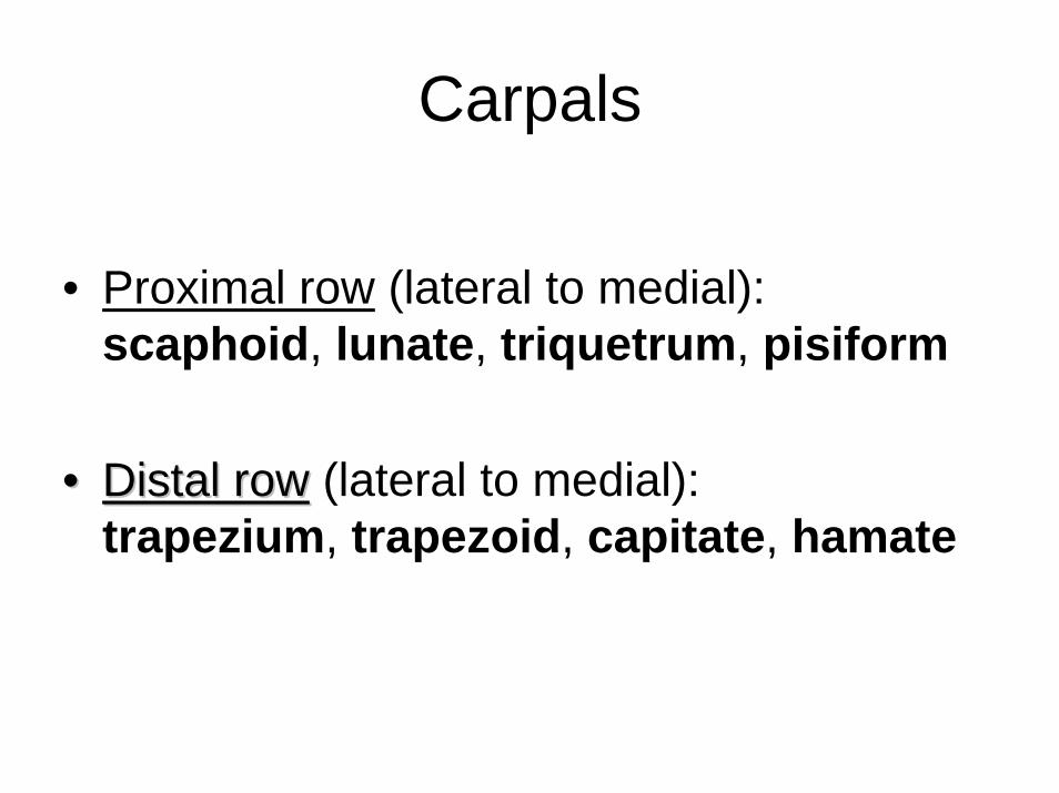
Carpals
• Proximal row (lateral to medial): scaphoid, lunate, triquetrum, pisiform
•• Distal rowDistal row (lateral to medial): trapezium, trapezoid, capitate, hamate

Metacarpals & phalanges
• Enlarged ends (epiphysis)• Distinct shaft (diaphysis)
• Numbered 1-5 from lateral to medial• 14 phalanges in each hand: 2 in thumb,
and 3 in each of the 4 digits
• Palpate: 1) the pisiform; 2) the scaphoid
Interphalaangeal joints (IPJ)
• Synovial
• Hinge joints…??

Extrinsic flexors of the digits
• Flexor digitorum superficialis: primary flexor of the proximal interphalangeal joints (PIPJ)
• Flexor digitorum profundus: primary flexor of the distal interphalangeal joints (DIPJ)
• Flexor pollicis longus: the only flexor of the IPJ of the thumb

Extrinsic extensors of the digits
• Extensor digitorum: extension of PIPJ & DIPJ of fingers 2-5
• Extensor pollicis longus: the only extensor of the IPJ of the thumb

Extensors of the digits
• Extension of IPJ of digits is also assisted by intrinsic muscles of the hand which are attached to the dorsal or extensor expansionof the fingers

Intrinsic extensors of the digits
• Lumbricals (1-4 lateral to medial): take their common attachment from the tendons of the flexor digitorum profundus
pull through the central and lateral slips of the extensor expansion
assist extension of both PIPJ & DIPJ

Intrinsic extensors of the digits
• Interossei:Attached to and lie between the metacarpals
pull through the central and lateral slips of the extensor expansion
assist extension of both PIPJ & DIPJ
Metacarpophalangeal joints (MPJ)
• Between the metacarpals & proximal phalanges
• Biaxial joints….?

Flexors of the MPJ
• Lumbricals & interossei: assist flexion of MPJ of digits 2-5
• Flexor digiti minimi: assist flexion of MPJ of finger 5
• Flexor pollicis brevis: assist flexion of MPJ of the thumb
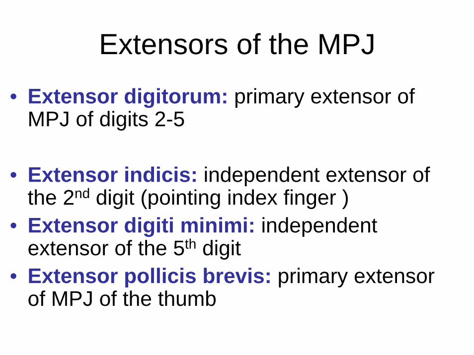
Extensors of the MPJ
• Extensor digitorum: primary extensor of MPJ of digits 2-5
• Extensor indicis: independent extensor of the 2nd digit (pointing index finger )
• Extensor digiti minimi: independent extensor of the 5th digit
• Extensor pollicis brevis: primary extensor of MPJ of the thumb
Abduction/adduction of the MPJ
• Described relative to the long axis of the hand which extends distally through the 3rd
or middle finger
• Frontal plane movement
• About an antero-posterior axis

Abduction/adduction of the MPJ
• Palmar interossei: adduct the fingers-move fingers 2, 4, & 5 toward the 3rd finger (fingers closing)
• Dorsal interossei: abduct the fingers- move fingers 2, 3, & 4 relative to the long axis of the hand (fingers spreading)
• The thumb & 5th digit have their own abductors


Flexors of the wrist & hand
• Lie within the anterior muscle compartment of the forearm
• Cross the wrist joint anterior to its axis of function
• Take their common origin from the medial epicondyle (site of golfer’s elbow = medial epicondylitis)
Flexors of the forearm
• Superficial muscles:Pronator teres
Palmaris longus
Flexor carpi radialis
Flexor carpi ulnaris
Pronator teres
• Origin:Medial epicondyle of humerusCoronoid process of ulna
• Insertion: Lateral surface of mid-shaft of radius

Pronator teres
• Action:
Pronation of forearm
• Innervation: median nerve

Palmaris longus
• Origin: medial epicondyle of humerus(common flexor tendon)
• Insertion: Palmar aponeurosis
• Action: Flexion of hand
• Innervation: median nerve

Flexor carpi radialis
• Origin: medial epicondyle of humerus(common flexor tendon)
• Insertion: base of 2nd metacarpal (possibly 3rd)
• Action: Flexion & abduction (radial deviation)
• Innervation: median nerve

Flexor carpi ulnaris
• Origin: medial epicondyle of humerus(common flexor tendon)
• Insertion: pisiform bone
• Action: Flexion & adduction (ulnar deviation)
• Innervation: ulnar nerve

Flexors of the forearm
• Intermediate muscles: Flexor digitorum superficialis
• Deep muscles:Flexor digitorum profundus
Flexor pollicis longus
Pronator quadratus

Flexor digitorum superficialis
• Origin: medial epicondyle of humerus(common flexor tendon)
• Insertion: base of middle phalanx of 4 digits
• Action: Flexion of PIPJ of medial 4 digits
• Innervation: median nerve
Flexor digitorum profundus
• Origin: anterior & medial surface of proximal ulna; aponeurosis of flexor carpi ulnaris
• Insertion: distal phalanx of medial 4 digits
• Action: Flexion of DIPJ of medial 4 digits
• Innervation: median & ulnar nerves

Flexor policis longus
• Origin: anterior surface of middle half of radius
• Insertion: distal phalanx of thumb
• Action: Flexion of DIPJ of thumb
• Innervation: median nerve

Pronator quadratus
• Origin: distal fourth of ulna
• Insertion: distal part of radius
• Action: pronation of forearm
• Innervation: median nerve


Extensors of the wrist & hand
• Lie within the posterior muscle compartment of the forearm
• Cross the wrist joint posterior to its axis of function
• Take their common origin from the lateral epicondyle (site of tennis elbow = lateral epicondylitis)

Extensors of the forearm
• Superficial muscles:BrachioradialisExtensor carpi radialis longusExtensor carpi radialis brevis
Extensor digitorumExtensor digiti minimi
Extensor carpi ulnaris

Brachioradialis
• Origin: lateral supracondylar ridge of humerus
• Insertion: lateral side of distal end of radius
• Action: flexion of forearm
• Innervation: radial nerve
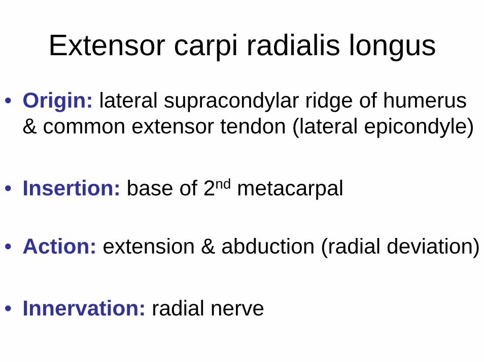
Extensor carpi radialis longus
• Origin: lateral supracondylar ridge of humerus& common extensor tendon (lateral epicondyle)
• Insertion: base of 2nd metacarpal
• Action: extension & abduction (radial deviation)
• Innervation: radial nerve

Extensor carpi radialis brevis
• Origin: common extensor tendon (lateral epicondyle)
• Insertion: base of 3rd metacarpal
• Action: extension of hand
• Innervation: radial nerve

Extensor digitorum
• Origin: common extensor tendon (lateral epicondyle)
• Insertion: middle & distal phalanges of 4 digits
• Action: extension of PIPJ & DIPJ of 4 digits
• Innervation: radial nerve

Extensor digiti minimi
• Origin: common extensor tendon
• Insertion: middle & distal phalanges of little finger
• Action: extension & abduction of little finger
• Innervation: radial nerve

Extensor carpi ulnaris
• Origin: common extensor tendon (lateral epicondyle)
• Insertion: base of 5th metacarpal
• Action: extension & adduction (ulnar deviation)
• Innervation: radial nerve
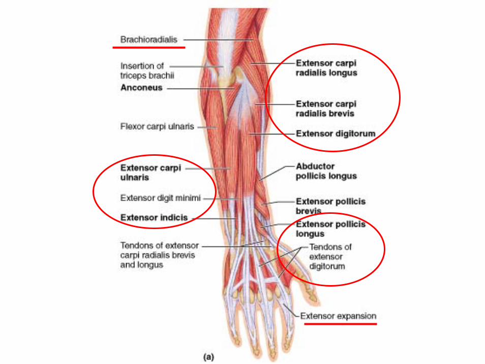


Extensors of the forearm
• Deep muscles:Supinator
Abductor pollicis longusExtensor pollicis brevisExtensor pollicis longus
Extensor indicis
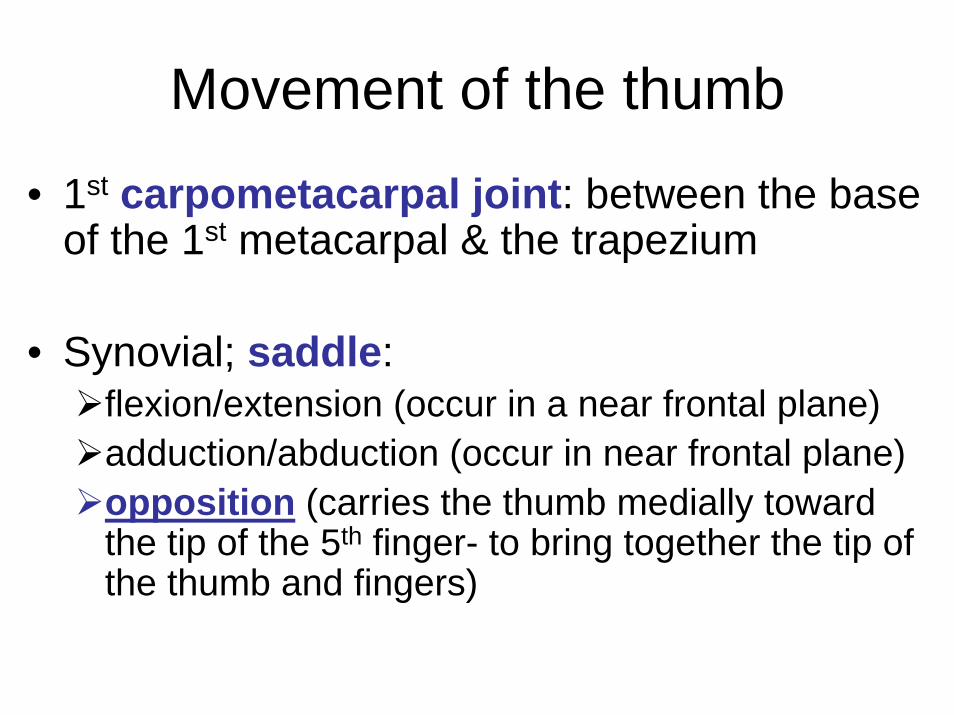
Movement of the thumb
• 1st carpometacarpal joint: between the base of the 1st metacarpal & the trapezium
• Synovial; saddle: flexion/extension (occur in a near frontal plane) adduction/abduction (occur in near frontal plane) opposition (carries the thumb medially toward the tip of the 5th finger- to bring together the tip of the thumb and fingers)

Primary movers of the thumb
• Flexors: flexor pollicis longusflexor pollicis brevis
• Extensors: extensor pollicis longusextensor pollicis brevis

Primary movers of the thumb
• Adductors: adductor pollicis
• Abductors: abductor pollicis longusabductor pollicis brevis
• Opposition: opponens pollicis

Vulnerable peripheral nervesUpper limb
• Median nerveMost common: compression at the wrist (carpal tunnel)Fluid retention in pregnancyRepetitive movement (flexor tenosynovitis)Sensory distribution: thumb, index, and middle fingers, half the ring finger

Vulnerable peripheral nervesUpper limb
• Ulnar nerve:Most common: irritation at the elbowSensory distribution: little finger & ulnar half of the ring finger
• Radial nerve:Vulnerable in the medial side of the upper arm (e.g., axillary crutches)Drop wrist (few sensory symptoms)

Fractures of the medial epicondyle causing an ulnarnerve paralysis.
Dislocation of the lunatecausing a median nervepalsy.

• Which muscles serve as the primary adductors of the wrist?
• Where is/are the axis/axes of movement of the IP joints, and which movement do they permit?