biomechanics and thermodynamics of nanoparticle interactions with plasma and endosomal membrane...
TRANSCRIPT
Biomechanics and Thermodynamics of Nanoparticle Interactionswith Plasma and Endosomal Membrane Lipids in Cellular Uptake andEndosomal EscapeChiranjeevi Peetla,† Shihua Jin,† Jonathan Weimer,† Adekunle Elegbede,† and Vinod Labhasetwar*,†,‡
†Department of Biomedical Engineering, Lerner Research Institute and ‡Taussig Cancer Institute, Cleveland Clinic, 9500 EuclidAvenue, Cleveland, Ohio 44195, United States
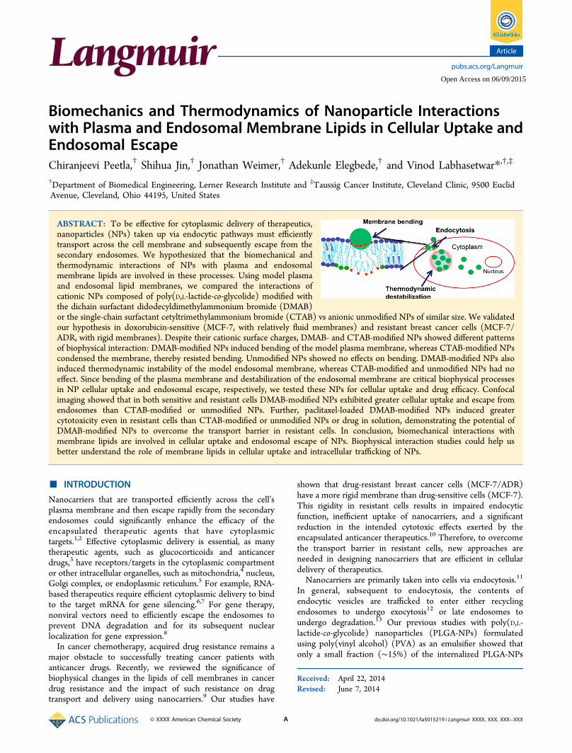
ABSTRACT: To be effective for cytoplasmic delivery of therapeutics,nanoparticles (NPs) taken up via endocytic pathways must efficientlytransport across the cell membrane and subsequently escape from thesecondary endosomes. We hypothesized that the biomechanical andthermodynamic interactions of NPs with plasma and endosomalmembrane lipids are involved in these processes. Using model plasmaand endosomal lipid membranes, we compared the interactions ofcationic NPs composed of poly(D,L-lactide-co-glycolide) modified withthe dichain surfactant didodecyldimethylammonium bromide (DMAB)or the single-chain surfactant cetyltrimethylammonium bromide (CTAB) vs anionic unmodified NPs of similar size. We validatedour hypothesis in doxorubicin-sensitive (MCF-7, with relatively fluid membranes) and resistant breast cancer cells (MCF-7/ADR, with rigid membranes). Despite their cationic surface charges, DMAB- and CTAB-modified NPs showed different patternsof biophysical interaction: DMAB-modified NPs induced bending of the model plasma membrane, whereas CTAB-modified NPscondensed the membrane, thereby resisted bending. Unmodified NPs showed no effects on bending. DMAB-modified NPs alsoinduced thermodynamic instability of the model endosomal membrane, whereas CTAB-modified and unmodified NPs had noeffect. Since bending of the plasma membrane and destabilization of the endosomal membrane are critical biophysical processesin NP cellular uptake and endosomal escape, respectively, we tested these NPs for cellular uptake and drug efficacy. Confocalimaging showed that in both sensitive and resistant cells DMAB-modified NPs exhibited greater cellular uptake and escape fromendosomes than CTAB-modified or unmodified NPs. Further, paclitaxel-loaded DMAB-modified NPs induced greatercytotoxicity even in resistant cells than CTAB-modified or unmodified NPs or drug in solution, demonstrating the potential ofDMAB-modified NPs to overcome the transport barrier in resistant cells. In conclusion, biomechanical interactions withmembrane lipids are involved in cellular uptake and endosomal escape of NPs. Biophysical interaction studies could help usbetter understand the role of membrane lipids in cellular uptake and intracellular trafficking of NPs.
■ INTRODUCTION
Nanocarriers that are transported efficiently across the cell’splasma membrane and then escape rapidly from the secondaryendosomes could significantly enhance the efficacy of theencapsulated therapeutic agents that have cytoplasmictargets.1,2 Effective cytoplasmic delivery is essential, as manytherapeutic agents, such as glucocorticoids and anticancerdrugs,3 have receptors/targets in the cytoplasmic compartmentor other intracellular organelles, such as mitochondria,4 nucleus,Golgi complex, or endoplasmic reticulum.5 For example, RNA-based therapeutics require efficient cytoplasmic delivery to bindto the target mRNA for gene silencing.6,7 For gene therapy,nonviral vectors need to efficiently escape the endosomes toprevent DNA degradation and for its subsequent nuclearlocalization for gene expression.8
In cancer chemotherapy, acquired drug resistance remains amajor obstacle to successfully treating cancer patients withanticancer drugs. Recently, we reviewed the significance ofbiophysical changes in the lipids of cell membranes in cancerdrug resistance and the impact of such resistance on drugtransport and delivery using nanocarriers.9 Our studies have
shown that drug-resistant breast cancer cells (MCF-7/ADR)have a more rigid membrane than drug-sensitive cells (MCF-7).This rigidity in resistant cells results in impaired endocyticfunction, inefficient uptake of nanocarriers, and a significantreduction in the intended cytotoxic effects exerted by theencapsulated anticancer therapeutics.10 Therefore, to overcomethe transport barrier in resistant cells, new approaches areneeded in designing nanocarriers that are efficient in cellulardelivery of therapeutics.Nanocarriers are primarily taken into cells via endocytosis.11
In general, subsequent to endocytosis, the contents ofendocytic vesicles are trafficked to enter either recyclingendosomes to undergo exocytosis12 or late endosomes toundergo degradation.13 Our previous studies with poly(D,L-lactide-co-glycolide) nanoparticles (PLGA-NPs) formulatedusing poly(vinyl alcohol) (PVA) as an emulsifier showed thatonly a small fraction (∼15%) of the internalized PLGA-NPs
Received: April 22, 2014Revised: June 7, 2014
Article
pubs.acs.org/Langmuir
© XXXX American Chemical Society A dx.doi.org/10.1021/la5015219 | Langmuir XXXX, XXX, XXX−XXX
Open Access on 06/09/2015
escape the above pathway to reach the cytoplasmic compart-ment, and the remaining NPs undergo exocytosis.12 Severalstrategies have been proposed to facilitate cellular uptake andescape of NPs from the endosomal pathway. Commonlyemployed approaches include (a) conjugating NPs to cell-penetrating peptides, thus bypassing the endocytic path-way;14,15 (b) modifying the NP surface with cationic polymersor surfactants to promote their interactions with the anionic cellmembrane;16 or (c) modifying the NP surface with pH-sensitive polymers that exploit the acidic pH in the endosomes,causing a change in the polymer configuration to facilitate NPinteraction with the endosomal membrane and escape into thecytoplasmic compartment.17
These strategies generally disregard the role of biophysics ofNP interactions with cell membrane lipids in intracellulartrafficking of NPs, particularly regarding their uptake into cellsand escape from endosomes. The role of cell membranebiophysical characteristics such as lipid composition andmembrane fluidity/rigidity on intracellular trafficking is onlybeginning to be known.18 Recent progress in membranebiology suggests that endocytosis can also be initiated bychanging lipid composition, by inducing differences betweenthe surface areas of outer and inner (cytoplasmic) lipidmonolayers in the cell membrane, or by inserting proteins thatcould act like wedges between the membrane.19 Additionally,studies have demonstrated that the endocytic process requireshigh local membrane curvature,20 which involves energy to actagainst the in-plane membrane tension and resistance tobending and stretching. The above studies clearly emphasizethe significance of cell membrane characteristics on theendocytosis of nanocarriers. Recently, several theoretical21
(molecular dynamic simulation) and experimental studies havedemonstrated that the attractive interactions between a particleand lipid head groups influence the mechanical andthermodynamic properties of the cell membrane, therebyaffecting the internalization of NPs via endocytosis.22
We propose that biomechanical and thermodynamicinteractions between NPs and plasma membrane lipids alterthe membrane’s curvature and the trafficking of NPs insidecells. We hypothesize that NP−lipid biophysical interactionsalter the bending energy associated with membrane wrappingduring endocytosis at the plasma membrane and that, followingendocytosis, NP−lipid interactions facilitate the endosomalescape of the NPs due to the unfavorable Gibbs energy (G) ofmixing of endosomal membrane lipids. Since these parameterscannot be assessed in live cells, we developed Langmuir modelsfor plasma and late endosomal membranes to test ourhypothesis. We then characterized biomechanical and thermo-dynamic properties to investigate the effects of differentcationic surfactant-modified NPs on those properties in thetwo model membranes.In this study, we analyzed the changes in surface pressure-
area (π−A) isotherms to understand the interfacial character-istics of NPs required to facilitate their cellular uptake andendosomal escape. Since biophysical interactions depend onboth the cell-membrane characteristics and surface properties ofNPs, we tested our hypothesis in resistant and sensitive breastcancer cells, using NPs modified with two different cationicsurfactants as well as unmodified NPs to determine whethercationic surfactant-modified NPs might be more effective inovercoming the transport barrier in resistant cells thanunmodified NPs. Cell-membrane fluidity is known to affectthe membrane’s bending energy associated with endocytosis;
therefore, intrinsic differences in the membrane characteristicsof sensitive and resistant cells could help us validate thecorrelation between NP-model membranes and the NPs’ abilityto achieve endosomal escape in live cells. We comparedunmodified NPs against NPs modified with two differentsurfactants: PVA, a nonionic surfactant conventionally used inthe formulation of PLGA-NPs, which served as our control(unmodified NPs) vs the single-chain surfactant cetyltrimethy-lammonium bromide (CTAB) and the dichain surfactantdidodecyldimethylammonium bromide (DMAB), both ofwhich are commonly used cationic surfactants for modifyingPLGA-NPs.16,23
Our data demonstrate that even though both DMAB- andCTAB-modified NPs carried a cationic surface charge, eachtype showed a different pattern of interaction with modelplasma and endosomal lipid membranes. DMAB-modified NPsbent the model plasma membrane, whereas CTAB-modifiedNPs created a resistance to bending in the model plasmamembrane; unmodified NPs showed no effect. DMAB-modified NPs also caused thermodynamic destabilization ofthe model endosomal membrane, whereas CTAB-modified andunmodified NPs had no effect. Confocal imaging data showedgreater uptake and endosomal escape of DMAB-modified NPsthan CTAB-modified and unmodified NPs in both sensitiveand resistant cells. Furthermore, DMAB-modified NPs loadedwith paclitaxel (PTX) demonstrated significantly greatercytotoxicity, particularly toward resistant cells, than didCTAB-modified, unmodified NPs, or drug in solution.
■ EXPERIMENTAL SECTIONMaterials. PLGA (50:50, inherent viscosity = 1.24 dL/g) was
purchased from DURECT Corp. (Cupertino, CA). The solventsmethanol (CH3OH), ethanol, and chloroform (CHCl3) were of highperformance liquid chromatography grade and purchased from FisherScientific (Pittsburgh, PA). Phospholipids, 1,2-dipalmitoyl-sn-glycero-3-phosphocholine (DPPC), 1,2-dipalmitoyl-sn-glycero-3-phosphoetha-nolamine (DPPE), 1,2-dipalmitoyl-sn-glycero-3-[phospho-L-serine](DPPS), L-α-phosphatidylinositol (DPPI), sphingomyelin (SM),cardiolipin (CL), and bis(monoacylglycero)phosphate (BMP) werepurchased from Avanti Polar Lipids (Alabaster, AL). D-PBS(Dulbecco’s phosphate-buffered saline) was used as a subphase formonolayer experiments. DMAB was obtained from Aldrich(Milwaukee, WI). CTAB and ammonium hydroxide (NH4OH) wereobtained from Acros Organics (Fair Lawn, NJ). Sucrose and PVA withan 87%−89% degree of hydrolysis were purchased from Sigma-Aldrich(St. Louis, MO). PTX was purchased from LC Laboratories (Woburn,MA). D-PBS and cell culture media were obtained from the CentralCell Services’ Media Laboratory of our institution.
Methods. Lipid Solutions. DPPC, DPPI, SM, CL, and BMP werediluted in CHCl3; DPPE and DPPS were diluted in a mixture ofCHCl3:CH3OH:H2O (65:35:8 v/v/v). The selection of appropriatesolvents for specific lipids is based on each lipid’s solubility in therespective solvent or mixture of solvents. The lipid mixture we usedwas prepared by mixing individual lipid solutions according to thepercentages shown in Table 1. These compositions are similar to thoseof the phospholipids present in cell plasma membranes and lateendosomal membranes in vivo.24
Formulation of NPs. PLGA-NPs were formulated using anemulsion-solvent evaporation technique. The procedure involvedemulsification of PLGA (60 mg) solution in chloroform (2 mL)into 16 mL of an aqueous phase (8 mL of 2% w/v PVA + 1 mL ofcationic surfactant at the desired concentration in ethanol + 7 mL ofMilli-Q water) (Table 2). Because of the higher critical micelleconcentration of CTAB than DMAB, at least 20 mM CTAB (vs 2 mMDMAB) was required to formulate cationic NPs. Unmodified NPswere prepared using the same protocol as above but without adding
Langmuir Article
dx.doi.org/10.1021/la5015219 | Langmuir XXXX, XXX, XXX−XXXB
the cationic surfactant in the PVA solution. Emulsification wasachieved using a probe sonicator set at 55 W energy output (XL 2015Sonicator ultrasonic processor, Misonix, Inc., Farmingdale, NY) for 3min over an ice bath to form an oil-in-water emulsion. The emulsionthus formed was stirred overnight at room temperature inside a hoodto allow evaporation of the organic solvent. NPs were recovered byultracentrifugation at 30000 rpm (∼80000g) for 30 min at <10 °C(Beckman Optima LE-80K, Beckman Instruments, Inc., Palo Alto,CA) and washed three times with distilled water to remove excesssurfactants. The pellet obtained was resuspended using 6 mL ofdistilled water, sonicated for 45 s, and further centrifuged at 1000 rpm(∼800g) for 10 min at 4 °C (Sorvall Legend RT, Thermo ElectronCorporation, Waltham, MA).To prevent aggregation of NPs, prior to lyophilization, 1 mL of 15%
w/v sucrose solution was added to a 4 mL of NP suspension. Thesuspension was lyophilized into preweighed cryovials at 3.5 Pa and−45 °C for about 48 h to obtain a dry powder. The number of NPs invials was estimated by separately lyophilizing the sucrose solutionadded into the NP suspension. The difference between the lyophilizedweight of the NPs with sucrose and the sucrose alone was used tocalculate approximately how many NPs were present. Based on theabove calculations, sucrose accounts for 3% w/w of NPs in thelyophilized samples. The amount of sucrose required to preventparticle aggregation was optimized. To prepare PTX-loaded NPs, 4 mgof drug was dissolved in a CHCl3 solution containing 60 mg of PLGA.The above procedure was repeated to prepare drug-loaded PLGA NPswith different modifications. To achieve similar dye loading in allformulations of NPs, typically 50 μg of dye (6-coumarin) was added tothe 60 mg of PLGA solution in CHCl3 to prepare the anionicunmodified NPs, whereas 60 μg of dye was used to prepare thecationic surfactant-modified NPs. Since cationic surfactants formmicelles, a greater fraction of the dye added to the polymer solutionescapes into the aqueous phase; this is in contrast to when unmodifiedNPs are formulated without a cationic surfactant. Similarly, lower drugentrapment efficiency was observed in CTAB-modified NPs thanunmodified or DMAB-modified NPs, due to drug partitioning intoCTAB micelles formed during the aqueous phase. Therefore, to obtaindrug loading similar to that in other formulations of NPs, the drugamount used during formulation was doubled to formulate CTAB-modified NPs (Table 2).Physical Characterization of NPs. The mean hydrodynamic
diameter of NPs was determined using a dynamic light-scatteringtechnique and the ζ-potential by using a phase-analysis light-scatteringtechnique (PSS/NICOMP 380/ZLS, Particle Sizing Systems, SantaBarbara, CA). A 50 μL aliquot of each NP suspension (5 mg/mL,sonicated for 30 s as above) was diluted to 3 mL with water and usedfor measuring the size and ζ-potential of NPs.
Surface Pressure−Area Isotherms for Plasma and EndosomalLipid Mixtures. A Langmuir balance (small Langmuir−Blodgetttrough, Biolin Scientific, Inc., Linthicum Heights, MD) was used tostudy the surface pressure−area (π−A) isotherms for plasma andendosomal lipid mixtures. In general, to obtain the π−A isotherm, 5μL of the lipid mixture was added dropwise (∼0.5 μL) onto the D-PBSsurface in the trough using a Hamilton digital microsyringe (Hamilton,Reno, NV). After waiting for 10 min to allow the chloroform toevaporate, the barriers were compressed at the rate of 5 mm/min untilthe collapse of the membrane. The π−A isotherm for plasmamembrane lipids was constructed on a buffer surface of pH 7, whereasthat of late endosomal membrane lipids was constructed on a buffersurface of pH 5 to mimic the endosomal pH. The pH of the D-PBSwas adjusted to pH 5 by adding 1 M HCl. All experiments involvinglate endosomal membrane lipids were carried out at pH 5.
Effect of Surfactant-Modified NPs on the π−A Isotherm ofPlasma and Endosomal Membrane Lipids. These experiments wereperformed to investigate the penetrability of modified and unmodifiedNPs into model plasma and endosomal membranes and to determinehow these interactions with NPs influence the mechanical stability ofboth model membranes. For this step, the plasma or endosomal lipidmixture was spread at a surface pressure of 0 mN/m; then a 500 μLaliquot of the NP suspension (5 mg/mL concentration in Milli-Qwater, sonicated for 30 s as above) was injected below the lipidmixture. A magnetic stir plate, located just below the trough as part ofthe Langmuir balance, was kept on to ensure a uniform distribution ofNPs into the subphase buffer. NPs were allowed to interact for 20 minwith the lipid mixture and were then compressed at the rate of 5 mm/min until the film collapsed.
Effects of Surfactant-Modified NPs on Surface Pressure of ModelPlasma and Endosomal Membranes. Plasma or endosomalmembrane lipids were spread on the buffer surface as above andthen compressed until the surface pressure of 30 mN/m. Since thearrangement of lipids at this surface pressure in the monolayers mimicsthe arrangement of lipids in the cell-membrane bilayer, hereafter weshall refer to the lipid monolayers constructed at the surface pressureof 30 mN/m as the model plasma or endosomal membrane. A 500 μLaliquot of NP suspension (5 mg/mL concentration) prepared as abovewas injected below the surface of the model plasma or endosomalmembranes through the injection port. The change in surface pressureof the model membrane was recorded immediately for a period of 20min. To ensure that the changes in surface pressure of the modelmembrane were due to the interactions of modified NPs, a controlexperiment with sucrose in Milli-Q water was carried out.
Analysis of Biomechanical and Thermodynamic Parameters ofInteractions with NPs. We used the isotherm data to investigate theeffects of NPs on the plasma and endosomal membranes’ bendingrigidity and thermodynamic stability, particularly the surface pressureat the point of the film’s collapse (collapse surface pressure) in thepresence of NPs. In the Langmuir monolayer, the collapse of a givenlayer is initiated by buckling or bending of the monolayer into thesubphase; therefore, the collapse surface pressure can be consideredthe minimum force required to bend the lipid monolayer at theinterface. Surface tension can also be defined as the force per unitlength, and since we are comparing the change is surface tension at aconstant length, we determined force using the following equation. Wecalculated the difference in the bending force in the absence vspresence of NPs using the formula
Table 1. Phospholipid Composition Used To Prepare ModelPlasma and Endosomal Membranesa
lipids PM (%) EM (%) lipids PM (%) EM (%)
DPPC 56 53 SM 6.0 3DPPE 24 19 CL 1.7DPPI 8.0 7 BMP 14DPPS 4.3 4
aPM, plasma membrane; EM, endosomal membrane.
Table 2. Physical Characteristics of Unmodified and Surfactant-Modified PLGA-NPsa
PVA(%) (w/v)
cationic surfactant(mM) size, nm (PI)
ζ-potential(mV)
paclitaxel EE(%)
paclitaxel loading(%) (w/w)
coumarin EE(%)
unmodified NPs 1 0 305.6 (0.02) −19.31 81.0 5.4 ± 0.1 88CTAB-modified NPs (Si-C16-NPs)
1 20 293.2 (0.15) +14.31 31.1 4.15 ± 0.62 75
DMAB-modified NPs (Di-C12-NPs)
1 2 240.1 (0.05) +39.91 67.5 4.5 ± 0.13 73
aPI, polydispersity index; EE, entrapment efficiency.
Langmuir Article
dx.doi.org/10.1021/la5015219 | Langmuir XXXX, XXX, XXX−XXXC
π πΔ = −F MMNP MMb c c (1)
where ΔFb is the difference in force required to bend the monolayer atthe interface, MMNP represents the model membrane with NPs, MMis the model membrane, and πc is the surface pressure at the point ofcollapse of the monolayer.The thermodynamic stability of the model membranes was
determined using the excess area (ΔA) and excess Gibbs energy(ΔG) of mixing. Both ΔA and ΔG provide the measure of relativestability of a model membrane by considering the energetics ofmiscibility of its pure lipid components. ΔA and ΔG are calculatedusing the following equations:
Δ = − −A A x A x A12 1 1 2 2 (2)
∫ πΔ = − − −π
G A x A x A x A( .... ) dn n n0
1,2... 1 1 2 2 (3)
where A1,2,...,n is the molecular area occupied by the mixed monolayer,A1, A2, ..., An are the area of per molecule in the pure monolayers ofcomponent 1, 2,..., n, x1, x2, ..., xn are the molar fractions of thecomponent, and dπ is the surface pressure. Integration was carriedbetween 0 and π. Data were calculated with vs without NPs at differentsurface pressures. The area per molecule (A) for different lipids used inmodel membranes was determined from the individual isotherm ofeach lipid. The isotherms were generated at pH 7 for lipids in plasmamodel membrane and at pH 5 for lipids in endosomal modelmembrane.Cell Culture. Doxorubicin-sensitive (MCF-7) and -resistant (MCF-
7/ADR) breast cancer cells were grown in Dulbecco’s modified Eagle’smedium (DMEM) supplemented with Earle’s salts, L-glutamine, 100μg/mL penicillin, and 100 μg/mL streptomycin. The mediumcontained 10% or 15% fetal bovine serum (FBS) for culturingsensitive and resistant cells, respectively. These conditions wereoptimized for growth of these cells. Drug resistance was maintained byexposing resistant cells to 100 ng/mL of doxorubicin (Drug SourceCo. LLC, Westchester, IL) after every two passages.Confocal Microscopy. Live cell imaging was performed using a
spinning disk confocal microscope (UltraView VoX, PerkinElmer,Waltham, MA). In a typical experiment, cells were seeded in 35 mmglass-bottom cell culture dishes (MatTek, Ashland, MA) at a density of45 000 cells/cm2 and were allowed to attach for 24 h prior to exposureto marker dye and fluorescent NPs (100 μg/mL and 2 mL,respectively). Confocal images were captured following incubation ofcells with NPs for 2 h. The interaction of NPs with the cell membranewas monitored by staining the membrane with Deep Red plasmamembrane dye (CellMask, Invitrogen/Life Technologies, Eugene,OR). For this step, 5 μg/mL of dye in cell culture media was added tothe cell culture dish 5 min prior to imaging the cells.
Under identical conditions as above, the escape of NPs fromendosomes was monitored by staining the late endosomes withLysoTracker Red DND-99 (Invitrogen/Life Technologies, Eugene,OR). For this step, 75 ng/mL of the dye solution in media was addedto the cell culture dish 30 min prior to imaging. Cells were washedtwice with PBS to remove excess NPs and dye, and fresh respectivemedia without NPs or dye were added to culture dish before imaging.Confocal images were captured by illuminating samples withrespective lasers for capturing NP signals (green filter, Ex λ 488)and dye signals (Red filter, Ex λ 561, for the endosomal compartmentor Deep Red filter, Ex λ 640, for the plasma membrane) in an alternatefashion. The processes of NP internalization and escape were recordedin z-planes (distance 0.25 μm) and presented as z-projections. Wehave previously used similar techniques to study cellular uptake andtrafficking of NPs.25
Cytotoxicity with Drug-Loaded NPs. The cytotoxicity of PTX-loaded NPs (PTX-NPs) vs control NPs was determined in both MCF-7 and MCF-7/ADR cells. It is known that MCF-7/ADR cells developresistance to multiple anticancer drugs, including PTX.26,27 In a typicalexperiment, cells were seeded in a 96-well plate (Microtest, BectonDickinson Labware, Franklin Lakes, NJ) at a density of 3000 cells perwell and allowed to attach for 24 h. For drug treatment, PTX stock inethanol was diluted (1:1000) with cell culture media to obtain the finalconcentration in the well. For MCF-7 cells, drug concentrationsranged from 0 to 500 ng/mL; for MFC-7/ADR cells, concentrationsranged from 0 to 20 000 ng/mL. For treatment with NPs, a stocksuspension of NPs was diluted in culture medium to obtain theappropriate equivalent drug concentrations in the well. Respective NPswithout drug were used as a control. Cells were incubated with drugfor 72 h and washed with D-PBS, and the medium in the plates wasreplaced with a drug-free medium. Cells were incubated for anadditional 48 h prior to determining cell viability using a standardMTS assay (CellTiter 96 AQueous, Promega, Madison, WI). Then 20μL of reagent was added to each well, and plates were incubated for 2h in a cell culture incubator. Color intensity was measured at 490 nmusing a microplate reader (Bio-Tek Instruments, Winooski, VT). Cellproliferation was calculated as the percentage of cell growth vs growthof respective controls. The drug concentration required for 50% celldeath (IC50) for each treatment was calculated using the equation
=−
++y
A Ax x
A1 ( / )p
1 2
02
(4)
where x is the drug concentration, y the % cell growth as determinedby MTS assay, A1 the % growth at the top plateau region of the growthcurve, A2 the % growth at the bottom plateau region of the curve, x0the inflection point of the curve, and p the slope. The data points werefit to this equation using OriginPro 8 (OriginLab Corp., Northampton,MA). IC50 was determined by using y = 50 in the above equation and
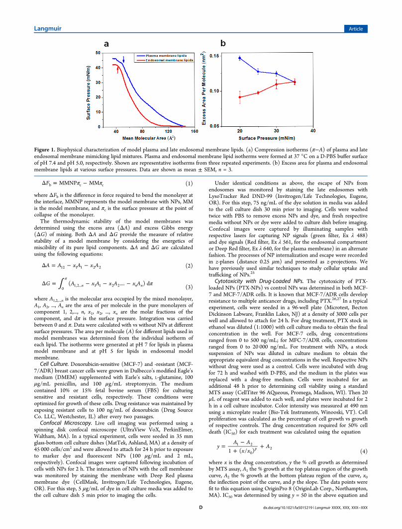
Figure 1. Biophysical characterization of model plasma and late endosomal membrane lipids. (a) Compression isotherms (π−A) of plasma and lateendosomal membrane mimicking lipid mixtures. Plasma and endosomal membrane lipid isotherms were formed at 37 °C on a D-PBS buffer surfaceof pH 7.4 and pH 5.0, respectively. Shown are representative isotherms from three repeated experiments. (b) Excess area for plasma and endosomalmembrane lipids at various surface pressures. Data are shown as mean ± SEM, n = 3.
Langmuir Article
dx.doi.org/10.1021/la5015219 | Langmuir XXXX, XXX, XXX−XXXD

calculating x using the parameters obtained after curve fitting. A meanof six replicates for each set of experiments was used to calculate IC50.
■ RESULTS
Characterization of NPs. Hydrodynamic diameters ofunmodified and cationic surfactant-modified NPs were in asimilar range; however, CTAB-modified NPs showed a slightlyhigher variance compared with unmodified and DMAB-modified NPs. The ζ-potential of the unmodified NPs wasanionic, whereas that of the cationic surfactant-modified NPswas positive, with DMAB-modified NPs showing a higher ζ-potential than CTAB-modified NPs (Table 2).Biophysical Characterization of Model Plasma and
Endosomal Membrane Lipids. In general, the isotherms forthe model plasma and endosomal membrane lipids showedthree phases with compression (Figure 1a): An initial lag phasewas followed by a steady increase in surface pressure and thencollapse. However, in the case of plasma membrane lipids, ascompression increased, there was a steady increase in surfacepressure with no significant lag phase, whereas the endosomalmembrane lipids showed a lag phase with compression untilreaching a mean molecular area (mmA) of 100 Å2, prior to asteady increase in surface pressure. In addition, the collapsesurface pressure for the plasma membrane lipids was 43 mN/m,corresponding to 49 Å2 mmA; in contrast, the collapse surface
pressure for the endosomal membrane lipids was 41.5 mN/m,corresponding to 43 Å2 mmA (Figure 1a). Interestingly, theplasma membrane lipids continued to show a further increasein surface pressure, even after the film’s collapse, whereas theendosomal membrane lipids reached a plateau with no furtherincrease in surface pressure with compression. The excess area(ΔA) values for the model plasma and endosomal membranelipids at various surface pressures, as calculated using eq 2, werehigher for the plasma membrane lipids than for the endosomalmembrane lipids at low surface pressures, but there was nodifference in ΔA between the two membrane lipids at highersurface pressures (Figure 1b).
Effects of NPs on Isotherms of Model Plasma andEndosomal Membrane Lipids. Both the plasma andendosomal membrane lipid isotherms in the presence ofunmodified or CTAB-modified NPs showed a slight shifttoward a higher mean molecular area with respect to lipidsalone (without NPs); however, this shift was significantlygreater in the presence of DMAB-modified NPs (Figure 2a).Most noticeable was the difference in the shape of theisotherms; model plasma and endosomal membranes bothshowed significantly higher surface pressures in the presence ofDMAB-modified NPs during the entire isotherm than in thepresence of unmodified or CTAB-modified NPs. The isothermsin the presence of CTAB-modified and unmodified NPs
Figure 2. Biophysical interactions of unmodified and modified NPs with model plasma and endosomal membrane lipids. (a) Compression isothermsof plasma and late endosomal membrane lipids in the presence of different formulations of NPs. (b) Change in surface pressures of model plasmaand late endosomal membranes following interaction with NPs. Shown are representative isotherms from three repeated experiments.
Langmuir Article
dx.doi.org/10.1021/la5015219 | Langmuir XXXX, XXX, XXX−XXXE
showed a shift toward higher surface pressure during the initialphase of compression, but at the later phase, the isothermsalmost overlapped with the isotherm of the lipids alone. Thesurface pressure at the point of collapse in the presence ofDMAB-modified NPs was significantly lower and at a highermean molecular area, whereas the collapse surface pressure wasat a higher surface pressure but a lower mean molecular area inthe presence of CTAB-modified NPs (Figure 2a). The collapsesurface pressure in the presence of unmodified NPs was almostthe same as that of lipids in the absence of NPs (Figure 2a).Effects of NPs on Surface Pressure of Model Plasma
and Endosomal Membranes. DMAB-modified NPs showeda gradual increase in surface pressure of both model plasma andendosomal membranes; the increase in surface pressure at 20min following interaction was greater for the plasma membranethan for the endosomal membrane (38.5 mN/m vs 40 mN/m;Figure 2b). CTAB-modified NPs also showed a very slowincrease in surface pressure of both the plasma and endosomalmodel membranes, but this increase was significantly lowerthan with DMAB-modified NPs. Unmodified NPs caused nochange in surface pressure in either of the model membranes(Figure 2b).Effects of NPs on the Bending Rigidity and
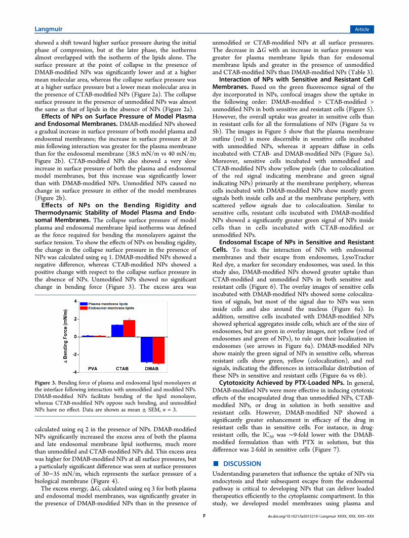
Thermodynamic Stability of Model Plasma and Endo-somal Membranes. The collapse surface pressure of modelplasma and endosomal membrane lipid isotherms was definedas the force required for bending the monolayers against thesurface tension. To show the effects of NPs on bending rigidity,the change in the collapse surface pressure in the presence ofNPs was calculated using eq 1. DMAB-modified NPs showed anegative difference, whereas CTAB-modified NPs showed apositive change with respect to the collapse surface pressure inthe absence of NPs. Unmodified NPs showed no significantchange in bending force (Figure 3). The excess area was
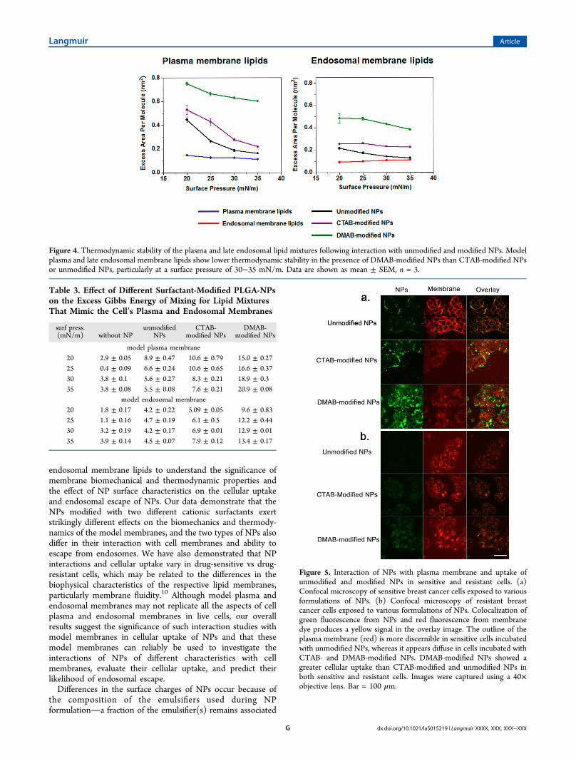
calculated using eq 2 in the presence of NPs. DMAB-modifiedNPs significantly increased the excess area of both the plasmaand late endosomal membrane lipid isotherms, much morethan unmodified and CTAB-modified NPs did. This excess areawas higher for DMAB-modified NPs at all surface pressures, buta particularly significant difference was seen at surface pressuresof 30−35 mN/m, which represents the surface pressure of abiological membrane (Figure 4).The excess energy, ΔG, calculated using eq 3 for both plasma
and endosomal model membranes, was significantly greater inthe presence of DMAB-modified NPs than in the presence of
unmodified or CTAB-modified NPs at all surface pressures.The decrease in ΔG with an increase in surface pressure wasgreater for plasma membrane lipids than for endosomalmembrane lipids and greater in the presence of unmodifiedand CTAB-modified NPs than DMAB-modified NPs (Table 3).
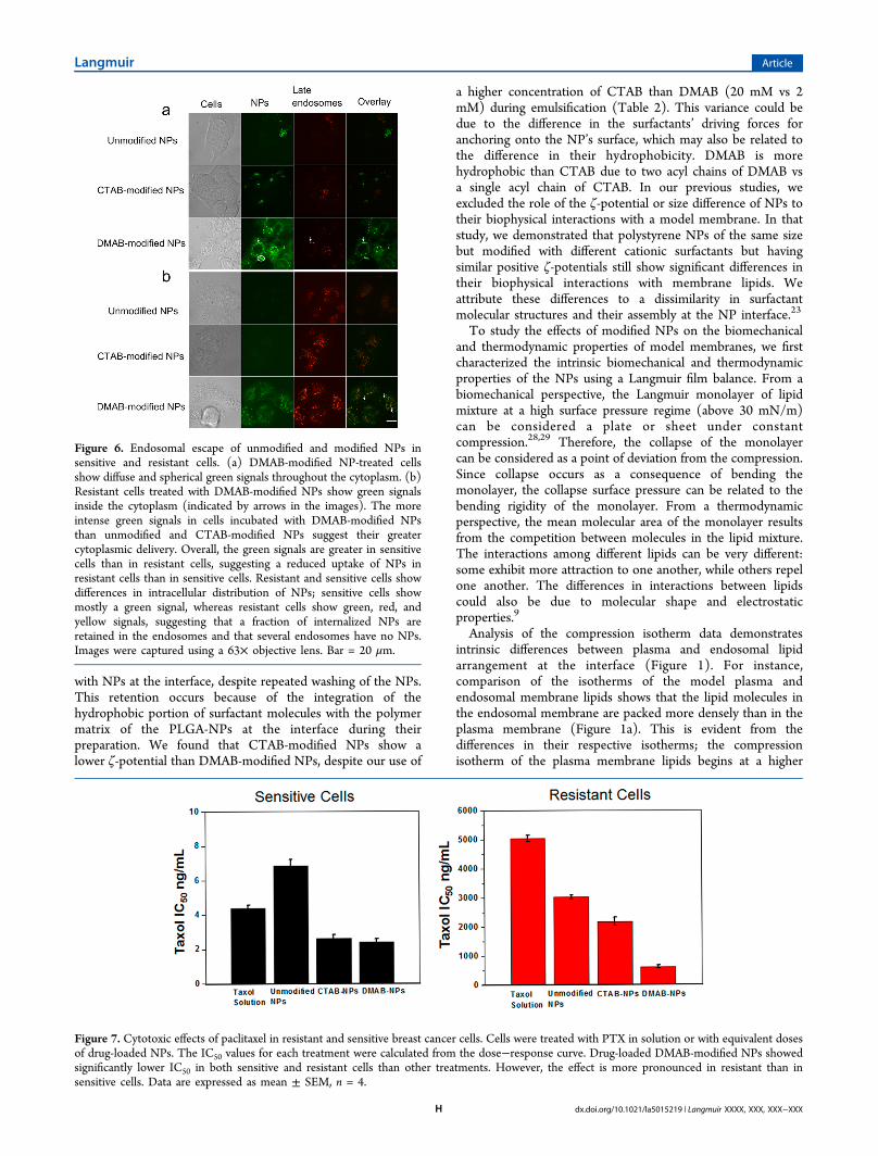
Interaction of NPs with Sensitive and Resistant CellMembranes. Based on the green fluorescence signal of thedye incorporated in NPs, confocal images show the uptake inthe following order: DMAB-modified > CTAB-modified >unmodified NPs in both sensitive and resistant cells (Figure 5).However, the overall uptake was greater in sensitive cells thanin resistant cells for all the formulations of NPs (Figure 5a vs5b). The images in Figure 5 show that the plasma membraneoutline (red) is more discernible in sensitive cells incubatedwith unmodified NPs, whereas it appears diffuse in cellsincubated with CTAB- and DMAB-modified NPs (Figure 5a).Moreover, sensitive cells incubated with unmodified andCTAB-modified NPs show yellow pixels (due to colocalizationof the red signal indicating membrane and green signalindicating NPs) primarily at the membrane periphery, whereascells incubated with DMAB-modified NPs show mostly greensignals both inside cells and at the membrane periphery, withscattered yellow signals due to colocalization. Similar tosensitive cells, resistant cells incubated with DMAB-modifiedNPs showed a significantly greater green signal of NPs insidecells than in cells incubated with CTAB-modified orunmodified NPs.
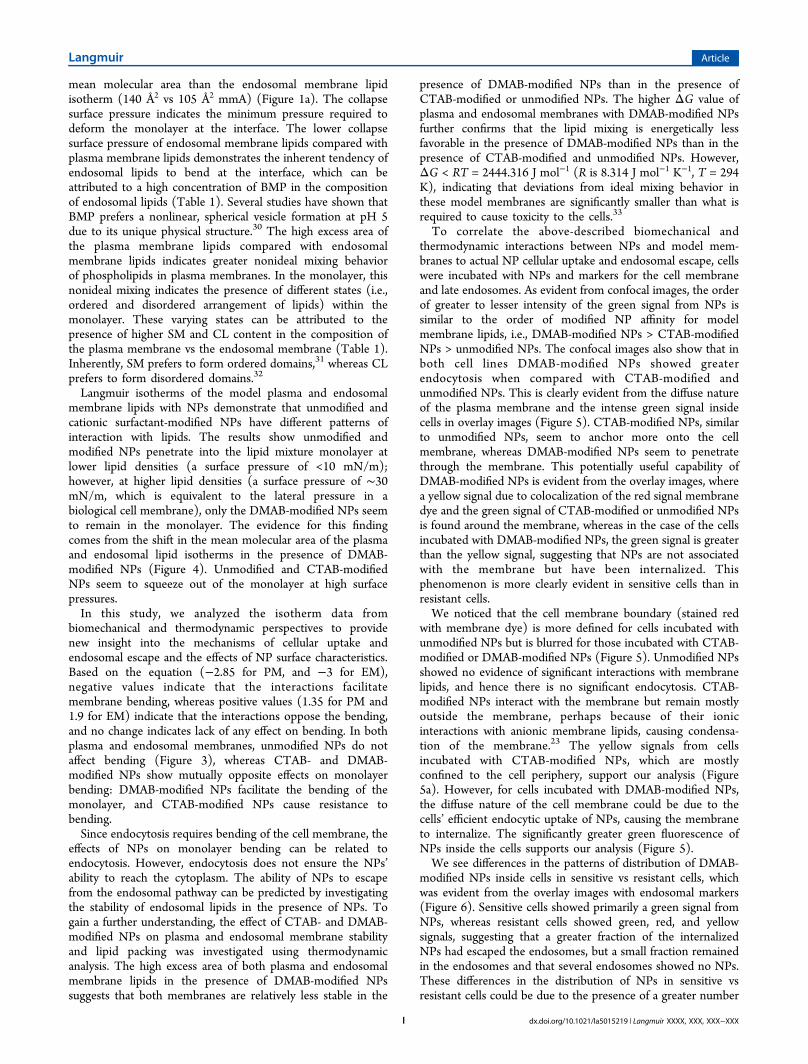
Endosomal Escape of NPs in Sensitive and ResistantCells. To track the interaction of NPs with endosomalmembranes and their escape from endosomes, LysoTrackerRed dye, a marker for secondary endosomes, was used. In thisstudy also, DMAB-modified NPs showed greater uptake thanCTAB-modified and unmodified NPs in both sensitive andresistant cells (Figure 6). The overlay images of sensitive cellsincubated with DMAB-modified NPs showed some colocaliza-tion of signals, but most of the signal due to NPs was seeninside cells and also around the nucleus (Figure 6a). Inaddition, sensitive cells incubated with DMAB-modified NPsshowed spherical aggregates inside cells, which are of the size ofendosomes, but are green in overlay images, not yellow (red ofendosomes and green of NPs), to rule out their localization inendosomes (see arrows in Figure 6a). DMAB-modified NPsshow mainly the green signal of NPs in sensitive cells, whereasresistant cells show green, yellow (colocalization), and redsignals, indicating the differences in intracellular distribution ofthese NPs in sensitive and resistant cells (Figure 6a vs 6b).
Cytotoxicity Achieved by PTX-Loaded NPs. In general,DMAB-modified NPs were more effective in inducing cytotoxiceffects of the encapsulated drug than unmodified NPs, CTAB-modified NPs, or drug in solution in both sensitive andresistant cells. However, DMAB-modified NP showed asignificantly greater enhancement in efficacy of the drug inresistant cells than in sensitive cells. For instance, in drug-resistant cells, the IC50 was ∼9-fold lower with the DMAB-modified formulation than with PTX in solution, but thisdifference was 2-fold in sensitive cells (Figure 7).
■ DISCUSSIONUnderstanding parameters that influence the uptake of NPs viaendocytosis and their subsequent escape from the endosomalpathway is critical to developing NPs that can deliver loadedtherapeutics efficiently to the cytoplasmic compartment. In thisstudy, we developed model membranes using plasma and
Figure 3. Bending force of plasma and endosomal lipid monolayers atthe interface following interaction with unmodified and modified NPs.DMAB-modified NPs facilitate bending of the lipid monolayer,whereas CTAB-modified NPs oppose such bending, and unmodifiedNPs have no effect. Data are shown as mean ± SEM, n = 3.
Langmuir Article
dx.doi.org/10.1021/la5015219 | Langmuir XXXX, XXX, XXX−XXXF
endosomal membrane lipids to understand the significance ofmembrane biomechanical and thermodynamic properties andthe effect of NP surface characteristics on the cellular uptakeand endosomal escape of NPs. Our data demonstrate that theNPs modified with two different cationic surfactants exertstrikingly different effects on the biomechanics and thermody-namics of the model membranes, and the two types of NPs alsodiffer in their interaction with cell membranes and ability toescape from endosomes. We have also demonstrated that NPinteractions and cellular uptake vary in drug-sensitive vs drug-resistant cells, which may be related to the differences in thebiophysical characteristics of the respective lipid membranes,particularly membrane fluidity.10 Although model plasma andendosomal membranes may not replicate all the aspects of cellplasma and endosomal membranes in live cells, our overallresults suggest the significance of such interaction studies withmodel membranes in cellular uptake of NPs and that thesemodel membranes can reliably be used to investigate theinteractions of NPs of different characteristics with cellmembranes, evaluate their cellular uptake, and predict theirlikelihood of endosomal escape.Differences in the surface charges of NPs occur because of
the composition of the emulsifiers used during NPformulationa fraction of the emulsifier(s) remains associated
Figure 4. Thermodynamic stability of the plasma and late endosomal lipid mixtures following interaction with unmodified and modified NPs. Modelplasma and late endosomal membrane lipids show lower thermodynamic stability in the presence of DMAB-modified NPs than CTAB-modified NPsor unmodified NPs, particularly at a surface pressure of 30−35 mN/m. Data are shown as mean ± SEM, n = 3.
Table 3. Effect of Different Surfactant-Modified PLGA-NPson the Excess Gibbs Energy of Mixing for Lipid MixturesThat Mimic the Cell’s Plasma and Endosomal Membranes
surf press.(mN/m) without NP
unmodifiedNPs
CTAB-modified NPs
DMAB-modified NPs
model plasma membrane20 2.9 ± 0.05 8.9 ± 0.47 10.6 ± 0.79 15.0 ± 0.2725 0.4 ± 0.09 6.6 ± 0.24 10.6 ± 0.65 16.6 ± 0.3730 3.8 ± 0.1 5.6 ± 0.27 8.3 ± 0.21 18.9 ± 0.335 3.8 ± 0.08 5.5 ± 0.08 7.6 ± 0.21 20.9 ± 0.08
model endosomal membrane20 1.8 ± 0.17 4.2 ± 0.22 5.09 ± 0.05 9.6 ± 0.8325 1.1 ± 0.16 4.7 ± 0.19 6.1 ± 0.5 12.2 ± 0.4430 3.2 ± 0.19 4.2 ± 0.17 6.9 ± 0.01 12.9 ± 0.0135 3.9 ± 0.14 4.5 ± 0.07 7.9 ± 0.12 13.4 ± 0.17
Figure 5. Interaction of NPs with plasma membrane and uptake ofunmodified and modified NPs in sensitive and resistant cells. (a)Confocal microscopy of sensitive breast cancer cells exposed to variousformulations of NPs. (b) Confocal microscopy of resistant breastcancer cells exposed to various formulations of NPs. Colocalization ofgreen fluorescence from NPs and red fluorescence from membranedye produces a yellow signal in the overlay image. The outline of theplasma membrane (red) is more discernible in sensitive cells incubatedwith unmodified NPs, whereas it appears diffuse in cells incubated withCTAB- and DMAB-modified NPs. DMAB-modified NPs showed agreater cellular uptake than CTAB-modified and unmodified NPs inboth sensitive and resistant cells. Images were captured using a 40×objective lens. Bar = 100 μm.
Langmuir Article
dx.doi.org/10.1021/la5015219 | Langmuir XXXX, XXX, XXX−XXXG
with NPs at the interface, despite repeated washing of the NPs.This retention occurs because of the integration of thehydrophobic portion of surfactant molecules with the polymermatrix of the PLGA-NPs at the interface during theirpreparation. We found that CTAB-modified NPs show alower ζ-potential than DMAB-modified NPs, despite our use of
a higher concentration of CTAB than DMAB (20 mM vs 2mM) during emulsification (Table 2). This variance could bedue to the difference in the surfactants’ driving forces foranchoring onto the NP’s surface, which may also be related tothe difference in their hydrophobicity. DMAB is morehydrophobic than CTAB due to two acyl chains of DMAB vsa single acyl chain of CTAB. In our previous studies, weexcluded the role of the ζ-potential or size difference of NPs totheir biophysical interactions with a model membrane. In thatstudy, we demonstrated that polystyrene NPs of the same sizebut modified with different cationic surfactants but havingsimilar positive ζ-potentials still show significant differences intheir biophysical interactions with membrane lipids. Weattribute these differences to a dissimilarity in surfactantmolecular structures and their assembly at the NP interface.23
To study the effects of modified NPs on the biomechanicaland thermodynamic properties of model membranes, we firstcharacterized the intrinsic biomechanical and thermodynamicproperties of the NPs using a Langmuir film balance. From abiomechanical perspective, the Langmuir monolayer of lipidmixture at a high surface pressure regime (above 30 mN/m)can be considered a plate or sheet under constantcompression.28,29 Therefore, the collapse of the monolayercan be considered as a point of deviation from the compression.Since collapse occurs as a consequence of bending themonolayer, the collapse surface pressure can be related to thebending rigidity of the monolayer. From a thermodynamicperspective, the mean molecular area of the monolayer resultsfrom the competition between molecules in the lipid mixture.The interactions among different lipids can be very different:some exhibit more attraction to one another, while others repelone another. The differences in interactions between lipidscould also be due to molecular shape and electrostaticproperties.9
Analysis of the compression isotherm data demonstratesintrinsic differences between plasma and endosomal lipidarrangement at the interface (Figure 1). For instance,comparison of the isotherms of the model plasma andendosomal membrane lipids shows that the lipid molecules inthe endosomal membrane are packed more densely than in theplasma membrane (Figure 1a). This is evident from thedifferences in their respective isotherms; the compressionisotherm of the plasma membrane lipids begins at a higher
Figure 6. Endosomal escape of unmodified and modified NPs insensitive and resistant cells. (a) DMAB-modified NP-treated cellsshow diffuse and spherical green signals throughout the cytoplasm. (b)Resistant cells treated with DMAB-modified NPs show green signalsinside the cytoplasm (indicated by arrows in the images). The moreintense green signals in cells incubated with DMAB-modified NPsthan unmodified and CTAB-modified NPs suggest their greatercytoplasmic delivery. Overall, the green signals are greater in sensitivecells than in resistant cells, suggesting a reduced uptake of NPs inresistant cells than in sensitive cells. Resistant and sensitive cells showdifferences in intracellular distribution of NPs; sensitive cells showmostly a green signal, whereas resistant cells show green, red, andyellow signals, suggesting that a fraction of internalized NPs areretained in the endosomes and that several endosomes have no NPs.Images were captured using a 63× objective lens. Bar = 20 μm.
Figure 7. Cytotoxic effects of paclitaxel in resistant and sensitive breast cancer cells. Cells were treated with PTX in solution or with equivalent dosesof drug-loaded NPs. The IC50 values for each treatment were calculated from the dose−response curve. Drug-loaded DMAB-modified NPs showedsignificantly lower IC50 in both sensitive and resistant cells than other treatments. However, the effect is more pronounced in resistant than insensitive cells. Data are expressed as mean ± SEM, n = 4.
Langmuir Article
dx.doi.org/10.1021/la5015219 | Langmuir XXXX, XXX, XXX−XXXH
mean molecular area than the endosomal membrane lipidisotherm (140 Å2 vs 105 Å2 mmA) (Figure 1a). The collapsesurface pressure indicates the minimum pressure required todeform the monolayer at the interface. The lower collapsesurface pressure of endosomal membrane lipids compared withplasma membrane lipids demonstrates the inherent tendency ofendosomal lipids to bend at the interface, which can beattributed to a high concentration of BMP in the compositionof endosomal lipids (Table 1). Several studies have shown thatBMP prefers a nonlinear, spherical vesicle formation at pH 5due to its unique physical structure.30 The high excess area ofthe plasma membrane lipids compared with endosomalmembrane lipids indicates greater nonideal mixing behaviorof phospholipids in plasma membranes. In the monolayer, thisnonideal mixing indicates the presence of different states (i.e.,ordered and disordered arrangement of lipids) within themonolayer. These varying states can be attributed to thepresence of higher SM and CL content in the composition ofthe plasma membrane vs the endosomal membrane (Table 1).Inherently, SM prefers to form ordered domains,31 whereas CLprefers to form disordered domains.32
Langmuir isotherms of the model plasma and endosomalmembrane lipids with NPs demonstrate that unmodified andcationic surfactant-modified NPs have different patterns ofinteraction with lipids. The results show unmodified andmodified NPs penetrate into the lipid mixture monolayer atlower lipid densities (a surface pressure of <10 mN/m);however, at higher lipid densities (a surface pressure of ∼30mN/m, which is equivalent to the lateral pressure in abiological cell membrane), only the DMAB-modified NPs seemto remain in the monolayer. The evidence for this findingcomes from the shift in the mean molecular area of the plasmaand endosomal lipid isotherms in the presence of DMAB-modified NPs (Figure 4). Unmodified and CTAB-modifiedNPs seem to squeeze out of the monolayer at high surfacepressures.In this study, we analyzed the isotherm data from
biomechanical and thermodynamic perspectives to providenew insight into the mechanisms of cellular uptake andendosomal escape and the effects of NP surface characteristics.Based on the equation (−2.85 for PM, and −3 for EM),negative values indicate that the interactions facilitatemembrane bending, whereas positive values (1.35 for PM and1.9 for EM) indicate that the interactions oppose the bending,and no change indicates lack of any effect on bending. In bothplasma and endosomal membranes, unmodified NPs do notaffect bending (Figure 3), whereas CTAB- and DMAB-modified NPs show mutually opposite effects on monolayerbending: DMAB-modified NPs facilitate the bending of themonolayer, and CTAB-modified NPs cause resistance tobending.Since endocytosis requires bending of the cell membrane, the
effects of NPs on monolayer bending can be related toendocytosis. However, endocytosis does not ensure the NPs’ability to reach the cytoplasm. The ability of NPs to escapefrom the endosomal pathway can be predicted by investigatingthe stability of endosomal lipids in the presence of NPs. Togain a further understanding, the effect of CTAB- and DMAB-modified NPs on plasma and endosomal membrane stabilityand lipid packing was investigated using thermodynamicanalysis. The high excess area of both plasma and endosomalmembrane lipids in the presence of DMAB-modified NPssuggests that both membranes are relatively less stable in the
presence of DMAB-modified NPs than in the presence ofCTAB-modified or unmodified NPs. The higher ΔG value ofplasma and endosomal membranes with DMAB-modified NPsfurther confirms that the lipid mixing is energetically lessfavorable in the presence of DMAB-modified NPs than in thepresence of CTAB-modified and unmodified NPs. However,ΔG < RT = 2444.316 J mol−1 (R is 8.314 J mol−1 K−1, T = 294K), indicating that deviations from ideal mixing behavior inthese model membranes are significantly smaller than what isrequired to cause toxicity to the cells.33
To correlate the above-described biomechanical andthermodynamic interactions between NPs and model mem-branes to actual NP cellular uptake and endosomal escape, cellswere incubated with NPs and markers for the cell membraneand late endosomes. As evident from confocal images, the orderof greater to lesser intensity of the green signal from NPs issimilar to the order of modified NP affinity for modelmembrane lipids, i.e., DMAB-modified NPs > CTAB-modifiedNPs > unmodified NPs. The confocal images also show that inboth cell lines DMAB-modified NPs showed greaterendocytosis when compared with CTAB-modified andunmodified NPs. This is clearly evident from the diffuse natureof the plasma membrane and the intense green signal insidecells in overlay images (Figure 5). CTAB-modified NPs, similarto unmodified NPs, seem to anchor more onto the cellmembrane, whereas DMAB-modified NPs seem to penetratethrough the membrane. This potentially useful capability ofDMAB-modified NPs is evident from the overlay images, wherea yellow signal due to colocalization of the red signal membranedye and the green signal of CTAB-modified or unmodified NPsis found around the membrane, whereas in the case of the cellsincubated with DMAB-modified NPs, the green signal is greaterthan the yellow signal, suggesting that NPs are not associatedwith the membrane but have been internalized. Thisphenomenon is more clearly evident in sensitive cells than inresistant cells.We noticed that the cell membrane boundary (stained red
with membrane dye) is more defined for cells incubated withunmodified NPs but is blurred for those incubated with CTAB-modified or DMAB-modified NPs (Figure 5). Unmodified NPsshowed no evidence of significant interactions with membranelipids, and hence there is no significant endocytosis. CTAB-modified NPs interact with the membrane but remain mostlyoutside the membrane, perhaps because of their ionicinteractions with anionic membrane lipids, causing condensa-tion of the membrane.23 The yellow signals from cellsincubated with CTAB-modified NPs, which are mostlyconfined to the cell periphery, support our analysis (Figure5a). However, for cells incubated with DMAB-modified NPs,the diffuse nature of the cell membrane could be due to thecells’ efficient endocytic uptake of NPs, causing the membraneto internalize. The significantly greater green fluorescence ofNPs inside the cells supports our analysis (Figure 5).We see differences in the patterns of distribution of DMAB-
modified NPs inside cells in sensitive vs resistant cells, whichwas evident from the overlay images with endosomal markers(Figure 6). Sensitive cells showed primarily a green signal fromNPs, whereas resistant cells showed green, red, and yellowsignals, suggesting that a greater fraction of the internalizedNPs had escaped the endosomes, but a small fraction remainedin the endosomes and that several endosomes showed no NPs.These differences in the distribution of NPs in sensitive vsresistant cells could be due to the presence of a greater number
Langmuir Article
dx.doi.org/10.1021/la5015219 | Langmuir XXXX, XXX, XXX−XXXI

of endocytic vesicles in resistant cells than in sensitive cells(Figure 5 vs 6). Another possibility could be the slower rate ofinternalization and escape of NPs in resistant cells than insensitive cells due to the relatively more rigid nature of theresistant cells’ membrane than that of sensitive cells.Differences in the ability of NPs with different surfactants to
get into the cell can be attributed to their different degrees ofsuccess in bending the monolayer, as seen in our modelmembrane studies. DMAB-modified NPs seem to facilitateendocytosis by bending the cell membrane. Further analysisshowed a more intense green signal compared with yellowsignal inside the cells. This difference suggests that the DMAB-modified NPs not only enhance endocytosis but also facilitateescape from the endocytic pathway (Figures 5 and 6). CTAB-modified NPs, which cause resistance to membrane bending,seem to remain mainly associated with the membrane.Endosomal escape requires either rupture or destabilization ofthe endosomal membrane. In contrast, DMAB-modified NPs’ability to escape from the endocytic pathway can be attributedto their favorable thermodynamic interactions with theendosomal membrane, as evident from the negative ΔG.Even though DMAB-modified NPs showed similar ΔG with
model plasma and endosomal membranes (Table 3), we believethat in live cells, DMAB-modified NPs do not affect the stabilityof the plasma membrane but exert significant effects on thestability of the endosomal membrane, for the following reasons:Cells maintain their integrity by continuous recycling of theplasma membrane by endocytosis and exocytosis of membranelipids. Since the deviation in ΔG with DMAB-modified NPs issmaller than the ΔG shown to cause toxicity,33 plasmamembrane stability is not affected. In contrast, if there is norecycling of the endosomal membrane, even a small change inΔG may have significant effects on the stability of themembrane.PTX-loaded DMAB-modified NPs caused greater cytotox-
icity in both sensitive and resistant breast cancer cells comparedwith PTX-loaded unmodified NPs or PTX-loaded CTAB-modified NPs (Figure 7). The differences in cytotoxic effects ofthe drug with unmodified vs modified NPs can be attributed tothe differences in their ability to interact with membrane lipidsand subsequently to escape from the endosomal compartment.The greater cytotoxicity of DMAB-modified NPs furtherconfirms the significance of the biomechanical and thermody-namic interactions of NP-model cell membranes in our studiesand validates our hypothesis. CTAB-modified NPs showedslightly better efficacy than unmodified NPs in both sensitiveand resistant cells. This effect could be because of the greaterability of the CTAB-modified NPs to bind to the membranedue to ionic interactions with anionic lipids of the membrane,causing more drug to diffuse inside cells through the membranethan with unmodified NPs which may be releasing the drugmostly in the media as there are not seen anchoring to themembrane to significant extent. As is evident from the confocalimages, the greater cytotoxic efficacy of the drug with DMAB-modified NPs could be due to their greater intracellular uptakeand escape from endosomes into cytoplasmic compartment(Figures 5−7). Recently, we showed that DMAB-modified NPsloaded with p53, a tumor suppressor gene, are more effective inachieving tumor regression in a prostate xenograft model thanCTAB-modified or unmodified NPs. This effect has beenattributed to selective biophysical interactions of DMAB-modified NPs with cancer cells than with normal cells.34
These studies clearly demonstrate the significance of
biophysical interaction studies of NPs with membrane lipidsin cellular uptake and efficacy of the encapsulated therapeuticsin vitro and in vivo.Since the order of “high to low” signals with both cell lines is
the same for different formulations of NPs, the more intensegreen signal seen due to NPs in sensitive than in resistant cellscould be attributed to differences in the biophysical character-istics of the cell membranes. We postulate that sensitive cellswith more fluid membranes facilitate efficient NP anchoring atthe cell membrane, intracellular uptake, and endosomal escapethan do resistant cells with a more rigid membrane (Figures 5and 6). Although drug-loaded DMAB-modified NPs achievedgreater cytotoxicity than unmodified or CTAB-modified NPs inresistant cells, the drug levels required to achieve IC50 inresistant cells were significantly higher than that required insensitive cells (Figure 7). This discrepancy suggests thatDMAB-modified NPs were partially effective in overcoming thetransport barrier across resistant cell membrane because of theirenhanced biophysical interactions but did not completelyachieve the same degree of uptake as in sensitive cells.Therefore, the other additional factor is the rigid nature of theresistant cells’ membrane, and hence one strategy to furtherimprove efficacy of DMAB-modified NPs could be to modulatethe membrane characteristics of resistant cells to enhancemembrane fluidity. In our recent studies, we have demonstratedthat treating resistant cells with the epigenetic drug decitabinecan alter membrane lipid synthesis, making the resistant cells’membrane more fluid.35 Increased membrane fluidity ofresistant cells following treatment with such epigenetic drugscould facilitate the endocytosis of DMAB-modified NPs andtheir subsequent escape from endosomes. Hence, it is possiblethat a combination treatment of an epigenetic drug plus drug-loaded DMAB-modified NPs would be more effective inovercoming the transport barrier further and thus overcomingdrug resistance.
■ CONCLUSIONS
Our data demonstrate that the biomechanics and thermody-namics of NP−cell membrane interactions play a significantrole in the endocytosis and endosomal escape of NPs. Ourresults show that these interactions depend on the biophysicalcharacteristics of both NPs and cell membranes. Although bothCTAB- and DMAB-modified NPs are cationic, they showdissimilar interactions with our model membranes and differentendocytic behavior in live cells. The correlation betweensurfactant-modified NPs-model membrane interactions and NPendocytosis and endosomal escape validates our hypothesis.Our results suggest that the interactions of NPs with the lipidsof model membranes could be used for optimizing the selectedcharacteristics of NPs to enhance endocytosis and endosomalescape. This ability would be particularly important in drug-resistant breast cancer cells, which have impaired endocyticfunction due to the rigid nature of the membrane. Furtherstudies with other acquired drug-resistant cells might generalizethe significance of the biomechanics and thermodynamics ofinteractions of modified NPs and their efficacy for cytoplasmicdelivery therapeutics.
■ AUTHOR INFORMATION
Corresponding Author*Tel (216) 445-9364; Fax (216) 444-9198; e-mail [email protected] (V.L.).
Langmuir Article
dx.doi.org/10.1021/la5015219 | Langmuir XXXX, XXX, XXX−XXXJ
NotesThe authors declare no competing financial interest.
■ ACKNOWLEDGMENTS
Funding for this study is gratefully acknowledged: Grant1R01CA149359 (to V.L.) from the National Cancer Institute(NCI) of the National Institutes of Health. Confocalmicroscopy studies were performed at the Cleveland ClinicImaging Core with guidance from Dr. Judith Drazba.
■ REFERENCES(1) Kievit, F. M.; Zhang, M. Cancer Nanotheranostics: ImprovingImaging and Therapy by Targeted Delivery across Biological Barriers.Adv. Mater. 2011, 23, H217−247.(2) Marrache, S.; Dhar, S. Engineering of Blended NanoparticlePlatform for Delivery of Mitochondria-Acting Therapeutics. Proc. Natl.Acad. Sci. U. S. A. 2012, 109, 16288−16293.(3) McQuade, R.; Young, A. H. Future Therapeutic Targets in MoodDisorders: The Glucocorticoid Receptor. Br. J. Psychiatry 2000, 177,390−395.(4) Fulda, S.; Galluzzi, L.; Kroemer, G. Targeting Mitochondria forCancer Therapy. Nat. Rev. Drug Discovery 2010, 9, 447−464.(5) Rajendran, L.; Knolker, H. J.; Simons, K. Subcellular TargetingStrategies for Drug Design and Delivery. Nat. Rev. Drug Discovery2010, 9, 29−42.(6) Asai, T.; Tsuzuku, T.; Takahashi, S.; Okamoto, A.; Dewa, T.;Nango, M.; Hyodo, K.; Ishihara, H.; Kikuchi, H.; Oku, N. Cell-Penetrating Peptide-Conjugated Lipid Nanoparticles for SiRNADelivery. Biochem. Biophys. Res. Commun. 2014, 444, 599−604.(7) Khormaee, S.; Choi, Y.; Shen, M. J.; Xu, B. Y.; Wu, H. T.;Griffiths, G. L.; Chen, R. J.; Slater, N. K. H.; Park, J. K. EndosomolyticAnionic Polymer for the Cytoplasmic Delivery of SiRNAs in Localizedin Vivo Applications. Adv. Funct. Mater. 2013, 23, 565−574.(8) Bally, M. B.; Harvie, P.; Wong, F. M. P.; Kong, S.; Wasan, E. K.;Reimer, D. L. Biological Barriers to Cellular Delivery of Lipid-BasedDNA Carriers. Adv. Drug Delivery Rev. 1999, 38, 291−315.(9) Peetla, C.; Vijayaraghavalu, S.; Labhasetwar, V. Biophysics of CellMembrane Lipids in Cancer Drug Resistance: Implications for DrugTransport and Drug Delivery with Nanoparticles. Adv. Drug DeliveryRev. 2013, 65, 1686−1698.(10) Peetla, C.; Bhave, R.; Vijayaraghavalu, S.; Stine, A.; Kooijman,E.; Labhasetwar, V. Drug Resistance in Breast Cancer Cells:Biophysical Characterization of and Doxorubicin Interactions withMembrane Lipids. Mol. Pharmaceutics 2010, 7, 2334−2348.(11) Sahay, G.; Alakhova, D. Y.; Kabanov, A. V. Endocytosis ofNanomedicines. J. Controlled Release 2010, 145, 182−195.(12) Panyam, J.; Labhasetwar, V. Dynamics of Endocytosis andExocytosis of Poly(D,L-Lactide-co-Glycolide) Nanoparticles in Vas-cular Smooth Muscle Cells. Pharm. Res. 2003, 20, 212−220.(13) Varkouhi, A. K.; Scholte, M.; Storm, G.; Haisma, H. J.Endosomal Escape Pathways for Delivery of Biologicals. J. ControlledRelease 2010, 151, 220−228.(14) Peetla, C.; Rao, K. S.; Labhasetwar, V. Relevance of BiophysicalInteractions of Nanoparticles with a Model Membrane in PredictingCellular Uptake: Study with TAT Peptide-Conjugated Nanoparticles.Mol. Pharmaceutics 2009, 6, 1311−1320.(15) Yuan, H.; Fales, A. M.; Vo-Dinh, T. TAT Peptide-Function-alized Gold Nanostars: Enhanced Intracellular Delivery and EfficientNIR Photothermal Therapy Using Ultralow Irradiance. J. Am. Chem.Soc. 2012, 134, 11358−11361.(16) Fay, F.; Quinn, D. J.; Gilmore, B. F.; McCarron, P. A.; Scott, C.J. Gene Delivery Using Dimethyldidodecylammonium Bromide-Coated PLGA Nanoparticles. Biomaterials 2010, 31, 4214−4222.(17) Tu, J.; Wang, T.; Shi, W.; Wu, G.; Tian, X.; Wang, Y.; Ge, D.;Ren, L. Multifunctional Znpc-Loaded Mesoporous Silica Nano-particles for Enhancement of Photodynamic Therapy Efficacy byEndolysosomal Escape. Biomaterials 2012, 33, 7903−7914.
(18) Ewers, H.; Helenius, A. Lipid-Mediated Endocytosis. ColdSpring Harb. Perspect. Biol. 2011, 3, a004721.(19) McMahon, H. T.; Gallop, J. L. Membrane Curvature andMechanisms of Dynamic Cell Membrane Remodelling. Nature 2005,438, 590−596.(20) Doherty, G. J.; McMahon, H. T. Mechanisms of Endocytosis.Annu. Rev. Biochem. 2009, 78, 857−902.(21) Deserno, M.; Gelbart, W. M. Adhesion and Wrapping inColloid-Vesicle Complexes. J. Phys. Chem. B 2002, 106, 5543−5552.(22) Xu, A.; Yao, M.; Xu, G.; Ying, J.; Ma, W.; Li, B.; Jin, Y. APhysical Model for the Size-Dependent Cellular Uptake of Nano-particles Modified with Cationic Surfactants. Int. J. Nanomedicine.2012, 7, 3547−3554.(23) Peetla, C.; Labhasetwar, V. Effect of Molecular Structure ofCationic Surfactants on Biophysical Interactions of Surfactant-Modified Nanoparticles with a Model Membrane and Cellular Uptake.Langmuir 2009, 25, 2369−2377.(24) Kobayashi, T.; Stang, E.; Fang, K. S.; de Moerloose, P.; Parton,R. G.; Gruenberg, J. A Lipid Associated with the AntiphospholipidSyndrome Regulates Endosome Structure and Function. Nature 1998,392, 193−197.(25) Panyam, J.; Sahoo, S. K.; Prabha, S.; Bargar, T.; Labhasetwar, V.Fluorescence and Electron Microscopy Probes for Cellular and TissueUptake of Poly(D,L-Lactide-co-Glycolide) Nanoparticles. Int. J. Pharm.2003, 262, 1−11.(26) Xue, X.; Liang, X. J. Overcoming Drug Efflux-Based MultidrugResistance in Cancer with Nanotechnology. Chin. J. Cancer 2012, 31,100−109.(27) Pires, M. M.; Emmert, D.; Hrycyna, C. A.; Chmielewski, J.Inhibition of P-Glycoprotein-Mediated Paclitaxel Resistance byReversibly Linked Quinine Homodimers. Mol. Pharmaceutics 2009,75, 92−100.(28) Ybert, C.; Lu, W. X.; M?ller, G.; Knobler, C. M. Collapse of aMonolayer by Three Mechanisms. J. Phys. Chem. B 2002, 106, 2004−2008.(29) Diamant, H.; Witten, T. A.; Ege, C.; Gopal, A.; Lee, K. Y. C.Topography and Instability of Monolayers near Domain Boundaries.Phys. Rev. E 2001, 63.(30) Frederick, T. E.; Chebukati, J. N.; Mair, C. E.; Goff, P. C.;Fanucci, G. E. Bis(Monoacylglycero)Phosphate Forms Stable SmallLamellar Vesicle Structures: Insights into Vesicular Body Formation inEndosomes. Biophy. J. 2009, 96, 1847−1855.(31) Ramstedt, B.; Slotte, J. P. Membrane Properties ofSphingomyelins. FEBS Lett. 2002, 531, 33−37.(32) Nichols-Smith, S.; Teh, S. Y.; Kuhl, T. L. Thermodynamic andMechanical Properties of Model Mitochondrial Membranes. Biochim.Biophys. Acta, Biomembr. 2004, 1663, 82−88.(33) Dennison, S. R.; Kim, Y. S.; Cha, H. J.; Phoenix, D. A.Investigations into the Ability of the Peptide, Hal18, to Interact withBacterial Membranes. Eur. Biophys. J. Biophy. 2008, 38, 37−43.(34) Sharma, B.; Peetla, C.; Adjei, I. M.; Labhasetwar, V. SelectiveBiophysical Interactions of Surface Modified Nanoparticles withCancer Cell Lipids Improve Tumor Targeting and Gene Therapy.Cancer Lett. 2013, 334, 228−236.(35) Vijayaraghavalu, S.; Peetla, C.; Lu, S.; Labhasetwar, V.Epigenetic Modulation of the Biophysical Properties of Drug-ResistantCell Lipids to Restore Drug Transport and Endocytic Functions. Mol.Pharmaceutics 2012, 9, 2730−2742.
Langmuir Article
dx.doi.org/10.1021/la5015219 | Langmuir XXXX, XXX, XXX−XXXK