membrane-remodeling by snx18 in endosomal transport and...
TRANSCRIPT
Membrane-remodeling by SNX18 in endosomal transport and autophagy
Karin Håberg
Department of Medical Biochemistry and Biophysics Umeå 2012
Responsible publisher under Swedish law: the Dean of the Medical Faculty This work is protected by the Swedish Copyright Legislation (Act 1960:729) ISBN: 978-91-7459-366-2 ISSN: 0346-6612 New series nr: 1478 Cover: © Karin Håberg Electronic version available at http://umu.diva-portal.org/ Printed by: VMC-KBC, Umeå Universitet Umeå, Sverige, 2012

Till min familj

i
Table of Contents
Abstract ii List of papers iv Abbreviations used v Sammanfattning på svenska vi 1. Introduction 1 2. Vesicular transport 1
The endomembrane system 1 Retrograde and anterograde transport pathways 2 Essential proteins in vesicular transport and sorting 3 Dynamin 3 Adaptor proteins 5
Adaptor protein complexes 5 GGAs 7 PACS adaptors 7 EpsinR 7 Retromer 7 Cargo recognition by the adaptors 8
Sorting nexins 8 The SNX9-family 9
3. Autophagy 12 Activation of autophagy through mTORC1 inhibition 13 Formation of the autophagosome 14 Initiation 14 Elongation and maturation 16 Autophagic receptors 17 Innate immunity and autophagy 18
4. Results and discussion 19 Paper I: SNX18 is an SNX9 paralog that acts as a membrane tubulator in AP-1-positive endosomal trafficking 19 Paper II: The membrane-remodeling PX-BAR protein SNX18 is required for autophagy 22 Concluding remarks and future perspectives 23
5. Acknowledgements 25 6. References 26
ii
Abstract
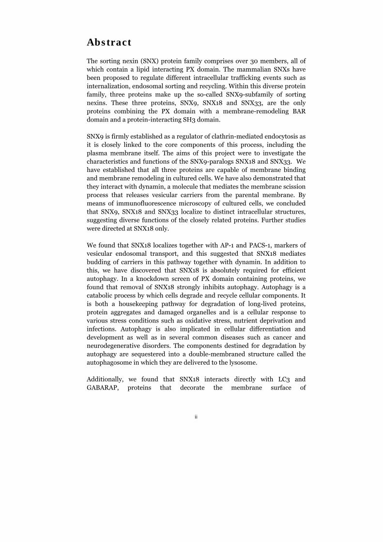
The sorting nexin (SNX) protein family comprises over 30 members, all of which contain a lipid interacting PX domain. The mammalian SNXs have been proposed to regulate different intracellular trafficking events such as internalization, endosomal sorting and recycling. Within this diverse protein family, three proteins make up the so-called SNX9-subfamily of sorting nexins. These three proteins, SNX9, SNX18 and SNX33, are the only proteins combining the PX domain with a membrane-remodeling BAR domain and a protein-interacting SH3 domain.
SNX9 is firmly established as a regulator of clathrin-mediated endocytosis as it is closely linked to the core components of this process, including the plasma membrane itself. The aims of this project were to investigate the characteristics and functions of the SNX9-paralogs SNX18 and SNX33. We have established that all three proteins are capable of membrane binding and membrane remodeling in cultured cells. We have also demonstrated that they interact with dynamin, a molecule that mediates the membrane scission process that releases vesicular carriers from the parental membrane. By means of immunofluorescence microscopy of cultured cells, we concluded that SNX9, SNX18 and SNX33 localize to distinct intracellular structures, suggesting diverse functions of the closely related proteins. Further studies were directed at SNX18 only.
We found that SNX18 localizes together with AP-1 and PACS-1, markers of vesicular endosomal transport, and this suggested that SNX18 mediates budding of carriers in this pathway together with dynamin. In addition to this, we have discovered that SNX18 is absolutely required for efficient autophagy. In a knockdown screen of PX domain containing proteins, we found that removal of SNX18 strongly inhibits autophagy. Autophagy is a catabolic process by which cells degrade and recycle cellular components. It is both a housekeeping pathway for degradation of long-lived proteins, protein aggregates and damaged organelles and is a cellular response to various stress conditions such as oxidative stress, nutrient deprivation and infections. Autophagy is also implicated in cellular differentiation and development as well as in several common diseases such as cancer and neurodegenerative disorders. The components destined for degradation by autophagy are sequestered into a double-membraned structure called the autophagosome in which they are delivered to the lysosome.
Additionally, we found that SNX18 interacts directly with LC3 and GABARAP, proteins that decorate the membrane surface of
iii
autophagosomes. Overexpression of SNX18 increased formation of autophagosomes, as seen by accumulation of GFP-LC3 spots in cells and we demonstrated that binding of SNX18 to the membrane is required for efficient autophagosome formation. By using mutants of SNX18, we were also able to demonstrate that the membrane-remodeling capability of SNX18 is a prerequisite to normal autophagosome formation. Moreover, the kinase TBK1 was identified in a cellular complex with SNX18. This kinase is both an inducer of interferon production in innate immunity responses and, together with several autophagy-related proteins, a key factor in the defense against intracellular bacteria.
Taken together, our results lead to the conclusion that SNX18 is a positive regulator of autophagy and that it is required for the formation of autophagosomes.
iv
List of papers
This thesis is based on the following papers:
I. Håberg, K., Lundmark, R., Carlsson, S.R. (2008) SNX18 is an SNX9 paralog that acts as a membrane tubulator in AP-1-positive endosomal trafficking. J Cell Sci. 121: 1495-1505.
II. Knævelsrud, H., Håberg, K., Søreng, K., Rasmuson, F., Raiborg, C., Liestøl, K., Stenmark, H., Carlsson, S.R., Simonsen, A. The membrane-remodeling PX-BAR protein SNX18 is required for autophagy. Manuscript.
v
Abbreviations used
AP – adaptor protein complex Atg – autophagy related gene bafA1 – Bafilomycin A1 BAR – Bin/amphiphysin/Rvs BFA – Brefeldin A CCP – clathrin-coated pit CCV – clathrin-coated vesicle CHC – clathrin heavy chain CK2 – casein kinase-2 CME – clathrin-mediated endocytosis dsDNA –double-stranded DNA ER – endoplasmic reticulum GDP – guanosine diphosphate GEF – guanine nucleotide exchange factor GGA – Golgi-localized, -ear containing, Arf binding protein GTP – guanosine triphosphate LC – low complexity LIR – LC3 interacting region MPR – mannose 6-phosphate receptor mTOR – mammalian target of rapamycin mTORC1 – mTOR complex 1 PAS – preautophagosomal structure PE – phosphatidylethanolamine PH – pleckstrin homology PRD – proline-rich domain PtdIns3K – phosphatidylinositol 3-kinase PtdIns3P – phosphatidylinositol 3-phosphate PtdIns(3,5)P2 - phosphatidylinositol 3,5-bisphosphate PtdIns(4,5)P2 – phosphatidylinositol 4,5-bisphosphate PX – phox homology SH3 – Src homology 3 SNX – sorting nexin STxB – Shiga toxin B-subunit TGN – trans-Golgi network ub-like – ubiquitination-like
vi
Sammanfattning på svenska
Alla celler är omgivna av ett cellmembran som avgränsar cellen och dess innehåll från omgivningen. Inuti cellerna finns organeller som också är inneslutna av membraner vilket gör att olika typer av biologiska reaktioner kan pågå på flera ställen i en cell samtidigt utan att störas av varandra. Varje organell har sin unika funktion. De cellulära membranerna är dynamiska och kan omformas på olika sätt; det kan knoppas av mindre bitar från organellerna likväl som de kan sammansmälta med varandra. Den första delen av den här avhandlingen handlar om membranproteiner som kan omforma membraner och bidrar till bildandet av små membran-avknoppningar. Dessa avknoppningar kallas vesiklar och används av cellen för att transportera molekyler mellan olika organeller.
SNX9 är ett protein som tillsammans med flera andra proteiner bildar vesiklar från cellmembranet. Denna process kallas endocytos och är en mekanism som celler använder för att ta upp material från utsidan, t.ex. näring och hormoner. Den endocytiska vesikeln skickas vidare in i cellen och sammansmälter med endosomer. I endosomerna sker en sortering av materialet. Vissa molekyler skickas tillbaka till cellmembranet, t.ex. receptorer som kan återanvändas för att samla in mer material till cellen. Andra molekyler ska förstöras och de sorteras till lysosomen, en organell som innehåller enzymer som bryter ner andra molekyler i mindre beståndsdelar, dessa delar kan sedan återanvändas av cellen för att bygga ihop nya molekyler. Material kan också sorteras in i andra transportvägar som leder till andra organeller, t.ex. till Golgikomplexet. SNX9 var känd sedan tidigare för sin roll i endocytos; syftet med den första studien var att studera dess närmaste släktingar SNX18 och SNX33. Vi upptäckte att SNX18 sitter på endosomer och att den interagerar med flera proteiner som är involverat vid bildandet av vesiklar från denna organell. När SNX-proteinerna binder till membraner så omformar de membranet till att bilda en smal tub. Därefter så rekryterar de ett enzym, dynamin, som kan knipsa av den smala membrantuben och på så sätt frisätts en transportvesikel som skickas iväg till en annan organell. Vi såg att SNX33 också kunde binda till membraner och interagera med dynamin.
Den andra delen av avhandlingen handlar om SNX18 och dess roll vid autofagi. Autofagi är en process för nedbrytning och återanvändning av cellulära komponenter. Autofagi pågår på en låg, basal nivå hela tiden för att omsätta gamla och skadade proteiner, aggregerade proteiner och även utslitna organeller som annars riskerar att ansamlas och skada cellen. Förekomsten av proteinaggregat är typiskt förknippade med flera typer av
vii
neurodegenerativa sjukdomar som t.ex. Parkinsons och Alzheimers sjukdomar. Autofagiprocessen kan också slås på i större skala när cellerna utsätts för stress, t.ex. oxidativ stress, näringsbrist eller infektioner. Det är även en livsnödvändig process för normal utveckling och differentiering. I djurmodeller har man sett att en stimulering av autofagi får djuren att leva längre.
Det material som ska brytas ner vid autofagi innesluts i en membranvesikel som sammansmälter med lysosomen. Vi fann att om vi stoppade uttrycket av SNX18-proteinet i våra cellodlingar så kunde cellerna inte längre starta igång autofagiprocessen. Å andra sidan kunde vi inducera högre nivåer än normalt av autofagi då vi överuttryckte SNX18 i cellerna. Vi införde mutationer i SNX18 och såg att om vi skapar en mutant som inte längre kan binda till membraner så försvinner även påverkan på autofagiprocessen. SNX18 är alltså på något sätt associerad med de autofagosomala membranerna, de molekylära detaljerna återstår att utreda. När vi sökte efter fler proteiner som interagerar med SNX18 fann vi TBK1, ett protein som är viktigt för det medfödda immunförsvaret och i skyddet mot bakterier som försöker infektera innandömet av våra celler. Intracellulära bakterier kan brytas ner av autofagisystemet och TBK1 är en faktor som ökar effektiviteten för detta, det är möjligt att även SNX18 har en roll här. Sammantaget så är SNX18 en positiv reglerare av autofagi, den tycks vara absolut nödvändig för att denna process ska fortlöpa normalt.

viii
1
1. Introduction
All cells are surrounded by a double layer of amphipathic lipids known simply as the cell membrane. This membrane encloses all cellular contents, creating an intracellular environment that is distinct from the outside. Amphipathic lipids, defined by the presence of both a hydrophobic and hydrophilic domain within the same molecule, spontaneously form lipid bilayers in an aqueous environment. The lipids expose their hydrophilic head groups to the water-based interior and exterior of the cells while their hydrophobic fatty acid tails are oriented towards the middle of the membrane, away from the water. As most biomolecules such as nucleic acids and proteins contain charged groups, they cannot diffuse through the hydrophobic core of biological membranes (1). This containment of biomolecules is a prerequisite for life as we know it.
Eukaryotic cells have taken this concept one step further, as they utilize membranes to divide their intracellular space into several smaller compartments. This compartmentalization allows these membrane-enclosed organelles to execute diverse enzymatic reactions separate from each other and from the cytosol. However, this higher order also requires an intricate network of specialized lipids and proteins to maintain organellar integrity and to allow for interorganellar communication (2).
This thesis addresses the regulation of intracellular membrane dynamics via investigations into the characteristics and functions of the membrane proteins belonging to the SNX9-protein family.
2. Vesicular transport
The endomembrane system
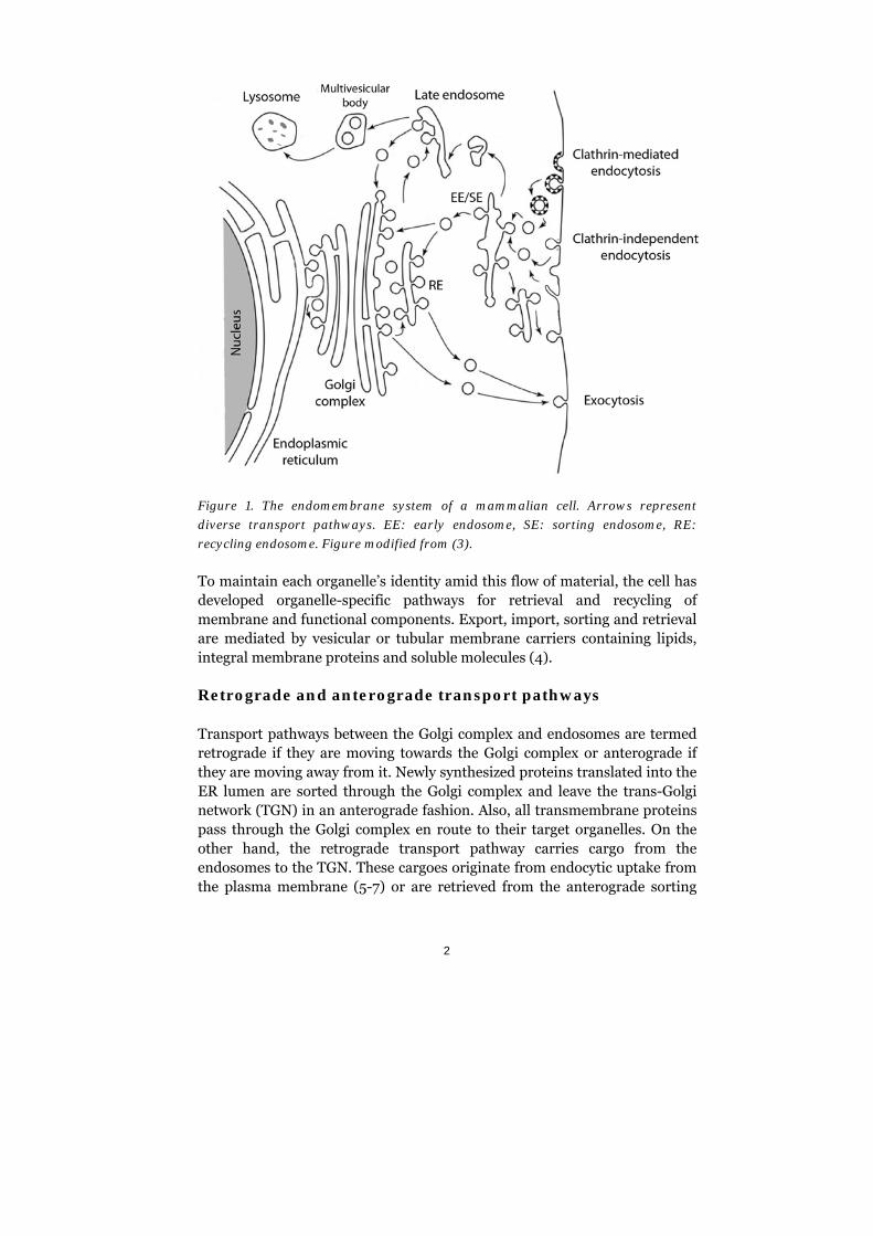
The endomembrane system comprises the membrane-bound compartments associated with endocytic, secretory and degradative processes in the cell. These processes are critical for a wide range of cellular functions, including nutrient uptake, signaling, and development. The organelles connected to this system are the endoplasmic reticulum (ER), Golgi complex, endosomes and lysosomes, each of which has its own unique identity and function. In addition, the endomembrane system includes the membrane-enclosed transport intermediates moving between these organelles and the plasma membrane (see figure 1). The main roles of this system are to export secretory cargo and to import and sort endocytic material.
2
Figure 1. The endomembrane system of a mammalian cell. Arrows represent
diverse transport pathways. EE: early endosome, SE: sorting endosome, RE:
recycling endosome. Figure modified from (3).
To maintain each organelle’s identity amid this flow of material, the cell has developed organelle-specific pathways for retrieval and recycling of membrane and functional components. Export, import, sorting and retrieval are mediated by vesicular or tubular membrane carriers containing lipids, integral membrane proteins and soluble molecules (4).
Retrograde and anterograde transport pathways
Transport pathways between the Golgi complex and endosomes are termed retrograde if they are moving towards the Golgi complex or anterograde if they are moving away from it. Newly synthesized proteins translated into the ER lumen are sorted through the Golgi complex and leave the trans-Golgi network (TGN) in an anterograde fashion. Also, all transmembrane proteins pass through the Golgi complex en route to their target organelles. On the other hand, the retrograde transport pathway carries cargo from the endosomes to the TGN. These cargoes originate from endocytic uptake from the plasma membrane (5-7) or are retrieved from the anterograde sorting
3
route (8). Endocytic material that is not sorted into the retrograde routes is delivered through the late endosomes to the lysosomes for degradation or retrieved for transport back to the plasma membrane, either directly or through the recycling compartment (9, 10).
Several distinct pathways for retrograde transport regulated by different protein complexes have been identified and characterized. These emerge from structures on early/sorting or late endosomes (11). Numerous endogenous proteins are sorted through these pathways, e.g., SNAREs, mannose 6-phosphate receptors (MPR), TGN46, mAtg9, sortilin, and furin. It has also been shown that bacterial, viral, and plant toxins can hijack these systems. After entering the cell via endocytosis, they follow the retrograde transport route through the Golgi complex and into ER from which they are retrotranslocated into the cytosol where they associate with their cellular targets (12).
Essential proteins in vesicular transport and sorting
Dynamin
As an essential step in membrane carrier formation, the newly formed membrane bud has to be released from the parental membrane. This process includes drastic remodeling of the lipid bilayer and the subsequent membrane fission is a thermodynamically unfavourable process that is poorly understood at the molecular level (13). The large GTPase dynamin is one of the proteins known to participate in membrane scission. Dynamin is expressed in three different isoforms in mammals; dynamin-1 is found in the brain, dynamin-2 is ubiquitously expressed and dynamin-3 is restricted to the brain, lung, heart, and testis (14). This 100 kDa protein contains a pleckstrin homology (PH) domain for binding to phosphoinositides in membranes and a proline-rich domain (PRD) for binding to Src homology (SH3) domains in accessory proteins (15). Dynamin also has a GTPase domain where binding and hydrolysis of guanine nucleotides takes place. Adjacent to this is a self-assembly domain simply referred to as the ‘middle’ domain. The C-terminally located GTPase effector domain also participates in self-assembly as well as the allosteric regulation of the GTPase activity (15).
Dynamin is well established as playing a role in the process of vesicle formation at diverse cellular sites that involve the plasma membrane, TGN, and endosomes. In addition, dynamin has been shown to play a role in caveolae budding, phagocytosis, cytokinesis, and actin rearrangements (16). Most studies, however, have focused on the role of dynamin in clathrin-
4
mediated endocytosis (CME), where it mediates membrane scission as it assembles around the necks of invaginated clathrin-coated pits (CCP) and pinches them off in a GTP hydrolysis-dependent mechanism (5, 17).
Dynamin is also required for budding and formation of transport carriers from endosomes, as it has been shown to regulate the retrograde transport of the bacterial Shiga toxin B-subunit (STxB) (18) and the recycling of transferrin receptor to the plasma membrane from the recycling endosomal compartment (19). Inhibition of dynamin in the cell results in endosomes with protruding membrane buds and tubules that cannot be released. This endosomal phenotype causes a block in endosomal maturation and acidification (20).
The exact mechanism of dynamin-mediated membrane fission is still a matter of debate and three different models have been proposed. All three models take into account the fact that dynamin undergoes major nucleotide- and membrane binding-dependent conformational changes (21-23). Dynamin oligomerizes as a spiral around tubular membranes and, upon GTP hydrolysis, has been observed to constrict the tube (24, 25), possibly leading to hemifusion of the inner lipid layer and subsequent membrane fission. GTP hydrolysis also leads to a lengthwise expansion of the dynamin spiral (21). Additionally, the conformational changes within the dynamin oligomer might exert a twisting force on the underlying membrane (26). These actions could possibly generate a mechanochemical force that drives vesicle release. In another proposed mechanism, dynamin serves as a PtdIns(4,5)P2-sequester, which, together with the GTP hydrolysis-dependent constriction of the vesicle neck, might cause membrane destabilization between different lipid phases and lead to spontaneous fission (27-29). The third model suggests that dynamin works as an effector recruiter in the same manner as most other classical GTPases (e.g., Rab, Ras). Such GTPases are considered active in the GTP-bound form and inactive when they are bound to GDP. Auxilin, for instance, binds only the GTP-bound form of dynamin (30) and SNX9 stabilizes the dynamin-membrane interaction only in the presence of GTP, not GDP (31). Taken together, this model suggests that dynamin functions as a molecular switch stabilizing and/or recruiting downstream effectors necessary for membrane fission.
Recruitment of dynamin is dependent on PRD-mediated interactions with accessory proteins containing SH3 domains. SH3 domains are found in many different proteins such as actin binding proteins and membrane active proteins (32).
5
Adaptor proteins
There are several types of adaptor proteins that sequester cargo and form the protein networks that orchestrate the production of intracellular transport vesicles in the endocytic and late secretory pathways. Adaptors relay signals between phosphoinositides in the membrane, the sorting signal of the sequestered cargo and accessory proteins found in the coat surrounding the forming vesicle.
Adaptor proteins in mammalian cells include the five adaptor protein (AP) complexes AP-1 through AP-5, the GGA family of monomeric adaptors, and several cargo-specific types of adaptors.
Adaptor protein complexes
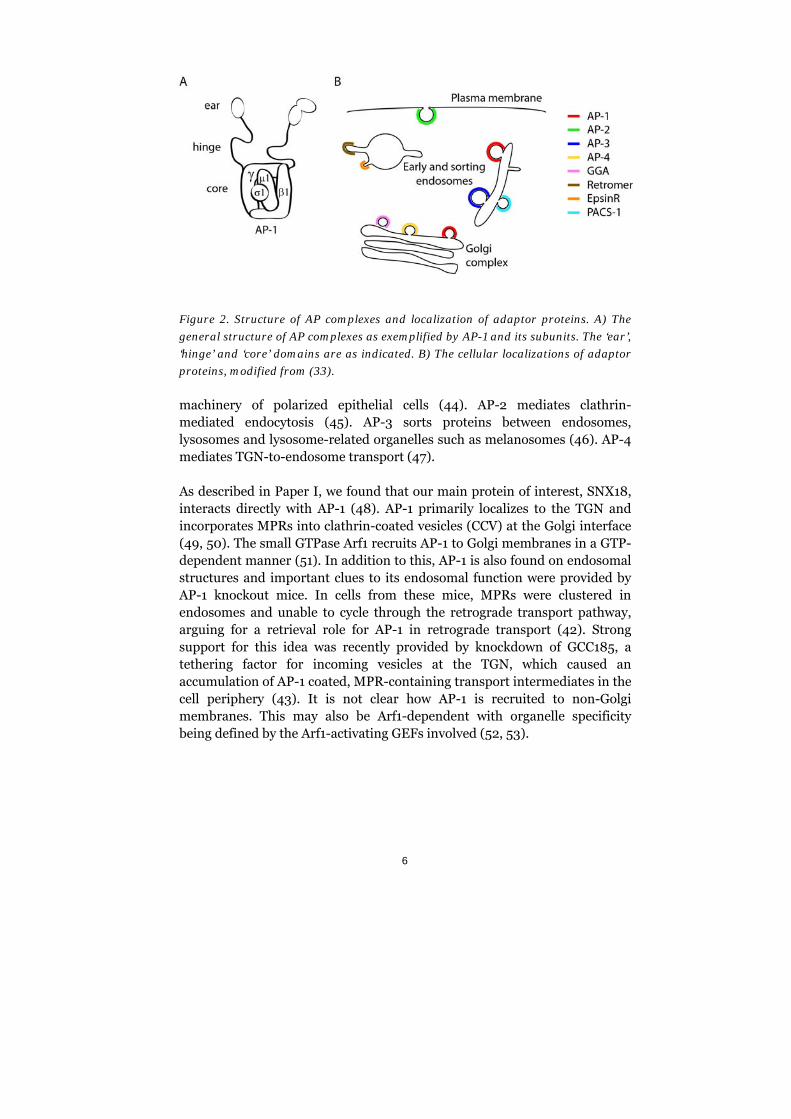
AP complexes are tetramers made up of two large and two small subunits called adaptins. The small subunits are termed 1 to 4 and 1 to 4 with the number corresponding to the AP complex to which they belong. The large adaptin subunits found in AP-1 to AP-4 are termed respectively, and 1 to 4. AP-5 has only recently been identified and its specific role and subunit structure are yet to be determined (33). Some subunits exist in several isoforms, e.g., 1b and 2 are associated with the AP-1 complex (34-36). The subunit organization of an AP complex is shown in figure 2A.
AP complexes are capable of membrane binding since they harbor phosphoinositide-binding motifs in their ‘core’ domains (figure 2A). The ‘core’ also mediates recognition and interaction with cargo whereas motifs for interactions with accessory proteins are located in the ‘hinge’ and ‘ear’ domains of the large adaptins (34, 36, 37).
Clathrin is a major binding partner of AP-1 and AP-2 at the TGN and plasma membrane, respectively. The connection between clathrin and AP-3 is unclear in physiological settings and AP-4 seems to be clathrin-independent. AP-1 and AP-3 have the ability to interact with an isoform of the clathrin heavy chain (38, 39) that primarily localizes to endosomal compartments and has been implicated in retrograde trafficking of MPRs and STxB (40).
The AP complexes in mammalian cells regulate distinct transport pathways (figure 2B). AP-1 is a clathrin-recruiter at the TGN and also coats tubulovesicular carriers at endosomes in retrograde pathways (41-43). The variant of AP-1 with the 1b subunit is found in the basolateral sorting
6
Figure 2. Structure of AP complexes and localization of adaptor proteins. A) The
general structure of AP complexes as exemplified by AP-1 and its subunits. The ‘ear’,
‘hinge’ and ‘core’ domains are as indicated. B) The cellular localizations of adaptor
proteins, modified from (33).
machinery of polarized epithelial cells (44). AP-2 mediates clathrin-mediated endocytosis (45). AP-3 sorts proteins between endosomes, lysosomes and lysosome-related organelles such as melanosomes (46). AP-4 mediates TGN-to-endosome transport (47).
As described in Paper I, we found that our main protein of interest, SNX18, interacts directly with AP-1 (48). AP-1 primarily localizes to the TGN and incorporates MPRs into clathrin-coated vesicles (CCV) at the Golgi interface (49, 50). The small GTPase Arf1 recruits AP-1 to Golgi membranes in a GTP-dependent manner (51). In addition to this, AP-1 is also found on endosomal structures and important clues to its endosomal function were provided by AP-1 knockout mice. In cells from these mice, MPRs were clustered in endosomes and unable to cycle through the retrograde transport pathway, arguing for a retrieval role for AP-1 in retrograde transport (42). Strong support for this idea was recently provided by knockdown of GCC185, a tethering factor for incoming vesicles at the TGN, which caused an accumulation of AP-1 coated, MPR-containing transport intermediates in the cell periphery (43). It is not clear how AP-1 is recruited to non-Golgi membranes. This may also be Arf1-dependent with organelle specificity being defined by the Arf1-activating GEFs involved (52, 53).
7
GGAs
The GGA (Golgi-localized, -ear containing, Arf binding protein) proteins are monomeric adaptors containing a domain homologous to the -subunit of AP-1. Mammals express three isoforms of GGAs: GGA1, GGA2 and GGA3. Their localizations in cells overlap with each other and to some extent with AP-1 (41, 49, 54, 55). GGAs participate in the budding of clathrin-coated carriers at the Golgi and mediate the binding between cargo, clathrin, and other accessory proteins (41). GGAs also interact with AP-1, but it is not clear whether GGAs work together with, or in parallel/anti-parallel pathways to, AP-1. Recent data suggests that GGAs and AP-1 function independently of each other, although all of them are recruited by Arf1 to membranes and interact with the same type of cargo and with clathrin (56).
PACS adaptors
PACS proteins are sorting proteins that work together with AP complexes and interact directly with cargo via phosphorylated acidic sequences, a type of sorting motif found in the cytosolic tail domains of several transmembrane proteins (57). PACS-1 was identified as an adaptor in retrograde sorting of furin and MPR (58). PACS-1 interacts with AP-1 and AP-3, but not clathrin, and it is absent in clathrin-enriched cell fractions (59, 60).
EpsinR
EpsinR is a cargo adaptor in the TGN-endosomal interface. It interacts with AP-1 (61, 62), GGA2 and clathrin (63, 64). Using STxB as a marker of retrograde transport, EpsinR was shown to be part of a clathrin-dependent endosome-to-TGN pathway that is also utilized by the endogenous proteins TGN46 and MPR (65).
Retromer
The retromer is a heteropentameric protein complex that mediates retrograde endosome-to-TGN transport. It is built up of a homo- or heterodimer of SNX1, SNX2, SNX5 or SNX6 that assembles with Vps26, Vps29 and Vps35. The SNX dimers form tubular protrusions at the endosome and recruit the cargo-sequestering complex made up of the three Vps proteins (66). These tubular carriers emanate from endosomal compartments that exhibit signatures of both early and late endosomes, and likely represent an intermediate between the two (67-69). Important cargoes
8
of these carriers are sortilins and MPRs, and STxB has been shown to hijack this system as well (70).
Cargo recognition by the adaptors
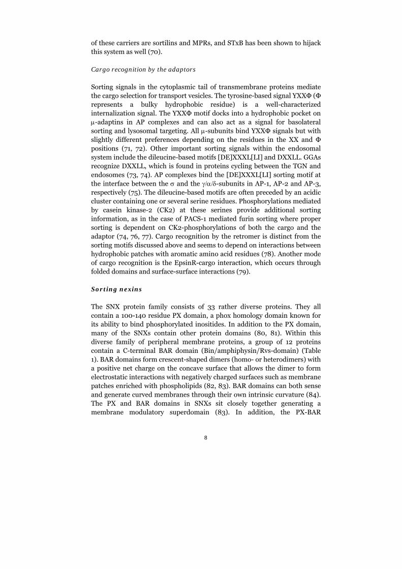
Sorting signals in the cytoplasmic tail of transmembrane proteins mediate the cargo selection for transport vesicles. The tyrosine-based signal YXXΦ (Φ represents a bulky hydrophobic residue) is a well-characterized internalization signal. The YXXΦ motif docks into a hydrophobic pocket on -adaptins in AP complexes and can also act as a signal for basolateral sorting and lysosomal targeting. All -subunits bind YXXΦ signals but with slightly different preferences depending on the residues in the XX and Φ positions (71, 72). Other important sorting signals within the endosomal system include the dileucine-based motifs [DE]XXXL[LI] and DXXLL. GGAs recognize DXXLL, which is found in proteins cycling between the TGN and endosomes (73, 74). AP complexes bind the [DE]XXXL[LI] sorting motif at the interface between the and the -subunits in AP-1, AP-2 and AP-3, respectively (75). The dileucine-based motifs are often preceded by an acidic cluster containing one or several serine residues. Phosphorylations mediated by casein kinase-2 (CK2) at these serines provide additional sorting information, as in the case of PACS-1 mediated furin sorting where proper sorting is dependent on CK2-phosphorylations of both the cargo and the adaptor (74, 76, 77). Cargo recognition by the retromer is distinct from the sorting motifs discussed above and seems to depend on interactions between hydrophobic patches with aromatic amino acid residues (78). Another mode of cargo recognition is the EpsinR-cargo interaction, which occurs through folded domains and surface-surface interactions (79).
Sorting nexins
The SNX protein family consists of 33 rather diverse proteins. They all contain a 100-140 residue PX domain, a phox homology domain known for its ability to bind phosphorylated inositides. In addition to the PX domain, many of the SNXs contain other protein domains (80, 81). Within this diverse family of peripheral membrane proteins, a group of 12 proteins contain a C-terminal BAR domain (Bin/amphiphysin/Rvs-domain) (Table 1). BAR domains form crescent-shaped dimers (homo- or heterodimers) with a positive net charge on the concave surface that allows the dimer to form electrostatic interactions with negatively charged surfaces such as membrane patches enriched with phospholipids (82, 83). BAR domains can both sense and generate curved membranes through their own intrinsic curvature (84). The PX and BAR domains in SNXs sit closely together generating a membrane modulatory superdomain (83). In addition, the PX-BAR
9
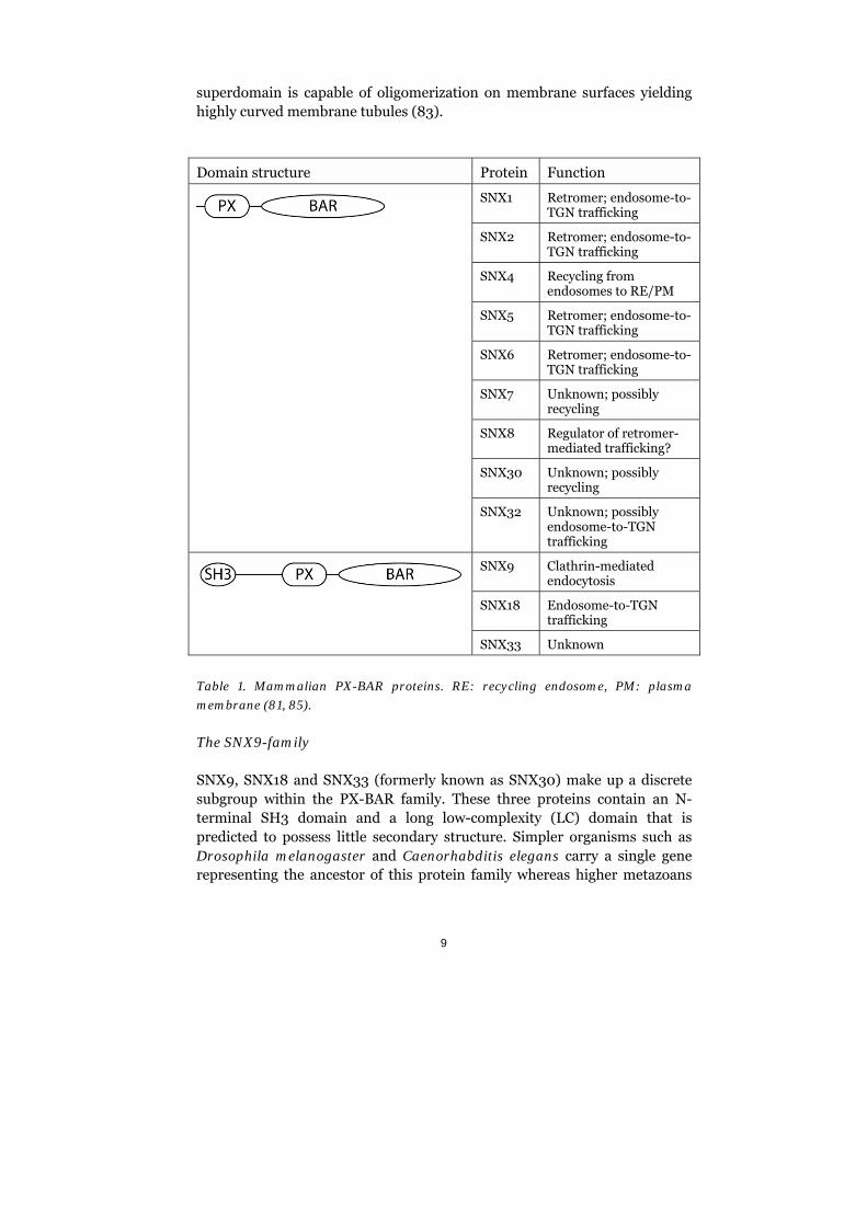
superdomain is capable of oligomerization on membrane surfaces yielding highly curved membrane tubules (83).
Domain structure Protein Function
SNX1 Retromer; endosome-to-TGN trafficking
SNX2 Retromer; endosome-to-TGN trafficking
SNX4 Recycling from endosomes to RE/PM
SNX5 Retromer; endosome-to-TGN trafficking
SNX6 Retromer; endosome-to-TGN trafficking
SNX7 Unknown; possibly recycling
SNX8 Regulator of retromer-mediated trafficking?
SNX30 Unknown; possibly recycling
SNX32 Unknown; possibly endosome-to-TGN trafficking
SNX9 Clathrin-mediated endocytosis
SNX18 Endosome-to-TGN trafficking
SNX33 Unknown
Table 1. Mammalian PX-BAR proteins. RE: recycling endosome, PM: plasma
membrane (81, 85).
The SNX9-family
SNX9, SNX18 and SNX33 (formerly known as SNX30) make up a discrete subgroup within the PX-BAR family. These three proteins contain an N-terminal SH3 domain and a long low-complexity (LC) domain that is predicted to possess little secondary structure. Simpler organisms such as Drosophila melanogaster and Caenorhabditis elegans carry a single gene representing the ancestor of this protein family whereas higher metazoans
10
express all three as a result of gene duplication. Sequence homology analysis reveals that the first duplication generated SNX9 and a SNX18/SNX33 predecessor, which was later functionally duplicated into SNX18 and SNX33 generating the three paralogs that we know of today. Unicellular eukaryotes lack apparent SNX9 homologues.
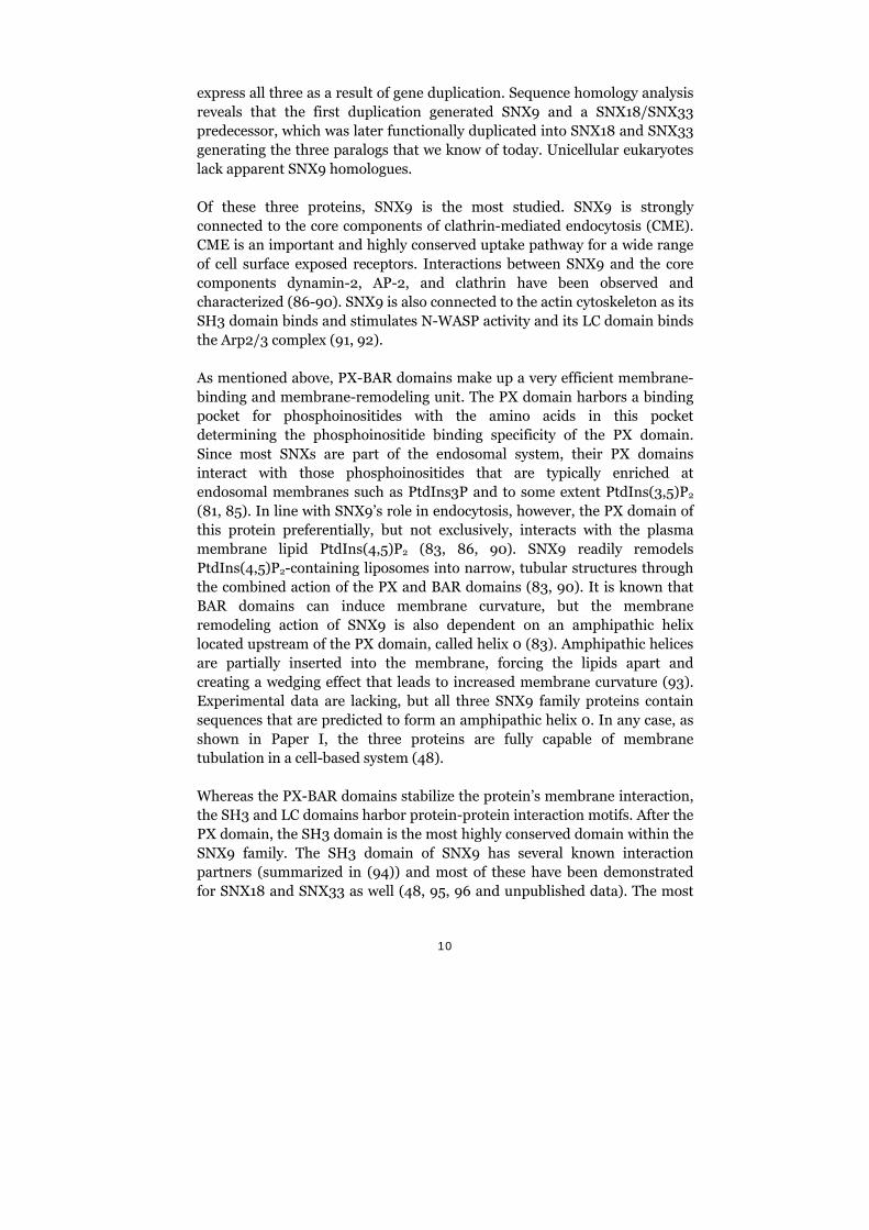
Of these three proteins, SNX9 is the most studied. SNX9 is strongly connected to the core components of clathrin-mediated endocytosis (CME). CME is an important and highly conserved uptake pathway for a wide range of cell surface exposed receptors. Interactions between SNX9 and the core components dynamin-2, AP-2, and clathrin have been observed and characterized (86-90). SNX9 is also connected to the actin cytoskeleton as its SH3 domain binds and stimulates N-WASP activity and its LC domain binds the Arp2/3 complex (91, 92).
As mentioned above, PX-BAR domains make up a very efficient membrane-binding and membrane-remodeling unit. The PX domain harbors a binding pocket for phosphoinositides with the amino acids in this pocket determining the phosphoinositide binding specificity of the PX domain. Since most SNXs are part of the endosomal system, their PX domains interact with those phosphoinositides that are typically enriched at endosomal membranes such as PtdIns3P and to some extent PtdIns(3,5)P2 (81, 85). In line with SNX9’s role in endocytosis, however, the PX domain of this protein preferentially, but not exclusively, interacts with the plasma membrane lipid PtdIns(4,5)P2 (83, 86, 90). SNX9 readily remodels PtdIns(4,5)P2-containing liposomes into narrow, tubular structures through the combined action of the PX and BAR domains (83, 90). It is known that BAR domains can induce membrane curvature, but the membrane remodeling action of SNX9 is also dependent on an amphipathic helix located upstream of the PX domain, called helix 0 (83). Amphipathic helices are partially inserted into the membrane, forcing the lipids apart and creating a wedging effect that leads to increased membrane curvature (93). Experimental data are lacking, but all three SNX9 family proteins contain sequences that are predicted to form an amphipathic helix 0. In any case, as shown in Paper I, the three proteins are fully capable of membrane tubulation in a cell-based system (48).
Whereas the PX-BAR domains stabilize the protein’s membrane interaction, the SH3 and LC domains harbor protein-protein interaction motifs. After the PX domain, the SH3 domain is the most highly conserved domain within the SNX9 family. The SH3 domain of SNX9 has several known interaction partners (summarized in (94)) and most of these have been demonstrated for SNX18 and SNX33 as well (48, 95, 96 and unpublished data). The most
11
prominent ones are dynamin-2 and N-WASP, which mediate membrane fission and actin assembly, respectively. The activities of these proteins are stimulated by the presence of SNX9, pointing to a regulatory role of the SNX9 family members (87, 91). The LC domain, located between the SH3 and PX-BAR domains, differs the most between SNX9, SNX18 and SNX33 with less than 25% sequence homology between the three proteins. This domain mediates the binding between clathrin and AP-2 in SNX9 (86) and AP-1 in SNX18 and SNX33 (48 and unpublished data). The association of actin-nucleating factor Arp2/3 to the LC domain provides an additional connection to the regulation of the actin cytoskeleton (92). The Arp2/3 binding sequence in SNX9 is also present in SNX18 and SNX33.
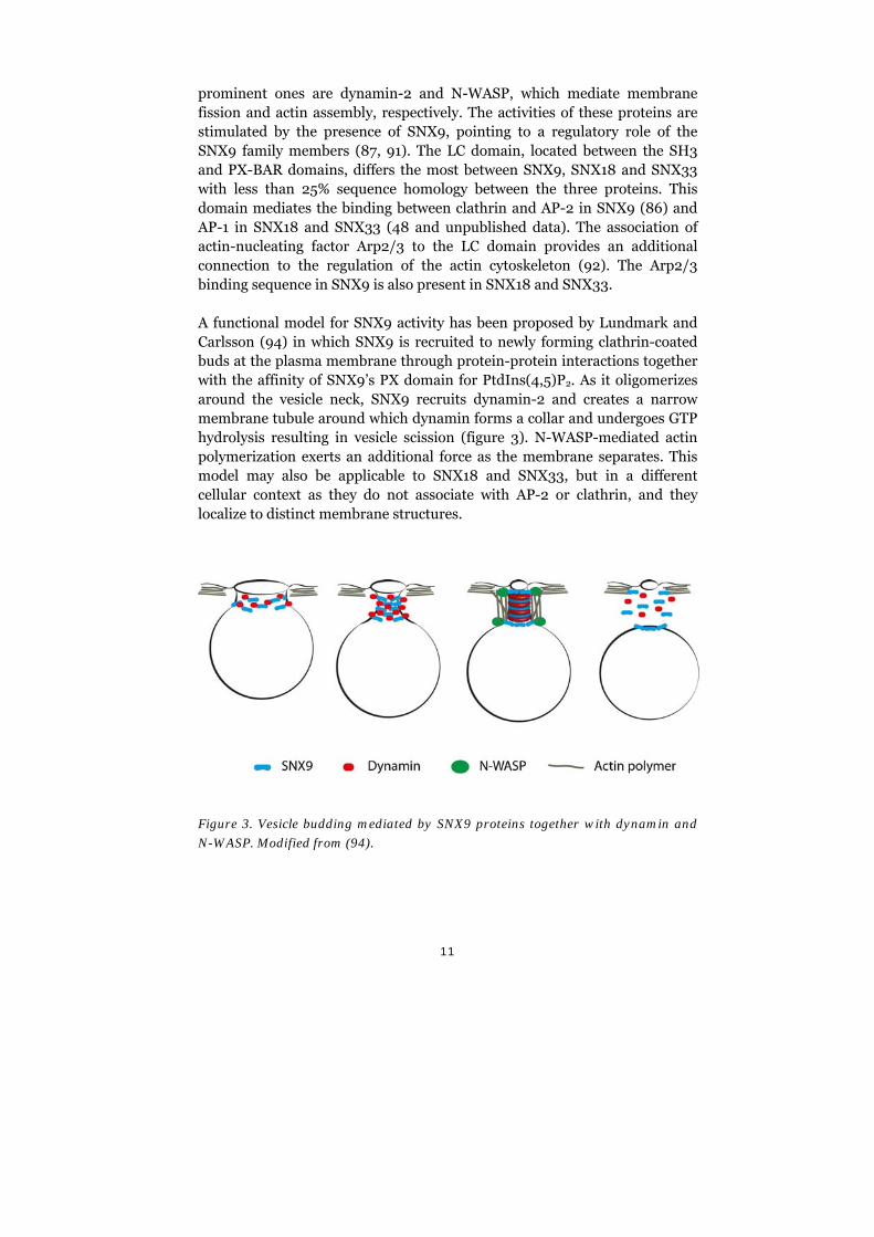
A functional model for SNX9 activity has been proposed by Lundmark and Carlsson (94) in which SNX9 is recruited to newly forming clathrin-coated buds at the plasma membrane through protein-protein interactions together with the affinity of SNX9’s PX domain for PtdIns(4,5)P2. As it oligomerizes around the vesicle neck, SNX9 recruits dynamin-2 and creates a narrow membrane tubule around which dynamin forms a collar and undergoes GTP hydrolysis resulting in vesicle scission (figure 3). N-WASP-mediated actin polymerization exerts an additional force as the membrane separates. This model may also be applicable to SNX18 and SNX33, but in a different cellular context as they do not associate with AP-2 or clathrin, and they localize to distinct membrane structures.
Figure 3. Vesicle budding mediated by SNX9 proteins together with dynamin and
N-WASP. Modified from (94).
12
3. Autophagy
Eukaryotic cells have two systems for proteolytic degradation: the lysosome and the proteasome. The proteasome degrades ubiquitinated proteins that are targeted to the barrel-shaped proteasomal complex, whereas extracellular components and transmembrane proteins are sorted as endocytic cargo into the lysosome. Additionally, intracellular proteins and organelles can be targeted for lysosomal degradation through autophagy (97). Autophagy can be divided into three main types: macroautophagy, microautophagy and chaperone-mediated autophagy. During macroautophagy, an expanding double membrane cup engulfs cytoplasmic material either selectively or unselectively. After closure, the autophagosome matures either through fusion with endosomes followed by lysosomes or directly with lysosomes, creating the autolysosome. In microautophagy, cytoplasmic material is engulfed directly by the lysosome itself. Chaperone-mediated autophagy targets proteins into the lysosome without membrane reorganization; the substrate is simply translocated into the lysosome (98). Hereafter, the term autophagy will refer to macroautophagy only.
Autophagy serves, in parallel with the proteasome, as a mechanism for the constant turnover of intracellular components. Basal autophagy acts as a quality-control system for cytoplasmic material, selectively degrading old components and providing the building blocks to synthesize new ones. Cellular stress, such as nutrient limitation and lack of growth factors or oxygen, induces higher levels of autophagy, which promotes survival under starvation conditions. Moreover, autophagy is also a defense mechanism against cytoplasmic pathogens. A general decline in autophagosomal activity is correlated with the cellular accumulation of damaged proteins and organelles that is a hallmark of ageing. Similarly, upregulation of autophagy is positively correlated to longevity, at least in model organisms.
Autophagy plays an essential role during development and differentiation. Knockout mice lacking key regulatory autophagic proteins die shortly after birth. In addition to this, autophagy has implications in a wide range of diseases including cancer and neurodegenerative syndromes (98-101). Initial mappings of the autophagic molecular machinery were carried out in yeast model systems. As autophagy is conserved among eukaryotes, the yeast mutant screens were of great value in understanding the mammalian autophagic process. The genes that were found to affect protein turnover in yeast are referred to as autophagy-related genes (Atg) and mammalian homologues have been identified for many of the yeast Atg proteins. Atg1, for
13
instance, is the yeast homologue of ULK1 and ULK2. Atg1 and ULK1/2 are, together with a number of other factors, part of the core molecular machinery essential for autophagosome formation in both yeast and mammals. The class III PtdIns3-kinase (PtdIns3K) complex is another of these factors. During autophagosome formation, two essential ubiquitination-like (ub-like) conjugation reactions are elicited involving Atg12 and Atg8, the latter having several identified mammalian counterparts in the LC3 and GATE16/GABARAP-protein families. Atg8 is conjugated to a membrane lipid in this highly conserved reaction. The only transmembrane protein found to be essential for autophagosome formation is Atg9, or mAtg9 as it is called in mammals (100). The control of the autophagic pathway and the formation of the autophagosome are discussed in greater detail below and are briefly summarized in figure 4.
Activation of autophagy through mTORC1 inhibition
mTOR (mammalian target of rapamycin) is a master regulator of a wide range of cellular activities including cell growth and proliferation, transcription and translation, cytoskeletal reorganization, and autophagy (102). The mTOR pathway is the most studied regulatory mechanism of autophagy. Nutrient limitation or rapamycin treatment result in induction of autophagy by inhibition of mTOR, more specifically the mTOR complex 1 (mTORC1), via a mechanism that is conserved from yeast to mammals (103-105).
Active mTOR is part of a complex consisting of ULK1/2, Atg13, and FIP200 in which it mediates phosphorylation-dependent inhibition of the kinases Atg13 and ULK1. Upon induction of the autophagic process, mTOR dissociates from these proteins, which allows for dephosphorylation of ULK1 to occur. Active ULK1 then phosphorylates Atg13, FIP200 and autophosphorylates ifself, eliciting the steps in the autophagic pathway downstream of mTORC1 (106-108). FIP200 translocates to pre-autophagosomal structures (PAS) and is essential for autophagosome formation (109). ULK1/2 also translocates to PAS in a FIP200-dependent manner and is responsible for the subsequent recruitment of the class III PtdIns3K-complex (109, 110). This complex, and its lipid kinase acitivity, is essential in the autophagic process.
14
Formation of the autophagosome
Initiation
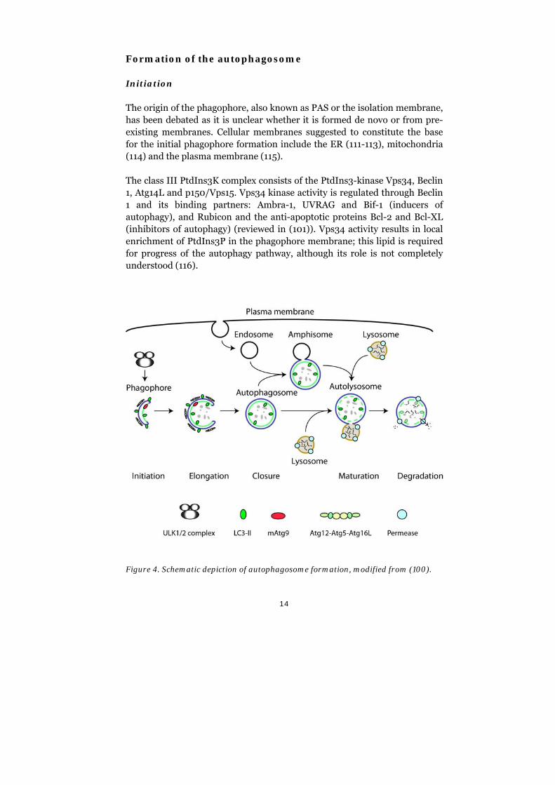
The origin of the phagophore, also known as PAS or the isolation membrane, has been debated as it is unclear whether it is formed de novo or from pre-existing membranes. Cellular membranes suggested to constitute the base for the initial phagophore formation include the ER (111-113), mitochondria (114) and the plasma membrane (115).
The class III PtdIns3K complex consists of the PtdIns3-kinase Vps34, Beclin 1, Atg14L and p150/Vps15. Vps34 kinase activity is regulated through Beclin 1 and its binding partners: Ambra-1, UVRAG and Bif-1 (inducers of autophagy), and Rubicon and the anti-apoptotic proteins Bcl-2 and Bcl-XL (inhibitors of autophagy) (reviewed in (101)). Vps34 activity results in local enrichment of PtdIns3P in the phagophore membrane; this lipid is required for progress of the autophagy pathway, although its role is not completely understood (116).
Figure 4. Schematic depiction of autophagosome formation, modified from (100).
15
Proteins binding to PtdIns3P have been described, mostly as part of endosomal machineries where this lipid is enriched. There are, however, a number of autophagic PtdIns3P effector proteins described as well. The WIPI-proteins are recruited early in the autophagic process due to their affinity for PtdIns3P in the phagophore (110, 117, 118). WIPI1 and WIPI2 act upstream of the ub-like conjugation systems described below and knockdown of WIPI-proteins results in a block of LC3 lipidation and inhibition of autophagosome formation (110, 118, 119). WIPI1 is only detected on early autophagosomal structures and seems to be excluded from the mature autophagosome (120).
Another PtdIns3P binding protein recruited to the phagophores originating from the ER is the double-FYVE containing protein, DFCP1 (111). FYVE domains interact with PtdIns3P and are found in many proteins related to endosomal sorting and autophagy. DFCP1 also contains an ER-targeting signal (111). DFCP1 localizes to structures in close proximity to WIPI1, Atg16L, Atg14L and LC3 during starvation-induced autophagy (110). Its exact role is not clear but temporal analyses show that DFCP1 puncta formation precedes accumulation of LC3 on the phagophore and DFCP1 disappears from these structures prior to the final maturation of the autophagosome (110, 111).
Atg16L is targeted to early phagophore structures as it participates in one of two ub-like conjugation systems that function during elongation of the phagophore. The first ub-like reaction creates an Atg12-Atg5-Atg16L complex, which localizes to the phagophore (121, 122). The second ub-like reaction results in targeting of LC3 to the autophagosomal membrane as cytosolic LC3-I becomes conjugated to phosphatidylethanolamine (PE) (123, 124). This lipidated form of LC3, called LC3-II, is anchored to the autophagosome throughout the elongation and maturation process. Luminally anchored LC3-II eventually becomes degraded together with the inner membrane of the autolysosome, whereas LC3-II on the cytoplasmic face can be de-lipidated before lysosomal fusion and recycled (125). The lipid modification and degradation of LC3 have proven to be very useful tools for monitoring the progress of the autophagosomal pathway. The PE-conjugated form of LC3 is detectable by a size shift on SDS-PAGE gels and its turnover can be detected in Western blotting. Fluorescently labeled LC3, such as GFP-LC3, is a useful marker for visualizing autophagosomal structures in microscopy as it is incorporated into these membranes as the endogenous protein (126).
16
The accumulation of PtdIns3P and recruitment of PtdIns3P-binding effector proteins is counteracted by PtdIns3P-phosphatases such as Jumpy and MTMR3 (127, 128).
Elongation and maturation
The phagophore expands by incorporation of additional membranes, both through addition of lipids from the parental organellar membrane and also through docking and fusion of vesicular membranes. One of the factors participating in the fusion process is Atg8, the yeast homolog of LC3, which has been shown to mediate vesicle tethering and hemifusion in vitro (129). Additionally, several studies have shown that Atg8 is involved in the expansion phase of the autophagosomal membrane in yeast cells (129, 130). The mammalian counterparts of Atg8, LC3 and GATE-16/GABARAP, are also capable of promoting membrane tethering and fusion (131). Silencing of LC3 in mammalian cells results in an accumulation of small, Atg5-labeled structures that likely represents a block in the early stages of phagophore expansion (132). Efficient membrane fusion, both as expansion and closure events, also requires SNARE proteins (133, 134). The Atg8/LC3 homologues in the GATE-16/GABARAP family are suggested to participate in later stages of autophagosome formation, possibly mediating closure of the autophagic membrane (132).
The transmembrane protein mAtg9 is required for autophagy in all species examined to date (135). Under nutrient-rich conditions, mAtg9 mostly resides in the TGN but is also found in peripheral endosomal structures (136). During autophagy, mAtg9-positive vesicles are formed at the TGN by the aid of the BAR domain containing Bif-1, after which mAtg9 becomes dispersed and co-localizes with LC3-positive structures (136, 137). These Golgi-derived vesicles likely fuse with the phagophore and/or the autophagosome, as these structures become Atg9-positive. In yeast, it has been shown that Atg9 is retracted from the double-membrane structure and is thought to cycle between the different membrane compartments (138, 139). Such a retrieval pathway may also exist in mammalian cells, as the starvation-induced dispersion of Atg9 can be reversed upon re-addition of nutrients (136). The trafficking events mediating the dispersion of mAtg9 during autophagy are dependent on a functional PtdIns3K complex (136). Inhibition of mAtg9 trafficking results in an autophagic block (140) and less LC3 lipidation is observed and autophagosomes are significantly smaller in size following mAtg9 depletion (136, 141). mAtg9 may act as an effector recruiter at early stages of autophagosome formation, but its exact function is not yet determined.
17
The autophagosome maturation process is also dependent on an intact microtubule system (142). Bidirectional movement along microtubules is important for efficient maturation of the forming autophagosome. FYCO1 links LC3-positive membrane structures to the plus end of microtubules via kinesin motor proteins (143), whereas minus-end directed dyneins transport autophagosomes to the centrally located microtubule-organizing center where late endosomes and lysosomes are clustered (144, 145). The autophagosome fuses with endosomes and later with lysosomes, creating the autolysosome in which the inner membrane and all protein contents are degraded, including the LC3-II that is still attached to the luminal membrane surface. The fusion step and final maturation rely on several groups of proteins that are also involved in the normal biogenesis of endosomes, such as ESCRT, SNAREs, Rab7, and class C Vps proteins, as reviewed in (101). The degradative function of the lysosomes can be inhibited by administration of Bafilomycin A1 (bafA1), which inhibits the H+-ATPase and thus inhibits the proper luminal acidification and activity of lysosomal enzymes (146).
Autophagic receptors
The cargo for the selective autophagy of protein aggregates, organelles, and intracellular pathogens has been found to be ubiquitinated and this ubiquitination is recognized by receptors that connect the cargo to the autophagosomal membrane. The most studied receptor is p62, which was identified in LC3- and ubiquitin-positive cytoplasmic aggregates (147). It contains a ubiquitin recognition domain, a domain for oligomerization and a LIR (LC3 interacting region) sequence (148). p62 links components destined for autophagic degradation with the autophagic machinery (147, 149). p62 itself is also degraded in the process and, as such, is useful for measurements of autophagic flux in general (126). p62 has been shown to be required for efficient autophagic clearance of ubiquitinated proteins (149).
Another autophagic receptor connecting ubiquitinated cargo and autophagosome-associated Atg8/LC3-family proteins is NBR1, which both cooperates with and works independently of p62 (150). Mitophagy, the selective autophagy of mitochondria, is mediated by Nix, which links Atg8/LC3-family members to the autophagic cargo, which in this case is damaged mitochondria (151). Selective autophagy is also mediated by NDP52, identified by Randow and colleagues as an adapter between ubiquitinated cytosolic bacteria and the autophagosomal machinery (152), a role that it shares with p62 (153). However, NDP52 and p62 seem to, at least in part, act independently of each other as they localize to distinct microdomains on the bacterial surface (154). Additionally, they can be
18
recruited independently of each other (154, 155). Besides their affinity for LC3 proteins and ubiquitin, p62 and NDP52 are part of distinct protein complexes. A unique feature of NDP52 is that it links a TBK1/Nap1/Sintbad-complex to cytosolic Salmonella (152).
Innate immunity and autophagy
TBK1 is a well-known signal transducer in innate immunity responses (reviewed in (156, 157)) and is also known to be involved in the defense against cytosolic bacteria (158). Recently, TBK1 has been shown to phosphorylate, and thereby enhance the efficiency of, autophagic receptor optineurin, which connects ubiquitinated bacteria to LC3 and GABARAP (159). Another connection between the innate immunity and autophagy through TBK1 was made by Akira and colleagues (160). They showed that STING, a double-stranded DNA (dsDNA)-induced mediator for interferon production, assembles in cytosolic TBK1-positive membrane-bound structures together with LC3 and mAtg9 upon dsDNA stimulation of cultured cells. This stimulation by dsDNA was shown to induce autophagy by monitoring the degradation of p62 (160). In line with this, the TBK1-related IB kinase IKK has been shown to participate in the induction of starvation-induced autophagy in a pathway unrelated to its more well known function as an inducer of the NF-B transcription factor (161). The connection between autophagy and innate immunity has attracted increased attention lately and will likely develop into a new immunological paradigm (162).
19
4. Results and discussion
Paper I: SNX18 is an SNX9 paralog that acts as a membrane tubulator in AP-1-positive endosomal trafficking
The aim of this study was to provide the first functional comparisons between the three proteins SNX9, SNX18 and SNX33 that make up the SNX9 protein family. SNX9 itself has been rather extensively investigated and shown to be a dynamin-recruiter and membrane-remodeler in clathrin-mediated endocytosis (94), whereas the functions of SNX18 and SNX33 (or SNX30 as it was previously known) were unknown.
The common denominators that we found within this protein family were the interactions between their SH3 domains and dynamin-2, and the ability of their PX-BAR domains to tubulate cellular membranes. The tubulation ability of SNX9, SNX18, and SNX33 were measured upon overexpression of their PX-BAR domains in HeLa cells. This had been demonstrated with SNX9-PX-BAR earlier (83), and we demonstrated similar activities for SNX18 and SNX33. SNX18 was also tested for phosphoinositide specificity and it almost exclusively interacted with bis-phosphorylated phosphoinositides, most notably PtdIns(4,5)P2.
An important question to answer was whether or not these related proteins could form functional units together, as has been reported for other BAR domain containing proteins. BAR domains always assemble as dimers and closely related BAR proteins can heterodimerize, e.g., SNX1 and SNX2 with each other as well as with SNX5 and SNX6. BAR domains within other protein families can also heterodimerize, such as is seen with amphiphysin 1 and 2 (66, 163, 164). In immunoprecipitations from cell lysates using affinity-purified antibodies against the individual SNX9 family proteins, we saw that SNX9, SNX18 and SNX33 precipitated independently of each other. Furthermore, immunofluorescent labeling of cells revealed that the three proteins localized differently within the cells. Both results indicated that these proteins act independently of each other. Subsequent studies from other labs questioned our results, as one group claimed that SNX9 and SNX18 are redundant in clathrin-mediated endocytosis and form heterodimers (165), and another study described interactions between SNX9 and SNX33 (96). However, thorough biochemical mapping of the dimer interface regions of the BAR domains in the SNX9 family strongly suggests that heterodimerization cannot happen; the lack of conservation of amino acid residues at the dimerization interfaces do not allow for the necessary intermolecular bonds to form (166). In addition to this, Dislich and
20
colleagues repeated and confirmed our immunoprecipitation results, strengthening the view that these proteins function in distinct complexes. Interestingly, artificial complexes can form if the proteins are present in non-physiological concentrations, for instance during high-level overexpression in cultured cells (166), which was the experimental setup used in both (165) and (96).
In order to limit the scope of this project, we decided to focus our efforts on SNX18. We screened the adaptor protein complexes AP-1 through AP-4 for SNX18 binding and found an interaction with -adaptin of AP-1. Co-localization studies in HeLa cells confirmed an in vivo association between AP-1 and SNX18 that preferentially occurred at endosomal structures. Treatment of cells with Brefeldin A (BFA) increased co-localization and enabled a co-immunoprecipitation of an AP-1/SNX18 complex from cell lysates. BFA inhibits Arf1-dependent recruitment of AP-1 to TGN (167). BFA is said to disperse AP-1 into the cytosol, but it also seems to increase AP-1’s localization to endosomes.
Since AP-1 is a clathrin adaptor and SNX9 is associated with the clathrin machinery at the plasma membrane, we investigated whether or not there is an association between SNX18 and clathrin. In pull-down experiments, we were not able to see a direct interaction between clathrin and SNX18. Moreover, by immunofluorescent double labeling, we also concluded that there is no apparent in vivo association. Thus we speculate that SNX18 and AP-1 regulate a trafficking pathway independent of clathrin. However, it should be noted that we only investigated one type of clathrin. There is another isoform of clathrin that has been described as strictly endosomal, regulating transport from endosomes to TGN (40). This isoform is known from earlier studies to interact only with AP-1 and AP-3, not AP-2 (38, 39).
SNX18-positive structures seen under the microscope rarely overlap with markers for early endosomes (EEA1), recycling endosomes (labeled by endocytosed transferrin), late endosomes (MPR), lysosomes (LAMP2) or overexpressed endosomal markers Rab5, Rab7, Rab9 and Rab11 (unpublished data). However, a study from the Gary Thomas laboratory demonstrated that AP-1 participates in an endosomal transport pathway together with the adaptor PACS-1, a pathway directing membrane carriers from late endosomal structures to the TGN in a retrograde manner (59). We saw that there was a partial overlap between SNX18 and PACS-1 in double labeled cells. We concluded, therefore, that SNX18 functions in AP-1/PACS-1-positive endosomal trafficking.
21
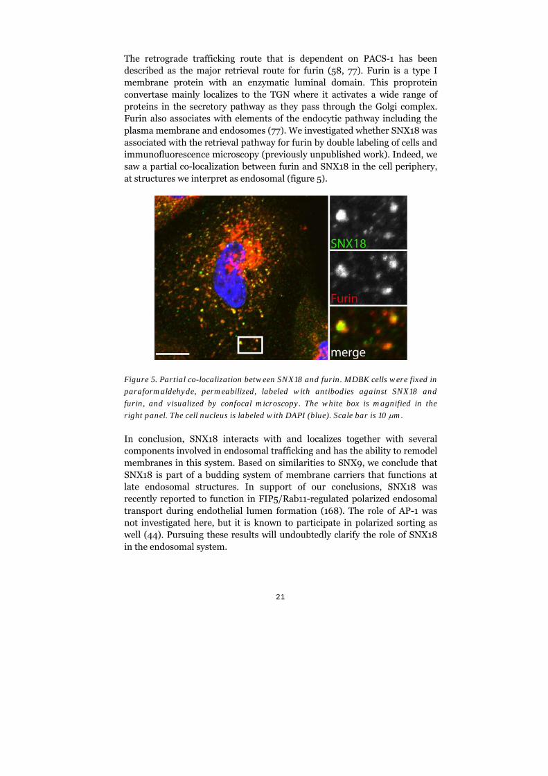
The retrograde trafficking route that is dependent on PACS-1 has been described as the major retrieval route for furin (58, 77). Furin is a type I membrane protein with an enzymatic luminal domain. This proprotein convertase mainly localizes to the TGN where it activates a wide range of proteins in the secretory pathway as they pass through the Golgi complex. Furin also associates with elements of the endocytic pathway including the plasma membrane and endosomes (77). We investigated whether SNX18 was associated with the retrieval pathway for furin by double labeling of cells and immunofluorescence microscopy (previously unpublished work). Indeed, we saw a partial co-localization between furin and SNX18 in the cell periphery, at structures we interpret as endosomal (figure 5).
Figure 5. Partial co-localization between SNX18 and furin. MDBK cells were fixed in
paraformaldehyde, permeabilized, labeled with antibodies against SNX18 and
furin, and visualized by confocal microscopy. The white box is magnified in the
right panel. The cell nucleus is labeled with DAPI (blue). Scale bar is 10 m.
In conclusion, SNX18 interacts with and localizes together with several components involved in endosomal trafficking and has the ability to remodel membranes in this system. Based on similarities to SNX9, we conclude that SNX18 is part of a budding system of membrane carriers that functions at late endosomal structures. In support of our conclusions, SNX18 was recently reported to function in FIP5/Rab11-regulated polarized endosomal transport during endothelial lumen formation (168). The role of AP-1 was not investigated here, but it is known to participate in polarized sorting as well (44). Pursuing these results will undoubtedly clarify the role of SNX18 in the endosomal system.

22
Paper II: The membrane-remodeling PX-BAR protein SNX18 is required for autophagy
As an essential part of autophagy, the membrane-remodeling and lipid composition of the autophagosomal structures have to be strictly controlled and regulated. Several membrane binding proteins and the lipid PtdIns3P have already been identified as essential in this process. In an attempt to identify additional membrane proteins in the autophagic process, we performed an siRNA knockdown screen targeting PX domain containing proteins in mammalian cells. PX domains are found in proteins interacting with phosphoinositides. A large fraction of human PX domain proteins belong to the SNX-family and several of these are known to mediate membrane-dependent cellular mechanisms. We used high-content immunofluorescence microscopy to monitor the knockdown effects in the siRNA screen by scoring the numbers of autophagosomes marked by GFP-LC3 in cultured mammalian cells, an established method to monitor autophagy (126).
In the siRNA screen, one set of siRNA oligonucleotides targeting the expression of SNX18 resulted in cells with less GFP-LC3 positive structures than the ULK1 knockdown cells (positive control). This drastic effect was confirmed in a follow-up experiment with individually distributed siRNA oligonucleotides. Apart from diminishing the amount of autophagosomes, as seen by the number of GFP-LC3 spots in cells, biochemical analysis of cell lysates showed an inhibition of the LC3-I to LC3-II conversion. Also, the amount of the autophagy receptor p62 remained high even after autophagy induction, which pointed to a severe degradation block caused by the lack of SNX18.
Using the same type of GFP-LC3 scoring system, SNX18 mutants were introduced into the cells to determine which properties of SNX18 that are important for autophagy. Wild-type SNX18 was also overexpressed as a control and we noted that in levels higher than normal SNX18 induced the formation of more autophagosomes and also increased the lipidation of LC3 resulting in higher amounts of LC3-II. In contrast to this, overexpressed SNX18 that had been mutated in a membrane interaction motif was incapable of autophagy induction. In addition to membrane binding, SNX18 might mediate membrane remodeling through insertion of an amphipathic helix into the lipid bilayer generating higher membrane curvature, as suggested from studies on SNX9 (83). This putative amphipathic helix in SNX18 contains a serine residue that is phosphorylated following rapamycin treatment of HeLa cells (169). To investigate its role in autophagosome formation, we mutated this serine and the resulting SNX18 mutant failed to
23
induce GFP-LC3 positive structures. We concluded that both the membrane binding and membrane remodeling capability of SNX18 are of importance for normal autophagosome biogenesis during starvation-induced autophagy.
The association between SNX18 and autophagy was further strengthened as we found direct interactions between the Atg8 homologues LC3/GABARAP and SNX18. Double labeling of cells also revealed co-localization between LC3 and SNX18 on peripheral punctate structures. In searching for additional protein interactions, we found that TBK1, a kinase mostly known for its implication in innate immune responses, co-immunoprecipitated with SNX18. This kinase is also connected to autophagy-mediated clearance of intracellular pathogens (159).
The indispensable role of SNX18 during autophagy in our experimental setting is striking, although the mechanism for this is still elusive. Nonetheless, it is clear that membrane association of this PX-BAR protein is of utmost importance for its function in this context. It is noteworthy to point out that SNX9 and SNX33 did not have any effect on autophagy in the siRNA screen of PX proteins. SNX18 is the first mammalian sorting nexin found to play a regulatory role in autophagy, while a yeast homolog of SNX4 has been shown to mediate membrane fusion during selective autophagy (170). It is possible that SNX4 or any of the other mammalian PX proteins in our siRNA screen are involved in other types of autophagy than the starvation-induced macroautophagy monitored in our study.
Concluding remarks and future perspectives
Shortly after the discovery of the first SNX in 1996 (171), this protein family attracted much attention and the mapping of these proteins quickly resulted in important insights into the internal membrane dynamics of cells. Paper I in this thesis was the first published article with a focus on SNX18 and, as for many of the other SNXs described, we located this protein in the endomembrane system. Because of the close relationship between SNX9, SNX18, and SNX33, initial investigations were aimed at characterizing both apparent similarities and conceivable differences between the proteins. We now consider these proteins as separate entities based on the results both from our and others’ laboratories. Future studies of SNX18 should aim for an extended understanding of its role in formation of endosomal membrane carriers. Use of selected markers for distinct endosomal pathways will reveal if SNX18 directs retrograde, polarized or perhaps both types of trafficking in conjunction with AP-1.
24
In addition to its role in endosomal carrier formation, it was very intriguing to find that SNX18 is a positive regulator of autophagy. This makes SNX18 yet another protein with dual roles in membrane trafficking connected to both endosomal and autophagic pathways. As so many question marks remain around the regulation of autophagic membranes, our findings regarding SNX18 will hopefully shed more light on these mechanisms. The membrane deformations during the assembly of the autophagic cup, its subsequent fission, and its final fusion with another organelle are complex processes that must be executed by several protein complexes. Understanding of this process also provides insights into many aspects of life, as autophagy controls development, homeostasis and ageing. As the research field around autophagy is rather young, the use of high-content screenings is a valuable tool that can quickly generate data in regards to the molecular regulation of this degradative pathway.
25
5. Acknowledgements
There are many people I would like to thank, without whom I would not have come this far and/or enjoyed the journey as much.
First of all, I want to acknowledge my supervisor Sven for your trust and patience, I truly admire your deep biochemical and cell biology knowledge as well as your teaching skills. Richard, on paper you were my co-supervisor but you were so much more in reality. You have been a true source of inspiration not only scientifically speaking. I wish to thank Dr. Anne Simonsen and her group in Oslo for a fruitful collaboration. Helene and Kristiane – it was great working with you! Our group was the smallest possible, so I really appreciated the occasional times with fellows in the lab: Erika, with your warm kindness it was a pleasure to work with you. Fredrik, you brought much needed energy into the lab. I also want to thank undergrads Rifat and Aziz for important contributions to the projects. And big hugs to my office mates, for the everyday small-talks, after works and sporadic scientific discussions; Björn, Monika, Mikkel, Jagan and Lars. Welcome to the new arrivals Lionel and Maite, hope you will enjoy your time here in Umeå. This department would not be the same without our hard-working T/A-personnel. Thanks Ingrid, Clas and Anna for always being there and taking care of us all and our paperwork, and Elisabeth for supplying us with clean labware. I also must say that I have really enjoyed the non-work related activities at this department. Thanks to all the innebandy players and members of the beer club. The whole Department of Medical Biochemistry and Biophysics deserves a Thank you! It has been a great workplace over the years and it just would not have been the same without all the past and present fellow workers. Sist men inte minst, ett stort tack till min familj. Mamma och pappa för att ni alltid funnits där, för allt stöd och uppmuntran jag fått. Mina storebrorsor Christer och Mikael för att ni alltid sett till att jag haft en fot kvar ute i den riktiga världen. Och slutligen, tack Tomas. Skulle aldrig ha fixat det här utan dig. Nu är det min tur att jag göra vad jag kan för att hjälpa dig fram till mållinjen.
26
6. References
1. Berg JM, Tymoczko JL, Stryer L, Stryer L. Biochemistry. 5th ed. New York: W.H. Freeman; 2002.
2. Alberts B. Molecular biology of the cell. 4th ed. New York: Garland Science; 2002.
3. Brighouse A, Dacks J, Field M. Rab protein evolution and the history of the eukaryotic endomembrane system. Cell Mol Life Sci. 2010; 67(20): 3449-65.
4. Lippincott-Schwartz J, Phair RD. Lipids and Cholesterol as Regulators of Traffic in the Endomembrane System. Annu Rev Biophys. 2010; 39(1): 559-78.
5. Conner SD, Schmid SL. Regulated portals of entry into the cell. Nature. 2003; 422(6927): 37-44.
6. Kirkham M, Parton RG. Clathrin-independent endocytosis: New insights into caveolae and non-caveolar lipid raft carriers. Biochim Biophys Acta. 2005; 1745(3): 273-86.
7. Mayor S, Pagano RE. Pathways of clathrin-independent endocytosis. Nat Rev Mol Cell Biol. 2007; 8(8): 603-12.
8. Rodriguez-Boulan E, Müsch A. Protein sorting in the Golgi complex: Shifting paradigms. Biochim Biophys Acta. 2005; 1744(3): 455-64.
9. Gruenberg J, Stenmark H. The biogenesis of multivesicular endosomes. Nat Rev Mol Cell Biol. 2004; 5(4): 317-23.
10. Maxfield FR, McGraw TE. Endocytic recycling. Nat Rev Mol Cell Biol. 2004; 5(2): 121-32.
11. Bonifacino JS, Rojas R. Retrograde transport from endosomes to the trans-Golgi network. Nat Rev Mol Cell Biol. 2006; 7(8): 568-79.
12. Johannes L, Popoff V. Tracing the Retrograde Route in Protein Trafficking. Cell. 2008; 135(7): 1175-87.
27
13. Kozlov MM, McMahon HT, Chernomordik LV. Protein-driven membrane stresses in fusion and fission. Trends Biochem Sci. 2010; 35(12): 699-706.
14. Cao H, Garcia F, McNiven MA. Differential Distribution of Dynamin Isoforms in Mammalian Cells. Mol Biol Cell. 1998; 9(9): 2595-609.
15. Ramachandran R. Vesicle scission: Dynamin. Semin Cell Dev Biol. 2011; 22(1): 10-7.
16. Praefcke GJK, McMahon HT. The dynamin superfamily: universal membrane tubulation and fission molecules? Nat Rev Mol Cell Biol. 2004; 5(2): 133-47.
17. Doherty GJ, McMahon HT. Mechanisms of Endocytosis. Annu Rev Biochem. 2009; 78(1): 857-902.
18. Lauvrak SU, Torgersen ML, Sandvig K. Efficient endosome-to-Golgi transport of Shiga toxin is dependent on dynamin and clathrin. J Cell Sci. 2004; 117(11): 2321-31.
19. van Dam EM, Stoorvogel W. Dynamin-dependent Transferrin Receptor Recycling by Endosome-derived Clathrin-coated Vesicles. Mol Biol Cell. 2002; 13(1): 169-82.
20. Mesaki K, Tanabe K, Obayashi M, Oe N, Takei K. Fission of Tubular Endosomes Triggers Endosomal Acidification and Movement. PLoS ONE. 2011; 6(5): e19764.
21. Stowell MHB, Marks B, Wigge P, McMahon HT. Nucleotide-dependent conformational changes in dynamin: evidence for a mechanochemical molecular spring. Nat Cell Biol. 1999; 1(1): 27-32.
22. Danino D, Moon K-H, Hinshaw JE. Rapid constriction of lipid bilayers by the mechanochemical enzyme dynamin. J Struct Biol. 2004; 147(3): 259-67.
23. Marks B, Stowell MHB, Vallis Y, Mills IG, Gibson A, Hopkins CR, et al. GTPase activity of dynamin and resulting conformation change are essential for endocytosis. Nature. 2001; 410(6825): 231-5.
28
24. Chen Y-J, Zhang P, Egelman EH, Hinshaw JE. The stalk region of dynamin drives the constriction of dynamin tubes. Nat Struct Mol Biol. 2004; v11(n6): 574-5.
25. Zhang P, Hinshaw JE. Three-dimensional reconstruction of dynamin in the constricted state. Nat Cell Biol. 2001; 3(10): 922-6.
26. Roux A, Uyhazi K, Frost A, De Camilli P. GTP-dependent twisting of dynamin implicates constriction and tension in membrane fission. Nature. 2006; 441(7092): 528-31.
27. Bethoney KA, King MC, Hinshaw JE, Ostap EM, Lemmon MA. A possible effector role for the pleckstrin homology (PH) domain of dynamin. Proc Natl Acad Sci USA. 2009; 106(32): 13359-64.
28. Roux A, Cuvelier D, Nassoy P, Prost J, Bassereau P, Goud B. Role of curvature and phase transition in lipid sorting and fission of membrane tubules. EMBO J. 2005; 24(8): 1537-45.
29. Liu J, Sun Y, Drubin DG, Oster GF. The Mechanochemistry of Endocytosis. PLoS Biol. 2009; 7(9): e1000204.
30. Newmyer SL, Christensen A, Sever S. Auxilin-Dynamin Interactions Link the Uncoating ATPase Chaperone Machinery with Vesicle Formation. Dev Cell. 2003; 4(6): 929-40.
31. Ramachandran R, Schmid SL. Real-time detection reveals that effectors couple dynamin's GTP-dependent conformational changes to the membrane. EMBO J. 2008; 27(1): 27-37.
32. Kim Y, Chang S. Ever-expanding network of dynamin-interacting proteins. Mol Neurobiol. 2006; 34(2): 129-35.
33. Hirst J, D. Barlow L, Francisco GC, Sahlender DA, Seaman MNJ, Dacks JB, et al. The Fifth Adaptor Protein Complex. PLoS Biol. 2011; 9(10): e1001170.
34. Robinson MS. Adaptable adaptors for coated vesicles. Trends Cell Biol. 2004; 14(4): 167-74.
35. Takatsu H, Sakurai M, Shin H-W, Murakami K, Nakayama K. Identification and Characterization of Novel Clathrin Adaptor-related Proteins. J Biol Chem. 1998; 273(38): 24693-700.
29
36. Bonifacino JS, Traub LM. Signals for sorting of transmembrane proteins to endosomes and lysosomes. Annu Rev Biochem. 2003; 72: 395-447.
37. Kelly BT, Owen DJ. Endocytic sorting of transmembrane protein cargo. Curr Opin Cell Biol. 2011; 23(4): 404-12.
38. Vassilopoulos S, Esk C, Hoshino S, Funke BH, Chen C-Y, Plocik AM, et al. A Role for the CHC22 Clathrin Heavy-Chain Isoform in Human Glucose Metabolism. Science. 2009; 324(5931): 1192-6.
39. Liu S-H, Towler MC, Chen E, Chen C-Y, Song W, Apodaca G, et al. A novel clathrin homolog that co-distributes with cytoskeletal components functions in the trans-Golgi network. EMBO J. 2001; 20(1/2): 272-84.
40. Esk C, Chen C-Y, Johannes L, Brodsky FM. The clathrin heavy chain isoform CHC22 functions in a novel endosomal sorting step. J Cell Biol. 2010; 188(1): 131-44.
41. Doray B, Ghosh P, Griffith J, Geuze HJ, Kornfeld S. Cooperation of GGAs and AP-1 in Packaging MPRs at the Trans-Golgi Network. Science. 2002; 297(5587): 1700-3.
42. Meyer C, Zizioli D, Lausmann S, Eskelinen E-L, Hamann J, Saftig P, et al. µ1A-adaptin-deficient mice: lethality, loss of AP-1 binding and rerouting of mannose 6-phosphate receptors. EMBO J. 2000; 19(10): 2193-203.
43. Brown FC, Schindelhaim CH, Pfeffer SR. GCC185 plays independent roles in Golgi structure maintenance and AP-1-mediated vesicle tethering. J Cell Biol. 2011; 194(5): 779-87.
44. Traub LM, Apodaca G. AP-1B: polarized sorting at the endosome. Nat Cell Biol. 2003; 5(12): 1045-7.
45. Loerke D, Mettlen M, Schmid SL, Danuser G. Measuring the Hierarchy of Molecular Events During Clathrin-Mediated Endocytosis. Traffic. 2011; 12(7): 815-25.
46. Dell´Angelica EC. AP-3-dependent trafficking and disease: the first decade. Curr Opin Cell Biol. 2009; 21(4): 552-9.
30
47. Burgos PV, Mardones GA, Rojas AL, daSilva LLP, Prabhu Y, Hurley JH, et al. Sorting of the Alzheimer's Disease Amyloid Precursor Protein Mediated by the AP-4 Complex. Dev Cell. 2010; 18(3): 425-36.
48. Håberg K, Lundmark R, Carlsson SR. SNX18 is an SNX9 paralog that acts as a membrane tubulator in AP-1-positive endosomal trafficking. J Cell Sci. 2008; 121(Pt 9): 1495-505.
49. Puertollano R, van der Wel NN, Greene LE, Eisenberg E, Peters PJ, Bonifacino JS. Morphology and Dynamics of Clathrin/GGA1-coated Carriers Budding from the Trans-Golgi Network. Mol Biol Cell. 2003; 14(4): 1545-57.
50. Waguri S, Dewitte F, Le Borgne R, Rouillé Y, Uchiyama Y, Dubremetz J-F, et al. Visualization of TGN to Endosome Trafficking through Fluorescently Labeled MPR and AP-1 in Living Cells. Mol Biol Cell. 2003; 14(1): 142-55.
51. Zhu Y, Traub LM, Kornfeld S. ADP-Ribosylation Factor 1 Transiently Activates High-Affinity Adaptor Protein Complex AP-1 Binding Sites On Golgi Membranes. Mol Biol Cell. 1998; 9(6): 1323-37.
52. D'Souza-Schorey C, Chavrier P. ARF proteins: roles in membrane traffic and beyond. Nat Rev Mol Cell Biol. 2006; 7(5): 347-58.
53. Braun V, Deschamps C, Raposo G, Benaroch P, Benmerah A, Chavrier P, et al. AP-1 and ARF1 Control Endosomal Dynamics at Sites of FcR–mediated Phagocytosis. Mol Biol Cell. 2007; 18(12): 4921-31.
54. Ghosh P, Griffith J, Geuze HJ, Kornfeld S. Mammalian GGAs act together to sort mannose 6-phosphate receptors. J Cell Biol. 2003; 163(4): 755-66.
55. Mardones GA, Burgos PV, Brooks DA, Parkinson-Lawrence E, Mattera R, Bonifacino JS. The Trans-Golgi Network Accessory Protein p56 Promotes Long-Range Movement of GGA/Clathrin-containing Transport Carriers and Lysosomal Enzyme Sorting. Mol Biol Cell. 2007; 18(9): 3486-501.
31
56. Hirst J, Sahlender DA, Choma M, Sinka R, Harbour ME, Parkinson M, et al. Spatial and Functional Relationship of GGAs and AP-1 in Drosophila and HeLa Cells. Traffic. 2009; 10(11): 1696-710.
57. Youker RT, Shinde U, Day R, Thomas G. At the crossroads of homoeostasis and disease: roles of the PACS proteins in membrane traffic and apoptosis. Biochem J. 2009; 421(1): 1-15.
58. Wan L, Molloy SS, Thomas L, Liu G, Xiang Y, Rybak SL, et al. PACS-1 Defines a Novel Gene Family of Cytosolic Sorting Proteins Required for trans-Golgi Network Localization. Cell. 1998; 94(2): 205-16.
59. Crump CM, Xiang Y, Thomas L, Gu F, Austin C, Tooze SA, et al. PACS-1 binding to adaptors is required for acidic cluster motif-mediated protein traffic. EMBO J. 2001; 20(9): 2191-201.
60. Lubben NB, Sahlender DA, Motley AM, Lehner PJ, Benaroch P, Robinson MS. HIV-1 Nef-induced Down-Regulation of MHC Class I Requires AP-1 and Clathrin but Not PACS-1 and Is Impeded by AP-2. Mol Biol Cell. 2007; 18(9): 3351-65.
61. Hirst J, Motley A, Harasaki K, Peak Chew SY, Robinson MS. EpsinR: an ENTH Domain-containing Protein that Interacts with AP-1. Mol Biol Cell. 2003; 14(2): 625-41.
62. Mills IG, Praefcke GJK, Vallis Y, Peter BJ, Olesen LE, Gallop JL, et al. EpsinR: an AP1/clathrin interacting protein involved in vesicle trafficking. J Cell Biol. 2003; 160(2): 213-22.
63. Kalthoff C, Groos S, Kohl R, Mahrhold S, Ungewickell EJ. Clint: A Novel Clathrin-binding ENTH-Domain Protein at the Golgi. Mol Biol Cell. 2002; 13(11): 4060-73.
64. Wasiak S, Legendre-Guillemin V, Puertollano R, Blondeau F, Girard M, de Heuvel E, et al. Enthoprotin: a novel clathrin-associated protein identified through subcellular proteomics. J Cell Biol. 2002; 158(5): 855-62.
65. Saint-Pol A, Yélamos B, Amessou M, Mills IG, Dugast M, Tenza D, et al. Clathrin Adaptor epsinR Is Required for Retrograde Sorting on Early Endosomal Membranes. Dev Cell. 2004; 6(4): 525-38.
32
66. Rojas R, Kametaka S, Haft CR, Bonifacino JS. Interchangeable but Essential Functions of SNX1 and SNX2 in the Association of Retromer with Endosomes and the Trafficking of Mannose 6-Phosphate Receptors. Mol Cell Biol. 2007; 27(3): 1112-24.
67. Arighi CN, Hartnell LM, Aguilar RC, Haft CR, Bonifacino JS. Role of the mammalian retromer in sorting of the cation-independent mannose 6-phosphate receptor. J Cell Biol. 2004; 165(1): 123-33.
68. Carlton J, Bujny M, Peter BJ, Oorschot VMJ, Rutherford A, Mellor H, et al. Sorting Nexin-1 Mediates Tubular Endosome-to-TGN Transport through Coincidence Sensing of High- Curvature Membranes and 3-Phosphoinositides. Curr Biol. 2004; 14(20): 1791-800.
69. Carlton JG, Bujny MV, Peter BJ, Oorschot VMJ, Rutherford A, Arkell RS, et al. Sorting nexin-2 is associated with tubular elements of the early endosome, but is not essential for retromer-mediated endosome-to-TGN transport. J Cell Sci. 2005; 118(19): 4527-39.
70. Utskarpen A, Slagsvold HH, Dyve AB, Skånland SS, Sandvig K. SNX1 and SNX2 mediate retrograde transport of Shiga toxin. Biochem Biophys Res Commun. 2007; 358(2): 566-70.
71. Ohno H, Aguilar RC, Yeh D, Taura D, Saito T, Bonifacino JS. The medium subunits of adaptor complexes recognize distinct but overlapping sets of tyrosine-based sorting signals. J Biol Chem. 1998; 273(40): 25915-21.
72. Hirst J, Bright NA, Rous B, Robinson MS. Characterization of a fourth adaptor-related protein complex. Mol Biol Cell. 1999; 10(8): 2787-802.
73. Puertollano R, Aguilar RC, Gorshkova I, Crouch RJ, Bonifacino JS. Sorting of mannose 6-phosphate receptors mediated by the GGAs. Science. 2001; 292(5522): 1712-6.
74. Braulke T, Bonifacino JS. Sorting of lysosomal proteins. Biochim Biophys Acta. 2009; 1793(4): 605-14.
75. Mattera R, Boehm M, Chaudhuri R, Prabhu Y, Bonifacino JS. Conservation and Diversification of Dileucine Signal Recognition by
33
Adaptor Protein (AP) Complex Variants. J Biol Chem. 2011; 286(3): 2022-30.
76. Schapiro FB, Soe TT, Mallet WG, Maxfield FR. Role of Cytoplasmic Domain Serines in Intracellular Trafficking of Furin. Mol Biol Cell. 2004; 15(6): 2884-94.
77. Molloy SS, Thomas L, Kamibayashi C, Mumby MC, Thomas G. Regulation of endosome sorting by a specific PP2A isoform. J Cell Biol. 1998; 142(6): 1399-411.
78. Bonifacino JS, Hurley JH. Retromer. Curr Opin Cell Biol. 2008; 20(4): 427-36.
79. Miller SE, Collins BM, McCoy AJ, Robinson MS, Owen DJ. A SNARE-adaptor interaction is a new mode of cargo recognition in clathrin-coated vesicles. Nature. 2007; 450(7169): 570-4.
80. Seet L-F, Hong W. The Phox (PX) domain proteins and membrane traffic. Biochim Biophys Acta. 2006; 1761(8): 878-96.
81. Teasdale RD, Collins BM. Insights into the PX (phox-homology) domain and SNX (sorting nexin) protein families: structures, functions and roles in disease. Biochem J. 2012; 441(1): 39-59.
82. Peter BJ, Kent HM, Mills IG, Vallis Y, Butler PJG, Evans PR, et al. BAR Domains as Sensors of Membrane Curvature: The Amphiphysin BAR Structure. Science. 2004; 303(5657): 495-9.
83. Pylypenko O, Lundmark R, Rasmuson E, Carlsson SR, Rak A. The PX-BAR membrane-remodeling unit of sorting nexin 9. EMBO J. 2007; 26(22): 4788-800.
84. Qualmann B, Koch D, Kessels MM. Let's go bananas: revisiting the endocytic BAR code. EMBO J. 2011; 30(17): 3501-15.
85. van Weering JRT, Verkade P, Cullen PJ. SNX–BAR proteins in phosphoinositide-mediated, tubular-based endosomal sorting. Sem Cell Dev Biol. 2010; 21(4): 371-80.
86. Lundmark R, Carlsson SR. Sorting nexin 9 participates in clathrin-mediated endocytosis through interactions with the core components. J Biol Chem. 2003; 278(47): 46772-81.
34
87. Soulet F, Yarar D, Leonard M, Schmid SL. SNX9 regulates dynamin assembly and is required for efficient clathrin-mediated endocytosis. Mol Biol Cell. 2005; 16(4): 2058-67.
88. Lundmark R, Carlsson SR. Regulated membrane recruitment of dynamin-2 mediated by sorting nexin 9. J Biol Chem. 2004; 279(41): 42694-702.
89. Lundmark R, Carlsson SR. The beta-appendages of the four adaptor-protein (AP) complexes: structure and binding properties, and identification of sorting nexin 9 as an accessory protein to AP-2. Biochem J. 2002; 362(Pt 3): 597-607.
90. Yarar D, Surka MC, Leonard MC, Schmid SL. SNX9 activities are regulated by multiple phosphoinositides through both PX and BAR domains. Traffic. 2008; 9(1): 133-46.
91. Yarar D, Waterman-Storer CM, Schmid SL. SNX9 Couples Actin Assembly to Phosphoinositide Signals and Is Required for Membrane Remodeling during Endocytosis. Dev Cell. 2007; 13(1): 43-56.
92. Shin N, Ahn N, Chang-Ileto B, Park J, Takei K, Ahn S-G, et al. SNX9 regulates tubular invagination of the plasma membrane through interaction with actin cytoskeleton and dynamin 2. J Cell Sci. 2008; 121(8): 1252-63.
93. Drin G, Antonny B. Amphipathic helices and membrane curvature. FEBS Lett. 2010; 584(9): 1840-7.
94. Lundmark R, Carlsson SR. SNX9 – a prelude to vesicle release. J Cell Sci. 2009; 122(1): 5-11.
95. Schöbel S, Neumann S, Hertweck M, Dislich B, Kuhn P-H, Kremmer E, et al. A Novel Sorting Nexin Modulates Endocytic Trafficking and α-Secretase Cleavage of the Amyloid Precursor Protein. J Biol Chem. 2008; 283(21): 14257-68.
96. Zhang J, Zhang X, Guo Y, Xu L, Pei D. Sorting Nexin 33 Induces Mammalian Cell Micronucleated Phenotype and Actin Polymerization by Interacting with Wiskott-Aldrich Syndrome Protein. J Biol Chem. 2009; 284(32): 21659-69.

35
97. Clague MJ, Urbé S. Ubiquitin: Same Molecule, Different Degradation Pathways. Cell. 2010; 143(5): 682-5.
98. Mizushima N, Komatsu M. Autophagy: Renovation of Cells and Tissues. Cell. 2011; 147(4): 728-41.
99. Rubinsztein David C, Mariño G, Kroemer G. Autophagy and Aging. Cell. 2011; 146(5): 682-95.
100. Yang Z, Klionsky DJ. Eaten alive: a history of macroautophagy. Nat Cell Biol. 2010; 12(9): 814-22.
101. Ravikumar B, Sarkar S, Davies JE, Futter M, Garcia-Arencibia M, Green-Thompson ZW, et al. Regulation of Mammalian Autophagy in Physiology and Pathophysiology. Physiol Rev. 2010; 90(4):1383-435.
102. Sarbassov DD, Ali SM, Sabatini DM. Growing roles for the mTOR pathway. Curr Opin Cell Biol. 2005; 17(6): 596-603.
103. Noda T, Ohsumi Y. Tor, a Phosphatidylinositol Kinase Homologue, Controls Autophagy in Yeast. J Biol Chem. 1998; 273(7): 3963-6.
104. Boland B, Kumar A, Lee S, Platt FM, Wegiel J, Yu WH, et al. Autophagy Induction and Autophagosome Clearance in Neurons: Relationship to Autophagic Pathology in Alzheimer's Disease. J Neurosci. 2008; 28(27): 6926-37.
105. Ravikumar B, Vacher C, Berger Z, Davies JE, Luo SQ, Oroz LG, et al. Inhibition of mTOR induces autophagy and reduces toxicity of polyglutamine expansions in fly and mouse models of Huntington disease. Nat Genet. 2004; 36(6): 585-95.
106. Ganley IG, Lam DH, Wang J, Ding X, Chen S, Jiang X. ULK1·ATG13·FIP200 Complex Mediates mTOR Signaling and Is Essential for Autophagy. J Biol Chem. 2009; 284(18): 12297-305.
107. Hosokawa N, Hara T, Kaizuka T, Kishi C, Takamura A, Miura Y, et al. Nutrient-dependent mTORC1 Association with the ULK1–Atg13–FIP200 Complex Required for Autophagy. Mol Biol Cell. 2009; 20(7): 1981-91.
36
108. Jung CH, Jun CB, Ro S-H, Kim Y-M, Otto NM, Cao J, et al. ULK-Atg13-FIP200 Complexes Mediate mTOR Signaling to the Autophagy Machinery. Mol Biol Cell. 2009; 20(7): 1992-2003.
109. Hara T, Takamura A, Kishi C, Iemura S-i, Natsume T, Guan J-L, et al. FIP200, a ULK-interacting protein, is required for autophagosome formation in mammalian cells. J Cell Biol. 2008; 181(3): 497-510.
110. Itakura E, Mizushima N. Characterization of autophagosome formation site by a hierarchical analysis of mammalian Atg proteins. Autophagy. 2010; 6(6): 764-76.
111. Axe EL, Walker SA, Manifava M, Chandra P, Roderick HL, Habermann A, et al. Autophagosome formation from membrane compartments enriched in phosphatidylinositol 3-phosphate and dynamically connected to the endoplasmic reticulum. J Cell Biol. 2008; 182(4): 685-701.
112. Hayashi-Nishino M, Fujita N, Noda T, Yamaguchi A, Yoshimori T, Yamamoto A. A subdomain of the endoplasmic reticulum forms a cradle for autophagosome formation. Nat Cell Biol. 2009; 11(12): 1433-7.
113. Yla-Anttila P, Vihinen H, Jokitalo E, Eskelinen EL. 3D tomography reveals connections between the phagophore and endoplasmic reticulum. Autophagy. 2009; 5(8): 1180-5.
114. Hailey DW, Rambold AS, Satpute-Krishnan P, Mitra K, Sougrat R, Kim PK, et al. Mitochondria Supply Membranes for Autophagosome Biogenesis during Starvation. Cell. 2010; 141(4): 656-67.
115. Ravikumar B, Moreau K, Jahreiss L, Puri C, Rubinsztein DC. Plasma membrane contributes to the formation of pre-autophagosomal structures. Nat Cell Biol. 2010; 12(8): 747-57.
116. Simonsen A, Tooze SA. Coordination of membrane events during autophagy by multiple class III PI3-kinase complexes. J Cell Biol. 2009; 186(6): 773-82.
117. Proikas-Cezanne T, Waddell S, Gaugel A, Frickey T, Lupas A, Nordheim A. WIPI-1[alpha] (WIPI49), a member of the novel 7-bladed WIPI protein family, is aberrantly expressed in human cancer
37
and is linked to starvation-induced autophagy. Oncogene. 2004; 23(58): 9314-25.
118. Mauthe M, Jacob A, Freiberger S, Hentschel K, Stierhof Y-D, Codogno P, et al. Resveratrol-mediated autophagy requires WIPI-1-regulated LC3 lipidation in the absence of induced phagophore formation. Autophagy. 2011; 7(12):1 448-61.
119. Polson HEJ, de Lartigue J, Rigden DJ, Reedijk M, Urbé S, Clague MJ, et al. Mammalian Atg18 (WIPI2) localizes to omegasome-anchored phagophores and positively regulates LC3 lipidation. Autophagy. 2010; 6(4): 506-22.
120. Proikas-Cezanne T, Ruckerbauer S, Stierhof Y-D, Berg C, Nordheim A. Human WIPI-1 puncta-formation: A novel assay to assess mammalian autophagy. FEBS Lett. 2007; 581(18): 3396-404.
121. Mizushima N, Sugita H, Yoshimori T, Ohsumi Y. A New Protein Conjugation System in Human. J Biol Chem. 1998; 273(51): 33889-92.
122. Mizushima N, Kuma A, Kobayashi Y, Yamamoto A, Matsubae M, Takao T, et al. Mouse Apg16L, a novel WD-repeat protein, targets to the autophagic isolation membrane with the Apg12-Apg5 conjugate. J Cell Sci. 2003; 116(9): 1679-88.
123. Kabeya Y, Mizushima N, Ueno T, Yamamoto A, Kirisako T, Noda T, et al. LC3, a mammalian homologue of yeast Apg8p, is localized in autophagosome membranes after processing. EMBO J. 2000; 19(21): 5720-8.
124. Tanida I, Ueno T, Kominami E. LC3 conjugation system in mammalian autophagy. Int J Biochem Cell Biol. 2004; 36(12): 2503-18.
125. Tanida I, Sou Y-s, Ezaki J, Minematsu-Ikeguchi N, Ueno T, Kominami E. HsAtg4B/HsApg4B/Autophagin-1 Cleaves the Carboxyl Termini of Three Human Atg8 Homologues and Delipidates Microtubule-associated Protein Light Chain 3- and GABAA Receptor-associated Protein-Phospholipid Conjugates. J Biol Chem. 2004; 279(35): 36268-76.
38
126. Klionsky DJ, Abeliovich H, Agostinis P, Agrawal DK, Aliev G, Askew DS, et al. Guidelines for the use and interpretation of assays for monitoring autophagy in higher eukaryotes. Autophagy. 2008; 4(2): 151-75.
127. Vergne I, Deretic V. The role of PI3P phosphatases in the regulation of autophagy. FEBS Lett. 2010; 584(7): 1313-8.
128. Noda T, Matsunaga K, Taguchi-Atarashi N, Yoshimori T. Regulation of membrane biogenesis in autophagy via PI3P dynamics. Semin Cell Dev Biol. 2010; 21(7): 671-6.
129. Nakatogawa H, Ichimura Y, Ohsumi Y. Atg8, a ubiquitin-like protein required for autophagosome formation, mediates membrane tethering and hemifusion. Cell. 2007; 130(1): 165-78.
130. Xie Z, Nair U, Klionsky DJ. Atg8 Controls Phagophore Expansion during Autophagosome Formation. Mol Biol Cell. 2008; 19(8): 3290-8.
131. Weidberg H, Shpilka T, Shvets E, Abada A, Shimron F, Elazar Z. LC3 and GATE-16 N Termini Mediate Membrane Fusion Processes Required for Autophagosome Biogenesis. Dev Cell. 2011; 20(4): 444-54.
132. Weidberg H, Shvets E, Shpilka T, Shimron F, Shinder V, Elazar Z. LC3 and GATE-16/GABARAP subfamilies are both essential yet act differently in autophagosome biogenesis. EMBO J. 2010; 29(11): 1792-802.
133. Nair U, Jotwani A, Geng J, Gammoh N, Richerson D, Yen WL, et al. SNARE proteins are required for macroautophagy. Cell. 2011; 146(2): 290-302.
134. Moreau K, Ravikumar B, Renna M, Puri C, Rubinsztein DC. Autophagosome precursor maturation requires homotypic fusion. Cell. 2011; 146(2): 303-17.
135. Tooze SA. The role of membrane proteins in mammalian autophagy. Semin Cell Dev Biol. 2010; 21(7): 677-82.
136. Young ARJ, Chan EYW, Hu XW, Köchl R, Crawshaw SG, High S, et al. Starvation and ULK1-dependent cycling of mammalian Atg9
39
between the TGN and endosomes. J Cell Sci. 2006; 119(18): 3888-900.
137. Takahashi Y, Meyerkord CL, Hori T, Runkle K, Fox TE, Kester M, et al. Bif-1 regulates Atg9 trafficking by mediating the fission of Golgi membranes during autophagy. Autophagy. 2011; 7(1): 61-73.
138. Reggiori F, Tucker KA, Stromhaug PE, Klionsky DJ. The Atg1-Atg13 Complex Regulates Atg9 and Atg23 Retrieval Transport from the Pre-Autophagosomal Structure. Dev Cell. 2004; 6(1): 79-90.
139. Geng J, Baba M, Nair U, Klionsky DJ. Quantitative analysis of autophagy-related protein stoichiometry by fluorescence microscopy. J Cell Biol. 2008; 182(1): 129-40.
140. Webber JL, Tooze SA. Coordinated regulation of autophagy by p38[alpha] MAPK through mAtg9 and p38IP. EMBO J. 2010; 29(1): 27-40.
141. Webber JL, Tooze SA. New insights into the function of Atg9. FEBS Lett. 2010; 584(7): 1319-26.
142. Köchl R, Hu XW, Chan EYW, Tooze SA. Microtubules Facilitate Autophagosome Formation and Fusion of Autophagosomes with Endosomes. Traffic. 2006; 7(2): 129-45.
143. Pankiv S, Alemu EA, Brech A, Bruun J-A, Lamark T, Øvervatn A, et al. FYCO1 is a Rab7 effector that binds to LC3 and PI3P to mediate microtubule plus end–directed vesicle transport. J Cell Biol. 2010; 188(2): 253-69.
144. Kimura S, Noda T, Yoshimori T. Dynein-dependent Movement of Autophagosomes Mediates Efficient Encounters with Lysosomes. Cell Struct Funct. 2008; 33(1): 109-22.
145. Ravikumar B, Acevedo-Arozena A, Imarisio S, Berger Z, Vacher C, O'Kane CJ, et al. Dynein mutations impair autophagic clearance of aggregate-prone proteins. Nat Genet. 2005; 37(7): 771-6.
146. Yamamoto A, Tagawa Y, Yoshimori T, Moriyama Y, Masaki R, Tashiro Y. Bafilomycin A(1) prevents maturation of autophagic vacuoles by inhibiting fusion between autophagosomes and
40
lysosomes in rat hepatoma cell line, H-4-II-E cells. Cell Struct Funct. 1998; 23(1): 33-42.
147. Bjørkøy G, Lamark T, Brech A, Outzen H, Perander M, Øvervatn A, et al. p62/SQSTM1 forms protein aggregates degraded by autophagy and has a protective effect on huntingtin-induced cell death. J Cell Biol. 2005; 171(4): 603-14.
148. Johansen T, Lamark T. Selective autophagy mediated by autophagic adapter proteins. Autophagy. 2011; 7(3): 279-96.
149. Pankiv S, Clausen TH, Lamark T, Brech A, Bruun J-A, Outzen H, et al. p62/SQSTM1 Binds Directly to Atg8/LC3 to Facilitate Degradation of Ubiquitinated Protein Aggregates by Autophagy. J Biol Chem. 2007; 282(33): 24131-45.
150. Kirkin V, Lamark T, Sou Y-S, Bjørkøy G, Nunn JL, Bruun J-A, et al. A Role for NBR1 in Autophagosomal Degradation of Ubiquitinated Substrates. Mol Cell. 2009; 33(4): 505-16.
151. Novak I, Kirkin V, McEwan DG, Zhang J, Wild P, Rozenknop A, et al. Nix is a selective autophagy receptor for mitochondrial clearance. EMBO Rep. 2010; 11(1): 45-51.
152. Thurston TLM, Ryzhakov G, Bloor S, von Muhlinen N, Randow F. The TBK1 adaptor and autophagy receptor NDP52 restricts the proliferation of ubiquitin-coated bacteria. Nat Immunol. 2009; 10(11): 1215-21.
153. Zheng YT, Shahnazari S, Brech A, Lamark T, Johansen T, Brumell JH. The Adaptor Protein p62/SQSTM1 Targets Invading Bacteria to the Autophagy Pathway. J Immunol. 2009; 183(9): 5909-16.
154. Cemma M, Kim PK, Brumell JH. The ubiquitin-binding adaptor proteins p62/SQSTM1 and NDP52 are recruited independently to bacteria-associated microdomains to target Salmonella to the autophagy pathway. Autophagy. 2011; 7(3): 341-5.
155. Mostowy S, Sancho-Shimizu V, Hamon MA, Simeone R, Brosch R, Johansen T, et al. p62 and NDP52 Proteins Target Intracytosolic Shigella and Listeria to Different Autophagy Pathways. J Biol Chem. 2011; 286(30): 26987-95.
41
156. Takeshita F, Ishii KJ. Intracellular DNA sensors in immunity. Curr Opin Immunol. 2008; 20(4): 383-8.
157. Clement JF, Meloche S, Servant MJ. The IKK-related kinases: from innate immunity to oncogenesis. Cell Res. 2008; 18(9): 889-99.
158. Radtke AL, Delbridge LM, Balachandran S, Barber GN, O'Riordan MXD. TBK1 Protects Vacuolar Integrity during Intracellular Bacterial Infection. PLoS Pathog. 2007; 3(3): e29.
159. Wild P, Farhan H, McEwan DG, Wagner S, Rogov VV, Brady NR, et al. Phosphorylation of the autophagy receptor optineurin restricts Salmonella growth. Science. 2011; 333(6039): 228-33.
160. Saitoh T, Fujita N, Hayashi T, Takahara K, Satoh T, Lee H, et al. Atg9a controls dsDNA-driven dynamic translocation of STING and the innate immune response. Proc Natl Acad Sci U S A. 2009; 106(49): 20842-6.
161. Criollo A, Senovilla L, Authier H, Maiuri MC, Morselli E, Vitale I, et al. The IKK complex contributes to the induction of autophagy. EMBO J. 2010; 29(3): 619-31.
162. Vojo D. Autophagy as an innate immunity paradigm: expanding the scope and repertoire of pattern recognition receptors. Curr Opin Immunol. 2011. Epub ahead of print. DOI 10.1016/j.coi.2011.10.006
163. Wassmer T, Attar N, Harterink M, van Weering JRT, Traer CJ, Oakley J, et al. The Retromer Coat Complex Coordinates Endosomal Sorting and Dynein-Mediated Transport, with Carrier Recognition by the trans-Golgi Network. Dev Cell. 2009; 17(1): 110-22.
164. Wigge P, Köhler K, Vallis Y, Doyle CA, Owen D, Hunt SP, et al. Amphiphysin Heterodimers: Potential Role in Clathrin-mediated Endocytosis. Mol Biol Cell. 1997; 8(10): 2003-15.
165. Park J, Kim Y, Lee S, Park JJ, Park ZY, Sun W, et al. SNX18 shares a redundant role with SNX9 and modulates endocytic trafficking at the plasma membrane. J Cell Sci. 2010; 123(10): 1742-50.
166. Dislich B, Than ME, Lichtenthaler SF. Specific amino acids in the BAR domain allow homodimerization and prevent
42
heterodimerization of sorting nexin 33. Biochem J. 2010; 433(1): 75-83.
167. Gillingham AK, Munro S. The small G proteins of the arf family and their regulators. Annu Rev Cell Dev Biol. 2007; 23: 579-611.
168. Willenborg C, Jing J, Wu C, Matern H, Schaack J, Burden J, et al. Interaction between FIP5 and SNX18 regulates epithelial lumen formation. J Cell Biol. 2011; 195(1): 71-86.
169. Chen RQ, Yang QK, Lu BW, Yi W, Cantin G, Chen YL, et al. CDC25B mediates rapamycin-induced oncogenic responses in cancer cells. Cancer Res. 2009; 69(6): 2663-8.
170. Ano Y, Hattori T, Oku M, Mukaiyama H, Baba M, Ohsumi Y, et al. A Sorting Nexin PpAtg24 Regulates Vacuolar Membrane Dynamics during Pexophagy via Binding to Phosphatidylinositol-3-Phosphate. Mol Biol Cell. 2005; 16(2): 446-57.
171. Kurten RC, Cadena DL, Gill GN. Enhanced degradation of EGF receptors by a sorting nexin, SNX1. Science. 1996; 272(5264): 1008-10.