breeding and feeding for elbow conformity - vet-iewg.org · klinik fűr kleintiere-chirurgie,...
TRANSCRIPT

PROCEEDINGS
32nd annual meeting of the
INTERNATIONAL ELBOW WORKING GROUP
September 13th 2018 WVOC 2018 congress,
Fiera de Barcellona, room 4, Barcelona, Spain.

32nd annual meeting IEWG, Barcelona Spain, September 13th 2018, p 2
Welcome IEWG-participants! The founding of the International Elbow Working Group (IEWG) took place in Davis (Ca) almost 30 years ago by an international group of veterinarians and interested breeders. Although the awareness of elbow dysplasia (ED) has increased considerably over the years, there is still a need to exchange the newest ideas and experiences regarding the pathogenesis of the different entities of ED, the etiological factors which play a role in the occurrence of ED, the details which may play a role in a more precise reading of the radiographs used for screening purposes, and the clinical use of the most appropriate imaging and surgical techniques. The board of the IEWG likes to thank the organisers of the World Veterinary Orthopaedic Congress (WVOC) for their kind invitation to the IEWG to have its annual meeting in conjunction with this prestigious joined congress of the Veterinary Orthopedic Society (VOS) and the European Society for Veterinary Orthopaedics and Traumatology (ESVOT), attended by veterinarians interested in orthopaedics and originating from the United States of America, a large variety of countries in Europe and also from Asian countries. In particular, we like to thank Prof. P. Böttcher, president of the WVOC and Dr A. Vezzoni (member of the organizing committee) for their kind invitation and their efforts to make it all happen. In addition, the board of the IEWG is most grateful to the selection of international and esteemed speakers who were enthusiastic when invited to speak at this years´ IEWG meeting, including Dr. Chico from the Spanish breeders association, Dr. Gielen from Ghent University who is known for her clear and complete overviews of a wide range of imaging techniques in use to investigate dogs lame from ED, Prof. Theyse from the University of Leipzig with his broad clinical experience as specialist in orthopaedic surgery, the very well appreciated speaker from the USA who participated so often in IEWG meetings Dr. Beale, and Dr. Von Pückler from Giessen University a very experience specialist in radiology and a very well appreciated speaker at international congress as the IEWG meeting. Advanced techniques of elbow surgery will be discussed during the main program of the WVOC and will thus not be a part of this IEWG-meeting. Interactive film reading will conclude the IEWG session in Barcelona. Participants from the IEWG are kindly invited to visit the web page of the IEWG (www.vet-iewg.org/). The secretary of the IEWG, Dr. Thijs How collected all proceeding texts of this and the past 17 years and made them thus available to the interested veterinarian. Also the older proceedings contain information which is still valuable in today´s veterinary and consulting practice. It also makes visible the large group of highly appreciated veterinarians who were involved in the IEWG meetings and helped to build its scientific reputation and its independent status. The IEWG as an affiliate of the World Small Animal Veterinary Association (WSAVA) feels obliged to be present at the WSAVA congress in Singapore, later in September this year. Those who cannot be in Barcelona at the IEWG meeting are invited to participate in the IEWG seminar in the pre-congress of the WSAVA-2018. We like to welcome all interested veterinarians from Spain, the rest of Europe and from overseas at the 32nd IEWG meeting in Barcelona, and wish you a fruitful meeting. Prof. dr. Herman A.W. Hazewinkel, President IEWG

32nd annual meeting IEWG, Barcelona Spain, September 13th 2018, p 3
International Elbow Working Group Meeting www.vet-iewg.org
Thursday September 13th 2018
pre-congress day WVOC 2018 congress Fira de Barcelona, room 4
Spain Scientific program 09.00 Opening by the president of the IEWG. 09.00 – 09.30 Prevalence of Elbow Dysplasia and pathogenesis of FCP in young Labradors, Prof. Dr. H.A.W. Hazewinkel [NL]. 09.30 – 09.50 Overview of Elbow Dysplasia screening in Spain, Dr. A. Chico [E]. 09.50 – 10.20 Strength and limitations of radiography, scintigraphy, ultrasound,CT, MRI and arthroscopy to diagnose Elbow Dysplasia in lame dogs, Dr. I. Gielen [B]. 10.20 – 11.00 Clinical signs, diagnostic and pathological findings of Elbow Dysplasia in dogs of different age groups, Prof. dr. L.F.H. Theyse [D]. 10.00 – 11.30 Break 11.30 – 11.50 Primary and secondary flexor enthesiopathy in the canine elbow, diagnostics and therapy, Dr. I. Gielen [B]. 11.50 – 12.10 The possible role of the biceps brachii and brachialis in fragmented medial coronoid, Dr. B. Beale [USA]. 12.10 - 12.40 Standardised grading of radiographs and CT-scan according to the IEWG for ED-screening, Dr. K.H. von Pückler [D]. 12.40 – 12.50 Interactive case discussion.

32nd annual meeting IEWG, Barcelona Spain, September 13th 2018, p 4
List of speakers Dr. B. Beale, DVM, Dipl. ACVS, Gulf Coast Veterinary Specialists, 1030 Wirt Rd, Houston, TX 77055-6849, USA, [email protected]. Dr. A. Chico, DVM, MVM, Director, Chico Veterinary Referral Centre, c/Napoleón Bonaparte, 4, 15008, La Coruña, Spain [email protected] Dr. I. Gielen, DVM, PhD, MSc, Clinical head CT-MR unit, Department of Medical Imaging & Small Animal Orthopaedics, Faculty of Veterinary Medicine, Ghent University, Salisburylaan 133, 9820 Merelbeke, Belgium, [email protected]. Dr. H.A.W. Hazewinkel, Prof. em., DVM, PhD, Dipl. ECVS & ECVCN, Department of Clinical Sciences of Companion Animals, Faculty of Veterinary Medicine, Utrecht University, PO box 80.154, 3508 TD Utrecht, The Netherlands, [email protected]. Dr. K.H. von Pückler, Dr. Med Vet., Dipl ECVDI, Justus-Liebig-Universität Giessen, Klinik fűr Kleintiere-Chirurgie, Frankfurter Strasse 108, D-35392 Giessen, Germany, [email protected]. Dr. L. F.H. Theyse, Prof., DVM, PhD, Dipl. ECVS, MRCVS, Faculty of Veterinary Medicine, University of Leipzig, Small Animal Clinic, Small Animal Surgery, An den Tierkliniken 23, D-04103 Leipzig, Germany, [email protected].

32nd annual meeting IEWG, Barcelona Spain, September 13th 2018, p 5
Prevalence of elbow dysplasia, and pathogenesis of FCP in young Labradors. Dr. H.A.W. Hazewinkel, Prof. em, DVM, PhD, Dipl. ECVS & ECVCN. Introduction Fragmented coronoid process (FCP) is a matter of concern of the veterinary and dog-world since its first publication by Tirgari in 1974. It has been the reason why the International Elbow Working Group (IEWG) has been founded in Davis, CA, U.S.A. in 1989 with the aim to increase the knowledge on and awareness of elbow disease in dogs, and to support all stakeholders in disseminating new knowledge in this field. At first, the focus was on the occurrence of the primary elbow diseases, grouped together as ´Elbow Dysplasia` (ED), a term chosen at that time to raise the awareness for newly recognised hereditary skeletal diseases, among breeders and veterinarians. Screening for ED was first limited to screening for osteoarthritis (OA) alone, but it revealed that there is a significant amount of dogs with ED, but without signs of osteophytosis (Lang et al. 1998) Therefore ED-screening has evaluated to screening for OA and primary signs of the different entities and to increase the sensitivity of registration. Statistics of ED and especially the prevalence of FCP may change by adapting the way of screening to new scientific insights; rather the detection of this obscure disturbance of development of the ulna than the statistics of it, is of importance to take the necessary breeding measures to decrease its occurrence and its consequences in the next generations. More knowledge about the pathogenesis of abnormal development of the medial coronoid process will help to understand the (absence of) radiological signs. Multiple research findings may help to explain the pathogenesis of FCP and/or eventually medial coronoid disease. 1.Prevalence of Elbow Dysplasia Different entities covered by the umbrella name “Elbow Dysplasia” are seen in particular in specific breeds, but can occur in the same dog as well like: Ununited Anconal Process (UAP) plus fragmented Coronoid Process (FCP) (Meyer-Lindenberg et al, 2006, Remy et al, 2004, Hazewinkel et al. 1998), FCP plus Elbow Incongruity (Samoy et al 2012, Ubbink et al 1999), and FCP plus Osteochondritis Dissecance (OCD) ( Meyer Lindenberg et al. 2006, Hazewinkel et al, 1998) have been registered as co-findings. The prevalence of ED or each of its separate entities as published in reports of Kennel Clubs, breeders clubs, or veterinary journals very much depend on the input data of the registration. Since the dogs with positive outcome during screening are often excluded from breeding anyway, not all owners will spend their effort and money to report their positive dog to the breeders club and thus to the Kennel Club. Paster et al (2005) reported that owners are more likely to submit for evaluation of hips those radiographs in which the hips appeared normal, and this might also be the case for ED screening. Although this intentional leaving positive dogs out of statistics and thus giving a false impression of the elbow status of a breed or breeding stock in a region, the final goal can be reached by keeping these positive dogs out of the breeding pool. Another influence of the statistics can be caused by changing the way screening takes place: the age of screening of certain dog breeds (Fig. 1), and the number and quality of radiological views, and the awareness of subtle changes or the introduction of new techniques, (Mostafa et al, 2018), may influence the registration of the (degree of ) osteoartrosis (OA), and the primary disease considerable. For example, for diagnosing OCD lesions, the APMO view was excellent and the AP view good for detection of the lesion (Fig.2) (Lappalainen 2013, Chanoit et al 2010), whereas the ML extended and MLflexed and/or an additional mediodistal-proximolateral oblique view gave no diagnostic information to the authors to register OC(D) lesions (Chanoit et al 2010). The dorsal rim of the anconeal process may give a false positive impression of new bone formation in certain breeds as was demonstrated by Lappalainen et al (2009) in 67% of the radiological investigated Belgium Shepherd dogs and controlled by CT, as was confirmed by others (Kunst el al, 2014) (Fig 3). This dorsal rim has long been considered to be the first and most characteristic osteophyte formation in case of ED. Lavrijsen et al (2014) analysed that the AP(MO) view detects more OA on `g´ and `f´ (Fig.2) than on the other locations as visible on the ML views, views which are considered superfluous in screening programs of major screening bodies. In a population genetic study of ED in Estrela Mountain dogs the authors conclude that the found low prevalence of ED in Estrela mountain dogs might be due to the minimal amount of one view (Alves-Pimenta et al, 2013). In future longitudinal studies, CT will be the golden standard for prevalence studies thus overcoming these false positive signs and will help to come to more accurate and complete diagnoses.

32nd annual meeting IEWG, Barcelona Spain, September 13th 2018, p 6
Fig.1 The % of affected elbows (IEWG grade ≥ 1) in Labradors, Golden Retrievers and Bernese Mountain dogs at screening site ‘g’ in four age groups at screening age of 12-24 months, 24-36 mo, 36-48 mo, and >48 mo. Number of dogs per group are given above the bar for each group. Site `g´ (see Fig. 2) was chosen since it is significant associated with OA in all 3 breeds. Age at screening was in Labradors and Bernese Mountain dogs significant associated with FCP (resp. p=0.002 and p=0.010), but not in Golden Retrievers, but with OC and OCD(-like-lesions in Golden Retrievers (p=0.011); the age association in these mature BMD with INC was not present (p=0.07) (Lavrijsen 2014).
Fig. 2. The mediolateral (ML) and craniocaudal (CrCd) views show the radiographic assessment locations for periarticular osteophytosis according to IEWG at the (a) proximal surface of the anconeal process, (b) the cranial aspect of the radial head, (c) the contour and contrast of the medial coronoid process, (d) the caudal surface of the lateral condylar ridge, (f) the medial contour of the humeral trochlea, and (g) the medial contour of the medial ulna. The trochlear notch at the base of the coronoid process is assessed for medullary sclerosis (e) and the subchondral bone of the humeral trochlea is assessed for an indentation (h) the so-called osteochondral (like-) lesion including kissing-lesions. (Courtesy Lau, Thesis 2013).
Fig. 3 Roughening at the dorsal margin of the anconeal process (“a” in Fig 2) can indicate the first sign of osteophytosis as can be seen in case of FCP, but can also be an indication of an anatomical rim dorsal at the anconeal process as demonstrated in CTs in a cohort of Belgium Shepherd dogs (Lippalainen et al, 2009). Other radiological views will help to differentiate..
Additional diagnostic techniques, including artroscopy and computer tomography (although normally not used for screening purposes) may change the score in either way (Lappalainen et al, 2009; Rau et al, 2011) and are discussed extensively by Dr I.Gielen at this meeting. Using publications in veterinary journals as indication of the elbow status in certain breeds, may be strongly influenced by the clientele of veterinary centre(s) involved in the study, the (evolution) of quality of the veterinary service, and the sensitivity for lameness of the breeds involved. An example of the latter is that Rottweilers screened in South Africa score high in the prevalence for ED and OA of the elbow joint (39% n=1148; in particular FCP) (Kirberger, 2017), but score quit low according to publications regarding this breeds and the correlation between radiological and physical signs of ED (Read et al, 1996). The same can be considered for Chow chows. Purebred dogs have a greater probability of expressing ED, although this does not exclude mixed breeds of developing ED (Bellumori et al, 2013). ED is mainly limited to particular subsets of related dogs (Ubbink, et al 1999) of the purebred population i.e., herding, sporting and working groups as divided by the American Kennel Club, but when assessing

32nd annual meeting IEWG, Barcelona Spain, September 13th 2018, p 7
the data for the haplotype share groupings (according to Wayne and Von Holdt, 2012), only retrievers (including Bernese MD, Newfoundland, Rottweiler, Golden and Labrador Retriever) and ‘working dog 2 groups’ exhibited increased risk for ED ( Oberbauer et al, 2015)
Belgiuma
2002-‘06 Belgiumb
2014 USAc
1974-2011 USAd
1970-2017 Netherlandse
2002- 2012 UKf
1999-2016
Bernese M.D.
20% (n=266)
19.5% 28.3% (n=11.685)
27.4% (n=16.897)
13,9% (n=1221)
36.7% (n=1922)
Labrador 13% (227)
21.4% 10.7% (59.832)
10.4% (91.188)
5.2% (3333)
13.4% (17.004)
Golden Retriever
18% (126)
26.3% 11.0% (n=28.923)
11.4% (45.948)
5.6% (1503)
21.2% (4296)
German Sh dog
12% (130)
12.1% 19.1 (32,937)
19.2% (46.055)
6.9% (480)
17.9% (3700)
Rottweiler 33% (135)
9.9% 39.7 (14172)
38.9% (19.235)
14% (314)
50.9% (1089)
a-Coopman et al Vet Rec 2008, 163, 654-658 b Coopman et al VCOT 2014,27, 395-397 c Orthop. Foundation for Animals (OFA) December 2011 d OFA August 2018 e Dutch Kennel Club 2002-2009 as in Lavrijsen et al. Vet J 2012, 193, 486-492 f Website Kennel Club August 2018 For that reason the statistics scored by different groups, should be compared with care. Even the results presented by the same groups can confuse the judgement of breeding results (Oberbauer et al, 2015). A more realistic impression of the incidence of ED (and other diseases) can be reached by evaluating a representative, preferably random, cohort of the breed, including dogs also not participating in the official screening program (Ubbink, 1998; Alves-Pimenta et al, 2013). An ideal data set would include ED evaluations for parent dogs and all their offspring. In the absence of direct genetic tests for ED, phenotypic selection has proven to be effective, possibly accelerated by the incorporation of estimated breeding values in the selection schemes (Oberbauer et al 2017). With the exception of a few breeds, the large majority of evaluated dog breeds reveal a positive correlation between the hip score and the elbow score (i.e. with an increasing hip score, the elbow score was more severe and visa versa), thus selection for the one will also improve the status of the other (Oberbauer et al 2017; Lavrijsen et al 2012; Hou, et al, 2013; Alves-Pimenta et al, 2013) although utilisation of both HD and ED scores presents a potential benefit of 14% over using hip data alone ( Lewis et al, 2011). 2. Pathogenesis of FCP in young Labradors For the possible etiology for FCP the following factors can be considered: (I) disturbance of endochondral ossification of the cartilaginous template of the coronoid process and/or its subchondral bone, and/or (II) excessive mechanical loading on the developing medial coronoid process. Endochondral ossification includes the process of growing cartilage developing into mineralized bone. Disturbance of this process as cause of FCP, as well as other entities of ED, have been suggested by the first authors on FCP Tirgari (1974) and Olsson (1981). Causes, but also the consequence of this disturbance of cartilage maturation can be failure of blood vessel ingrowth (Ytrehus et al, 2004), and/or by nutritional and hormonal delay of chondrocyte differentiation in case of overfeeding (Hedhammar at el, 1974), or more specific dietary calcium excess (Goedegebuure et al, 1986) and/or vitamin D excess (Tryfonidou et al, 2002). For a detailed description of a study in Labrador puppies followed during their growth from 6 weeks till 28 weeks of age or till the moment they revealed signs on CT of medial coronoid disease (MCD), microc-CT and histological description of the developing MCD are given in the proceedings of the IEWG-2017 meeting in Verona (Early development of FCP and the value of radiology and CT to detect FCP by Hazewinkel (http://www.vet-iewg.org/wp-content/uploads/2017/09/IEWGproc.2017.pdf ).
32nd annual meeting IEWG, Barcelona Spain, September 13th 2018, p 8
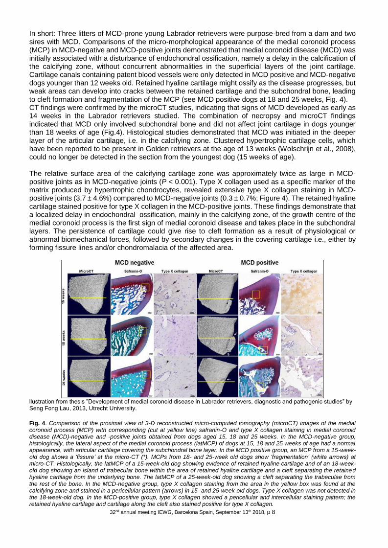
In short: Three litters of MCD-prone young Labrador retrievers were purpose-bred from a dam and two sires with MCD. Comparisons of the micro-morphological appearance of the medial coronoid process (MCP) in MCD-negative and MCD-positive joints demonstrated that medial coronoid disease (MCD) was initially associated with a disturbance of endochondral ossification, namely a delay in the calcification of the calcifying zone, without concurrent abnormalities in the superficial layers of the joint cartilage. Cartilage canals containing patent blood vessels were only detected in MCD positive and MCD-negative dogs younger than 12 weeks old. Retained hyaline cartilage might ossify as the disease progresses, but weak areas can develop into cracks between the retained cartilage and the subchondral bone, leading to cleft formation and fragmentation of the MCP (see MCD positive dogs at 18 and 25 weeks, Fig. 4). CT findings were confirmed by the microCT studies, indicating that signs of MCD developed as early as 14 weeks in the Labrador retrievers studied. The combination of necropsy and microCT findings indicated that MCD only involved subchondral bone and did not affect joint cartilage in dogs younger than 18 weeks of age (Fig.4). Histological studies demonstrated that MCD was initiated in the deeper layer of the articular cartilage, i.e. in the calcifying zone. Clustered hypertrophic cartilage cells, which have been reported to be present in Golden retrievers at the age of 13 weeks (Wolschrijn et al., 2008), could no longer be detected in the section from the youngest dog (15 weeks of age). The relative surface area of the calcifying cartilage zone was approximately twice as large in MCD-positive joints as in MCD-negative joints (P < 0.001). Type X collagen used as a specific marker of the matrix produced by hypertrophic chondrocytes, revealed extensive type X collagen staining in MCD-positive joints (3.7 ± 4.6%) compared to MCD-negative joints (0.3 ± 0.7%; Figure 4). The retained hyaline cartilage stained positive for type X collagen in the MCD-positive joints. These findings demonstrate that a localized delay in endochondral ossification, mainly in the calcifying zone, of the growth centre of the medial coronoid process is the first sign of medial coronoid disease and takes place in the subchondral layers. The persistence of cartilage could give rise to cleft formation as a result of physiological or abnormal biomechanical forces, followed by secondary changes in the covering cartilage i.e., either by forming fissure lines and/or chondromalacia of the affected area.
llustration from thesis ”Development of medial coronoid disease in Labrador retrievers, diagnostic and pathogenic studies” by Seng Fong Lau, 2013, Utrecht University.
Fig. 4. Comparison of the proximal view of 3-D reconstructed micro-computed tomography (microCT) images of the medial coronoid process (MCP) with corresponding (cut at yellow line) safranin-O and type X collagen staining in medial coronoid disease (MCD)-negative and -positive joints obtained from dogs aged 15, 18 and 25 weeks. In the MCD-negative group, histologically, the lateral aspect of the medial coronoid process (latMCP) of dogs at 15, 18 and 25 weeks of age had a normal appearance, with articular cartilage covering the subchondral bone layer. In the MCD positive group, an MCP from a 15-week-old dog shows a ‘fissure’ at the micro-CT (*). MCPs from 18- and 25-week old dogs show ‘fragmentation’ (white arrows) at micro-CT. Histologically, the latMCP of a 15-week-old dog showing evidence of retained hyaline cartilage and of an 18-week-old dog showing an island of trabecular bone within the area of retained hyaline cartilage and a cleft separating the retained hyaline cartilage from the underlying bone. The latMCP of a 25-week-old dog showing a cleft separating the trabeculae from the rest of the bone. In the MCD-negative group, type X collagen staining from the area in the yellow box was found at the calcifying zone and stained in a pericellular pattern (arrows) in 15- and 25-week-old dogs. Type X collagen was not detected in the 18-week-old dog. In the MCD-positive group, type X collagen showed a pericellular and intercellular staining pattern; the retained hyaline cartilage and cartilage along the cleft also stained positive for type X collagen.

32nd annual meeting IEWG, Barcelona Spain, September 13th 2018, p 9
Recently the connection between FCP in Labradors and a significant lowered content of zinc (Zn), copper (Cu) and sulphur (S) in hair has been published, reflecting a chronic malnutrition, malabsorption, dietary content or disturbed metabolism of the macro (sulphur) or micro (Zn and Cu) minerals (Davies et al, 2017). Since the Labrador-puppies litters developing MCD as described by Lau were raised on exactly the same food (Fig. 4; Lau et al 2013), a dietary cause seems not likely. In addition, all these pups affected and non-affected with MCD had the same Zn, Cu and S plasma content (R.J. Corbee, personal communication), and the G.Danes with severe osteochondrosis as described by Goedegebuure et al (1986) due to excessive Ca intake, had the same Cu and Zn liver content (Hazewinkel, personal observations ), reflecting chronic Cu and Zn status, comparable with hair analysis. MMP-9 is specifically inhibited by tissue inhibitor of matrix , which may be vital in the regulation of connective tissue metabolism and remodelling in numerous different organ systems, including hair follicle (Chun et al, 2016); this might be a possible explanation for delayed endochondral ossification and lowered mineral content of hair in case of FCP. Chondromalacia as described in cases of MCD, and revealing softening on probing of the joint cartilage due to thickened cartilage and/or necrosis of the subchondral bone, can be due to failure of blood vessel ingrowth (Wolschijn et al, 2005; Mairee et al, 2014), with secondary necrosis bordering the thickened or detached cartilage (in case of OCD), bordering the instable fragment of the fractured coronoid process, or bordering the area of disturbed endochondral ossification, thus indicating that this bone necrosis is secondary (Ytrehus 2004). The combination of necropsy and microCT findings in the Labrador puppies ( Lau et al 2013 ) indicated that MCD only involved subchondral bone; eventually it did cause fissure lines and fracturing of overlying joint cartilage in dogs older than 18 weeks of age (Fig. 4). The pathological findings of medial coronoid processes removed during subtotal coronoidectomy, is discussed by Prof Theyse at this meeting. The instability due to subchondral bone softening and/or joint cartilage fissuring/fracturing will eventually lead to joint irritation, synovial inflammation and pain. The pathological medial coronoid process can be detached by a variety of biomechanical causes, including(1) a disparity in the length of radius and ulna, as known entity in 80% of the Bernese Mountain dogs with ED (Lavrijsen et al, 2012), causes impairment of mechanical loading of radius and ulna with overloading of the ulna (Samoy et al, 2006). (2) an underdevelopment of the trochlear notch, as known entity in German Shepherd dogs
Fig. 5 The radial incisure of the ulna can be incongruent with the radial head (R) and thus causing increased pressure on the medial coronoid process causing fracturing especially in case of weakening of the subchondral bone. This incongruity of the subchondral bone can better be noticed on CT (left; arrow) than on artroscopy, artrotomy or gross pathology due to the covering cartilage layer (right) (courtesy Thesis Lau 2013).
(Wind 1986), causing pressure on both the coronoid process and the anconeal process causing UAP and/or FCP even in the same dog (Meyer-Lindenberg et al, 2003) (3) physiological incongruity (House et al, 2009) during weight bearing, (4) increased tensile forces during pronation by the annular ligament (Wolschijn et al, 2004), (5) repeated shear stress between the contact area of the proximal radial head and the axial border of the medial coronoid process during pronation and supination (Hulse, 2010), the latter possibly increased at the ulnar radial incisure by the biceps brachii/ brachialis muscle complex during supination (Fitzpatrick and Yeadon, 2009). This incisure is often irregular on CT pictures of the radioulnar joint in case of a proven fragmented coronoid process, although it should be realized that only the subchondral bone contour can be made visible on CT-scanning of the elbow joint (Fig. 5, courtesy Thesis Lau 2013)
Trauma as a cause of FCP in mature dogs has been published in a few papers. Meyer-Lindenberg et al 2002 published a study of FCP in total 332 affected joints and in 5 lame dogs a FCP without any radiological signs of OA in dogs >3 years of age; these 2% might have suffered a traumatic fragmentation of the coronoid, or the FCP has caused minimal arthritic changes (Meyer-Lindenberg et al 2002). In a more recent publication (Tan et al, 2016), 24 of 1011 dogs treated arthroscopically for ED with mean age of 48 months revealed a unilateral, single, large, displaced or non-displaced fracture of the medial coronoid process which was considered to be of traumatic origin, although disputed by others (von Pfeil

32nd annual meeting IEWG, Barcelona Spain, September 13th 2018, p 10
et al, 2017). This distinct group of dogs may have a better prognosis that the dysplasia-related dogs with a FCP and/or MCD (not proven yet) and thus may be of influence in the conversation between veterinary surgeon and owner; this notion is not of influence on grading the primary or secondary signs, since the ethiology does not play a role in grading the elbow joint for ED. In conclusion, the vulnerability for disturbance of the process of endochondral ossification, (either as breed characteristic, or caused by genetic factors related to MMP or other metabolic process, or as part of a pathological process due to non-genetic factors as nutrition), in combination with relative over-loading due to hyperactivity, elbow joint incongruities or increased tensile or compression forces can be the combined etiological background of the MCD. These factors can be of value to take into account for further molecular genetic research and breeding measures as well as for surgical intervention in clinical cases. References Alves-Pimenta S, Colaco B, Silvestre AM et al Prevalence and breeding values of ED in the Estrela mountain dog Vet Medicina 2013: 58; 484-490 Bellumori TP, Famula TR, Bannasch DL et al Prevalence of inherited disorders among mixed-breed and purebred dogs: 27,254 cases (1995-2010) JAVMA 242; 1549-1555 Beuing R, Mues CH, Tellhelm B et al Prevalence and inheritance of canine ED in German Rottweiler J Anim Breed and Genetics 2000: 117; 375-383. Cachon T, Genevois JP, Remy D et al Risk of simultaneous expression of hip and elbow dysplasia in dogs, a study of 1,411 radiographic examinations sent for official scoring VCOT 2010:23; 28-30 Chanoit G, Singhani NN, Marcelin-Little DJ et al. Comparison of five radiographic views for assessment of the medial aspect of the humeral condyle in dogs with osteochondritis dissecans. Am J Vet Res 2010; 71: 780-783 Chun H, Yong M, Xue W. et al Expression of matrix metalloproteinases and tissue inhibitor of matrix metalloproteinases in the hair cycle. Exp. Ther Med 2016; 12: 231-237 Coopman F, Verhoeven G, Saunders J et al Prevalence of HD, ED and humeral head OCD in dog breeds in Belgium. Vet Rec 2008; 163: 654-658 Coopman F, Broeckx B, Verelst E et al Combined prevalence of inherited skeletal disorders in dog breeds in Belgium VCOT 2014;27:395-397 Davies M, West J, Williams C et al. Selected minerals in hair of healthy dogs with or without medial coronoid process disease Vet Rec 2017; 180: 448-454. Fitzpatrick, N, Yeadon, R. Working algorithm for treatment decision making for developmental disease of the medial compartment of the elbow in dogs. Vet Surg 2009; 38: 285-300 Goedegebuure S, Hazewinkel H. Morphological findings in young dogs chronically fed a diet containing excess calcium. Vet. Pathol. 1986; 23: 594-605. Hazewinkel HAW, Kantor A, Meij BP, et al. Fragmented coronoid process and osteochondritis dissecans of the medial humeral condyle. Tijdschr Diergen. 1998; 113 Suppl 1, 41-46. Hazewinkel HAW, Meij BP, Theyse LF Surgical treatment of elbow dysplasia Vet Q 20 suppl I: 29-31. Hedhammar A, Wu F, Krook L et al. Overnutrition and skeletal disease; an experimental study in growing Great Dane dogs Cornell Vet 1974; 64 (suppl 5): 5-160 House MR, Marino DJ, Lesser ML. Effect of limb position on elbow congruity with CT evaluation. Vet Surg 2009; 38: 154-160 Hou Y, Wang Y, Lu X et al Monitoring hip and elbow dysplasia achieved modest genetic improvement of 74 dog breeds over 40 years in USA PLos ONE 2013: 8; 1-12 (e76390) Hulse D, Young B, Beale B, et al., Relationship of the biceps-brachialis complex to the medial coronoid process of the canine ulna. VCOT 2010; 23: 173-176. Kirberger RM Phenotypic hip and elbow dysplasia trends in Rottweilers and Labrador retrievers in South Africa (2007-2015): Are we making progress? J S Afr Vet Assoc. 2017; 88: 1-10 Lappalainen A. Radiographic Screening for Hereditary Skeletal Disorders in Dog, Thesis Helsinki University 2013 Lappalainen A, Mölsä s, Liman A et al Radiographic and computed tomography findings in Belgium shepherd dogs with mild elbow dysplasia Radiol Ultrasound 2009; 54; 364-369 Lang J, Busato A, Baumgarter D, Flückiger M, Weber UT. Comparison of two classification protocols in the evaluation of elbow dysplasia in the dog J Small Anim Pract 39; 169-174, 1998 Lau Seng Fong. Development of medial coronoid disease in Labrador retrievers: Diagnostic and pathogenic studies, Thesis Utrecht University 2013 Lau SF, Hazewinkel HAW, Grinwis GCM et al. Delayed endochondral ossification in early medial coronoid disease: a morphological and immune-histochemical evaluation in growing Labrador retrievers Vet J 2013;197:731-738 Lavrijsen IC, Heuven HCM, Voorhout G, et al. Phenotypic and genetic evaluation of ED in Dutch Labrador retrievers, Golden Retrievers, and Bernese Mountain dogs. Vet J 2012: 193; 486-492. Lavrijsen IC, Heuven HC, Meij BP et al. Prevalence and co-occurrence of HD and ED in Dutch pure-bred dogs. Prev Vet Med. 2014;114:114-122

32nd annual meeting IEWG, Barcelona Spain, September 13th 2018, p 11
Lavrijsen IC Genetics of HD, Ed and patellar luxation in purebred dogs Thesis Utrecht University 2014 Lewis TW, Ilska JJ, Blott SC et al Genetic evaluation of elbow scores and the relationship with hip scores in UK Vet J 2011; 187: 227-233. Mairee IC, Gröne A, Theyse LF The role of osteonecrosis in canine coronoid dysplasia; arthroscopic and histopathological findings Vet J 2014; 2014: 382-386. Meyer-Lindenberg A, Langham A, Fehr M et al. Prevalence of fragmented medial coronoid process of the ulna in lame adult dogs Vet Rec 2002; 151: 230-234. Meyer-Lindenberg A, Fehr M, Nolte I. Co-existence of ununited anconeal process and fragmented medial coronoid process of the ulna in the dog J Small Anim Pract 2006; 47: 61-65 Mostafa A, Nolte I, Wefstaedt P The prevalence of medial coronoid process disease is high in lame large breed dogs and quantitative radiographic assessments contribute to the diagnosis. Vet Radiol Ultrasound 2018; 1-13 Oberbauer AM, Keller GG, Famula TR. Long-term genetic selection reduced prevalence of HD and ED in 60 dog breeds PLoS ONE 12; e0172918 Oberbauer AM, Belanger JM, Bellumori DL et al. Ten inherited disorders in purebred dogs by functional breed groupings. Canine Genetics and Epidemiology 2015; 2: 9-12 Olsson, S. E. (1974) En ny typ av armbågsledsdysplasi hos hund? Svensk Veterinärtidning 26, 152-157 Paster ER, LaFond E, Biery DN et al Estimates of prevalence of HD in Golden Retrievers and Rottweilers and the influence on bias on published prevalence figures JAVMA 2005; 226: 387-392 Rau FC, Wigger A, Tellhelm B et al Observer variability and sensitivity of radiological diagnosis of canine medial coronoid disease Tierztl Prax Ausg Kleintiere Heimtiere 2011;39:313-322. Read RA, Armstrong SJ, Black AP et al Relationship between physical signs of ED and radiological score in growing Rottweilers. JAVMA 1996; 209: 1427-1430 Remy D, Neuhart L, Fau D, Genevois JP Canine elbow dysplasia and primary lesions in German Shepherd dogs in France. J Small Anim Pract 2004; 45: 244-248 Samoy Y, Gielen I, Van Caelenberg A, et al. Computed tomography findings in 32 joints affected with severe elbow incongruity and FMCP. Vet Surg 2012 41; 486-494 Tan DK, Canapp SO, Leasure CS et al Traumatic fracture of the medial coronoid process in 24 dogs VCOT 2016; 29: 325-329 Tirgari M. Clinical radiographical and pathological aspects of arthritis of the elbow joint in dogs. J Small Anim Pract 1974; 15: 671-679. Tryfonidou MA, Holl MS Stevenhagen JJ et al Dietary 135-fold cholecalciferol supplementation severely disturbs the endochondral ossification in growing dogs. Domest Anim Endocrinol 2003; 4: 265-285. Ubbink GJ, Hazewinkel HAW, van de Broek J, et al Familial clustering and risk analysis for fragmented coronoid process and elbow incongruity in Bernese Mountain dogs in The Netherlands. Am J Vet Res 1999 60; 1082-1087. Von Pfeil DJF, Albrecht M, Glassman M. A letter to the editor VCOT 2017; 30: 88 Wayne RK, Von Holdt BM, Evolutionary genomics of dog domestication Mamm Genome 2012; 23; 3-18 Wind AP, Packard ME, Elbow incongruity and developmental elbow diseases in the dog: part II. JAVMA 1986; 22: 725-730. Wolschrijn CF, Weijs WA. Development of the trabecular structure within the ulnar medial coronoid process of young dogs. The Anatomical Record. Part A, Discoveries in Molecular, Cellular, and Evolutionary Biology 2004; 278: 514-519. Ytrehus B, Ekman S, Carlson CS, et al. Focal changes in blood supply during normal epiphyseal growth are central in the pathogenesis of osteochondrosis in pigs. Bone 2004; 35: 1294-1306.

32nd annual meeting IEWG, Barcelona Spain, September 13th 2018, p 12
OVERVIEW OF ELBOW DYSPLASIA SCREENING IN SPAIN. Dr. A. Chico, DvM, MVM. AVEPA (Spanish Association of small animal veterinarians) is the most important organization of small animal veterinarians in Spain, with over 4.700 members. The AVEPA Orthopedics and Traumatology working group (G.E.V.O.) is one of the largest groups devoted to orthopedics in Europe, (more than 250 associates). Since 2007 GEVO carries out a service for Elbow Dysplasia screening. Almost 2.800 dogs have been evaluated throughout these years and the number of studies performed is rising quickly , with a mean of 325 dogs screened in the last five-year period as a consequence of the increasing concern of pet owners and breeders. Evaluating Committee is formed by three recognized experts in the subject during a 2- 4 year period. All breeds are examined by this panel. The cost of the service is 45€ and the results are usually ready in 45 days. With the dogs under sedation or anaesthesia two radiographs of each elbow are required, (one lateral 110-140º and one craniocaudal view). Minimum age should be 1 year. Sixty percent of the dogs evaluated were females.
The 10 most represented breeds are as follows: Rottweiler (17,3%), Labrador ( 17,3%), Border Collie (9,2%), Golden R. (7,8%), German Shepherd (4,2%), Swiss White Shepherd (3,6%), Great Dane (3,2%), Newfoundland (3,2%), Czech Shepherd (3,1%), Am Stafford (2,2%). Using IEWG scoring system, most of the dogs were classified as Grade 0 (87%) . Grade 1 accounted for 9%, Grade 2 for 2% and Grade 3 for 1% In the event of a claim by the owner, AVEPA accepts to revise the radiographs. If the result is finally changed, there are no extra charges for
the owner. In the opposite case the cost is 40 €. Usually no more than 3 claims per year are attended. There are other organizations in Spain that also run an elbow dysplasia scheme (AMVAC, SETOV), sometimes through agreements with certain breeds. Challenges for coming years are to expand the screening program to breeds now underrepresented making efforts to reach more breeders and canine clubs through lectures and online education. Additionally, all institutions involved in elbow dysplasia screening in Spain should work together so that more homogenous diagnosis can be achieved. Finally, the issue of accepting CT scans when there is profound disagreement between the reading panel and the owner has to be further discussed in our country since there is not an “official “ position yet.
0
100
200
300
400
20
07
20
08
20
09
20
10
20
11
20
12
20
13
20
14
20
15
20
16
20
17
1st
hal
f 2
01
8
NUMBER OF DOGS SCREENED (AVEPA-GEVO)

32nd annual meeting IEWG, Barcelona Spain, September 13th 2018, p 13
Strength and limitations of radiography, scintigraphy, ultrasound, CT, MRI and arthroscopy to diagnose Elbow Dysplasia in lame dogs.
Dr. I. Gielen, DVM, PhD, MSc, Dr. A. Villamonte‐Chevalier, DVM, PhD.
The diagnosis of elbow dysplasia (ED) in lame dogs is made from a combination of clinical signs, palpation of the joints, and medical imaging. A wide range of imaging options are now available but the “perfect” imaging protocol does not exist because each modality has his strengths and limitations. Although radiography is still the standard technique for diagnosing elbow disorders in the dog, other imaging techniques like scintigraphy, ultrasound, computed tomography (CT) and magnetic resonance imaging (MRI) can be useful.
In diagnosing ED there a two different issues: there is the need for selecting ED free breedingstock and there is the diagnosis of the condition in the individual patient presented for forelimb lameness. For selection purposes, most of the time the secondary degenerative joint (DJD) changes are scrutinised by means of radiographs and mostly the individuals are not suffering lameness. For the individual patient the early diagnosis of the primary lesion is very important because an early treatment guaranties a better prognosis. Although the most important cause of elbow lameness in dogs is medial coronoid disease (MCD), recently flexor enthesopathy (FE) has been recognized as an elbow disorder in medium and large breed dogs and is characterized by lesions of the medial epicondyle and the attaching flexor muscles. The differential diagnosis between both elbow disorders is not obvious and a combination of these two elbow diseases is possible. The challenge in these cases is to define the cause of the elbow pain in order to make the correct treatment decision. In both, MCD and FE, the radiographic features may be minimal and indistinct. A recent study, compared radiographic, CT and arthroscopic findings in a population of 90 dogs that presented with elbow lameness. Three standard radiographic views (lateral extension, lateral flexion and a 15° oblique cranio-medial caudo-lateral) when compared with CT presented a sensitivity of 97% and a specificity of 64%. In another recent study in 424 elbows screened for ED, the sensitivity and specificity were 65% and 93% respectively. Based on these results radiography remains a good imaging tool for screening purposes, however in cases where radiographic signs of MCD are not clear; CT remains as the imaging technique of choice
In cases where the clinical examination is not providing a clear localisation or in case of uncertain radiographic findings, scintigraphy is a useful technique to localise the cause of lameness. Although it is very sensitive, it is not very specific and the spatial resolution offered, is not well enough to specify anatomic structures. A micro-single photon emission tomography (μ-SPECT) technique has been described. HiSPECT has a much higher resolution and allows better differentiation of the anatomical areas in the elbow joint. A major drawback to joint imaging by scintigraphy is the normal uptake at the end of long bones, especially in immature animals. In some instances it is difficult to determine whether a difference in counts between two joints represents a meaningful finding. Comparison of bilateral images, acquired over the same time, and quantitative analysis of joint images by computer can provide diagnostic guidelines. In cases of flexor enthesopathy (FE), HiSPECT, reveals focal increased bone tracer uptake in the region of the medial humeral epicondyle. Ultrasound (US) is a potential valuable imaging technique of the musculoskeletal system in small animals. High frequency linear transducers are used because of their flat application surface and high resolution power. Accurate examination of joints requires substantial ultrasonographic experience and a standardised examination procedure. In most of the joints even small amounts of fluid accumulation (hypo- to anechoic) can be easily demonstrated in the area of the joint pouches. Although a thorough US study of the normal elbow joint has been conducted, US is only of limited use in the diagnosis of a fragmented coronoid process. Only large displaced fragments can be diagnosed with certainty. Also US is helpful in diagnosing flexor tendon pathology.

32nd annual meeting IEWG, Barcelona Spain, September 13th 2018, p 14
Computed Tomography (CT) can help significantly in establishing a definite diagnosis. The positioning of the patient is very important and CT of both elbow joints extended with the head pulled back outside the gantry results is better quality images and less artefacts. A recent study was performed, where lateral positioning (with both thoracic limbs extended symmetrically cranially and the head pulled back out of the gantry) was compared with sternal positioning. Results demonstrated that on lateral recumbency less artefacts appear on the images, on the other hand images from sternal recumbency presented in most cases streak artefacts. Moreover, the quality of images (differentiation between cortical and subchondral bone, bone structures contours) was superior on images obtained on lateral recumbency. Lateral positioning is in general more reliable when it comes to image quality and absence of artefacts which is highly significant in the accurate detection of discrete lesions. The scan parameters kV and mA should be high and thin slices with an overlap are preferred. Images should be obtained in bone algorithm and proper windowing during the evaluation of a study is a necessity. The modality of multiplanar reconstructions in different planes is useful in order to evaluate the complete joint surface. Abnormalities in the area of the medial coronoid process include: fragmentation (displaced or nondisplaced), fissure, abnormal shape, sclerosis, osteophytes, and lucencies. A recent study attempts to make objective the measurement of sclerosis. By means of CT, and specific regions of interest measurements, sclerosis can be evaluated in a reproducible way. The findings of this study suggest that an increase or a decrease of values of HU and bone density (BD) can be associated with the presence of elbow pathology. In particular that an increase in HU and BD values in regions such as the MCP base and MCP apex would be related to MCD. In the area of the medial humeral condyle sclerosis, lucency, and/or flattening can be evaluated and a differential diagnosis between kissing lesions and real OCD lesions can be made All these abnormalities can be diagnosed on the transverse and reconstructed images. In several cases CT findings, like fissures at medial coronoid process and subchondral luscencies at medial humeral condyle, were useful for decision making in the arthroscopic treatment of these lesions. A recent study shows that CT is a very reliable technique to evaluate fragmented coronoid process with a sensitivity and specificity of over 90% when compared to arthroscopy; whereas, the methods showed an almost perfect agreement (kappa = 0.959) between CT and arthroscopy which is still considered to be the “gold standard”. Ununited anconeal process with or without humeroulnar incongruity can be appreciated and the incidence of incongruities of the humeroradial, humeroulnar, and/or radioulnar joints can be accurately appreciated. On transverse CT slices, at the level of the trochlear notch of the ulna and the humerus, the fitting of the joint space can be noticed. On the reconstructions in the sagittal and dorsal plane, at the level of the trochlea humeri and the lateral compartment the incidence of a step between the ulna and radial head, the shape of the trochlear notch and the fitting of the humeral condyle in the trochlear notch can be evaluated. In cases of FE, the medial epicondyle appears sclerotic and shows a clear periosteal reaction in all cases. Mineralized opacities can be present within the flexor tendons. CT also shows concomitant lesions like coronoid disease whenever present. The soft tissue studies presents a thickening of the involved tendons and IV administration of contrast shows enhancement in the affected tendons and fluid pockets can easily be visualised. Arthro-CT can be used to evaluate loss of cartilage in cases of medial compartment syndrome.
Magnetic Resonance Imaging (MRI) has limitations for imaging the canine elbow based on
the relatively small size of the joint and complex articulations in conjunction with the thin articular cartilage surfaces of the humerus, radius, and ulna. These limitations depend also of the field strength of the MR device. All MRI planes, dorsal, sagittal, and axial/transverse, are potentially useful for diagnosis of elbow disorders. The incidence of subchondral bone pathology and oedema can be diagnosed. This technique offers a great visualisation of the soft tissues around the elbow joint and in cases of pathology within the flexor tendons its application can be very useful. On Magnetic Resonance Imaging (MRI), the sagittal T2- weighted sequence reveals a hyperintense signal around the proximal aspect of the flexor muscles extending in the muscle bellies. This signal can be confirmed as being a fluid signal on the fat suppressed STIR sequence. The T1 and T2 studies showed a thickening and irregular delineation of the involved tendons. There is obvious enhancement on T1 contrast studies.
32nd annual meeting IEWG, Barcelona Spain, September 13th 2018, p 15
As well as providing valuable diagnostic information about the elbow, arthroscopy also allows minimally-invasive treatment of coronoid disease. It allows us to obtain a magnified panoramic view of the inside of a joint. The drawback of arthroscopy is that it only allows the inspection of the articular surface. The combination of CT and arthroscopy allows a more complete diagnosis of ED. In cases of FE, arthroscopy shows the presence of loose fibres, degenerated tendinous tissue, cartilage loss and/or local synovitis at the attachment of the flexor muscles to the medial humeral epicondy Suggested reading: Y. Baeumlin, L. De Rycke, A. Van Caelenberg, H. Van Bree, I Gielen. Magnetic Resonance Imaging of the Canine Elbow: An Anatomic Study. Vet Surg. 2010, 39(5): 566-573. De Rycke LM, Gielen IM, van Bree H, et al. Computed tomography of the elbow joint in clinically normal dogs. American Journal of Veterinary Research 2002, 63: 1400-1407. de Bakker, Evelien, Gielen Ingrid, Kromhout Kaatje, van Bree Henri, and Van Ryssen Bernadette. Magnetic Resonance Imaging of Primary and Concomitant Flexor Enthesopathy in the Canine Elbow. Veterinary Radiology & Ultrasound 2014. 55 (1): 56–62. de Bakker, Evelien, Gielen Ingrid, Van Caelenberg Annemie, van Bree Henri, and Van Ryssen Bernadette. Computed Tomographic Findings of Canine Elbow Joints Affected by Primary and Concomitant Flexor Enthesopathy. Veterinary Radiology & Ultrasound 2014.55 (1): 45–55. de Bakker, Evelien; Saunders, Jimmy H.; van Bree, Henri; Gielen, Ingrid; Van Ryssen, Bernadette. Radiographic features of primary and concomitant flexor enthesopathy in the canine elbow. Vet Radiol Ultrasound. 2013;54(2):107-13. de Bakker E, Samoy Y, Gielen I, et al. Medial humeral epicondylar lesions in the canine elbow. A review of the literature. Vet Comp Orthop Traumatol 2011;24:9–17. Debruyn K, K Peremans, E Vandermeulen, B Van Ryssen, and J Saunders. Evaluation of semi- quantitative bone scintigraphy in canine elbows. Veterinary Journal 2013, 196:424–30. Griffon, Dominique J., Mostafa, Ayman A., Blond, L., Schaeffer, David J. Radiographic, computed tomographic, and arthroscopic diagnosis of radioulnar incongruence in dogs with medial coronoid disease. VETERINARY SURGERY Volume: 47, Issue: 3, Pages: 333-342. House, Mark R.; Marino, Dominic J.; Lesser, Martin L. .Effect of Limb Position on Elbow Congruity with CT Evaluation VETERINARY SURGERY , 2009, Volume: 38 Issue: 2 Pages: 154-160. Kunst CM, Pease AP, Nelson NC, Habing G, Ballegeer EA. Computed tomographic identification of dysplasia and progression of osteoarthritis in dog elbows previously assigned OFA grades 0 and 1. Veterinary Radiology and Ultrasound. 2014, 55(5): 511-20. Lamb CR, Wong K. Ultrasonographic anatomy of the canine elbow. Vet Radiol Ultrasound 46:319-25, 2005. S.F. Lau a,e,, C.F. Wolschrijn , H.A.W. Hazewinkel , M. Siebelt , G. Voorhout The early development of medial coronoid disease in growing Labrador retrievers: Radiographic, computed tomographic, necropsy and micro-computed tomographic findings. The Veterinary Journal 197 (2013) 724–730. Seng Fong Lau, Lars F.H. Theyse, George Voorhout, and Herman A.W. Hazewinkel. Radiographic, Computed Tomographic, and Arthroscopic Findings in Labrador Retrievers With Medial Coronoid Disease. Veterinary Surgery, 2014, 1–10. K. Peremans, S. Vermeire, A. Dobbeleir, I. Gielen, Y. Samoy, K. Piron, E. Vandermeulen, G. Slegers, H. van Bree, B. De Spiegeleer, K. Dik. Recognition of anatomical predilection sites in canine elbow pathology on bone scans using micro-single photon emission tomography. The Veterinary Journal 2011,188(1): 64-72. Samoy Y, Van Ryssen B, Gielen I, et al. Review of the literature: Elbow incongruity in the dog. Vet Comp Orthop Traumatol 2006; 19: 1-8. Seyrek-Intas D, Michele U, Tacke S, et al. Accuracy of ultrasonography in detecting fragmentation of the medial coronoid process in dogs. J Am Vet Med Assoc 2009;234:480–5. N. Shimizu, C. M. Warren-Smith, S. J. Langley-Hobbs, N. J. Burton, E. Kulendra, K. Bradley, E. Bowen, A. Holdsworth and K. J. Parsons. Inter- and intraobserver agreement in interpretation of CT features of medial coronoid process disease. Journal of Small Animal Practice2015, 56, 707–713. T.C. Tromblee, J.C. Jones, A.M. Bahr, et al. Effect of computed tomography display window and image plane on diagnostic certainty for characteristics of dysplastic elbow joints in dogs. American Journal of Veterinary Research 2007, 68: 858-871. van Bree H, Van Ryssen B. Diagnostic imaging of the canine elbow including radiology, arthroscopy and computed tomography (CT). Oral Abstracts, 10th IRVA Meeting. Veterinary Radiology and Ultrasound. 1994, 35: p 248, nr. 069.
32nd annual meeting IEWG, Barcelona Spain, September 13th 2018, p 16
van Bree H, Gielen I, Van Ryssen B, et al. Comparative joint imaging in small animals. The European Journal of Companion Animal Practice 2002, 12: 25-36. van Bree H, Gielen I, Van Ryssen B, De Rooster H. Early diagnosis of fragmented coronoid process in the dog: elbow arthroscopy compared to radiographic signs of degenerative joint disease. The European Journal of Companion Animal Practice 2012. 22(4). p.6-14. A. Villamonte Chevalier, H. van Bree, B.J.G. Broeckx, W. Dingemanse, M. Soler, B. Van Ryssen, I. Gielen. 2015. Assessment of Medial Coronoid Disease in 180 Canine Lame Elbow Joints: A Sensitivity and Specificity Comparison of Radiographic, Computed Tomographic and Arthroscopic Findings. BMC Veterinary Research 2015, 11:243, DOI 10.1186/s12917-015- 0556-9. A. Villamonte-Chevalier, W. Dingemanse, A. Van Caelenberg, B. Broeckx, H. van Bree, I. Gielen. 2016. Bone density of elbow joints in Labrador retrievers and Golden retrievers: Comparison of healthy joints and joints with medial coronoid disease. Veterinary Journal, 2016, 216, 1-7. A. Villamonte Chevalier , H. van Bree , B.J.G. Broeckx , W. Dingemanse, I. Gielen. 2016. Agreement between radiography and computed tomography for medial coronoid disease in the screening of 424 canine elbow joints. 6th CT-User Meeting, 2nd International Veterinary CT- User Meeting Ghent University-DVG, 9-10 December 2016, Ghent, Belgium

32nd annual meeting IEWG, Barcelona Spain, September 13th 2018, p 17
Clinical, diagnostic and pathological findings in Canine Elbow Dysplasia. Prof. dr. L.F.H. Theyse, DVM, PhD, Dipl. ECVS, MRCVS. Canine elbow dysplasia (CED) is the most common developmental disorder of the elbow joint and results in forelimb lameness in juvenile and adult medium and large breed dogs. CED is a syndrome which consists of several conditions, including fragmentation of the medial coronoid process of the ulna also described as coronoid dysplasia (CD), osteochondritis dissecans of the medial part of the humeral condyle, ununited anconeal process and elbow joint incongruity. These conditions can occur as single traits or in combination. Each can cause irreversible elbow osteoarthrosis (OA), due to bone and cartilage damage, medial joint instability, and chronic synovitis. Symptoms of CD may be subclinical initially but will result in marked lameness eventually. In the long term OA will develop progressively. Canine elbow dysplasia is a polygenic trait whereby both environmental and hereditary influences play a role in its development. The aetiology of CD is undetermined at present. Several hypotheses concerning the pathogenesis of CD have been proposed, most of which involve abnormal endochondral ossification and abnormal mechanical forces arising from incongruity of radius, ulna and humeral condyle. In addition, abnormalities in the bone structure and density of the coronoid have been described. Coronoid dysplasia usually presents as fragmentation of a central part of the medial coronoid process of the ulna with osteomalacia and chondromalacia as the most common arthroscopic findings. Coronoid dysplasia can result in fragmentation of the coronoid with a fragment in situ and partially intact joint cartilage or complete disruption with a displaced fragment. Coronoid dysplasia is presumed to start in immature animals and the first clinical signs of lameness due to CD may appear at four to six months of age. The dogs usually have a history of forelimb lameness. In contrast, CD is also seen in middle-aged and older dogs without any symptoms early in live and even without typical signs of OA. The diagnosis CD is based on clinical signs including joint distension, and pain during combined flexion, pronation, and supination of the effected joint. In more severe and chronic cases secondary signs of OA can be present. The radiographic evaluation of CD typically follows the guidelines of the International Elbow Working Group (IEWG. Computed tomographic (CT) evaluation enables more sensitive visualization of the coronoid process including multiplanar reconstructed images. Bony abnormalities associated with CD include structural changes, sclerosis, osteophytosis, fissure formation, and fragmentation. Arthroscopy is considered to be the ‘golden standard’ as it is the most valuable diagnostic and therapeutic tool for elbow dysplasia, due to its direct visualization and assessment of the articular cartilage and subchondral bone. A dual approach of CT assessment and arthroscopy of the elbow joint provides the most accurate evaluation of pathological changes in the coronoid. In general, CD is treated by removing fragments, dysplastic bone and cartilage using curettage and partial ostectomy. Based on our arthroscopic experiences during treatment of CD, we hypothesized that CD is primarily located in the subchondral bone and not in the cartilage. In a prospective study, we evaluate the arthroscopic changes in elbows affected by CD and assessed the dysplastic bone and cartilage removed during arthroscopic intervention using histopathology. The osteochondral samples collected during subtotal coronoidectomy, primarily showed osteonecrosis of subchondral bone. Vascular necrosis within the subchondral bone was a consistent finding. Articular cartilage appeared to be normal or presented with degenerative changes as chondron formation and focal necrosis. The osteonecrotic bone was mature lamellar bone implying initial normal bone formation within the coronoid with secondary necrosis. These findings support the hypothesis that CD primarily develops in the subchondral bone and that cartilage pathology is secondary to osteonecrosis. This also implies that evaluating elbow joint cartilage status using the modified Outerbridge score as a means of determining the severity of CD has clear limitations. The combination of mature necrotic bone and vascular necrosis would mean that vascular compromise precedes osteonecrosis and plays a critical role in the pathogenesis CD. This also means that physiological loading of weakened subchondral bone can result in fragmentation of the coronoid and degeneration of the associated cartilage. Local vascular compromise during the development of the elbow joint could also explain the malformation of

32nd annual meeting IEWG, Barcelona Spain, September 13th 2018, p 18
radial and ulnar joint surfaces resulting in elbow joint incongruity. These findings are consistent with the recent insights on the etiology of osteochondritis dissecans which is also part of CED. In a second study, we described the radiographic, CT and arthroscopic findings in different age groups of Labrador Retrievers diagnosed with CD and compared the ulnar subtrochlear sclerosis (STS) observed on radiographs with the ratio between the mean attenuation of the ulnar subtrochlear bone and the mean attenuation of the cortical bone measured on CT. Ulnar STS (88%) was the most common radiographic findings in dogs ≤12 months and blurring of the cranial edge of the medial coronoid process (67%) was the most common radiographic findings in dogs >12 months. In contrast with radiographic evaluation, CT imaging showed ulnar STS to be present in 56% of dogs ≤12 months and to be absent in dogs >12 months. This means that radiographic ulnar STS has to be interpreted as a summation of true intramedullary STS and osteophytosis. Fragmentation was the most common CT finding in both age groups with 94% in dogs ≤12 months and 67% in dogs >12 months. A displaced fragment (69%) was the most common arthroscopic finding in dogs ≤12 months whereas osteochondromalacia (53%) was the most common finding in dogs >12 months. Full-thickness cartilage lesions of the medial part of the humeral condyle consistent with modified Outerbridge score 4 were a consistent finding with an equal distribution in both age groups. Typically, these lesions are located on the humeral condyle without direct contact with the CD lesion in the standing and walking joint angle. Contact with the typical CD lesion does occur during flexion of the joint beyond 90 degrees as occurs when a dog lies down but also would mean that mechanical load is then minimal. This finding supports the idea that the cartilage damage on the medial part of the humeral condyle is also related to initial subchondral bone pathology linked to vascular compromise. Although several CT imaging studies have described changes in the radioulnar joint responsible for pronation and supination, arthroscopic findings have been focussing on the coronoid and humeral condyle. In the author’s experience many of the changes seen in CD can also be recognized in the central part of the radius adjacent to the medial coronoid. Although real fragmentation of the radius is rare, the presence of osteomalacia, osteonecrosis and chondromalacia is a consistent finding. Again a primary mechanical etiology would seem unlikely leaving vascular deficiency as a causative factor. An abnormal development of the medial part of the radius could also explain the typical incongruity of the elbow joint with a congruent lateral humeroradial joint in combination with central radioulnar and humeroradial incongruity In conclusion, vascular compromise causing deficiencies in bone development and bone metabolism ultimately leading to osteonecrosis and subchondral bone collapse seem to play a key role in the etiology of CD and associated elbow joint pathology. References: Lau SF, Theyse LF, Voorhout G, Hazewinkel HA. Radiographic, computed tomographic, and arthroscopic findings in labrador retrievers with medial coronoid disease. Vet Surg. 2015 May;44(4):511-20. Mariee IC, Gröne A, Theyse LF. The role of osteonecrosis in canine coronoid dysplasia: arthroscopic and histopathological findings. Vet J. 2014 Jun;200(3):382-6. Griffon DJ, Mostafa AA, Blond L, Schaeffer DJ. Radiographic, computed tomographic, and arthroscopic diagnosis of radioulnar incongruence in dogs with medial coronoid disease. Vet Surg. 2018 Apr;47(3):333-342. Rohwedder T, Fischer M, Böttcher P. In vivo fluoroscopic kinematography of dynamic radio-ulnar incongruence in dogs. Open Vet J. 2017;7(3):221-228. Franklin SP, Burke EE, Holmes SP. Utility of MRI for Characterizing Articular Cartilage Pathology in Dogs with Medial Coronoid Process Disease. Front Vet Sci. 2017 Feb 24;4:25. Davies M, West J, Williams C, Gardner DS. Mineral status in canine medial coronoid process disease: a cohort study using analysis of hair by mass spectrometry. Vet Rec. 2017 May 6;180(18):448.

32nd annual meeting IEWG, Barcelona Spain, September 13th 2018, p 19
Primary and secondary flexor enthesopathy (FE) in the canine elbow, diagnostics and therapy. Dr. I. Gielen, DVM, PhD, MSc. Although the most important cause of elbow lameness in dogs is medial coronoid disease (MCD), recently flexor enthesopathy (FE) has been recognized as an elbow disorder in medium and large breed dogs and is characterized by lesions of the medial epicondyle and the attaching flexor muscles. The differential diagnosis between both elbow disorders is not obvious and a combination of these two elbow diseases is possible. The challenge in these cases is to define the cause of the elbow pain in order to make the correct treatment decision. In both, MCD and FE, the radiographic features may be minimal and indistinct. The aim of this study is to describe the imaging features in dogs suffering FE. In literature radiographic signs of flexor pathology have mainly been described as a calcified body and less frequently as spur formation. Radiographically, an irregular outline of the medial humeral epicondyle, a calcified body and a spur are regarded as radiographic signs of flexor enthesopathy. These radiographic changes found in primary flexor enthesopathy are not significantly different from those found in concomitant flexor enthesopathy and can be incidental findings. However, radiography is unable to detect soft tissue pathology of the flexor muscles. The main ultrasonographic findings of flexor enthesopathy are pre-insertional hypoechoic swelling, outward bowing and thickening of the common tendon of the flexor muscles. The tendon appears to be heterogenous with decreased echogenicity and focal or diffuse areas of irregular fibrillar appearance and ill-defined margins with partial or complete tears. Additionally cortical irregularities at the medial epicondyle (spur formation) and intra-tendinous calcifications can be detected. HiSPECT, a refined scintigraphy technique, which enables more detailed anatomical localization of pathology within the elbow joint reveals focal increased bone tracer uptake in the region of the medial humeral epicondyle. Computerised tomography (CT) reveals new bone formation in all effected joints. The medial epicondyle appears sclerotic and shows a clear periosteal reaction in all cases. Mineralized opacity can be present within the flexor tendons. CT also shows concomitant lesions like coronoid disease whenever present. The soft tissue studies presents a thickening of the involved tendons in and IV administration of contrast shows enhancement in the affected tendons. On Magnetic Resonance Imaging (MRI), the sagittal T2-weighted sequence reveals a hyperintense signal around the proximal aspect of the flexor muscles extending in the muscle bellies. This signal can be confirmed as being a fluid signal on the fat suppressed STIR sequence. The T1 and T2 studies showed a thickening and irregular delineation of the involved tendons. There is obvious enhancement on T1 contrast studies. Arthroscopy shows the presence of loose fibres, degenerated tendinous tissue, cartilage loss and/or local synovitis at the attachment of the flexor muscles to the medial humeral epicondyle. Usually, treatment options derived from human medicine are performed: intra-‐articular injection of corticosteroids or surgical removal of the affected tissue.

32nd annual meeting IEWG, Barcelona Spain, September 13th 2018, p 20
Lateral radiographic view of an elbow affected by primary flexor enthesopathie. An irregular outline of the medial humeral epicondyle (black arrow), a small spur (white arrow) and a large, elongated calcified body (black arrowhead) are visible. A moderate degree of subtrochlear sclerosis is visible (black star).
After intravenous injection of contrast medium clear enhancement on the soft tissue CT image is seen within the flexor muscles (yellow circle). The calcification within the flexor muscles can be noticed (black arrow).
T2 weighted sagittal MRI image showing fluid between the flexor muscles (purple arrows).

32nd annual meeting IEWG, Barcelona Spain, September 13th 2018, p 21
References Y. Baeumlin, B. Van Ryssen, E. De Bakker, I. Gielen, J. Saunders, H. van Bree. 2009. Imaging findings in 5 dogs with insertional flexor tendon disorders. “25th International Workshop for Small Animal Arthroscopy, advanced course”, January 23-24th, 2009, Department of Medical Imaging and Small Animal Orthopaedics , Faculty of Veterinary Medicine, Ghent University, Merelbeke, Belgium. p. 14-16. de Bakker E, Samoy Y, Gielen I, Van Ryssen B. Medial humeral epicondylar lesions in the canine elbow: a review of the literature. Veterinary and Comparative Orthopaedics and Traumatology. 2011;24(1):9-‐17. de Bakker E, Saunders JH, Gielen I, van Bree H, Coppieters E, Van Ryssen B. Radiographic findings of the medial humeral epicondyle in 200 canine elbow joints. Veterinary and Comparative Orthopaedics and Traumatology 2012; 25(5): 359-65. E. De Bakker, I. Gielen, J. Sauders I. Polis, S.Vermeire, K. Peremans, J. Dewulf, H. van Bree, B. Van Ryssen. 2013. Primary and concomitant flexor enthesopathy of the canine elbow. Veterinary and Comparative Orthopaedics and Traumatology, 26, 6 :425-34. E. de Bakker, J. Saunders, H. Van Bree, I. Gielen, B. Van Ryssen. 2013. Radiographic features of primary and concomitant flexor enthesopathy in the canine elbow. Veterinary Radiology & Ultrasound, 54, 2, 107-13. E. de Bakker, I. Gielen, A. Van Caelenberg, H. van Bree, B. Van Ryssen. 2014. Computed tomography of canine elbow joints affected by primary and concomintant flexor enthesopathy. Veterinary Radiology & Ultrasound, 2014, 55, 1,45-55. E. de Bakker, I. Gielen, K. Kromhout, H. van Bree, B. Van Ryssen. 2014. Magnetic resonance imaging of primary and concomitant flexor enthesopathy in the canine elbow. Veterinary Radiology & Ultrasound, 2014, 55, 1, 56-62. Fox SM, Bloomberg MS, Bright RM. Developmental anomalies of the canine elbow. Journal of the American Animal Hospital Association. 1983;19:605-‐15.
Meyer-‐Lindenberg A, Heinen V, Hewicker-‐Trautwein M, Nolte I. Vorkommen und Behandlung von knochern Metaplasien in den am medialen Epikondylus des Humerus entspringenden Beugesehnen beim Hund. Tierarztliche Praxis Ausgabe Kleintiere Heimtiere. 2004;32:276-‐85. Morgan JP, Wind A, Davidson AP. Elbow Dysplasia. Hereditary Bone and Joint Diseases. Hanover: Manson Publishing Ltd; 2003. p. 41-‐94. Van Ryssen B, de Bakker E, Baeumlin Y, Samoy Y, Van Vynckt D, Gielen I, Ducatelle R, van Bree H. Primary flexor enthesopathy of the canine elbow: imaging and arthroscopic findings in eight dogs with discrete radiographic changes. Veterinary and Comparative Orthopaedics and Traumatology 2012; 25: 239-245.

32nd annual meeting IEWG, Barcelona Spain, September 13th 2018, p 22

32nd annual meeting IEWG, Barcelona Spain, September 13th 2018, p 23
International Elbow Working Group
The International Elbow Working Group [IEWG] was founded in 1989 by a small group of canine elbow experts from the USA and Europe to provide for dissemination of elbow information and to develop a protocol for screening that would be acceptable to the international scientific community and breeders. The annual meeting is organized for the purpose of exchanging information and reviewing the Protocol. All interested persons are invited to attend the meeting and to participate in its activities. The IEWG is an affiliate of the WSAVA. IEWG meetings were held in
Davis 1989 San Francisco 1990 Vienna 1991 Rome 1992 Berlin 1993 Philadelphia 1994 Konstanz 1995 Jeruzalem [cancelled] 1996 Birmingham 1997 Bologna 1998 Orlando 1999 Amsterdam 2000 Vancouver 2001 Granada 2002 Estoril 2003 Bangkok 2003 Rhodes 2004 Amsterdam 2005 Mexico 2005 Munich 2005 Prague 2006 Munich 2007 Dublin 2008 Sao Paulo 2009 Bologna 2010 Amsterdam 2011 Birmingham 2012 Cape Town 2014 Bangkok 2015 Vienna 2016 Verona 2017
IEWG 2018 president Herman Hazewinkel [email protected] treasurer Bernd Tellhelm [email protected] secretary Thijs How [email protected]
website: www.vet-iewg.org