capk-phosphorylation controls the interaction of the regulatory domain of cardiac myosin binding...
TRANSCRIPT

cAPK-phosphorylation controls the interaction of the regulatory domainof cardiac myosin binding protein C with myosin-S2 in an on-o¡ fashion
Mathias Gruen, Heino Prinz, Mathias Gautel1;*Max-Planck-Institute fu«r molekulare Physiologie, Abt. Physikalische Biochemie, Otto-Hahn-Strasse 11, 44227 Dortmund, Germany
Received 31 March 1999; received in revised form 19 May 1999
Abstract Myosin binding protein C is a protein of the myosinfilaments of striated muscle which is expressed in isoformsspecific for cardiac and skeletal muscle. The cardiac isoform isphosphorylated rapidly upon adrenergic stimulation of myocar-dium by cAMP-dependent protein kinase, and together with thephosphorylation of troponin-I and phospholamban contributes tothe positive inotropy that results from adrenergic stimulation ofthe heart. Cardiac myosin binding protein C is phosphorylated bycAMP-dependent protein kinase on three sites in a myosinbinding protein C specific N-terminal domain which binds tomyosin-S2. This interaction with myosin close to the motordomain is likely to mediate the regulatory function of the protein.Cardiac myosin binding protein C is a common target gene offamilial hypertrophic cardiomyopathy and most mutationsencode N-terminal subfragments of myosin binding protein C.The understanding of the signalling interactions of theN-terminal region is therefore important for understanding thepathophysiology of myosin binding protein C associatedcardiomyopathy. We demonstrate here by cosedimentationassays and isothermal titration calorimetry that the myosin-S2binding properties of the myosin binding protein C motif areabolished by cAMP-dependent protein kinase-mediated tris-phosphorylation, decreasing the S2 affinity from a Kd of WW5 WWMto undetectable levels. We show that the slow and fast skeletalmuscle isoforms are no cAMP-dependent protein kinasesubstrates and that the S2 interaction of these myosin bindingprotein C isoforms is therefore constitutively on. The regulationof cardiac contractility by myosin binding protein C thereforeappears to be a `brake-off' mechanism that will free a specificsubset of myosin heads from sterical constraints imposed by thebinding to the myosin binding protein C motif.z 1999 Federation of European Biochemical Societies.
Key words: Myosin binding protein C; Myosin regulation;Protein interaction; Phosphorylation
1. Introduction
Myosin binding protein C (MyBP-C) is a modular muscleprotein of the intracellular immunoglobulin superfamily (re-viewed in [1]) which is expressed in at least three isoforms, itscardiac isoform being strictly speci¢c for heart muscle inmammals [2,3]. The C-terminal region interacts with the lightmeromyosin portion (LMM) of myosin as well as with titin,thus anchoring the protein to the thick ¢lament shaft and
specifying its sarcomeric localization [1]. MyBP-C is localizedto the central region of the thick ¢lament (C-zone) in regularstripes spaced 43 nm apart [4^6]. There are 11 C-zone stripes.However, in mammalian muscle, only stripes 3^11 are usuallyoccupied by MyBP-C, stripe 3 can also be occupied by MyBP-H. The proteins of stripes 1 and 2 are unknown. Due to the 43nm spacing, only every third level of myosin heads in the C-zone associated with a MyBP-C molecule [4] and the functionof the protein may involve cooperative changes over largerdistances. Furthermore, the localization of MyBP-C to thecentral region of the thick ¢lament implies that its unknownregulatory functions can only a¡ect myosin heads when theoverlap of actin and myosin ¢laments extends into the C-zone.The presence of the protein may therefore help to sense thedegree of actomyosin overlap.
Cardiac MyBP-C is phosphorylated in a dynamic way bycAMP-dependent protein kinase (cAPK), suggesting a role inthe L-adrenergic regulation of muscle contraction [7^12].Phosphorylation occurs on three sites in a MyBP-C-speci¢cdomain in the N-terminal region, the MyBP-C motif [12]. Thisregion is highly conserved between all isoforms of MyBP-Cand between species [12,13]. The high degree of sequence iden-tity and cross-species conservation between various MyBP-Cisoforms suggests that the function of the MyBP-C motif isspeci¢ed by the conserved regions and that the three phos-phorylation sites found experimentally in the cardiac isoformrepresent an additional feature. We could show recently thatthe MyBP-C motif binds to the proximal 126 residues of themyosin-S2 segment, close to the lever-arm domain of the my-osin head [13]. This segment of myosin-S2 is almost com-pletely identical between all sarcomeric myosin isoforms andvertebrate species [13]. The interaction with MyBP-C is henceindependent of the MyBP-C or myosin isoform and theMyBP-C motifs of both cardiac as well as slow skeletalMyBP-C were demonstrated experimentally to bind to theN-terminal S2 segment with comparable a¤nities [13]. Theinteraction with S2 directs the A-band association of N-ter-minal cardiac MyBP-C fragments in neonatal rat cardiomyo-cytes [13,14]. It has been proposed that the interaction withmyosin-S2 could modulate the head-tail motility of the two-headed sarcomeric myosin [13], based on the observation thatantibody Fab-fragments against the same region of S2 con-strain the movement of the myosin heads [15]. The control ofsuch a constraint by phosphorylation of MyBP-C might there-fore release this `brake' and recruit myosin heads for activeforce production, possibly in a cooperative way along severalcrowns of myosin heads.
MyBP-C has been shown to be one major protein involvedin the pathophysiology of familial hypertrophic cardiomyop-athy [1]. However, its physiological role is still poorly under-stood. Most MyBP-C mutations that cause FHC are pre-
0014-5793 / 99 / $20.00 ß 1999 Federation of European Biochemical Societies. All rights reserved.PII: S 0 0 1 4 - 5 7 9 3 ( 9 9 ) 0 0 7 2 7 - 9
*Corresponding author. Fax: (49) (6221) 387 306.E-mail: [email protected]
1 Present address: European Molecular Biology Laboratory, Struc-tural Biology Division, Postfach 10 22 09, 69012 Heidelberg, Ger-many.
FEBS 22182 18-6-99
FEBS 22182FEBS Letters 453 (1999) 254^259

dicted to result in N-terminal fragments of the protein [1].Therefore, the e¡ect of cardiac MyBP-C phosphorylation onthe interaction with myosin-S2 is of great importance tounderstand the basis of the regulatory mechanism of this pro-tein and to gain insight into possible pathological mechanismsin FHC.
2. Materials and methods
2.1. Bu¡ersBu¡er A (myosin mini¢laments): Tris-citrate pH 8.0 10 mM. Bu¡er
B (cosedimentation assay): Tris-HCl pH 7.0 20 mM, NaCl 200 mM,DTE 1 mM, EDTA 1 mM. Bu¡er C (isothermal titration calorimetry(ITC)): MES/NaOH pH 7.0 20 mM, NaCl 50 mM, DTE 1 mM,EDTA 1 mM)
2.2. Protein expression, puri¢cation and phosphorylationMyBP-C and L-myosin-S2 fragments were expressed soluble as de-
scribed [12,13,16] using the pET expression system [17]. His6-taggedproteins were puri¢ed on Ni2� NTA columns following the manufac-turer's instructions (Qiagen, Germany) and further puri¢ed by anionexchange chromatography on a monoQ column (Pharmacia, Sweden).The His6 tag was cleaved o¡ by recombinant TEV protease (GibcoBRL, UK). Myosin (rabbit, skeletal) was prepared according to themethod of [18]. Myosin mini¢laments were prepared as described [19]from myosin which was previously puri¢ed by three cycles of high-and low salt precipitation. The content of endogeneous MyBP-C afterthis scheme was estimated by Western blotting using a speci¢c MyBP-C antibody [3] and by gel densitometry to be below 5%.
Preparative phosphorylation of cardiac C1C2 (c-C1C2) was carriedout in a volume of 10 ml at a concentration of 1 mg/ml c-C1C2,100 U/ml bovine brain cAPK catalytic subunit (Sigma, Germany)and 0.7 mM ATP in 20 mM HEPES pH 7.0, 5 mM MgCl2, 1 mMdithiothreitol at 25³C. Samples of 5 Wl were taken at regular intervalsand the degree of phosphorylation was monitored by mass spectrom-etry. Complete tris-phosphorylation was observed after about 3 h.
2.3. Cosedimentation assayAll proteins (except myosin mini¢laments) were dialyzed into bu¡er
B and centrifuged prior to use at 4³C for 30 min at 400 000Ug using aBeckman TLA 100.1 rotor and an Optima TL ultracentrifuge. Appro-priate amounts were mixed in Beckman polycarbonate centrifugetubes (number 343776) and the volume was made up to 25 Wl withbu¡er B. Myosin mini¢laments (25 Wl of 5 WM in bu¡er A) wereadded to the mixture to give a ¢nal volume of 50 Wl, pH 7.3, and aNaCl concentration of 100 mM. The mixture was incubated at 4³Cfor 30 min and subsequently centrifuged for 20 min at 400 000Ug.The supernatant was removed and the pellet was washed twice with100 Wl of a 1:1 mixture of bu¡er A and bu¡er B. The pellet wasredissolved in 50 Wl of 7 M urea and appropriate amounts of super-natant and pellet were analyzed by SDS-PAGE as described [20]. Gelswere stained for 24 h in staining solution (1.5% w/v Coomassie bril-liant blue R250, 40% ethanol, 10% acetic acid) and subsequently de-stained for 48 h.
2.4. ITCAll proteins were dialyzed into bu¡er C and centrifuged at
100 000Ug for 10 min immediately prior to use. Calorimetric experi-ments were carried out using a titration calorimeter from Microcal,USA, with a 250 Wl injection syringe while stirring at 400 rpm. Theconcentrations of the myosin-S2 constructs in the syringe were gen-erally 10^20 times higher than the C1C2 concentrations in the reac-tion cell. The reference cell was ¢lled with 1 mM sodium azide. Aninitial injection was performed with a small volume of 0.25 times theexperimental injection volume (5^10 Wl). Data analysis was performedwith the manufacturer's software. Experimental values for the bindingconstant, heat of binding and stoichiometric ratio are from deconvo-lution using non-linear least-squares minimization. The heats of dilu-tion were determined in independent experiments and subtracted fromthe raw data prior to data analysis.
2.5. Mass spectrometryNano-electrospray mass spectroscopy was performed with a Finni-
gan LCQ mass spectrometer equipped with a micromanipulator for
the correct positioning of the nanospray needle. The needles and theion source were made following the original design of Mann et al.[21]. In short, capillaries (GC120F from Clark Instruments, Reading,UK) were pulled using the DMZ-Universal puller (Zeitz-InstrumenteGmbH, Mu«nchen, Germany). They were coated with gold using aPolaron SC7610 Sputter Coater (VG Microtech, Uck¢eld, UK) anda gold target. These needles were ¢lled with 1^4 Wl of aqueous so-lution and centrifuged for several seconds using a PicoFuge (TomyCapsule, Tomy Tech, Palo Alto, CA, USA). The needles weremounted on the micromanipulator and were carefully brought in con-tact with the surface of the mass spectrometer. Air pressure was ap-plied to the needle until a small droplet appeared on the tip. Theneedle was then adjusted 2.5 mm opposite to the inlet of the heatedcapillary of the mass spectrometer. A potential of 900 V was appliedto the needle and the spectrum was recorded. The original data (i.e.abundance of the multiple charged protein populations versus mass/charge) were deconvoluted into mass spectra (relative abundance ver-sus mass) by means of the deconvolution software BioExplore (Fin-nigan). The error of the mass spectrometer was 100 ppm in the nor-mal mass range (150^2000 mu), which was used for all experimentsshown here.
3. Results
3.1. Phosphorylation by cAPK is restricted to the cardiacisoform of MyBP-C
Phosphorylation by cAPK as well as by an associated cal-cium-calmodulin-dependent protein kinase is well-documentedfor cardiac MyBP-C. We wanted to establish experimentallywhether also the known skeletal isoforms from slow and fastmuscle contain accessible substrate sites for cAPK which arenot evident from multiple sequence comparison [12,13,22]. Wetherefore expressed the C1C2 fragments of human fast andslow skeletal MyBP-C (f-C1C2 and s-C1C2), which containthe phosphorylatable MyBP-C motif [12], and compared theirsubstrate properties for cAPK phosphorylation with c-C1C2.We observed phosphorylation for the c-C1C2 fragment butnot for the two skeletal isoforms (Fig. 1). Quantitative anal-ysis of the kinetics of c-C1C2 phosphorylation was carried outby mass spectrometry under preparative conditions. After2 min of phosphorylation, conversion to the monophospho-rylated form was visible with no detectable bis- or tris-phos-
Fig. 1. Phosphorylation by cAPK results in phosphate incorporationinto the cardiac MyBP-C motif (lanes 1 and 4), but not the slow(lanes 2 and 5) or fast (lanes 3 and 6) skeletal isoforms. This dem-onstrates that adrenergic regulation can act exclusively on the car-diac isoform. Lanes 1^3: Coomassie-stained gel; Lanes 4^6: autora-diograph of duplicate. M: marker proteins.
FEBS 22182 18-6-99
M. Gruen et al./FEBS Letters 453 (1999) 254^259 255
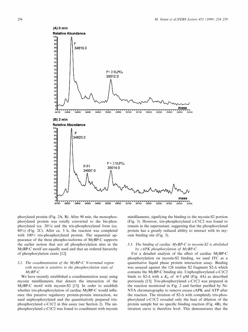
phorylated protein (Fig. 2A, B). After 90 min, the monophos-phorylated protein was totally converted to the bis-phos-phorylated (ca. 20%) and the tris-phosphorylated form (ca.80%) (Fig. 2C). After ca. 5 h, the reaction was completedwith 100% tris-phosphorylated protein. The sequential ap-pearance of the three phospho-isoforms of MyBP-C supportsthe earlier notion that not all phosphorylation sites in theMyBP-C motif are equally used and that an ordered hierarchyof phosphorylation exists [12].
3.2. The cosedimentation of the MyBP-C N-terminal regionwith myosin is sensitive to the phosphorylation state ofMyBP-C
We have recently established a cosedimentation assay usingmyosin mini¢laments that detects the interaction of theMyBP-C motif with myosin-S2 [13]. In order to establishwhether tris-phosphorylation of cardiac MyBP-C would in£u-ence this putative regulatory protein-protein interaction, weused unphosphorylated and the quantitatively prepared tris-phosphorylated c-C1C2 in this assay (see Section 2). The un-phosphorylated c-C1C2 was found to cosediment with myosin
mini¢laments, signifying the binding to the myosin-S2 portion(Fig. 3). However, tris-phosphorylated c-C1C2 was found toremain in the supernatant, suggesting that the phosphorylatedprotein has a greatly reduced ability to interact with its my-osin binding site (Fig. 3).
3.3. The binding of cardiac MyBP-C to myosin-S2 is abolishedby cAPK phosphorylation of MyBP-C
For a detailed analysis of the e¡ect of cardiac MyBP-Cphosphorylation on myosin-S2 binding, we used ITC as aquantitative liquid phase protein interaction assay. Bindingwas assayed against the 126 residue S2 fragment S2-v whichcontains the MyBP-C binding site. Unphosphorylated c-C1C2binds to S2-v with a Kd of W5 WM (Fig. 4A) as describedpreviously [13]. Tris-phosphorylated c-C1C2 was prepared inthe reaction monitored in Fig. 2 and further puri¢ed by Ni-NTA chromatography to remove excess cAPK and ATP afterthe reaction. The titration of S2-v with completely tris-phos-phorylated c-C1C2 revealed only the heat of dilution of theprotein sample but no speci¢c binding reaction (Fig. 4B), thetitration curve is therefore level. This demonstrates that the
FEBS 22182 18-6-99
M. Gruen et al./FEBS Letters 453 (1999) 254^259256

interaction of c-C1C2 with myosin-S2 is abolished by tris-phosphorylation or reduced to levels undetectable by ITC orcosedimentation.
4. Discussion
Regulation of protein-protein interactions by phosphoryla-tion is a common mechanism to control cell function in eu-karyotes. Both the response of assembled cellular structures aswell as their assembly itself are regulated by phosphorylationof speci¢c proteins by dedicated kinases. Sarcomeres, the con-tractile units of muscle, represent a complex and dynamicmacromolecular assembly that responds to changes in phys-iological demands on the molecular level. Long-term regula-tion is achieved by changes in gene expression patterns [23],whereas short-term dynamic adaptations are mediated by pro-tein phosphorylation. MyBP-C is one of three proteins rapidly
phosphorylated when myocardium is stimulated by adrenergicagonists [10]. This stimulation results in an increase in con-traction force and speed.
We demonstrated recently that the regulatory domain of c-MyBP-C interacts with a short segment of myosin-S2 close tothe motor domain. This interaction raises the possibility thatthe regulation of cardiac contractility by MyBP-C could in-volve the mobility of the two myosin heads at their head-tailjunction [13]. Recent ultrastructural analysis of phospho-rylated and unphosphorylated native thick ¢laments suggesteda swinging out of myosin heads concomitant with the phos-phorylation of cardiac MyBP-C [24,25].
We demonstrate here that the interaction of the N-terminalregulatory domain of cardiac MyBP-C (MyBP-C motif) withthe myosin-S2 domain is sensitive to cAPK phosphorylationof the MyBP-C motif. The interaction of both proteins, asjudged from cosedimentation assays and ITC, is regulated in
Fig. 2. Time-course of the sequential phosphorylation of c-C1C2 by cAPK. The control (reaction mixture at 0 min) shows the unphosphoryl-ated protein (P) with a mass of 34 819 Da and several ion adducts (A). After 2 min incubation, monophosphorylated protein appears (P-P1)(B). After 90 min, the pools of unphosphorylated and monophosphorylated protein have disappeared and both bis- and tris-phosphorylatedprotein (P-P2 and P-P3) are detected (C). After 5.5 h, the reaction led to a complete conversion to tris-phosphorylated protein P-P3).
FEBS 22182 18-6-99
M. Gruen et al./FEBS Letters 453 (1999) 254^259 257

an on-o¡ fashion by tris-phosphorylation. It is possible, how-ever, that in vivo phosphorylation intermediates may occurwhich result in a more subtle change in a¤nity. This on-o¡regulation suggests that whatever steric constraints would beimposed by MyBP-C either on the mobility of the myosinhead-tail junction or the swinging out of the myosin headfrom the thick ¢lament shaft, would be relieved by MyBP-Cphosphorylation. This could (i) recruit heads for actin bindingand hence force generation or (ii) alter the binding state andcycling rates of the two-headed myosin. Both mechanismswould be synergistic to the increased generation of activeforce that is achieved by the cAPK-mediated phosphorylationof sarcomeric proteins in cardiac muscle. The major phos-phorylation site in cardiac MyBP-C is a cardiac-speci¢cloop insertion in the MyBP-C motif of the sequenceLAGGGRRIS [12]. Removal of this site restricts the accessi-bility to the two other cardiac cAPK sites. All three sites arenot found in the skeletal isoform sequences. Since sequencecomparison may overlook physiologically relevant phospho-rylation sites, we tested the skeletal isoforms of MyBP-C ex-perimentally for cAPK phosphorylation and found them to beno substrates. Recent work identi¢ed two phosphorylationsites for protein kinase C (PKC) in the cardiac MyBP-C motif[22]. These sites are also not found in the skeletal isoforms.Furthermore, investigations on intact heart muscle show noPKC-mediated phosphorylation of MyBP-C under physiolog-ical conditions [26,27]. Therefore, the selected interaction ofMyBP-C with only few myosin heads must have a ¢bre-type-dependent modulating e¡ect on contractility independent ofthe phosphorylation-dependent on-o¡ switch of this interac-tion, which is a speci¢c feature of cardiac muscle.
All isoforms of MyBP-C bear a speci¢c sequence at the veryN-terminus which, in the cardiac isoform, comprises a pro-line-alanine rich stretch and an additional Ig domain [16]. In
addition, a short isoform-speci¢c charged sequence is found inthe MyBP-C motif [13]. The interactions of these regions areyet unknown. Some sequence homology to the N-terminalregions of regulatory light chains (not shown) and the spatialproximity to the myosin light chains suggest that this regionmight contribute to isoform-speci¢c regulation possibly inconcert with the regulatory light chain.
It is interesting to note that at least one point mutation inL-myosin-S2 which causes FHC has an identical net e¡ect onthe interaction of myosin and MyBP-C. The exchange of ly-sine-924 for glutamate also abolishes their interaction withouta¡ecting the dimeric coiled-coil structure of S2 [13]. Possibly,
Fig. 3. Cosedimentation binding assay of unphosphorylated (lanes1 and 2) and tris-phosphorylated c-C1C2 (lanes 3 and 4) to myosinmini¢laments. Supernatants: lanes 1 and 3; pellets: lanes 2 and 4.Whereas the unphosphorylated control binds to myosin and isfound in the pellet (lane 2), tris-phosphorylated c-C1C2 does not co-sediment and stays in the supernatant of the reaction (lane 3).M: marker proteins.
Fig. 4. The binding of c-C1C2 to myosin-S2 is regulated by phos-phorylation. Isothermal calorimetry demonstrates the liquid phaseinteraction of S2-v with C1C2. S2-v is titrated against C1C2 in reg-ular time intervals and the reaction heat is monitored as a changeof the heating current (Wcal/s, upper panels). Integration of the re-sulting peaks yields the titration plot (lower panel). (A) 20 WMc-C1C2 and 350 WM S2-v, (B) 20 WM tris-phosphorylated c-C1C2and 350 WM S2-v. The experiments demonstrate the interaction ofboth proteins in a 1:1 complex in the unphosphorylated state andyield a dissociation constant of 5.2 WM for the interaction (A). Thebinding of both proteins is abolished after tris-phosphorylation of c-C1C2. only the heat of dilution is detectable (B).
FEBS 22182 18-6-99
M. Gruen et al./FEBS Letters 453 (1999) 254^259258

therefore, some of the FHC-associated mutations in L-myo-sin-S2 could result in the constitutive mimicry of the phos-phorylated state of MyBP-C and result in chronic dysregula-tion of contractility. Indeed, hypercontractility is a featureobserved in several mutations causing FHC [28]. Our resultsprovide the molecular basis for a functional investigation ofthe phosphorylation regulation of cardiac contractility in nor-mal and diseased heart by the interaction of myosin andMyBP-C.
Acknowledgements: we are greatly indebted to Roger Goody for hisgenerous support. We are most grateful to the EMBL for providingadministrative support that made this project possible. Supported bythe Deutsche Forschungsgemeinschaft (Grant Ga405/3-6).
References
[1] Carrier, L., Bonne, G. and Schwartz, K. (1998) Trends Cardio-vasc. Med. 8, 151^157.
[2] Fougerousse, F., Delezoide, A.L., Fiszman, M.Y., Schwartz, K.,Beckmann, J.S. and Carrier, L. (1998) Circ. Res. 82, 130^133.
[3] Gautel, M., Fu«rst, D.O., Cocco, A. and Schia¤no, S. (1998)Circ. Res. 82, 124^129.
[4] O¡er, G. (1972) Cold Spring Harbor Symp. Quant. Biol. 37, 87^95.
[5] Craig, R. and O¡er, G. (1976) J. Mol. Biol. 102, 325^332.[6] Bennett, P., Craig, R., Starr, R. and O¡er, G. (1986) J. Muscle
Res. Cell Motil. 7, 550^567.[7] Jeacocke, S. and England, P. (1980) FEBS Lett. 122, 129^132.[8] Hartzell, H.C. and Titus, L. (1982) J. Biol. Chem. 257, 2111^
2120.
[9] Hartzell, H.C. and Glass, D.B. (1984) J. Biol. Chem. 259, 15587^15596.
[10] Garvey, J.L., Kranias, E.G. and Solaro, R.J. (1988) Biochem. J.249, 709^714.
[11] Schlender, K.K. and Bean, L.J. (1991) J. Biol. Chem. 266, 2811^2817.
[12] Gautel, M., Zu¡ardi, O., Freiburg, A. and Labeit, S. (1995)EMBO J. 14, 1952^1960.
[13] Gruen, M. and Gautel, M. (1999) J. Mol. Biol. 286, 933^949.[14] Sebillon, P., Bonne, G., Flavigny, J., Venin, S., Wisnewsky, C.,
Vikstro«m, K., Leinwand, L., Carrier, L. and Schwartz, K. (1998)Circulation 98, 235.
[15] Knight, P.J. (1996) J. Mol. Biol. 255, 269^274.[16] Freiburg, A. and Gautel, M. (1996) Eur. J. Biochem. 235, 317^
323.[17] Studier, F.W., Rosenberg, A.H., Dunn, J.J. and Dubendor¡,
J.W. (1990) Methods Enzymol. 185, 60^89.[18] Margossian, S.S. and Lowey, S. (1982) Methods Enzymol. 85,
55^71.[19] Reisler, E., Smith, C. and Seegan, G. (1986) J. Mol. Biol. 143,
129^145.[20] Laemmli, U.K. (1970) Nature 227, 680^685.[21] Mann, M. and Wilm, M. (1995) TIBS 20, 219^224.[22] Mohamed, A.S., Dignam, J.D. and Schlender, K.K. (1998) Arch.
Biochem. Biophys. 358, 313^319.[23] Schia¤no, S. and Reggiani, C. (1996) Physiol. Rev. 76, 371^423.[24] Weisberg, A. and Winegrad, S. (1996) Proc. Natl. Acad. Sci.
USA 93, 8999^9003.[25] Weisberg, A. and Winegrad, S. (1998) Circ. Res. 83, 60^72.[26] Edes, I. and Kranias, E.G. (1990) Circ. Res. 67, 394^400.[27] Talosi, L. and Kranias, E.G. (1992) Circ. Res. 70, 670^678.[28] Bonne, G., Carrier, L., Richard, P., Hainque, B. and Schwartz,
K. (1998) Circ. Res. 83, 580^593.
FEBS 22182 18-6-99
M. Gruen et al./FEBS Letters 453 (1999) 254^259 259