conserved neural mechanisms for regulation of food …
TRANSCRIPT

CONSERVED NEURAL MECHANISMS FOR REGULATION OF FOOD INTAKE IN FLY
AND RODENT MODELS
(Under the Direction of Ping Shen)
ABSTRACT
Obesity and excess body fat are the result of caloric intake that exceeds metabolic need, and their
development is correlated with insufficient exercise and overeating. Evolutionary pressures tend
to promote biological mechanisms which enhance an organism’s ability to obtain nutrients and
minimize excessive energy utilization. However, the worldwide trend toward positive energy
balance has led to an increased prevalence of obesity. It is a quiet killer, predisposing individuals
to a variety of diseases including cardiovascular and metabolic disorders, increasing rates of
morbidity and mortality (1). The financial consequences of obesity in terms of medical expenses
and loss of productivity has deleterious social costs (2).
In order to understand the mechanisms that promote overeating and obesity at the behavioral
level, it is informative to make use of invertebrate and mammalian models of feeding behavior.
Using a Drosophila larvae model system, we have demonstrated that norepinephrine-like
octopamine (OA) neurons in the fly are acutely required for homeostatic food intake regulation,
and selectively enhance intake of sweet, palatable foods. The feeding regulatory effects of OA
neurons were determined to dependent on Octβ3R and OAMB receptors, respectively. In
addition, we have demonstrated that OA neurons are regulated by a vascular endothelial growth
factor receptor (dVegfr, or Pvr), whose activity is required for OA dependent feeding rate
increases (3). Based on the homology of the fly OA and norepinephrine systems in mammals,
and the similarity of fly dVegfr to mammalian VEGFR2, we hypothesized that VEGF/VEGR2
may have a homologous role mammalian food intake regulation. To investigate this possibility,
young rats were treated intracerebroventrically (i.c.v.) with a single dose of VEGFR2 Tyrosine
Kinase Inhibitor V (VTKI-V). Treated rats displayed decreased food intake and weight gain.
These effects are selectively manifested in different feeding regimens. Animals treated with
VTKI-V showed markedly attenuated consumption of palatable food that is sweet and fatty.
Meal pattern analysis reveals that this is achieved by consumption of smaller, shorter meals.
Overall, our results suggest that VTKI-V treatment may interfere with two separate VEGFR-
mediated mechanisms: one which promotes overconsumption of palatable food, and the other
mediates body weight gain.
INDEX WORDS: Octopamine, norepinephrine, neuropeptide Y, neuropeptide F, Oamb, Octβ3R,
vascular endothelial growth factor receptor (VEGFR), subesophogeal ganglion, food intake,
appetitive motivation, hyperphagia, palatable diet, Drosophila melanogaster, rodents.

CONSERVED NEURAL MECHANISMS FOR REGULATION OF FOOD INTAKE IN FLY
AND RODENT MODELS
by
Audrey Eileen Branch
A Dissertation Submitted to the
Graduate Faculty of the University of Georgia
In Partial Fulfillment of the Requirements for the Degree
DOCTOR OF PHILOSOPHY
Athens, GA
December, 2014

© 2014
Audrey Branch
All Rights Reserved

CONSERVED NEURAL MECHANISMS FOR REGULATION OF FOOD INTAKE IN FLY
AND RODENT MODELS
by
Audrey Eileen Branch
Major Professor: Ping Shen
Committee Members: James Lauderdale
Silvia Giraudo
Gaylen Edwards
Electronic Version Approved:
Julie Coffield
Interim Dean of the Graduate School
University of Georgia
December 2014

iv
DEDICATION
This work is dedicated to Meredith, Trip, Joyce, and Drew, my wonderful family, and to Ana
and Peter, for helping me get through it.
“Always you have been told that work is a curse and labour a misfortune.
But I say to you that when you work you fulfil a part of earth's furthest dream, assigned to you
when that dream was born,
And in keeping yourself with labour you are in truth loving life,
And to love life through labour is to be intimate with life's inmost secret.
You have been told also life is darkness, and in your weariness you echo what was said by the
weary.
And I say that life is indeed darkness save when there is urge,
And all urge is blind save when there is knowledge,
And all knowledge is vain save when there is work,
And all work is empty save when there is love;
Work is love made visible.”
~Khalil Gibran, “On Work”

v
ACKNOWLEDGEMENTS
I would like to express my deepest appreciation to all of those who assisted me with the
experiments described in these pages, and those who helped me as a person along the way.
For my labmates: Thank you: to Ting Zhang for your mentorship and friendship, to Yonghua
Wang for your humor, insight, and instructing me to think for myself, to Yuhan Pu for always
being willing to share your knowledge and skills, and always being willing to help everyone
around you, to Yiwen Zhang for your energy, determination, wit, and for reminding me to try
new things, and to Melissa Masserant for all your help and positive energy. For the members of
the Lauderdale lab (Rebecca, Barbara, Kenji, Vani, VJay, Lindsey, Ana, and Tyler) for always
being willing to give help and advice, sometimes on very short notice. To Kimberly Freeman and
Kathie Wickwire, I cannot express how grateful I am for your time, help, and expertise. Thank
you for assisting me with both my successes and my struggles, and for teaching me how to learn
from both. Thank you Ariel VanLeuvan for your unwavering dedication to learning and helping.
You are a bright light that inspires people around you to do their best and be their best.
I am enormously grateful for the support of my committee, all of whom have gone above and
beyond in my years working with them to assist me as a researcher and a person. Thank you to
Ping Shen for your mentorship, for allowing me take my project in different directions, for your
humor and insight, for teaching me how to think, design, troubleshoot, fix, and write (concisely),
and for your extraordinary patience and creativity. Thank you to Silvia Giraudo for your
patience, instruction, for making my rodent work possible, for the use of your lab space and
feeding cages, and for making me believe I could succeed. Thank you to Gaylen Edwards for

vi
your astounding breadth and depth of knowledge, for always being willing to share it and your
expertise, for use of your lab space, and for taking so much time to listen and instruct. Thank you
to James Lauderdale for the countless times you made things possible, for the use of your lab
space, for encouraging me, for listening, for finding people to help me, for helping me to put it
all in perspective, and for trusting and believing in me.

vii
TABLE OF CONTENTS
Page
ACKNOWLEDGMENTS …………………………………………………................... v
LIST OF FIGURES………………………………………………………. …………….. ix
CHAPTER 1: INTRODUCTION………………………………………………………. 1
CHAPTER 2: OCTOPAMINE-MEDIATED CIRCUIT MECHANISM UNDERLYING
CONTROLLED APPETITE FOR PALATABLE FOOD IN DROSOPHILA
2.1 INTRODUCTION……………………………………………………………….. 22
2.2 RESULTS………………………………………………………………………… 24
2.3 DISCUSSION……………………………………………………………………. 31
2.4 MATERIALS AND METHODS……………………………………………….. 34
CHAPTER 3: AN OAMB/NPF PATHWAY IN LARVAL SOG PREVENTS SWEET
FOOD OVERCONSUMPTION
3.1 INTRODUCTION……………………………………………………………….. 61
3.2 RESULTS AND DISCUSSION……………………………………………….... 63
3.3 MATERIALS AND METHODS……………………………………………….. 66
viii
CHAPTER 4: PREVENTION OF PALATABLE DIET-INDUCED HYPERPHAGIA
IN RATS BY CENTRAL INJECTION OF A VEGFR KINASE INHIBITOR
4.1 INTRODUCTION……………………………………………………………….. 78
4.2 RESULTS………………………………………………………………………… 79
4.3 DISCUSSION……………………………………………………………………. 83
4.4 MATERIALS AND METHODS……………………………………………….. 86
CHAPTER 5: DISCUSSION AND GENERAL CONCLUSIONS…………………… 101
REFERENCES…………………………………………………………………………… 117

ix
LIST OF FIGURES
Figure 2.1. Quantification of hunger-driven feeding responses of Drosophila larvae…. 39
Figure 2.2. Control of appetite for liquid sugar food by the OA system…………………. 41
Figure 2.3. Functional mapping of tdc2-Gal4 neurons by targeted laser lesion……….. 43
Figure 2.4. The neural activity of drk in appetitive motivation……………………………. 45
Figure 2.5. A unique neural activity of the Pvr pathway in appetitive motivation……… 47
Figure 2.6. Schematic model for differential regulation of two hunger-driven appetitive
motivations………………………………………………………………………………………… 49
Figure 2.S1. Quantification of hunger-driven feeding responses to liquid sugar food…. 51
Figure 2.S2. Role of tdc-Gal4 neurons in hunger-driven feeding responses to liquid
sugar food…………………………………………………………………………………. 52
Figure 2.S3. NPF activity is dispensable for hunger-driven feeding of liquid
sugar food………………………………………………………………………… 53
Figure 2.S4. Functional mapping of tdc2-Gal4 neurons by targeted laser lesion……….. 54
Figure 2.S5. Characterization of the neural activity of drk in larval feeding response
to solid sugar food………………………………………………………………………. 55
Figure 2.S6. Characterization of the activity of Pvr in larval feeding responses…………. 56
Table 2.S1. Functional analysis of four OA receptors via RNA interference dsRNA specific to
individual OA receptors………………………………………………………………….. 58

x
Table 2.S2. Functional analysis of the activities of 14 receptor tyrosine kinases in tdc-Gal4
neurons via RNA interference…………………………………………………………… 59
Table 2.S3. Feeding responses to liquid sugar food by fasted Pvf1, Pvf2, and
Pvf3 larvae………………………………………………………………………… 60
Figure 3.1. Oamb is required for suppression of food intake in fed larvae……………….. 70
Figure 3.2. Oamb mutant larvae display enhanced basal rates of feeding………………… 72
Figure 3.3. Oamb is required in npf-Gal4 and oaFS-Gal4 neurons for feeding
suppression.……………………………………………………………………………… 74
Figure 3.4. NPF SOG neurons are required for suppression of sweet food intake and co-express
Oamb ………………………………………………………………………………………………. 76
Figure 4.1. Cumulative daily effects on body weight and intake following treatment….. 91
Figure 4.2. Five day average light and dark phase differences in meal variables……… 93
Figure 4.3. Meal pattern for ten consecutive light periods of rats fed CHOW or
HC diet…………………………………………………………………………… 95
Figure 4.4. Effects of Drosophila UAS/Gal4 mediated dVegfr knockdown on adult
body size…………………………………………………………………………. 97
Figure 4.5. Conditioned taste aversion test…………………………………………………… 99

1
CHAPTER 1:
INTRODUCTION
You can tell a lot about a fellow’s character by the way he eats jellybeans ~ Ronald Reagan.
The developed world is in the midst of an epidemic of obesity. A great deal of research has been
performed in the study of food intake in both human and rodent models, providing evidence of
environmental, genetic, epigenetic, endocrine, and neural mechanism which may predispose
individuals to weight gain and obesity and contribute to its manifestation and persistence.
However, in the case of humans, it is abundantly clear that the obesogenic environment of the
developed world plays an enormous role as it limits energy expenditure and provides easy access
to cheap, tasty, high energy foods. The specter of starvation has been a vastly greater threat to
human survival over the course of our evolution, thus mechanisms for preventing overeating
during abundance have been subjected to less pruning. There is a great interest and need for
thorough characterization of biological systems involved in the prevention of palatable food
overconsumption in order to combat and prevent obesity.
Due to the difficulty of controlling the many factors involved in the development of obesity,
animal models, particularly mice and rats and more recently the fruit fly, have served as
reasonable surrogate models. Earlier studies making use of broad physical, pharmacological, and
electrolytic techniques for disruption of neural circuit components identified key regions of the
hypothalamus and hindbrain as critical regulators of energy homeostasis. Such work revealed the

2
presence of homeostatic control centers within the brain which operate to match short term and
long term food intake with metabolic output such that body weight remains stable over time
despite variations in food availability and quality. In addition, it is now increasingly recognized
that the brainstem and several cortico-limbic systems coordinating cognitive, hedonic, and
emotional neural processes also play important roles in food selection and ingestion (4).
While these studies provide important evidence of the identity of the anatomical location of
neural substrates promoting or preventing food intake and weight gain, they were limited in their
capacity to elucidate circuit level connectivity and physiologically relevant circuit activity. In
recent decades, the advent of genetic tools for targeting and manipulating neural circuits at the
cellular level has allowed for a greater spatial and temporal control over circuit dynamics. While
our map of feeding neurocircuitry remains far from complete, it has become clear that the
division of food intake regulation into “homeostatic” and “hedonic” categories is an
oversimplification. Both homeostatic and hedonic circuits appear to adaptively respond to
palatable food consumption. Furthermore, there is a considerable amount of cross-talk between
hypothalamic, cortical, and limbic structures, and both are responsive to circulating nutritive and
endocrine factors(4). The predisposition of individuals to overeating and obesity can
theoretically develop from any pathological disruption or maladaptation of this complex system
to alterations of internal and external factors.
Summary of Primary Findings
This introduction will provide an outline of the major theories of how short and long term body
weight and food intake are controlled, followed by an overview of the main neurotransmitter
systems which are investigated in the experiments. The main chapters will describe the

3
characterization of an invertebrate neural circuit required for control of homeostatic control of
palatable food intake. The circuit relies on signaling by norepinephrine-like octopamine (OA)
neurons in the larval subesophogeal ganglion, which acts as an integration center between
olfactory and gustatory inputs and the expression of ingestive motor patterns. The activity of
separate subsets of these neurons are required for the efficient re-establishment of energy balance
following food deprivation, and for the suppression of palatable food consumption during satiety.
These selective roles are linked to separate downstream pathways mediated Octβ3R and
Oamb/NPF, respectively. Furthermore, we have identified an upstream regulatory pathway
mediated by a VEGF/VEGFR/Ras-like signaling molecules which is required in OA neurons for
appropriate modulation of palatable food intake. This finding prompted an investigation into the
role of the VEGFR receptor in the rodent brain, which demonstrated that inhibition of this
receptor results in reduced body weight gain and prevention of diet induced hyperphagia by
reducing the caloric size of meals. The relevance of these findings to current knowledge
regarding the relevant systems is presented in the discussion.
Motivated Behaviors
In order for a species to survive, the individual of the species must be able to execute adaptive
motivated behaviors such that its internal needs are resolved with its external environment. This
requires that individuals engage in goal-oriented behaviors that allow it to obtain resources
necessary for its survival, such as food, water, and sexual partners, as well as to avoid potentially
harmful foods or predators (5). “Drives” are excitatory pressures exerted by homeostatic
disturbances (biological, emotional, social, and/or cognitive) which have the power to compel an
individual to coordinate their behaviors in order to obtain a particular goal (6). Motivated

4
behaviors are those that are guided by internal drives in order obtain internal or external goals.
As they are guided by internal forces, such behaviors present an inherent difficulty as objects of
study: the underlying internal variables driving motivated behaviors, such as thirst or hunger, are
not directly observable or measurable. Thus, in order to attempt to identify and characterize the
biological mechanism(s) underlying motivated behaviors, scientists must focus on the observable
phenomena that result from internal states (7). The three broad classes of motivated behaviors
that are essential for the survival of an organism and/or species are ingestion, defense, and
reproduction.
The Meal
Like other classes of motivated behaviors, ingestive behaviors occur in cycles or phases. Though
the character and sequence of the behaviors performed are highly individualized and species
specific, the main phases are similar. Ingestive behaviors begin with an initiation phase, followed
by procurement, consumption and termination, and one full cycle of these phases is referred to as
a meal (8). The meal initiation phase begins with the physiological deficit signals encoding
internal energy status, which are integrated with sensory input. Together this information is
centrally processed to encode the incentive value of the goal object, in this case palatable
foodstuffs. The output of this processing provides a value for the “drive” associated with feeding,
and this is the basis for selection of an appropriate motor program to execute in order to obtain
the goal object, which begins the “procurement” phase. When food is located via sensory
mechanisms, the consummatory phase begins, which involves the execution of stereotypical
rhythmic motor programs such as chewing and swallowing such that the individual is able to
consume the selected food. During this phase, the animal structures its meal (in terms of

5
duration, quantity ingested, etc.) via dynamic integration of sensory and endocrine inputs which
communicate with central mechanisms regulating motor output. As the consummatory phase
continues, internal sensory feedback signals arise that either sustain ingestive behaviors or
promote termination of consumption. Termination occurs either when satiation is achieved or at
any time during this sequence if a second motivated behavior becomes more imperative or
appropriate (8).
Like other animals, humans and other mammals eat in an episodic fashion, and these episodes
are referred to as “meals” or “snacks” (9, 10). Once a meal is initiated, it tends to continue until a
sensation of “fullness” or “satiation” is attained, which is followed by a non-feeding period
referred to as satiety (11, 12). During the satiety phase, the drive to eat is very low, and this drive
must build up again in order to initiate the next feeding episode (13). The internal force
providing the drive to search, chose, and ingest food is referred to as “appetite” (13).
The cycle of ingestive behaviors is also demonstrated in invertebrate species, such as
Drosophila. During both larval and adult stages, flies execute a number of distinct, highly
stereotyped, and quantifiable behaviors relating to meal initiation, procurement, ingestion, and
termination. For example, the proboscis extension reflex in adult flies can be stimulated by the
application of sweet compounds to the chemosensory bristles of the leg, which is used in assays
of meal initiation and food preference (14). In the larval stages, actively foraging larvae feed
steadily when provided with soft, sweet media. Larval food ingestion rate and feeding
preferences can be assessed by the use of counting the rate of bites or measuring the intake of
dyed feeding media (3, 15, 16), while their persistence in continuing to feed on a particular
media can be used as a readout of foraging and procurement (15). Using assays such as these,

6
researchers have investigated mechanisms underlying both homeostatic and hedonic food intake
regulation in the fly which share homology with those found in vertebrates.
Short and Long Term Regulation of Body Weight and Food Intake in Mammals and Flies
Energy homeostasis is a balance between energy intake, expenditure, and storage, which is
achieved by continual dialogue between multiple brain and body communication networks. The
concept was first proposed 60 years ago by Kennedy (17), who described energy homeostasis as
a system whereby circulating signals provide the brain information regarding available energy
stores, and in response the brain makes corrective adjustments to food intake and energy
expenditure. When a negative energy balance occurs within an organism, nutrient deprivation
signals from the periphery are relayed to brain where several overlapping circuits are triggered to
implement foraging, appetitive, and ingestive behaviors to obtain nutrients and to simultaneously
transition to an energy conservation mode via autonomic, endocrine, and peripheral cellular
mechanisms (18). In mammals, the hypothalamus and dorsal vagal complex of the hindbrain are
two neural regions that are critical in regulating these responses. These neurons project to areas
important to homeostatic maintenance via behavioral, physiological, and endocrine effects (19-
21). The disregulation of these pathways has been demonstrated in mice experiencing diet
induced obesity (20, 22), ob/ob mice (23), and in diabetic patients (24-26).
Mammalian Systems
In the mammal homeostatic system, afferent projections from peripheral organs as well as
humoral signals, peptides, converge on neurons in the hypothalamus, hindbrain, and other areas
which form an interwoven yet distributed system of neurocircuits (27). For each of its regulatory
7
functions, the hypothalamus senses environmental or body signals and uses this information to
organize an appropriate response, and subsequently to command other brain regions to
implement that response. Ultimately, the autonomic nervous system is recruited to implement the
appropriate responses via descending autonomic projections originating the in the hypothalamus
and brainstem (28). In regards to the regulation of food intake, nutrient deprivation signals are
sent to the hypothalamus to implement foraging, appetitive, and ingestive behaviors to obtain
nutrients and to simultaneously transition to an energy conservation mode via autonomic,
endocrine, and peripheral cellular mechanisms (18). Specialized metabolic sensors within these
regions receive and integrate these inputs and respond by coordinating various downstream
systems involved in food intake, metabolism, and energy storage. One such signal is leptin, a
hormone secreted by adipocytes into plasma at levels that are proportional to body fat stores
(29), enters the brain in proportion to its level in plasma (30), and acts on key neurons that
regulate energy balance (31, 32). The pancreatic hormone insulin is another example, as it also
circulates at levels proportional to body fat (33) and directly acts on neural targets to reduce food
intake (34), while reduced neuronal insulin signaling results in a mild expansion of fat mass (35).
Leptin and insulin are thus both implicated in this adiposity negative feedback control system,
although the feeding effect of leptin is quantitatively much greater than that of insulin (36). Both
leptin and insulin cross the blood brain barrier and interact with neurons in the arcuate nucleus of
the hypothalamus (ARC) which produce and secrete neuropeptide Y (NPY), agouti-related
peptide (AgRP), and proopiomelanocortin (POMC) (37-40).
Early studies based on gross anatomical lesions or neuropharmacological manipulations
identified key areas in the brain mediate hyper or hypophagic behaviors, including the PVN,
LHA, ARC, and hindbrain adrenergic bundles. The ARC is located in the periventricular region
8
of the hypothalamus, where the blood-brain barrier is relatively weak, allowing access to
circulating signals relating to energy balance. This location allow the ARC to act as a relay
center through which information regarding internal energy status and external energy options
are integrated. The ARC receives strong intrahypothalmic inputs from other peri- and
paraventricular nuclei and LHA, and sensory inputs (olfactory, taste, visual), but in regards to
food intake, the most important inputs are hormonal (27). Leptin from adipocytes provides input
to the ARC regarding long term energy status, acting centrally as a potent inhibitor of food
intake. Direct administration of leptin to the brain reduces food intake and body weight gain (41,
42), while reduced or impaired leptin signaling promotes hyperphagia and weight gain (43, 44).
In addition, the neurons in the ARC are receptive to ghrelin produced by the enteroendocrine
cells of the stomach in response to fasting. Ghrelin promotes food intake, and therefore acts
opposite to leptin and insulin (28). The ARC also receives input regarding immediately available
energy stores via neurons which are responsive to insulin, glucose, free fatty acids, and other
metabolites circulating in the plasma (27). Though other regions of the brain and hypothalamus
may also be sensitive to these signals, ARC projections to the paraventricular nucleus (PVN) and
lateral hypothalamic area (LHA) are key to transmitting their effects on energy balance and food
seeking behavior (28).
In terms of food intake regulation, the ARC contains two major populations of neurons. The first
are neurons that release orexigenic peptides, such as those expressing neuropeptide Y (NPY) and
agouti-related protein (AgRP), which project the PVN and LHA in the periventricular zone (27).
These neurons express both the leptin and ghrelin receptors, which act to inhibit or activate the
release of NPY/AgRP, respectively. The second major group are those that release anorexgenic
peptides including those expressing pro-opiomelanocortin (POMC) and cocaine- and

9
amphetamine regulated transcript (CART). Melanocortins, such as α-melanocyte stimulating
hormone (α-MSH), are processed from the POMC gene. CART and α-MSH are both
anorexigenic peptides, and their expression levels positively correlate with circulating levels of
leptin (45), while POMC neuron activity is inhibited by activation of the ghrelin receptor (46).
NPY producing neurons in the ARC project to the paraventricular nucleus (PVN) and lateral
hypothalamic area (LHA) (47, 48) where NPY binding to its Y1 receptors triggers increases in
food intake and reduction in energy expenditure (49). POMC neurons also project to the PVN
and LHA where they release alpha-melanocyte-stimulating hormone (α-MSH) (50), which acts
via melanocortin-4 receptor neurons to decrease intake and increase expenditure (51). These
neurons also respond to AgRP released from ARC NPY/AgRP neurons, which acts as an
antagonist of the functions of α-MSH signaling through melanocortin receptors (38). Together,
leptin and insulin signaling in the ARC acts as a negative feedback system which prevents
against weight gain and obesity. Increases in adipose tissue results in an increase in leptin and
insulin levels, which then inhibit orexigenic NPY/AgRP neurons while activating the
anorexogenic pathway mediated by POMC neurons (40).
Numerous short term hormonal and nutrient related signals also potently influence feeding, such
as gut peptides involved in the perception of satiety and thus promote termination of individual
meals. The decision to initiate a meal is influenced by many external factors, while the amount
eaten (meal size) is primarily determined by internal signals (36). The major internal signals
affecting meal size are peptides, such as cholecystokinin (CCK) and GLP-1 which are secreted
from gastrointestinal tract in response to food ingestion (52, 53). Satiety information is conveyed
by these peptides as well as by neural signals generated by gastric distention, to the CNS through
afferent fibers of the vagus nerve that project from the gut to the nucleus of the solitary tract

10
(NTS) in the caudal hindbrain to trigger short lived food intake inhibition (36) at the level of the
meal.
Long term regulation of food intake is dependent in part on adiposity negative feedback systems
which reduce food intake in part by increasing brain responsiveness to satiety signals (54), such
as leptin and insulin, while weight loss lowers the plasma levels of these same hormones, thereby
increasing meal size by reducing the satiating effect of food (36). For example, leptin enhances
the brain’s response to satiety signals (55), which decreases meal size (56); conversely, low
levels of leptin signaling reduces the responsiveness of central circuits to CCK, which leads to an
increase in meal size (54). This interaction between leptin and satiety signals is dependent on
activation of leptin receptors in the hindbrain (NTS), and hypothalamic neurons (ARC nucleus)
which project directly or indirectly to the NTS (40). Ultimately, this interaction enables food
consumption during individual meals to be adjusted to compensate for changes in body fat mass
(36).
Drosophila Systems
The fly fat body is homologous to adipose tissue in mammals, acting as a storage compartment
for lipids and as a regulator of lipid homeostasis. Carbohydrate and lipid homeostasis in the fly
are regulated by insulin and adipokinetic hormone (AKH) which performs a similar function as
mammalian glucagon (57). Insulin producing cells (IPCs) are located in the median
neurosecretory region of the brain, and act in a manner similar to that of pancreatic beta cells,
while AKH is produced by corpora cardiac (CC) cells in the neuroendocrine ring gland, which
correspondingly act as pancreatic alpha cells (57). Like their mammalian counterparts, insulin is
upregulated following food ingestion and acts to promote uptake of sugar from circulating
11
hemolymph for storage as glycogen and fats, while AKH is secreted during starvation and breaks
down glycogen and fats (58). IPCs produce several isoforms of insulin-like peptides (dILPs;
isoforms dILP2 dILP3 and dILP5 are produced by IPCs) (59, 60). dILPs are secreted when
nutrient levels are high, and ablation of IPCs or dILP expression in these cells disrupts
carbohydrate homeostasis (61), body size regulation (62, 63), and feeding behavior (16). The fly
fat body responds to nutrient deprivation triggers mobilization of fats by remotely stimulating
AKH release from CC cells, which is then secreted into hemolymph to target peripheral energy
stores (58).
Like mammals, adult flies and larvae must determine when to eat, what to eat, and how much to
eat, and these decisions are determined by the coordinated actions of peripheral signals of
indicating the levels of internal energy stores, as well as external cues of nutrient availability.
The decision to initiate a meal is largely determined by the detection of nutritive compounds in
the environment by gustatory receptor neurons (64, 65), while detection of noxious stimuli such
as aversive taste (66) or temperature (67) elicit rejection behaviors. Once a feeding period is
initiated, the rate of ingestion and duration are dynamically modulated based on internal
metabolic status via circulating hormones from neuroendocrine cells and fat body tissue such as
the Drosophila insulin-like peptides (16, 61), adipokinetic hormone (58), and leptin homolog
Unpaired 2 act as coding mechanisms for the availability of carbohydrate and lipid stores (57).
Post-ingestive feedback from the gut following a meal may also inhibit feeding via the recurrent
nerve or medial abdominal nerve (68), acting similarly to gut distention signals in mammals.
In previous investigations in our lab, we have developed a behavioral experimental paradigm to
evaluate larval food intake and willingness to work for food (66). These studies demonstrated
that, like other animals, fly larvae demonstrate adaptive behavioral responses to foods which are

12
dependent on both their internal satiety status, and external sensory cues of food quality and
nutritional value. For example, satiated larvae tend to eat at a steady baseline rate, and prefer
soft, sweet foods but decline those that are difficult to masticate or have aversive taste (15, 16,
66). However, these preferences are discarded in a dose dependent fashion as larvae are withheld
from food for increasingly long periods. Larvae which have been starved for 2.5 hours double
their feeding rate on attractive (16) or aversive (66) foods in an effort to quickly regain energetic
equilibrium, returning to their baseline feeding rate and preferences when satiated. The behavior
of satiated larvae is similarly responsive to modification by external cues indicating the presence
of preferred foods. For example, satiated larvae display a significant increase of their rate of
ingestion when they are in the presence of food related odors, such as banana-like amyl acetate
(69), or when media has a sweet taste, even if sugar is not present. These observations
demonstrate that flies, like mammals, are capable of modulating their behavioral output
according to both their internal energy status and environmental cues of nutrient availability in
order to maintain energetic homeostasis by maximize consumption of nutritive foods and
minimize energy expenditure.
Hedonic Regulation of Food Intake in Mammals and Flies
The hedonic aspects of food intake regulation are evident in rodents and humans studies which
demonstrate that drugs of abuse and consumption of highly palatable foods converge on a shared
pathway within the limbic system to mediate motivated behaviors (70, 71). Most motile animals
display active foraging behavior to locate and obtain energy resources in their environment, and
will act to avoid stimuli that are harmful. Rewarding stimuli are characterized as having a

13
reinforcing property, in that almost all motile animals studied will learn to repeat actions that
bring about or bring them closer to a rewarding outcome (72, 73).
Mammalian Systems
Rewarding stimuli are characterized as having a reinforcing property, in that when they are
presented, animals will learn to repeat actions that will obtain them or bring them closer to
obtaining them. (73, 74). Natural rewards such as food are able to influence output effector
pathways controlling food intake (70). Dopamine (DA) has long been known to act as a key
regulator in motor function in mammals, but began to receive attention as a mediator of goal-
driven or “rewarding” behavior when it was shown that administration of dopamine receptor
antagonists blocked the motivation to respond to food rewards (72, 73, 75), which appeared to be
due to a reduction in the reinforcing properties of rewarding stimuli (75). Dopamine is now
thought to regulate learning processes that encode stimuli which animals associate with a
previously experienced reward (72). Stimulation of midbrain neurons expressing dopamine, such
as the VTA and LHA, has a reward reinforcing effect, and recordings from these areas show
strong responses following presentation of primary rewards like food and water or conditioned
rewards (72). Projections from midbrain dopamine neurons terminate on areas of the nucleus
accumbens (NAC) and frontal cortex, and display a tonic baseline firing rate unless subjects are
presented with rewarding stimuli, which results in phasic bursts of activation, the intensity of
which is thought to be modulated by the prediction of reward value (74).
When food is consumed, gustatory and viscerosensory pathways are activated which project
information to the NTS and amygdala (76). The signals are processed by a system that included
the nucleus accumbens, ventral pallidum, and the ventral tegmental area which is located in the

14
midbrain and projects via the mesolimbic dopamine system back to the nucleus accumbens, the
prefrontal cortex, the hippocampus, and the amygdala (4). The mesolimbic dopamine pathway is
thought to play a central role in both reward driven and addictive behaviors due to the fact that
all common drugs of abuse increase dopamine signaling from terminals in the ventral tegmental
area which project to neurons in the nucleus accumbens (77). This increase in dopamine
signaling is due to direct activation of dopamine neurons, or indirectly by inhibition of gabaergic
interneurons located in the VTA (70, 71). Presentation of highly palatable foods induces release
of dopamine into the nucleus accumbens (70), which acts to coordinate several aspects of an
animal’s efforts to procure food rewards such as increased arousal, psychomotor activation, and
conditioned learning (77).
Drosophila Systems
In vertebrates, the mesolimbic dopamine pathway has long been considered the major system
underlying reward driven learning and motivated behavior. In Drosophila, however, OA has
historically been thought to be the major neurotransmitter required for coordination of reward
learning and behavioral responses to reward while dopamine has been strongly connected to
aversive learning (78-82). This connection is based primarily studies involving odor learning
paradigms where adult flies learn to associate food related odor cues with unconditioned stimuli
that are either appetitive (sucrose) or aversive (electric shock) (83, 84). Using appetitive and
aversive learning assays in adult flies, it was shown that OA injection into the mushroom body
(MB) or antennal lobe (AL) could substitute for sucrose presentation or pairing in conditioned
learning paradigms (85). The MB acts as a protocerebral higher brain center similar to the
striatum, hippocampus, and prefrontal cortex in mammals, and is best characterized for its roles

15
in olfactory processing, learning and memory (86), decision making (87), and associative
learning (88). In addition, flies lacking OA due to a mutation in the tyramine-beta-hydroxylase
enzyme showed no impairment in aversive tests involving the pairing of a novel odor with a
shock, but did not learn to associate a sugar reward with odor (78). However, recent studies have
revealed that both DA and OA are required signals for reward learning, and that DA is sufficient
for both short and long term memory encoding even in flies lacking OA (89). Emerging evidence
now indicates that the role OA in learning may be restricted to its ability to code for the gustatory
detection of “sweet” tastes (90). In addition, the NPY-like neuropeptide F (NPF) has been
characterized as a major modulator of DA dependent reward learning and olfactory coding in
flies(88), as well as in olfactory coding for food intake regulation (69)
The role of norepinephrine in mammalian food intake regulation
Norepinephrine is a catecholamine and signals through two classes of G protein coupled
receptors which are designated α and β based on their rank order potency of agonists (91) and
were further divided into subcategories including α1, α2, β1, β2 based on binding and functional
characteristics (92). All of the subtypes bind NE and epinephrine with varying affinities. α1
receptors excite neurons by increasing IP3, Ca2+, and DAG and α2 receptors inhibit neurons by
decreasing cAMP. β1, β2, and β3 receptors both act to increase cyclic AMP (92).
The central effects of NE on feeding are antagonistically mediated in part by α1 and α2 receptors
(93), which display differences in function, spatial and temporal expression patterns, and profiles
of drug responsiveness (94). Their roles in the regulation homeostasis are primarily linked to
their expression within the nucleus of the tractus solitarius (NTS), dorsal motor nucleus of the
vagus (DMV), and the hypothalamic paraventricular nucleus (PVN) and ventral medial nucleus

16
(VMH). NTS cells in the brainstem receive visceral and gustatory sensory input from the facial,
glossopharyngeal, and sensory vagal nerves. Information from the NTS and hypothalamus are
sent to the DMV where they are integrated to regulate various visceral organ secreto-motor
functions such as pancreatic hormone secretion, gastric motility, and gastric acid secretion. The
interconnected medullary noradrenergic cell groups A1, A2, and A5 innervate the NTS and
DMV, and α1- and α2-noradrenergic receptors excite and inhibit these neurons respectively, thus
medullary noradrenergic pathways are thought to be important in processing and integrating
sensory and motor vagal information, which may allow them to modulate autonomic reflexes or
information coming from the hypothalamus (95).
Thus NE plays a role in in the regulation of homeostatic energy balance by mediating
hypoglycemic responses, and in coupling signals regarding nutrient levels to appropriate
behaviors. Glucose availability is critical to brain function and thus glucose sensitive neurons are
stimulated during hypoglycemia to activate glucoprivic counter-regulatory mechanisms (96),
such as activation of the sympathoadrenal system and hypothalamo-pituitary axis (97, 98). In the
hindbrain, glucose sensing neurons are found in the substantia nigra (99, 100), the NTS (101,
102), DVM (103), the area postrema (104), and NE neurons in the locus coeruleus (105, 106).
Thus NE efferents from the locus coeruleus and brainstem are both responsive to glucose levels,
and also modulate the glucose sensitive NTS and DMV neurons. These efferents project to
several areas, but their primary effects on feeding behavior are mediated by terminals in the PVN
and VMH hypothalamic nuclei, areas which are known to receive and integrate inputs from
metabolic and neuroendocrine systems regarding the nutritional status of an animal and to
translate them into appropriate mechanisms for maintaining homeostasis (19, 107). Thus NE

17
plays a role in integrating sensory input and energy balance signals with digestive functions
through its regulation of the NTS and DMV.
In the hypothalamus, α1- and α2-noradrenergic receptors are found in the PVN and VMH and act
to excite or inhibit descending pathways that regulate feeding. In the PVN, α2 receptors have
been demonstrated to promote feeding, presumably by inhibiting “satiety” neurons that project
from the PVN to the brainstem (108), whereas α1 receptors in the PVN and VMH have an
opposite effect (109, 110). α2 receptors are considered to be the primary effectors of NE’s
enhancement of feeding in the PVN as their expression levels peak immediately prior to feeding
onset and fall after meal consumption, and NE stimulation of feeding is mimicked by α2 agonists
and not α1 agonists (19, 109). Disregulation of the binding affinity of α2 receptors is seen in the
PVN of ob/ob mice, indicating that this pathway may be important in obesity (23). However, α1
receptors seem to play a more dominant role in regulation of the VMH. Lesions of the VMH or
chronic infusion of NE induce hyperphagia, hyperinsulinemia, and obesity, and this has been
found to be dependent on glucose and insulin sensitive neurons in this region (111, 112). These
glucose responsive neurons respond to NE, and the direction of the response is dependent on the
relative levels of α1 and α2 receptors. Studies in rats that developed dietary induced obesity
(DIO) as a result of a sweet condensed milk diet display decreased binding of α1 receptors in the
VMH, which was inversely correlated to body weight gain over several months, regardless of
diet (20). Thus α1 receptors appear to be linked to predisposing individual rats to develop DIO.
Decreases were also seen in α2 receptor binding in the DMV and hypothalamus, indicating that
both receptors contribute to this effect but have different roles (22). Taken together, these
observation indicate that NE pathways are important in the regulation of homeostatic feeding as
they act to promote energy intake during times of energy depletion, their activity is linked to

18
circulating glucose levels, and they stimulate behaviors which promote reestablishment of energy
balance.
The Role of Octopamine in Drosophila Food Intake Regulation
Among invertebrate species, octopamine (OA), a molecule chemically similar to dopamine and
norepinephrine (Fig. X). OA is produced from stepwise from tyramine via tyramine
decarboxylase (tdc) and tyramine beta hydroxylase (TβH) (113), and signals through four closely
related G protein coupled receptors with homology to mammalian α and β noradrenergic
receptors (114, 115). NE and OA exhibit a high degree of similarity, differing only in that NE
contains one additional hydroxyl group in the 3-position of its phenolic ring (cite). OA receptors
also have a high degree of homology to those of NE in terms of sequence, pharmacological
properties, and activation of downstream pathways (116, 117). The similarities between OA
receptor subtypes in invertebrates and adrenergic receptors in vertebrates suggest these two
systems may have diverged from a common evolutionary origin (74).
Immunohistochemical analyses of the expression of tdc2-Gal4 construct and TβH have revealed
that OA cell bodies cell bodies are located in the ventral nerve cord, which is critical in
locomotion, and the subesophogeal ganglion, an area that is important in integrating gustatory
input and feeding regulation (118). Flp-out analysis of the OA circuit has characterized the
projection pattern of each of the 137 OA neurons, and identified that they are organized in
various clusters, the majority of which receive synaptic input in the area surrounding the
esophageal foramen, and each cluster projects to widely varying areas including the
neuromuscular junction, segmental nerves, antennal lobe, and to the mushroom body (119). They
are divided into ventral medial (1-3), dorsal medial (1-2), and paramedial groups based on their
19
location (118). In regards to feeding, their postsynaptic input regions are ideally positioned to
receive input from the esophagus as well as from gustatory receptor neurons (GRNs), and send
projections to regions involved in the regulation of appetitive memory and control of muscle
contraction (65, 120). This positioning strongly suggests that OA neurons may act as a weigh
station centered between detection of gustatory signals and behavioral output. However, the
upstream and downstream connectivity of OA neurons has only been inferred from their
anatomical location and receptor distribution and from phenotypic analysis of OA receptor
subtypes. Further, how separate subsets are independently regulated or coupled to physiological
signals is an enticing underexplored area, which may illuminate how similar but unique OA
neurons mediate their many effects on physiological processes.
Invertebrate and Vertebrate VEGF/VEGFR Pathways
Vesicular endothelial growth factor (VEGF) has angiogenic (121), mitogenenic (122), and
vascular permeability enhancing properties (123) and has become a target of study due to its
angiogenic role in tumor formation (124, 125). VEGF exerts its effects via binding with receptor
tyrosine kinases (RTK) which dimerize and autophosphorylate intracellular tyrosine residues
upon ligand binding, ultimately activating PI3-kinase, PKC, and Ras/MAPK pathways (126).
Five types of VEGF (A-D, PLGF) have been identified which are produced as monomers that
combine to form homo- or hetero-dimeric molecules (126-128). The receptors (VEGFRs)
likewise come in the monomeric isoforms, which dimerize to form homo- or hetero-complexes
upon activation (126, 127). Different combinations of ligands and receptors are expressed in
different cell types and exhibit distinctive roles. For example, VEGFR1 is required in
macrophage migration (129), and VEGFR3 regulates lymphatic system (130), while VEGFR2 is
20
the primary regulator of vascular permeability (123). The Drosophila PDGF- and VEGF-related
Receptor (Pvr) has been demonstrated to exhibit similar roles in its regulation of border cell
migration in ovaries (131), hemocyte proliferation (132), and axonal growth (133), and also
displays conserved signaling properties (133). Three Drosophila proteins have been identified as
containing PDGF/VEGF homology domains (Pvf1, Pvf2, Pvf3) (131, 134, 135). Only one
receptor (Pvr) has been identified, though it can be alternatively spliced to produce four different
isoforms (136).
The invertebrate Pvr/Pvf2 display homology to VEGF/VEGFR pathways, which have been
implicated in regulating neuronal processes, such as axonal outgrowth (137, 138) and
neuroprotection (139), and behavior (140). However, though several families of growth factors
have been implicated in the regulation of food intake, including PDGF (141), EGF (141), FGF
(142, 143), and BDNF (144), the role of VEGF and VEGFR in feeding behavior has not been
explored. Some evidence exists for interaction between VEGF/VEGFR pathways and major
feeding regulatory circuits. For example, NPY also acts as a mediator of neurogenic
angiogenesis, in particular by modulating sympathetic neurotransmission (145). The increased
levels of NPY following hypoxia, exercise, and cold allow the body to respond to ischemic
signals via the sympathetic nervous system to stimulate angiogenesis, which is dependent on
VEGF signaling (145, 146). Also, NE signaling is also known to have neuroprotective effects
during hypoglycemia, and this effect has been linked to the upregulation of VEGF which
promotes the translocation of GLUT1 transporters in neural epithelial fenestrations (147) and
provides neuroprotective effects (148) . Obesity is also accompanied by alterations in VEGF
levels. Obese patients often display increased VEGF levels (149). Ghrelin also promotes
angiogenesis in persistent ischemia by upregulating VEGF expression (150). High VEGF levels
21
in diabetic patients is correlated with decreased intake of carbohydrates, presumably by
enhancement of GLUT1 and glucose uptake in vascular endothelia tissue surround the brain
(151). Furthermore, VEGF/VEGFR2 signaling has been linked to low carbohydrate intake (6),
the suppression of sucrose preference (7), and modulation of glucose transport in neuronal tissue
(8, 9). However, the direct effects of VEGFR signaling on behavioral regulatory circuits is not
well understood.

22
CHAPTER 2:
OCTOPAMINE-MEDIATED CIRCUIT MECHANISM UNDERLYING CONTROLLED
APPETITE FOR PALATABLE FOOD IN DROSOPHILA
2.1 INTRODUCTION
The easy accessibility of energy-rich palatable food makes it difficult to resist food temptation.
Drosophila larvae are surrounded by sugar-rich food most of their lives, raising the question of
how these animals modulate food-seeking behaviors in tune with physiological needs. Here we
describe a circuit mechanism defined by neurons expressing tdc2-Gal4 (a tyrosine decarboxylase
2 promoter-directed driver) that selectively drives a distinct foraging strategy in food-deprived
larvae. Stimulation of this otherwise functionally latent circuit in tdc2-Gal4 neurons was
sufficient to induce exuberant feeding of liquid food in fed animals, whereas targeted lesions in a
small subset of tdc2-Gal4 neurons in the subesophageal ganglion blocked hunger-driven
increases in the feeding response. Furthermore, regulation of feeding rate enhancement by tdc2-
Gal4 neurons requires a novel signaling mechanism involving the VEGF2-like receptor,
octopamine, and its receptor. Our findings provide fresh insight for the neurobiology and
evolution of appetitive motivation.
The adaptive control of foraging decisions is crucial to survival and reproduction and is mediated
by complex brain mechanisms. For example, in hungry animals, feeding behaviors can be
modulated by diverse neural systems including those responsible for receiving and processing
sensory properties and assigning reward and motivational significance of food stimuli (152-154).

23
At present, elucidation of molecular and circuit mechanisms underlying the adaptive control of
feeding behavior remains highly challenging.
Our previous studies have shown that Drosophila larvae, like mammals, display diverse adaptive
foraging strategies in response to appetizing odors or satiety state and food quality (66, 69, 155).
For example, larvae fed for ad libitum intake tend to prefer soft, liquid sugar media that contain
readily ingestible sugar solution but decline solid media in which sugar solution is embedded in
gelled agar and is less accessible (66). However, as food deprivation is prolonged, larvae will
become increasingly persistent in extracting the sugar solution from solid media (16). We have
also shown that an evolutionarily conserved signaling cascade, involving neuropeptide F (NPF,
the fly homolog of neuropeptide Y, or NPY) and insulin-like peptides (dILPs), selectively
integrates motivational state (hunger) with persistence to pulverize solid food (16, 66).
The observation that the conserved NPY-like system selectively promotes food acquisition
behaviors that require high energetic cost has led us to postulate that fly larvae may use other
conserved neural mechanisms to regulate acquisition of readily accessible palatable food. In this
work, we provide evidence which supports this hypothesis. We show that an octopamine (OA)/β-
adrenergic-like receptor (Octß3R)-dependent circuit mechanism selectively regulates appetite for
soft sugar media. This circuit mechanism seems to involve two subsets of tdc2-Gal4 neurons in
the subesophogeal ganglia (SOG). One of them mediates the hunger-driven increase of feeding
and is modulated by a novel activity of the VEGF2-like receptor pathway (134). The other is
required for preventing excessive appetite in fed larvae. This and our previous findings provide
fresh mechanistic insights into how brain mechanisms differentially organize appetitive
motivations in responses to high- and low quality food sources under different energy states.

24
2.2 RESULTS
2.2.1 Hunger-Driven Appetite for Liquid Sugar Media in Fly Larvae
Larvae fed for ad libitum consumption display a baseline level of feeding responses to readily
accessible palatable food (e.g., 10% glucose agar paste), which can be quantified by counting the
number of larval mouth hook contractions (MHCs) during a 30-s test period (Fig. 1). This
baseline level of MHC rate increases in a dose dependent manner when larvae are deprived of
food (Fig. 1A) and is accompanied by an increase in ingestion rate (Fig. 1B). Hungry larvae
display a slightly higher peak speed of MHC (11%) relative to fed larvae (maximum number of
contractions per 3 s) (Fig. S1A), as well as a large increase in persistence of feeding, as
evidenced by the shorter time intervals between bites (Fig. 1C and Fig. S1B). Together, these
results indicate that fasted larvae, like hungry mammals, execute a behavioral program to
effectively restore energy balance following food deprivation.
2.2.2 Role of tdc2-Gal4 Neurons in Appetitive Motivation
Our previous study of the NPF system suggests that feeding incentives of hungry larvae might be
regulated by distinct neural mechanisms (16, 155). The insect OA system has been implicated in
behaviors associated with seeking food rewards (156). The tdc2-Gal4 driver directs reporter
expression in central neurons producing OA and/or tyramine (157). We found that blocking tdc2-
Gal4 neuronal activity by expressing an inwardly rectifying potassium channel protein (UAS-
Kir2.1) completely abolished hunger-induced MHC rate increases in liquid food (Fig. 1D) (158).
Importantly, fasted tdc2-Gal4/UAS-Kir2.1 larvae showed normal hunger-driven feeding
responses to less-accessible solid sugar food, opposite to the behavioral phenotypes of fasted
larvae expressing UAS-Kir2.1 in NPF neurons (Fig. 1E and Fig. S2A). We also transiently
25
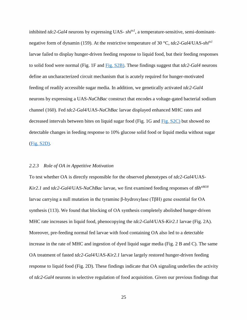
inhibited tdc2-Gal4 neurons by expressing UAS- shits1, a temperature-sensitive, semi-dominant-
negative form of dynamin (159). At the restrictive temperature of 30 °C, tdc2-Gal4/UAS-shits1
larvae failed to display hunger-driven feeding response to liquid food, but their feeding responses
to solid food were normal (Fig. 1F and Fig. S2B). These findings suggest that tdc2-Gal4 neurons
define an uncharacterized circuit mechanism that is acutely required for hunger-motivated
feeding of readily accessible sugar media. In addition, we genetically activated tdc2-Gal4
neurons by expressing a UAS-NaChBac construct that encodes a voltage-gated bacterial sodium
channel (160). Fed tdc2-Gal4/UAS-NaChBac larvae displayed enhanced MHC rates and
decreased intervals between bites on liquid sugar food (Fig. 1G and Fig. S2C) but showed no
detectable changes in feeding response to 10% glucose solid food or liquid media without sugar
(Fig. S2D).
2.2.3 Role of OA in Appetitive Motivation
To test whether OA is directly responsible for the observed phenotypes of tdc2-Gal4/UAS-
Kir2.1 and tdc2-Gal4/UAS-NaChBac larvae, we first examined feeding responses of tßhnM18
larvae carrying a null mutation in the tyramine β-hydroxylase (TβH) gene essential for OA
synthesis (113). We found that blocking of OA synthesis completely abolished hunger-driven
MHC rate increases in liquid food, phenocopying the tdc2-Gal4/UAS-Kir2.1 larvae (Fig. 2A).
Moreover, pre-feeding normal fed larvae with food containing OA also led to a detectable
increase in the rate of MHC and ingestion of dyed liquid sugar media (Fig. 2 B and C). The same
OA treatment of fasted tdc2-Gal4/UAS-Kir2.1 larvae largely restored hunger-driven feeding
response to liquid food (Fig. 2D). These findings indicate that OA signaling underlies the activity
of tdc2-Gal4 neurons in selective regulation of food acquisition. Given our previous findings that

26
tdc2-Gal4 neuron activity stimulates a behavioral program distinct from that of npf-gal4 neurons,
we investigated how simultaneous increases of OA and NPF signaling levels might affect food
motivation in fed larvae. To this end, OA was introduced orally to pre-fed elav-Gal4/UAS-
npfcDNA larvae that overexpress NPF. We found that fed elav-Gal4/UAS-npfcDNA larvae treated
with OA behaved similarly to OA-treated fed control larvae in liquid food (Fig. S3A). However,
in solid food, OA-treated elav-Gal4/UAS-npfcDNA larvae behaved similarly to untreated elav-
Gal4/UAS-npfcDNA larvae (Fig. S3B). These results support the notion that the OA-mediated
circuit mechanism for feeding of liquid sugar media functions independently from the NPF
circuit mechanism.
2.2.4 OA Enhancement of Feeding Requires Octβ3R
Four different OA receptors have been identified in Drosophila (114, 116). To determine the
downstream effectors of the OA feeding pathway, we used a mifepristone (RU486)-inducible
pan-neural GS-elav-Gal4 driver to perform dsRNA-mediated conditional knockdown of
individual OA receptor activity (Table S1). We found that only disruption of the β-adrenergic–
like Octβ3R receptor (116) blocked starvation-induced MHC rate increases. Unlike in tdc2-Gal4/
UAS-Kir2.1 larvae, oral introduction of OA failed to rescue the defect of feeding response in
fasted GS-elav-Gal4/ UAS-Octβ3RdsRNA larvae (Fig. 2E). Furthermore, Octβ3RMB04794 larvae
containing a transposable element that disrupts Octβ3R are also deficient in the hunger-driven
feeding response (Fig. 2F). These results suggest that OA and Octβ3R define a circuit
mechanism that enhances feeding of liquid sugar media in fasted larvae.
27
2.2.5 Subsets of tdc2-Gal4 Neurons Differentially Control Appetite
Tdc2-Gal4 is expressed in multiple clusters of OA neurons in the larval brain lobes and the
ventral ganglia (Fig. 3A). Immunostaining of the larval central nervous system with anti-TβH
antibodies further suggests that the somata of all central OA neurons are located in the ventral
ganglia (Fig. 3B) (81). OA neurons in the larval SOG respond to gustatory inputs from gustatory
receptor neurons, and unlike the OA neurons in the abdominal ganglia, they are not motor
neurons (81, 161). Anatomical mapping of tdc2-Gal4 neurons at the single-cell resolution in
adult flies revealed that the ventral unpaired median neurons (VUMs) in the anterior
compartment (VUM1) and middle compartment (VUM2) of SOG seem to project to several
common areas of the protocerebrum, whereas those in the posterior compartment of SOG
(VUM3) project to the ventral ganglia(119, 162). The tsh-Gal80 construct blocks GAL4
activities in the thoracic and abdominal ganglia (163) (Fig. 3C). Fasted tsh-Gal80/tdc2-Gal4/
UAS-shits larvae showed reduced MHC rates in response to liquid glucose media (Fig. 3D),
suggesting that tdc2-Gal4 neurons in the SOG are required for hunger-driven response to liquid
food. The SOG is proposed to act as a feeding control center in the central nervous system of
insects (14, 164). To test which tdc2-Gal4 neurons in the SOG are important for OA-dependent
feeding activity, we generated targeted lesions in the subsets of tdc2-Gal4 neurons using focused
laser beams (165). Targeted lesions in five VUM1 with or without four ventral paired median
(VPM1) neurons in SOG1 caused a significant increase in the feeding activity of fed larvae (Fig.
3E and Fig. S4A). However, this increased feeding activity was abolished when additional
neurons from SOG2 (five VUM1 plus six VUM2) were lesioned. In fasted larvae, targeted
lesions in six VUM2 neurons alone attenuated hunger-elicited increases of feeding response (Fig.
3F), suggesting that proper control of appetitive motivation under fed and fasted conditions may

28
require the negative and positive regulatory activities from VUM1 and VUM2 neurons,
respectively. Lesions in all OA neurons (five VUM3 plus two VPM3) in SOG3 had no effects on
larval feeding response (Fig. S4 A and B). In addition, conditioned excitation of tdc2-Gal4
neurons in fed larvae by expressing UAS-dTrpA1 triggered increased feeding response to liquid
sugar media. However, this dTrpA1-stimulated effect was completely abolished by targeted
lesions in VUM2 and VPM2 neurons (Fig. 3G). Together, our findings suggest that VUM1
neurons may restrict appetite for liquid sugar media in fed larvae by suppressing feeding
enhancement by VUM2 neurons.
2.2.6 Drk Mediates Hunger-Induced Appetitive Motivation
From a previous genetic screen, we isolated a candidate gene downstream receptor kinase (drk),
the fly homolog of human growth factor receptor-bound protein 2 (Grb2) (166), whose mutations
affect larval feeding response to liquid, but not solid, food (Fig. 4). Under fed conditions, larvae
transheterozygous for three independent loss-of-function drk alleles, drkΔP24/drkR1,
drkΔP24/drk10626 (167, 168), showed basal levels of feeding activity similar to wild-type larvae.
However, after 150-min deprivation, the mutant larvae exhibited significantly attenuated feeding
responses to liquid food (Fig. 4A). In addition, expression of UAS-drkdsRNA driven by elav-Gal4
also led to significantly reduced hunger-driven responses to liquid food. Conversely,
overexpression of drk-cDNA in fed larvae (elav-Gal4/UAS-drkcDNA) caused excessive feeding
response to liquid food (Fig. 4B). Importantly, both fasted elav-Gal4/UAS-drkdsRNA and fed elav-
Gal4/UAS-drkcDNA showed normal responses to solid food (Fig. S5A). These findings suggest
that the neural activity of drk is a positive regulator of hunger-driven feeding response to liquid
sugar food.

29
Because loss of the neural activity of drk or OA signaling led to similar feeding behavioral
defects, we investigated whether drk regulates OA neuronal signaling. Indeed, larvae expressing
UAS-drkdsRNA in tdc2-Gal4 but not npf-Gal4 neurons displayed attenuated hunger-driven
approaching response to liquid food, but showed normal food response under fed conditions
(Fig. 4C). Moreover, oral administration of OA to tdc2-Gal4/UAS-drkdsRNA larvae largely
restored their deficiency in food motivation (Fig. 4D). In addition, both fasted tdc2-Gal4/UAS-
drkdsRNA and fed tdc2-Gal4/UAS-drkcDNA showed normal responses to solid food (Fig. S5B).
These findings suggest that drk regulates feeding of liquid food through its modulation of OA
neuronal signaling.
2.2.7 Tdc2-Gal4 Neuronal Signaling Requires Pvr
The fact that Drk is a SH2/SH3 adaptor protein that directly binds to activated receptor tyrosine
kinase (RTK) strongly implicates the involvement of an as of yet-unknown RTK in modulation
of OA neuronal activity. At least 14 RTK genes have been identified in the Drosophila genome
(131, 169-174). To identify which RTK(s) are involved, we performed dsRNA-mediated
knockdown of the 14 known RTK genes in tdc2-Gal4 neurons (Table S2). This initial screening
has led to the identification of three candidates, Eph receptor tyrosine kinase (Eph), heartless
(htl), and PDGF/VEGF-receptor-related (Pvr), which is a VEGF2-like receptor (134). To assess
whether the effects of these three RTKs on tdc2-Gal4 neurons are physiological, we
conditionally knocked down the individual activity of Pvr, Eph, or htl using the GS-elav-Gal4
driver. We found that only GS-elav-Gal4/UAS-PvrdsRNA larvae showed significantly attenuated
hunger-driven response to liquid food (Fig. 5A). Similarly, GS- elav-Gal4/UAS-drkdsRNA larvae
also showed significantly attenuated hunger-driven response to liquid food. Moreover, the

30
phenotype of tdc2-Gal4/ UAS-PvrDN larvae expressing a dominant negative form of Pvr provides
further verification of the essential role of Pvr in appetitive motivation (131) (Fig. 5B and Fig. S6
A and D). Furthermore, expression of a dominant-active Pvr (PvrACT) in tdc2-Gal4/UAS-PvrACT
fed larvae caused excessive feeding of liquid sugar media, as evidenced by increased rate of
MHC and ingestion of dyed food (Fig. 5C and Fig. S6C). These findings indicate that the Pvr
pathway in tdc2-Gal4 neurons has a previously uncharacterized role in the physiological
regulation of hunger-driven food motivation. The Drosophila genome encodes three
PDGF/VEGF homologs (Pvf1–3) that function as the ligands of Pvr (131). We tested the three
different mutant larvae (Pvf1MB01242, Pvf2d02444, and Pvf3EY09531), each carrying a transposon that
disrupts pvf1, pvf2, and pvf3, respectively (Table S3). We have found that pvf2d02444 larvae
showed attenuated hunger-driven feeding response (Fig. 5D).
To provide evidence for the functional interaction between drk and Pvr in tdc2-Gal4 neurons, we
co-expressed UAS-PvrdsRNA and UAS-drkcDNA under the direction of tdc2-Gal4. We found that
expression of UAS-drkcDNA in fasted tdc2-Gal4/UAS-PvrdsRNA larvae completely restored the
deficiency in approaching response to liquid food (Fig. 5E). Because drk signaling is mediated
by Ras85D GTPase (175), we also co-expressed a dominant-negative form of mammalian Ras
protein (UAS-RasDN (176)) with UAS-drkcDNA in tdc2-Gal4 neurons. As expected, expression of
UAS-RasDN blocked the excessive food response in fed tdc2-Gal4/UAS-drkcDNA larvae (Fig. 5F).
Together, these findings raise the possibility that Drk and Ras may function in the Pvr pathway
to regulate OA neuronal signaling.

31
2.3 DISCUSSION
2.3.1 A Neural Network That Differentially Regulates Appetitive Motivations
Modulation of feeding responses to food sources is heavily influenced by nutritional quality,
taste, and the energy costs of foraging. Our findings suggest that Drosophila larvae have evolved
a complex neural network to regulate appetitive motivations (Fig. 6). In hungry fly larvae, OA
neurons seem to mediate a specialized circuit that selectively promotes persistent feeding of
readily ingestible sugar food. This OA circuit functions in parallel to the previously characterized
mechanism co-regulated by the fly insulin and NPY-like systems that drives feeding response to
non-preferred solid food (16, 66). Because food deprivation triggers simultaneous activation of
both circuits, hungry larvae become capable of adaptively responding to diverse energy sources
of high or low quality. It remains to be determined how OA signaling promotes persistent
feeding response to liquid sugar food in hungry larvae. One possible scenario is that OA neurons
in the SOG may be conditionally activated by gustatory cues associated with rich palatable food
to promote appetitive motivation.
2.3.2 Two Opposing OA Activities in Regulation of Appetite Control
We have provided evidence, at both molecular and neuronal levels, that the OA-mediated
feeding circuit has two opposing effects on food motivation. When surrounded by liquid sugar
media, the OA circuit is essential to prevent fed animals from excessive feeding. Because
targeted lesions in VUM1 neurons caused excessive feeding response, these neurons may define
an inhibitory subprogram within the OA feeding circuit (Fig. 3E). However, targeted lesions in
VUM2 neurons attenuated hunger-induced increases of feeding response, suggesting that VUM2
neurons, along with the OA receptor Octβ3R, may define a subprogram that enhances feeding in

32
fasted larvae (Fig. 3F). Several lines of evidence suggest that the VUM2 neuron-mediated
subprogram may be suppressed by the VUM1 neuron-mediated subprogram. First, fed larvae
with double lesions in both VUM1 and VUM2 neurons failed to display excessive feeding,
suggesting that increased feeding response of fed larvae deficient for VUM1 neuronal signaling
requires VUM2 neurons. Second, targeted lesions in VUM2 neurons of fed tdc2-Gal4/UAS-
dTrpA1 larvae completely blocked the increased feeding response induced by genetic activation
of tdc2-Gal4 neurons. Finally, the anatomical data also show that VUM1 and VUM2 neurons
project to many common regions of the larval brain implicated in the control of feeding (Fig. 3).
Future work will be needed to determine whether VUM1 neurons inhibit directly or indirectly
the activity of VUM2 neurons.
2.3.3 Functional Parallels Between OA and Norepinephrine Systems
We have obtained genetic and pharmacological evidence for the critical role of OA in the
regulation of acquiring readily accessible sugar media. OA has been reported to mediate diverse
neurobiological functions including appetitive memory formation and modulation of the dance of
honey bee foragers to communicate floral or sucrose rewards (78, 79, 156). We postulate that the
different OA receptors may mediate diverse OA-dependent behavioral responses to high-quality
foods.
Norepinephrine (NE), the vertebrate counterpart of OA, has been shown to promote ingestion of
carbohydrate-rich food at the beginning of a natural feeding cycle (177, 178). This feeding
activity of NE resides in the paraventricular nucleus (PVN) of the feeding control center. In the
PVN, α1 and α2 adrenergic receptors are organized in an antagonistic pattern (179). Activation
of α1 receptor inhibits food intake (180), whereas activation of the α2 receptor stimulates food

33
intake (181). Our results suggest that the insect OA system, like the NE system in mammals,
exerts both positive and negative effects on the intake of preferred food. The activity of NE in
PVN has been shown to antagonize that of 5-HT, which suppresses intake of carbohydrate rich
food (182). In Drosophila, 5-HT is also known to suppress feeding response (183). These
findings suggest that the homeostatic control of intake of preferred food is likely mediated by a
conserved neural network in flies and mammals.
2.3.4 The Role of Pvr in OA Neurons
We have identified a unique role of Pvr in physiological regulation of hunger-motivated feeding
of preferred food (liquid sugar media). The feeding-related activity of the Pvr pathway involves
two regulatory proteins, Drk and Ras, and oral introduction of OA restores the hunger-driven
feeding response in tdc2-Gal4/ UAS-drkdsRNA larvae. Together, these results suggest that the Pvr
pathway positively regulates OA release by tdc2-Gal4 neurons. Among the three identified
ligands of Pvr (131, 134), Pvf2 is enriched in the larval CNS. Our finding suggests that Pvf2
regulates the feeding-related activity of the Pvr pathway. It is possible that Pvf2 may transduce a
metabolic stimulus to Pvr/tdc2-Gal4 neurons that signals the energy state of larvae. In the honey
bee brain, OA neurons from the SOG have been reported to respond to sugar stimulation (184,
185). Therefore, it would be interesting to test whether the Pvf2/ Pvr pathway is responsive to
sugar stimuli.
Our previous studies showed that the fly insulin and NPY-like systems co-regulate hunger-
elicited motivation to acquire solid sugar media (16, 66). We have now provided evidence that
the fly VEGFR2- and NE-like systems control larval motivation to acquire liquid sugar media.
These findings strongly suggest that the neural activities of different RTK systems play critical

34
roles in different aspects of adaptive feeding decisions under various food and metabolic
conditions. Therefore, further investigation of the mechanistic details of the food-related
functions of RTK systems in the Drosophila model may provide novel insights into the
neurobiology and evolution of appetitive control as well as pathophysiology of eating-related
disorders.
2.4 MATERIALS AND METHODS
2.4.1 Fly Strains, Media, and Larval Growth
The fly rearing and the egg collections were performed as previously described (186). After a
2.5-h synchronized egg collection, eggs were kept in a 12-h light/dark cycle in an incubator at 25
°C. Larvae were transferred to a fresh apple juice plate with yeast paste at the age of 48–52 h
(<80 larvae per plate). The fly lines used include tdc2-Gal4 (157), npf-Gal4, UAS-npf cDNA(155),
UAS-Kir2.1 (158), UAS-shits1 (159), tßhnM18 (113), drkR1, drkΔP24 (167), tsh-Gal80 (163), UAS-
drkcDNA (discussed below), UAS-drkdsRNA (187), GS-elav-Gal4 (188), UAS-PvrDN, UAS- PvrACT
(131), UAS-dTrpA1 (189), UAS-Octß3RdsRNA, drk10626, actin-Gal4, UAS-GFP.nls, UAS-
NaChBac, elav-Gal4, UAS-RasDN, UAS-dTrpA1, Pvf1MB01242, Pvf2d02444, Pvf3EY09531, and
Octß3RMB04794 (Bloomington Drosophila Stock Center at Indiana University). The following lines
were obtained from the Vienna Drosophila RNAi Center: UAS-PvrdsRNA (105353), UAS-EphdsRNA
(4771), UAS-htldsRNA (27180), and UAS-OambdsRNA (106511).
2.4.2 Transgenic Constructs
The UAS-drkcDNA was made by ligating drkcDNA construct into the pUAST vector. The drkcDNA
clone was acquired from Berkeley Drosophila Genome Project. This vector was digested with

35
EcoRI and XhoI (New England Biolabs), and the resulting 1,558-bp sequence covered the whole
drk coding sequence. This sequence was subcloned into the EcoRI and Xhol sites of pUAST
vector, which is at the downstream of the UAS promoters. Purified drkcDNA/pUAST was injected
to w1118 (BestGene Inc.)
2.4.3 Behavioral Assays
The rate of larval food intake was quantified by following a previously published protocol with
slight modification (16, 190). Liquid glucose agar was prepared by mixing 45 mL ddH2O, 5g D-
glucose (Fisher Chemical), and 6 g agar powder (US Biological). Solid glucose agar was
prepared by melting a mixture of 90 mL ddH2O, 10 g D-glucose (Fisher Chemical), and 2.3 g
agar powder (US Biological). The feeding assay was performed in a 35-mm Petri dish containing
1g of glucose–agar paste or 2 mL of solid glucose agar. Solidified glucose agar was further cut
into eight blocks as described (190) and allowed to dry in a 70% humidity chamber overnight.
For assays, early third-instar larvae (10–20) were transferred to the center of the assay plate, and
then each plate was videotaped for 2 min. The number of MHCs per 30 s was scored and
analyzed. The dynamic patterns of larval MHC were generated with a computer program using
the MatLab software (MatLab Inc.). All assays were analyzed under blind conditions. At least
three separate trials were conducted for each test. Statistical analyses were performed using one-
way ANOVA followed by the Student–Newman–Keuls or Dunn test. The food ingestion assay
was carried out by feeding a group of 30 larvae 10% (wt/vol) glucose liquid media containing
1% food dye FD&C No. 1 (Sigma-Aldrich) for 3 min. After rinsing with a copious amount of
water, larvae were quickly frozen in liquid nitrogen and homogenized in 100 μL 0.1 M
phosphate buffer (pH 7.2). The homogenates were centrifuged at 30,000 × g for 10 min, and the

36
supernatants were analyzed with a spectrophotometer for absorbance at 625 nm. When the fly
line UAS-shits1 was used in the liquid food assay, the permissive temperature was room
temperature, 23 °C, and the restrictive temperature was 30 °C. Twenty minutes before video
recording, the experimental larva groups were pre-warmed on a 30 °C heat plate. The liquid food
plate was pre-warmed on the heat plate for 3 min before use, and the videos were recorded on the
heat plates. The control group assays were performed at 23 °C. To activate GS-elav-Gal4, larvae
were fed with deactivated yeast paste containing 1 mM RU486 (Cayman Chemical) and 4%
(vol/vol) ethanol as solvent, and the mock group used deactivated yeast paste containing only 4%
ethanol. Ten to 20 second-instar larvae were transferred to RU486 or ethanol yeast plates 20 h
before assay.
2.4.4 Pharmacological Treatment
OA stock solution was 500 mM OA in double distilled H2O. When used, the solution was diluted
to desired concentration with deactivated yeast or with double-distilled H2O, dependent on the
larval food deprivation conditions. Larvae treated with OA were fed 5 mM or 10 mM OA, in
yeast or water, 30 min before video recording. For assays using GS-elav-Gal4, the stock RU486
solution was 20 mM RU486 in 80% ethanol. Yeast was deactivated by mixing 5 g yeast powder
with 10 mL boiling water and was then mixed with RU486 stock solution or 80% ethanol at the
ratio of 1:20 after it cooled. RU486 and ethanol yeast were spread on apple juice plates in Petri
dishes and allowed to dry for 24 h before use to evaporate extra water and ethanol.

37
2.4.5 Immunohistochemistry
Brains from larvae 76 h after egg lay were dissected out and the immunostaining were performed
as previously described (24) by using chicken anti-GFP (1:1,000; Invitrogen), rabbit anti-ßgal
(1:1,000; Promega), rabbit anti-tyromine-β-hydroxylase (1:500; gift from Vivian Budnik,
University of Massachusetts Medical School, Worcester, MA), Alexa 488-goat anti-chicken
(1:2,000; Invitrogen), Alexa Fluor-568 goat anti-rabbit (1:2,000; Invitrogen), and rhodamine
conjugated phalloidin (1:1,000; Cytoskeleton). Images were collected using a Zeiss LSM510
META confocal microscope.
2.4.6 Targeted Laser Lesion
The 337-nm nitrogen laser unit (337-USAS; Micro Point) was calibrated and adjusted as
previously described (165). The laser lesion was performed using a previously published
protocol with slight modification (69, 191). Selected tdc2-Gal4 neurons in the subesophageal
ganglion (SOG) were lesioned, using UAS-GFP.nls as an indicator. In early second-instar larvae
(52 h after egg lay (AEL)), the tdc2- GFP.nls in the SOG was visible and distinguishable in vivo
under 40× magnification. Six to nine second-instar larvae were rinsed briefly and transferred to
150 μL double-distilled H2O (ddH2O) on a microscope slide. Ether (250 μL) was added to the
anesthetization chamber (90-mm Petri dish with two strips folded four times to hold microscope
slide above the ether), the slide was placed inside the chamber, and the chamber was covered
immediately. After 3 min, slides were removed and larvae were aligned with a brush, covered
with a coverslip, and would remain anesthetized/immobile for roughly 10 min. The laser beam
was focused on individual nuclei and was applied as two bursts of 15 shots at a rate of three
shots per second. Roughly 50% of larvae on each slide were at the proper angle and displayed

38
clearly distinguishable SOG tdc2-GFP.nls neurons, which were lesioned with laser and showed
the loss of GFP signal. The rest served as the mock group. After the laser treatment, the larvae
were allowed to recover on fresh apple juice plates with yeast paste for 24 h before the assay.
Each experimental or mock group contained at least 12 individual animals pooled from three
separate trials.
2.4.7 Quantitative Real-Time PCR Analysis
For estimation of the UAS-PvrdsRNA efficiency, quantitative real-time PCR was conducted on the
whole body of early third-instar actin-q larvae. Total RNA of five early third-instar larvae was
extracted with TRIzol (Invitrogen). Five micrograms total RNA was reverse transcribed to
cDNA using the kit of SuperScript III FirstStrand Synthesis System for RT-PCR (Invitrogen).
One primer was designed to bind cDNA specifically rather than genomic DNA, which
overspaned a Pvr intron. RpS17 was used as the endogenous control. Quantitative real-time PCR
was performed with Maxima/ROX SYBR Green qPCR Master Mix (Fermentas) in the 7500
Real Time PCR System (Applied Biosystems). Data were calculated with the 7500 System
Software and analyzed with unpaired t test using Microsoft Excel.

39

40
Figure 2.1. Quantification of hunger-driven feeding responses of Drosophila larvae. (A) The
rate of larval MHC linearly increases as food deprivation prolongs up to 150 min; the correlation
coefficient R = 0.99. (B) The amount of ingested food increased after food deprivation. N = 10
trials. *P < 0.05; **P < 0.01. (C) Each dot represents one bite. The typical feeding patterns
sampled from eight fed or 150-min-deprived larvae are shown. (D) Inhibition of tdc2-Gal4, but
not npf-Gal4, neuronal activity using UAS-Kir2.1 blocked hunger-induced feeding rate increases
in liquid food (P < 0.01). Columns with identical letters indicate differences are statistically
insignificant. (E) In contrast, inhibition of npf-Gal4 not tdc2-Gal4 neurons attenuated
hungerinduced feeding rate increases in solid food; P < 0.01. (F) Transient inhibition of
neurotransmission by tdc2-Gal4 neurons by expressing UAS-shits1 at 30 °C abolished the hunger-
elicited approaching response to liquid food; P < 0.01. (G) Stimulation of tdc2-Gal4 neurons by
expressing UAS-NaChBac increased feeding response to liquid sugar but not sugar-free media; P
< 0.01. All of the behavioral assays in this and following figures were analyzed under blind
conditions. Unless stated otherwise, at least 12 larvae were tested for each group (n ≥ 12), and
statistical analyses were performed using one-way ANOVA followed by Student–Newman–
Keuls test in all figures. Error bars represent the SE (SEM) in this and all other figures.

41

42
Figure 2.2. Control of appetite for liquid sugar food by the OA system. (A) The null mutant
tßhnM18 is deficient in hunger-driven feeding response; columns with identical letters indicate
differences are statistically insignificant analyzed with one-way ANOVA followed by Dunn’s
test. (B) Prefeeding of fed wild-type larvae with media containing10 mM OA for 30 min
increased larval MHC rate (P < 0.01) and (C) ingestion rate on liquid glucose media (P < 0.05).
(D) Oral OA treatment significantly restored the hunger-driven feeding response of fasted tdc2-
Gal4/UAS-Kir2.1 larvae; P < 0.05. (E) Conditional knockdown of Octß3R, but not Oamb, in the
nervous system attenuated hunger-driven approaching of liquid food. OA treatment failed to
rescue the behavioral phenotype; columns with identical letters indicate differences are
statistically insignificant (P > 0.05). (F) Octß3RMB04794 larvae showed attenuated hunger-driven
approaching of liquid food (P < 0.05).

43

44
Figure 2.3. Functional mapping of tdc2-Gal4 neurons by targeted laser lesion. (A) Expression
of nuclear LacZ in the tdc2-Gal4 neurons in the central nervous system of tdc2-Gal4/UAS-
nls.LacZ larvae. The tissue is counterstained with phalloidin. (Inset)The distribution of tdc2-Gal4
neurons in three SOG compartments (SOG1–3). Five VUM1 and four VPM1 neurons (arrows) in
the SOG1; six VUM2 and two VPM2 neurons (arrowheads) in the SOG2; five VUM3 and two
VPM3 neurons (asterisks) in the SOG3. (Scale bar, 50 μm.) (B) Expression of TβH in the larval
brain lobes and ventral nerve cord. No TβH-positive somata were detected in the brain lobes.
(Scale bar, 50 μm.) (C) Expression of a nuclear GFP in the central nervous system of tdc2-
Gal4/tsh-Gal80/UAS-nlsGFP larvae (lateral view). The GFP expression in the OA neurons from
the thoracic and ventral ganglia is blocked by tsh-Gal80. The numbers denote SOG1 to 3,
respectively. (Scale bar, 50 μm.) (D) At 30 °C, fed larvae expressing tdc2-Gal4/UAS- shits1/tsh-
Gal80 remained deficient in hunger-driven feeding response; **P < 0.01. (E and F) Analysis of
feedingrelated activities of OA neurons in the SOG in fed and fasted larvae by generating
targeted lesions using focused laser beams (P < 0.01). (G) tdc2Gal4/UAS-dTrpA1 larvae showed
abnormally high feeding response, which was blocked by targeted lesions in VUM2 and VPM2
neurons; P < 0.01. At least 12 larvae were tested for each group (n ≥ 12) in three separate trials.

45

46
Figure 2.4. The neural activity of drk in appetitive motivation. (A) The drk transheterozygous
mutant larvae are deficient in hunger-driven food response; *P < 0.05 and **P < 0.01, compared
with +/+. (B) Knockdown of drk in the nervous system reduced hunger-driven food response (P <
0.05), whereas its overexpression increased feeding response in fed larvae (one-way ANOVA
followed by Dunn’s test; P < 0.05). (C) Knockdown of drk activity in tdc2-Gal4 neurons, but not
npf-Gal4 neurons, attenuated hunger-driven food response. One-way ANOVA followed by
Dunn’s test; P < 0.05. (D) OA treatment of tdc2-Gal4/UAS-drkdsRNA larvae rescued the
deficiency of hunger-driven response to liquid food. Columns with identical letters indicate
differences are statistically insignificant.
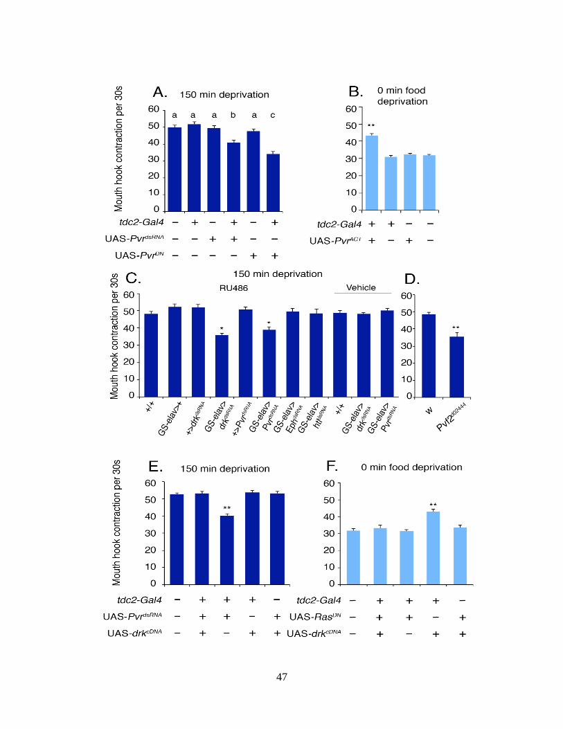
47

48
Figure 2.5. A unique neural activity of the Pvr pathway in appetitive motivation. (A) Conditional
pan-neuronal expression of drkdsRNA and PvrdsRNA but not EphdsRNA or htldsRNA reduced hunger-
driven food response. One-way ANOVA followed by Dunn’s test; P < 0.05. (B) Expression of
UAS-PvrdsRNA and UAS-PvrDN in tdc2-Gal4 neurons attenuated hunger-driven feeding response
to liquid media. Columns with identical letters indicate differences are statistically insignificant.
(C) Expression of UAS-PvrACT-caused increased feeding in fed larvae; P < 0.01. (D) Fasted
Pvf2d02444 larvae showed attenuated feeding response; P < 0.01. (E) Fasted larvae expressing both
drkcDNA and PvrdsRNA in tdc2-Gal4 neurons displayed normal liquid food response; P < 0.01. (F)
Fed larvae expressing both drkcDNA and RasDN in tdc2-Gal4 neurons also displayed normal liquid
feeding response; P < 0.01.

49

50
Figure 2.6. Schematic model for differential regulation of two hunger-driven appetitive
motivations. The OA-mediated circuit, regulated by the Pvf2/Pvr signaling pathway, selectively
controls appetite for readily accessible liquid sugar media. This circuit functions independently
of the previously identified the dILPs/insulin receptor-regulated NPF circuit that selectively
promotes appetite for solid sugar media.

51
Figure 2.S1. Quantification of hunger-driven feeding responses to liquid sugar food. (A) The
rates of mouth hook contractions (MHCs) of fed and fasted larvae during the 3 s of undisrupted
meal time showed only a small difference (ca. 10%). n ≥ 12; three separate trials. (B) Box plot
showing variations of time intervals between bites during a 30-s period, which are larger in fed
animals. Each box represents one individual larva. (Inset) The SD of intervals from eight
individual larvae. Statistical analyses were performed using one-way ANOVA followed by
Dunn’s test; P < 0.05.

52
Figure 2.S2. Role of tdc-Gal4 neurons in hunger-driven feeding responses to liquid sugar food.
(A) The dynamic patterns of MHCs of tdc2-Gal4/ UAS-Kir2.1, npfGal4/UAS-Kir2.1, and wild-
type larvae. Each dot represents one bite. The larvae were fasted for 150 min, and feeding assay
was performed on 10% glucose solid agar food. (B) Transient inhibition of neurotransmission in
tdc2-Gal4/UAS-shits1 larvae at 30 °C caused no significant difference in solid food response
relative to control larvae. (C) The dynamic patterns of MHCs of fed and fasted wild-type larvae
and fed tdc2-Gal4/ UAS-NaChBac in 10% liquid glucose food. (D) Stimulation of tdc2-Gal4
neurons with UAS-NaChBac failed to increase larval MHC rate on 10% glucose solid food. n ≥
12; three separate trials.

53
Figure 2.S3. NPF activity is dispensable for hunger-driven feeding of liquid sugar food. (A)
Overexpression of NPF in elav-Gal4/UAS-npfcDNA larvae failed to further increase feeding
response to liquid food by octopamine (OA)-treated fed larvae. (P < 0.01). (B) Fed elav-
Gal4/UAS-npfcDNA larvae showed elevated feeding response to solid food (P < 0.01), which was
not further enhanced by OA treatment. n ≥ 12; three separate trials.

54
Figure 2.S4. Functional mapping of tdc2-Gal4 neurons by targeted laser lesion. Analysis of
feeding-related activities of OA neurons in the SOG in (A) fed and (B) fasted larvae by
generating targeted lesions using focused laser beams (*P < 0.05, **P < 0.01). n ≥ 15; three
separate trials.

55
Figure 2.S5. Characterization of the neural activity of drk in larval feeding response to solid
sugar food. Manipulation of drk activity in (A) pan neurons using elavGal4/UAS-drkcDNA/UAS-
drkdsRNA or in (B) tdc2-Gal4 neurons using tdc2-Gal4/UAS- drkdsRNA/UAS- drkcDNA caused no
significant difference in solid food response relative to control larvae. n ≥ 12; three separate
trials.

56

57
Figure 2.S6. Characterization of the activity of Pvr in larval feeding responses. (A) Actin-
Gal4/PvrdsRNA larvae showed a reduced Pvr transcripts level. n = 9; one-way ANOVA followed
by Student–Newman–Keuls test; P < 0.001. Total RNAs from actin-Gal4/UAS-pvrRNAi and
control third-instar larvae (74 h AEL) were extracted using TRIzol (Invitrogen), and 5 μg of each
RNA sample was reverse-transcribed to cDNA using the SuperScript III First-Strand Synthesis
Kit (Invitrogen). The forward and reverse primer sequences are as
follows:TGCACCGCGATACGAATG and CGAATTTCTGCGATGAGTTG. The level of
RpS17 transcripts was used as the endogenous control. The real-time PCR was performed using
Maxima/ROX SYBR Green qPCR Master Mix (Fermentas) in the 7500 Real Time PCR System
(Applied Biosystems). (B) Down-regulation of Pvr activity in tdc2-Gal4 neurons using tdc2-
Gal4/UAS-PvrdsRNA caused no significant change in larval feeding response to solid sugar food (n
≥ 12). (C) Enhancement of Pvr activity in tdc2-Gal4 neurons using tdc2-Gal4/UAS-PvrACT
increased the amount of ingested food; P < 0.01. (D) Down-regulation of Pvr activity in tdc2-
Gal4 neurons using tdc2-Gal4/UAS-PvrDN inhibited the increase of hunger-driven food ingestion;
P < 0.05.

58
Table 2.S1. Functional analysis of four OA receptors via RNA interference dsRNA specific to
individual OA receptors. MHCs per 30 s ± SEM, 150-min food deprivation. Feeding responses
to liquid sugar food by fasted larvae expressing dsRNA specific to each of four OA receptors
driven by GS-Elav-Gal4. Statistical analysis was performed using one-way ANOVA followed by
Student–Newman–Keuls test. n ≥ 10, P < 0.01.

59
Table 2.S2. Functional analysis of the activities of 14 receptor tyrosine kinases in tdc-Gal4
neurons via RNA interference. Feeding responses to liquid sugar food by fed or fasted larvae
expressing dsRNA specific to each of 14 receptor tyrosine kinases (RTKs) driven by tdc-Gal4.
Statistical analysis was performed using one-way ANOVA followed by Student–Newman–Keuls
test. n ≥ 5, P < 0.01.
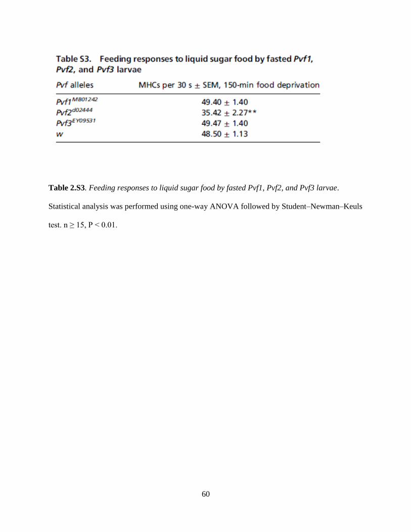
60
Table 2.S3. Feeding responses to liquid sugar food by fasted Pvf1, Pvf2, and Pvf3 larvae.
Statistical analysis was performed using one-way ANOVA followed by Student–Newman–Keuls
test. n ≥ 15, P < 0.01.

61
CHAPTER 3:
AN OAMB/NPF PATHWAY IN LARVAL SOG PREVENTS SWEET FOOD
OVERCONSUMPTION
3.1 INTRODUCTION
The adaptive control of foraging decisions is crucial to survival and reproduction and requires
selective activation of motor programs which best fit both nutritional need and environmental
availability of nutrients. Feeding behaviors can be modulated by diverse neural systems
including those responsible for receiving and processing sensory properties and assigning reward
and motivational significance of food stimuli (152-154). Foraging motor programs that require
high energetic costs, risk, or low nutritional yield must be reserved for situations of high
energetic need. This is demonstrated by young fruit fly larvae which prefer to feed on soft, sweet
foods but when deprived of food will actively forage on non-preferred food and will increase
their rate of feeding.
Our recent study of neurons expressing tdc2-Gal4 (a tyrosine decarboxylase 2 promoter-directed
driver) demonstrates that activity of OA neurons is capable of providing a switching mechanism
between the exuberant feeding of palatable foods demonstrated by fasted larvae and the slower
feeding rate of satiated larvae. In contrast, the manipulation of NPF signaling in the larval
nervous system was demonstrated to selectively modulate starvation dependent alterations in
motivated feeding, as evidenced by its effect on the feeding of difficult to masticate feeding
media. The role of NPF in the modulation of motivated behaviors has been well characterized.

62
Young, actively foraging larvae display high levels of NPF expression in the CNS, which
decreases as larvae age and move toward pupation. Global disruption of NPF signaling in the
brains of young larvae triggers precocious movement out of food and early onset of pupation;
conversely, elevated expression of NPF in older larvae suppresses pupation. In addition, larval
NPF activity is required for starvation dependent motivational processes. As larvae prefer to feed
on soft, sweet media, they display aversion for foods that are hard and difficult to masticate.
Their willingness to feed on such non-preferred foods increases with the duration of food
deprivation. Lack of NPF or its receptor in the larval CNS diminishes the ability of larvae to
display such motivated behaviors, and overexpression of the same enhances motivated foraging
in satiated larvae. Together, these results suggest that starvation dependent enhancement of NPF
signaling is required for appetitive motivation in adverse conditions. To date, however, very little
is known regarding signals that may directly enhance or suppress NPF neuron activity or NPF
release, and recent studies demonstrate that NPF/NPFR1 signaling selectively alters separate
behavioral processes in different regions of the brain. For example, in the mushroom bodies, an
area important for learning and memory, NPF/NPFR1 signaling is required for inhibition of tonic
dopamine (DA) release in order to gate the expression of starvation dependent learning
enhancement (88), whereas in the lateral horn it gates the perception of olfactory inputs (69).
In this work, we provide evidence for a putative pathway acting upstream of NPF neurons. We
show that an octopamine (OA)/OAMB dependent mechanism operates through NPF neurons
located in the subesophogeal ganglia (SOG) which acts to prevent excessive appetite in fed
larvae. Given our previous finding that the Oamb receptor is required to suppress faster rates of
feeding in satiated larvae, we hypothesized that OA signaling may act through a selective
pathway to suppress feeding rate under times when internal energy stores are sufficient. The
63
selective activation of this subprogram in satiated larvae may be due to selective suppression of
OA neuron subsets, or by selective activation of an OA/OAMB dependent inhibitory pathway.
To investigate this, we began with an UAS-OambdsRNA mediated screen to identify types of
neurons required for Oamb feeding rate suppression.
3.2 RESULTS AND DISCUSSION
3.2.1 Baseline Rate of Sweet Food Intake Requires Oamb
Four different OA receptors have been identified in Drosophila (114, 116). In previous work, we
identified a specific role for the B-adrenergic-like OctB3R receptor in promotion of food intake
under conditions of food deprivation (3). In order to identify other downstream effectors of the
OA feeding pathway, we used a mifepristone (RU486)-inducible pan-neural GS-elav-Gal4 driver
to perform dsRNA-mediated conditional knockdown of individual OA receptor activity. Third
instar mutant larvae were then tested for behavioral responses to sweet, palatable media under
fed and starved conditions (Fig.1, Table S1). We found that only disruption of Oamb receptor
(116) was sufficient to trigger feeding rate enhancement in satiated larvae (Fig. 1A).
Furthermore, larvae carrying a Minos transposable element insertion at the Oamb locus,
Mi[ET1]OambMB00297(192, 193), also displayed an enhanced feeding response (Fig. 2A), as did
Oamb286 larvae (194, 195) (Fig. 2B).
In our previous work, we have shown that tdc2-Gal4/UAS-NaChBac larvae expressing a
bacterial sodium channel construct in OA neurons displayed increased feeding rates only when
the assay media contained glucose (3), indicating that OA dependent enhancement of larval
mouth hook contraction rate was specific to nutritive food rather than a non-specific increase in
locomotion. Similarly, Oamb286 larvae were also tested for responses to media containing 0%,

64
5%, and 10% glucose (Fig. 2B), and demonstrated a graded increase in mouth hook contraction
rate as the concentration of glucose was increased in the media, indicating that lack of Oamb
specifically enhances feeding responses to sweet food. These results suggest the presence of an
OA/Oamb mechanism that maintains the baseline feeding rate of larvae on sweet foods by
suppressing the response to sugar.
3.2.2 OAFS-Gal4 Larvae Display Enhanced Basal Feeding Rate on Sweet Food
The OA receptor Oamb is expressed as two isoforms (K3 and AS), both of which have been
shown to mediate increases in intracellular calcium, similar to the mammalian a1-adrenergic
receptor (196). Expression of Oamb-K3 and Oamb-AS has been demonstrated in the mushroom
body where they are required for appetitive olfactory memory formation in response to sugar
(196) and courtship behavior (194). In an effort to identify and target neurons expressing the
Oamb receptor underlying the suppression of food intake in fed larvae, we constructed an Oamb
enhancer based Gal4 line using a 1.6 kb fragment upstream of the Oamb gene locus. This oaFS-
Gal4 (OA feeding suppression, (oaFS)) line drives expression in the brain lobes, in four pairs of
neurons in the SOG, and in the posterior portion of the VNC (Fig. 3B). To test whether these
cells underlie Oamb-dependent feeding regulation, we tested oaFS-Gal4/UAS-OambdsRNA larvae
for response to sugar media and observed an increase in feeding rate (Fig. 3A). This
demonstrates that the oaFS-Gal4 targets expression to neurons which functionally mediate the
effects of the Oamb receptor on feeding rate suppression in satiated larvae.

65
3.2.2 Suppression of Sweet Food Consumption Requires NPF/Oamb
A number of neurotransmitter systems have been implicated in the regulation of food intake,
appetitive motivation, and appetitive learning in Drosophila . We speculated that OA may exert
suppression in food intake in satiated larvae by activating the Oamb receptor on neurons which
regulate food intake. To functionally map the activity of Oamb, we performed a dsRNA-based
screen with a library of Gal4 lines which drive expression in known feeding regulatory neurons
(Fig.3B). Of the chosen lines used, only npf-Gal4/UAS-OambdsRNA larvae phenocopied the
increase of feeding rate on sweet media seen in Oamb mutants. This suggests that NPF neurons
mediate the effects of the Oamb receptor.
3.2.3 NPF SOG neurons are required to suppress food intake
Drosophila npf-Gal4 is expressed in six neurons in the protocerebrum and subesophogeal
ganglion of larval brain (Fig. 4B). OA neurons in larval SOG respond to gustatory inputs from
gustatory receptor neurons (81, 161), and the SOG is proposed to act as a feeding control center
in the central nervous system of insects (14, 164). When oaFS-Gal4/UAS-mCD8GFP larvae
were co-stained with anti-NPF, co-localization was seen only in NPF neurons located in the SOG
region. To determine whether npf-Gal4 neurons in the SOG are important for the Oamb
dependent suppression of feeding rate, we generated targeted lesions in the DM, DL, and SOG
subsets of npf-Gal4 neurons using focused laser beams (165). Following lesioning of the DM or
DL pairs, npf-Gal4/UAS-GFP.nls larvae behaved similarly to mock treated controls, however,
lesions of the SOG subset caused a significant increase in the feeding activity (Fig. 4A). We have
previously shown that alterations in NPF or NPFR1 activity in the larval CNS is sufficient to
disrupt starvation dependent motivated feeding behaviors, such the willingness to work to

66
procure food (15) or to consume foods with aversive tastes (66) or temperature. These findings
present the first evidence of a role for NPF signaling in the regulation of intake of preferred
foods. The reduction of Oamb levels in the CNS or specifically in OaFS-Gal4 and npf-Gal4
neurons results in starvation-like feeding behavior in larvae. In conjunction with our finding that
VUM1 OA neurons mediate a feeding suppression mechanism, these results suggest that OA
signaling in satiated larvae acts to suppress overconsumption of sweet foods via a local circuit
involving OA and NPF neurons located in the SOG.
3.4 MATERIALS AND METHODS
3.4.1 Fly Strains, Media, and Larval Growth
The fly rearing and the egg collections were performed as previously described (186), with a
slight modification for glucose feeding assays using 0%, 5%, and 10% glucose. After a 2.5-h
synchronized egg collection, eggs were kept in a 12-h light/dark cycle in an incubator at 25 °C.
Larvae were transferred to a fresh apple juice plate with yeast paste at the age of 48–52 h (<80
larvae per plate). The fly lines used included tdc2-Gal4 (157), npf-Gal4 (15), Oamb286 (194,
195), npfr1-Gal4 (69), UAS-dilp2-Gal4 (16). UAS-GFP.nls, Mi[ET1]OambMB00297 (192, 193),
UAS-mCD8-GFP, GS-elav-Gal4, UAS-OctB2RdsRNA, UAS-OctB3RdsRNA, TH-Gal4, vglut-Gal4,
TRH-Gal4, were obtained from Bloomington Drosophila Stock Center at Indiana University.
UAS-OambdsRNA (#2861) (197), UAS-oa2dsRNA (#47896) (197) were obtained from the Vienna
Drosophila RNAi Center.

67
3.4.2 Transgenic Constructs
To construct he oaFS-Gal4 driver line, a 1.6 kb DNA fragment containing the 5’ regulatory
sequence for the Oamb gene was amplified by 5’-
ATACATACTAGAATTCTCTGAAAGCTGCGGGATA-3’ and 5’-
GGGCGAGCTCGAATTCCGGCAAGAACCGTTAGTTC-3’ and cloned into the pCaSpeR-
Gal4 vector at the EcoR1 site. The purified construct was injected to w1118 background
(BestGene Inc.).
3.4.3 Behavioral Assays
The rate of larval food intake was quantified by following a previously published protocol with
slight modification (16, 190). Liquid glucose agar was prepared by mixing 45 mL ddH2O, 5g D-
glucose (Fisher Chemical), and 6 g agar powder (US Biological). Solid glucose agar was
prepared by melting a mixture of 90 mL ddH2O, 10 g D-glucose (Fisher Chemical), and 2.3 g
agar powder (US Biological). The feeding assay was performed in a 35-mm Petri dish containing
1g of glucose–agar paste or 2 mL of solid glucose agar. Solidified glucose agar was further cut
into eight blocks as described (190) and allowed to dry in a 70% humidity chamber overnight.
For assays, early third-instar larvae (10–20) were transferred to the center of the assay plate, and
then each plate was videotaped for 2 min. The number of MHCs per 30 s was scored and
analyzed. All assays were analyzed under blind conditions. At least three separate trials were
conducted for each test. Statistical analyses were performed using one-way ANOVA followed by
the Student–Newman–Keuls or Dunn test.

68
3.4.5 Immunohistochemistry
Brains from larvae 76 h after egg lay were dissected out and the immunostaining were performed
as previously described (24) by using chicken anti-GFP (1:1,000; Invitrogen), rabbit anti-NPF
(cite), Alexa 488-goat anti-chicken (1:2,000; Invitrogen), Alexa Fluor-568 goat anti-rabbit
(1:2,000; Invitrogen), Images were collected using a Zeiss LSM510 META confocal microscope.
3.4.6 Targeted Laser Lesion
The 337-nm nitrogen laser unit (337-USAS; Micro Point) was calibrated and adjusted as
previously described (165). The laser lesion was performed using a previously published
protocol with slight modification (69, 191). Selected npf-Gal4 neurons in the subesophageal
ganglion (SOG) were lesioned, using UAS-GFP.nls as an indicator. In early second-instar larvae
(52 h after egg lay (AEL)), the npf- GFP.nls in the SOG was visible and distinguishable in vivo
under 40× magnification. Six to nine second-instar larvae were rinsed briefly and transferred to
150 μL double-distilled H2O (ddH2O) on a microscope slide. Ether (250 μL) was added to the
anesthetization chamber (90-mm Petri dish with two strips folded four times to hold microscope
slide above the ether), the slide was placed inside the chamber, and the chamber was covered
immediately. After 3 min, slides were removed and larvae were aligned with a brush, covered
with a coverslip, and would remain anesthetized/immobile for roughly 10 min. The laser beam
was focused on individual nuclei and was applied as two bursts of 15 shots at a rate of three
shots per second. Roughly 50% of larvae on each slide were at the proper angle and displayed
clearly distinguishable SOG npf-GFP.nls neurons, which were lesioned with laser and showed
the loss of GFP signal. The rest served as the mock group. After the laser treatment, the larvae
were allowed to recover on fresh apple juice plates with yeast paste for 24 h before the assay.

69
Each experimental or mock group contained at least 12 individual animals pooled from three
separate trials.

70
B
A

71
Figure 3.1. Oamb is required for suppression of food intake in fed larvae. A) Conditional
knockdown of Oamb, but not other octopaminergic receptors, with the GS-elav-Gal4 inducible
pan-neural driver line resulted in enhancement of feeding rate in satiated larvae (** p<0.01). B)
Expression of two different OambdsRNA constructs similarly triggered increased feeding response.
columns with identical letters indicate differences are statistically insignificant analyzed with
one-way ANOVA followed by Dunn’s test (** p<0.01).

72
***
*** ***
B
A
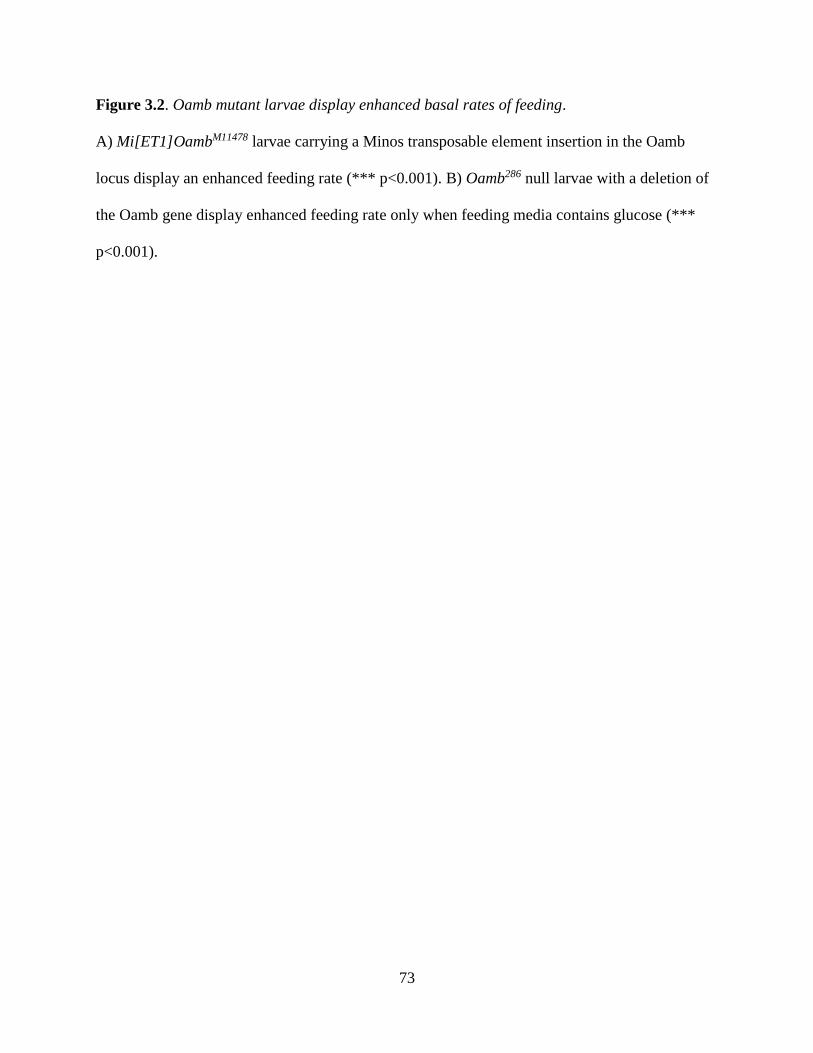
73
Figure 3.2. Oamb mutant larvae display enhanced basal rates of feeding.
A) Mi[ET1]OambM11478 larvae carrying a Minos transposable element insertion in the Oamb
locus display an enhanced feeding rate (*** p<0.001). B) Oamb286 null larvae with a deletion of
the Oamb gene display enhanced feeding rate only when feeding media contains glucose (***
p<0.001).

74
**
***
C
B A

75
Figure 3.3. Oamb is required in NPF and oaFS-Gal4 neurons for feeding suppression. A)
OaFS-Gal4/UAS-OambdsRNA larvae display increased feeding rate on sweet food, similar to
Oamb mutants (*** p<0.001). B) Expression pattern of OaFS-Gal4/UAS-mCD8GFP.
C) Functional mapping of Oamb regulation of feeding via Gal4 mediated screen in cells
expressing feeding regulatory neuropeptides. Only larvae expressing npf-Gal4/UAS-OambdsRNA
displayed increased ingestion rate (** p<0.01).

76
*
α-NPF α-NPF
OaFS-Gal4>
UAS-CD8GFP
UAS-mCD8GFP
OaFS-Gal4>
UAS-CD8GFP
UAS-mCD8GFP
DM
DL
SOG
A B
C
B
D
B
E
B
npf-Gal4<UAS-nls,GFP

77
Figure 3.4. NPF SOG neurons are required for suppression of sweet food intake and co-express
Oamb. A) Targeted laser lesion of dorsal medial (DM), dorsal lateral (DL), and subesophogeal
ganglion (SOG) subsets of npf-Gal4/UAS-nls.GFP neurons in fed larvae. Only lesion of the
SOG subset resulted in an enhancement of sweet food intake (*p<0.05). B) Expression pattern of
npf-Gal4/UAS-mCD8GFP shows DM, DL, and SOG NPF neuron subsets. C) Anti-NPF
immunoreactivity, D) expression of OaFS-Gal4/UAS-mCD8GFP, E) merged images. Co-
localization is observed only in the SOG subset of NPF neurons.

78
CHAPTER 4:
PREVENTION OF PALATABLE DIET-INDUCED HYPERPHAGIA IN RATS BY
CENTRAL INJECTION OF A VEGFR KINASE INHIBITOR
4.1 INTRODUCTION
Humans and animals alike are vulnerable to the temptation of overeating appetizing foods. Many
studies have implicated the “rewarding” nature of tasty foods in the drive to overeat (77, 198).
Palatable foods are more likely to be eaten in excess relative to bland foods, and the pleasurable
effects of consuming them may override homeostatic signals (77, 198-200). It is speculated that
obese individuals may be more susceptible to the temptations of food. In support of this, obese
humans display heightened activity in neural reward circuits in response to palatable food or
food-associated cues (201, 202). The taste of palatable food may also contribute to overeating by
suppressing satiety. For example, in the presence of palatable food, endocannabinoids increase
appetite by stimulating the desire to eat while also blocking signals for meal termination (203).
Our recent study using the Drosophila larva model points to the essential roles of a vesicular
endothelial growth factor (VEGF)-like ligand (Pvf2) and its receptor (dVegfr, also known as Pvr)
in promotion of hunger-driven intake of palatable food (3). Furthermore, the enhancement of
feeding by Pvf2/dVegfr signaling positively correlated with the concentration of sugar in the
media. These findings have led us to postulate that a homologous VEGF signaling pathway in
mammals may have a parallel function in regulation of consumption of palatable food. An N-
cyclopropylnaphthamide compound, named as VEGFR Tyrosine Kinase Inhibitor V (VTKI-V),
79
has been shown to selectively inhibit VEGF2 receptor kinase at subnonamolar concentrations
(204). Oral and intraperitoneal administration of VTKI-V in mice has been demonstrated to
inhibit angiogenesis and to promote regression of established HT29 tumors (205). In this work,
we examined its potential effect on the feeding behavior of male Sprague Dawley rats. We find
that i.c.v. injection of VTKI-V at a lower single dose attenuates overeating induced by sweet and
fatty food. The same drug treatment also reduced body weight gain of animals fed with CHOW
or sweet and fatty food. Our results suggest that i.c.v. VTKI-V may interfere two separate
VEGFR-mediated mechanisms that promote overconsumption of palatable food and body weight
gain, respectively. Therefore, components of the VEGFR pathway may have potential diagnostic
and therapeutic applications for prevention of overeating and obesity.
4.2 RESULTS
4.2.1 Effects of i.c.v. Injection of VTKI-V on CHOW-Fed Rats
Our previous finding of the role of neural VEGFR-like signaling in promoting hunger-driven
feeding of Drosophila larvae led us to speculate that a mammalian VEGFR system(s) might
perform a similar neural function. To test this hypothesis, we selected a small molecule inhibitor
of VEGFR tyrosine kinase, VTKI-V, which has been characterized pharmacologically for its
selectivity profile against tyrosine kinase receptors and its efficacy in inhibition of VEGF
dependent VEGFR2 phosphorylation and tumor cell growth (204). In order to target the neural
activity of the VEGFR system, the compound was injected i.c.v. In a pilot dose response study,
a single dosage of 2 µg dissolved in 2 µl of DMSO was determined to be sufficient to elicit a
reduction in weight gain (data not shown). On a per animal basis, this dose is >500 times lower

80
than the minimal effective oral dose (3 mg/kg) demonstrated to block VEGF mediated processes
(205). This dosing regimen was subsequently adopted throughout the following experiments.
Following recovery from surgery, rats of roughly equal starting weight (control group: 288 ±
22.1; drug group: 287 ± 25.8) were habituated to home cages for one week. Rats were then
injected with one dose of VTKI-V, followed by monitoring for body weight, food intake, and
water intake for five days post injection (Fig. 1). The treatment resulted in a rapid reduction in
the rate of body weight gain detectable within 24 hours, and it persisted during the test period
(Fig. 1A). This was accompanied by a slight but significant reduction in cumulative daily intake
(p<0.05 for day 3 and 5; Fig. 1B). By five days post injection, animals treated with VTKI-V
gained approximately 35% less weight than vehicle treated controls and reduced their caloric
intake by roughly 10%. However, no significant alteration of water intake was observed (Fig.
1C). These results suggest that a single dose of i.c.v. VTKI-V is sufficient to inhibit the weight
gain in the male rats over at least 5 days. It also appears to cause a slight but significant reduction
in cumulative consumption of CHOW.
4.2.2 Effects of i.c.v. Injection of VTKI-V on Rats Fed a Palatable Diet
We previously demonstrated that reduced VEGFR-like signaling in Drosophila larvae selectively
attenuated feeding rate when hungry larvae were placed on media with higher sugar
concentrations. Therefore, we postulate that a mammalian VEGFR system might have a similar
function. To test this, we acclimated animals to a palatable diet rich in fat and sugar (Cat No.
RD12506; Research Diets). This high-calorie diet (HC) has been widely used in studies of diet
induced obesity (206) and hedonic feeding (144). Animals with roughly equal body weight
(DMSO group: 346.9 ± 5.7; VTKI-V group: 366.6 ± 3.4) were injected with either VTKI-V or

81
vehicle as described before. Treatment again resulted in a rapid as well as persistent reduction in
the daily rate of body weight gain (Fig. 1D). By five days post injection, animals treated with
VTKI-V gained approximately 60% less weight than vehicle treated controls (p < 0.0001).
Surprisingly, however, a more pronounced effect on cumulative daily intake was observed under
this condition (Fig. 1E). The drug-treated animals consumed 30% less calories than the controls
(p < 0.0001). Again, the VTKI-V treatment had no significant effect on water intake (Fig. 1F).
Together, these results indicate that the VTKI-V treatment results in more drastic attenuation of
food intake when the fat and sugar content of the diet is increased. However, the effect of the
drug on body weight gain remains substantial under both feeding conditions.
4.2.3 Effect of Drug Treatment on Conditioned Taste Aversion
The experience of DMSO or VTKI-V injection (unconditioned stimulus) was paired with a novel
neutral taste (conditioned stimulus) with the experimental paradigm outlined in Figure 5A.
Briefly, prior to drug administration, animals were trained to drink sweetened condensed milk
from 25-mL graduated drinking tubes presented for one hour periods in a pattern of two days
on/one day off, and 30 minute ingestion amount was recorded. Once animals reached a stable
baseline of ingestion of the solution, the conditioning trial was performed in which novel orange
flavoring was added (0.5 ml/liter). Immediately following the removal of the orange flavored
milk, animals received and injection of DMSO (n=7) or VTKI-V (n=8). Three days later,
animals were again presented with orange flavored milk solution and ingestion was recorded.
Animals displayed no aversion to the orange flavored milk after treatment with either DMSO or
VTKI-V (Fig. 5). This demonstrates that the effects of VTKI-V on body weight gain and food
intake are not due to nausea or malaise.

82
4.2.4 Effects of i.c.v. VTKI-V on Meal Pattern
To better understand the behavioral effects of VTKI-V, we assessed potential changes in meal
pattern that contributed to altered food intake. CHOW fed control animals treated with DMSO or
VTKI-V ate similar amounts of food in diurnal and nocturnal phases of the five-day period in
terms of calories or mass (Fig. 2A, B). This observation is largely consistent with the minor
effect of VTKI-V on cumulative food intake shown in Fig. 1B. In addition, the meal patterns of
both groups showed no significant changes, as evidenced by the meal number, size and duration
as well as bouts per meal (Fig. 2C-F).
However, control animals given access to the palatable diet exhibited hyperphagic behavior.
Their diurnal and nocturnal caloric intake increased by roughly 50%, although the mass of food
consumed remained the same (Fig. 2A, B). We found that this increased calorie intake is due to
an increase in average meal size rather than meal number (Fig. 2C, D). Contrastingly, VTKI-V-
treated animals showed reduction in the mass of palatable food ingested, bringing their caloric
intake down to a level comparable to that of CHOW fed animals (Fig. 2A, B). Furthermore, we
observed that the drug treatment reduced meal size and duration as well as the number of bouts
per meal (Fig. 2C, F). However, the meal number remained unaltered (Fig. 2D). Together, these
results suggest that i.c.v. VTKI-V may affect meal consumption but not initiation in both dark
and light phases.
To evaluate the time course of VTKI-V effects, we further analyzed the meal patterns of the four
animal groups by individual 12-hour periods over five consecutive days. We found that VTKI-V
injection caused no significant changes in all parameters examined under CHOW fed conditions
(Fig. 3A-E). In contrast, the drug treatment resulted in significant reductions in food intake, meal
size, bouts per meal, and meal duration. These effects were evident within 12 hours and persisted

83
for at least five days (Fig. 3F-I). Again, the average number of meals for the light or dark period
remained constant throughout the 5-day test period (Fig. 3J). These findings suggest that a single
dose of VTKI-V is sufficient to inhibit feeding of palatable food over a significant length of time
without detectable compensatory consumption as normally seen in calorie restricted animals
(207). These results also reveal that the magnitudes of the drug effects on meal consumption and
duration were comparable for five consecutive days.
4.2.5 Effects of Reduced Neural dVEGFR Activity on Body Weight Gain in Drosophila
Given that i.c.v. injection of VTKI-V resulted in a significant reduction of body weight gain in
rodents, we sought to investigate whether reduction in neural activity of the fly VEGF-like
pathway had a similar effect. Activity of dVegfr was knocked down by expressing its double-
stranded RNA (UAS-dVegfrdsRNA) using a pan-neural elav-Gal4 driver. The body mass and
protein content of elav-Gal4/ UAS-dVegfrdsRNA and male flies were significantly lower relative to
the controls in 4 day-old adults (Fig. 4A, B). These results suggest that the neural VEGFR
systems in insects and mammals may have parallel regulatory functions in the central control of
body weight as well as intake of palatable food.
4.3 DISCUSSION
We have shown that i.c.v. injection of a single dose of a VEGFR tyrosine kinase inhibitor causes
reduced body weight gain and attenuated consumption of palatable food, and that these effects
are both rapid and persistent. Therefore, these results raise the possibility that VTKI-V may
interfere with two separate functions of VEGF system(s) that acutely regulate body weight gain
and consumption of palatable food, respectively. In our assay, VTKI-V is administered into the

84
lateral ventricles at a single dose of 2 µg. This is >500 fold lower than the minimal effective
intraperitoneal dose shown to reduce VEGF-mediated angiogenesis and tumor growth (205).
This result argues for a neural target(s) that mediates the behavioral effects of i.c.v. VTKI-V.
Very little is known regarding central regulation of feeding behavior by VEGFR. Inhibition of
VEGFR2 has been demonstrated to block the effects of fluoxetine on sucrose preference (140).
VEGFRs are essential for a number of neuronal activities such as axonal outgrowth and
membrane excitability (208). VTKI-V may exert its effects by altering activity of a neural
system (e.g., sympathetic nervous system) to regulate peripheral metabolism of stored fat and/or
regulation of energy expenditure (209). It is also possible that VTKI-V might interfere with a
central feeding circuit (e.g., the norepinephrine circuit implicated in intake of carbohydrates) that
promotes appetitive response to food rich in sugar and fat (210, 211). Future determination of the
target(s) of i.c.v. VTKI-V will likely yield fresh insights into novel strategies for prevention of
overeating and the development of obesity.
This and other studies have shown that animals fed with palatable diets exhibit rapid alterations
in meal pattern, including increased meal size and duration (206, 212). These changes persist
over time and lead to diet induced obesity (206, 213). In our studies, the control animals
acclimated to the palatable diet displayed a similar trend. VTKI-V injection led to a roughly 35%
reduction of the average mass of food ingested, bringing their caloric meal size down to a similar
level as CHOW fed animals. Intriguingly, the drug treatment caused at most a minor reduction in
CHOW consumption, which is only detectable by measuring cumulative intake over more than
three days. These findings suggest that i.c.v. VTKI-V may selectively affect a VEGFR-mediated
mechanism that regulates palatable-diet induced hyperphagia.

85
An in-depth analysis of meal pattern reveals that the reduced consumption of palatable food is
due to smaller, shorter meals with fewer bites. We find that control animals fed on the rich diet
behave similarly to the CHOW fed animals in terms of meal number, duration and mass. This
translates to a 55% increase in daily caloric intake over the testing period. Following VTKI-V
treatment, animals consumed a smaller mass of food, and therefore fewer calories. This appears
to be the result of eating shorter and smaller meals (Fig. 2, 3). Together, these findings suggest
that VTKI-V treatment selectively alters the mass of food ingested, but only when the energy
density of that food exceeds a baseline need of calories.
We also observed that VTKI-V treatment caused a reduction in the rate of weight gain. This
effect was rapid and persistent, with a five day reduction of 35% and 60% for rats fed on CHOW
or the rich diet, respectively. Given that drug-treated rats fed with CHOW diet display a modest
reduction of cumulative food intake, this suggests that the comparatively large effect of VTKI-V
on body weight is largely due to its interference with a physiological process that controls body
weight gain. In contrast, the more pronounced reduction in body weight gain for animals fed
with the rich diet could be due to the compounding effects of the drug on mechanisms underlying
both feeding and metabolism. These results are reminiscent of diet-induced obesity resistant rats
that display a reduction of meal size and caloric intake when fed with palatable diets relative to
normal controls (206).
In Drosophila larvae, attenuation of neural signaling by the VEGFR-like system blocks hunger-
driven feeding activity in sugar-rich media, and the magnitude of this effect becomes smaller as
the concentration of sugar is reduced (3). Interestingly, we find that i.c.v. injection of VTKI-V
caused pronounced reduction in consumption of the food rich in sugar and fat, but showed much
less effect on CHOW intake. These parallel observations across phylogenetically divergent

86
species raise the possibility that the VEGFR system may play a conserved role in neural circuitry
that underlying palatable food-induced overeating. In addition, the regulatory activity of the fly
VEGFR system for feeding of sugar media has been mapped to a subset of neurons producing
octopamine. Future work will test whether the effect of i.c.v. VTKI-V on overeating of palatable
food is mediated by norepinephrine neurons.
4.4 MATERIALS AND METHODS
4.4.1 Animals and Diet
Sixty male Sprague-Dawley rats approximately 7-8 weeks old (250 – 270g of initial body
weight) purchased in batches of twelve from Harlan Inc. (Indianapolis, IN, USA) were
individually housed in plastic shoebox cages equipped with BioDAQ® Food Intake Monitors
(Research Diets, Inc., New Brunswick, NJ). Rats were maintained on a 12/12-hr light-dark cycle
(lights on at 06.00h) in a temperature controlled environment (23 ± 1˚C) with ad libitum access
to food and water throughout the study. CHOW fed animals received 5053-Rodent Diet
(LabDiet, St. Louis, MO) (3.07 kcal/g, 13% fat, 5% sugar) and HC groups received 12451-
Research Diets (4.73 kcal/g, 45% fat, 17% sucrose). Rats were removed from and returned to
their homecages daily (between 09.00h and 10.00h) for food hopper maintenance and monitoring
of body weights and water bottle weights. Food intake was monitored as described below. All
experimental and surgical procedures were approved prior to initiation of the study by the
Institutional Animal Care and Use Committee of the University of Georgia.
Fly rearing and egg collections were performed as previously described (3). Following a 2.5-h
synchronized egg collection, petri dishes containing egg plates were maintained in a 12-h
light/dark cycle incubator at 25 C. Larvae were transferred to fresh apple juice plates at 48-52h
87
(>80 per plate), and returned to incubator until adults began to emerge (9-10 days). Male adults
were collected and transferred to standard yeast, cornmeal, and molasses fly media for 4-5 days.
Samples were prepared with five flies per Eppendorf tube, which were flash frozen in liquid
nitrogen. Following freezing, flies were thawed and weighed to determine average body mass.
Flies were then homogenized and average protein content per fly was determined using a BCA
protein assay (Peirce, Cat No. 23225). Fly lines included Canton S (BL1) and elav-Gal4
(BL8760) (Bloomington Stock Center at Indiana University (BL)). UAS-dVegfrdsRNA (105353)
was obtained from the Vienna Drosophila RNAi Center.
4.4.2 Drug Treatment
VEGFR2 Tyrosine Kinase Inhibitor V or VTKI-V (Cat No. 676501) was purchased from EMD
Millipore, Darmstadt, Germany. It is a cell-permeable, ATP-binding pocket-targeting N-
cyclopropylnaphthamide compound with high selectivity against VEGFR2 (IC50 = 0.6 nM)
(204). The compound was dissolved in DMSO (Sigma) at a concentration of 1 µg/µl, and
aliquots were stored at -80 ˚C. For all injections, 2 µL of VTKI-V solution or vehicle (DMSO)
were delivered via i.c.v. cannula at a rate of 1 µL/min.
4.4.3 Surgical Procedures
Following a one-week acclimation period, anesthetized animals were implanted with a chronic
22 gauge i.c.v. cannula (Plastics One, Roanoke, VA) stereotaxically positioned in the lateral
ventricle with the coordinates 1.0 mm posterior, 1.5 mm lateral, and 3.2 mm ventral to bregma.
The cannula was affixed to the skull by three stainless steel screws and cranioplastic cement.

88
Following surgery, animals were transferred back to their home cages and allowed to recover for
three days prior to treatment.
4.4.4 Cannula placement verification
Angiotensin II drinking tests were performed one week after surgery. Animals were given one
intracerebroventricular (i.c.v.) injection of 100 ng human angiotensin II (Sigma, St. Louis, MO)
dissolved in 5µL volume of saline. Rats that drank >5 mL of water in 30 minutes were
considered to have the cannula correctly placed.
4.4.5 Conditioned taste aversion test
Behavioral testing was performed during the light cycle between 10.00 and 12.00 h. On days 1,
2, and 4 of behavioral testing, food and water were removed for one hour and animals presented
with sweetened condensed milk (diluted, 1 part milk: 2 parts distilled water) in 25 mL calibrated
drinking tubes secured to the homecage. Consumption of solution was measured after 30
minutes. On day 5 of the experiment a conditioning trial was performed in which animals were
offered the sweetened condensed milk solution flavored with orange extract (0.5 mL/liter).
Consumption was recorded at the time points of 15 and 30 minutes. Immediately following
removal of the drinking tubes, animals received an injection (2 µl of 1 µg/µl VTKIIV in DMSO).
On day 8, animals were again offered the orange flavored solution, and ingestion was recorded
after 30 minutes. Conditioned taste aversion was calculated as the percentage of orange flavored
SCM ingested on the test day relative to baseline ingestion of non-flavored SCM during the
training period (214).

89
4.4.6 Food Intake Monitoring and Analysis
All animals were individually housed in homecages equipped with the BioDAQ® Food Intake
Monitoring System (215). It provides accurate and continuous collection of food intake with
minimal experimenter intervention. The system consists of individual low spill food hoppers
attached to the outside of each homecage which rests on a strain gauge-based load cell
(peripheral sensor controller, PSC) that measures the weight of the hopper 50 times/second. Each
PSC is linked to a central controller, which transmits raw data to a peripheral computer where
the mean mass and standard deviation of the hopper weight is calculated each second. When the
computer detects a difference in hopper weight of ≥0.01g for a minimum of five seconds, it is
recorded as the beginning of feeding event (bout). Termination of a feeding event is reached
when the hopper is undisturbed minimum of five seconds. Upon termination of a bout, the
computer records the difference in beginning and ending weight (bout grams), and the duration
of the event. This information is stored using BioDAQ® Monitoring Software 2.1.00 which
allows viewing, filtering, and export of the raw data for further analysis.
4.4.7 Data Processing and Statistics
Continuous raw data for each animal exported in 5 day blocks from BioDAQ® Monitoring
Software 2.1.00. Data was imported into MATLAB, in which all filtering, bout, and meal
calculations were performed. Minimal filtering was applied to remove data points indicative of
mechanical error or human intervention (values between 0.01/-0.01, negative values, and bouts
≥3g). Data occurring within the hopper and animal maintenance period (9.00 – 10.00h) was also
filtered.

90
Bouts were defined as a difference in hopper weight of >0.03g, and bouts were binned to define
a meal when a net food intake of ≥0.23 g occurred within 15 min (206, 215). A meal was
considered terminated when no bouts occurred for a period of 15 minutes. Bouts not occurring
within a meal were included in calculation of total intake, but excluded from meal pattern
analyses. Duration of a meal includes bouts that are not separated for more than 15 minutes.
Meal size is calculated based on calorie intake (food mass multiplied by its caloric content) per
meal.
Feeding data for each animal was binned by 24 h (daily) or 12 h (light-dark phases) to calculate
total, diurnal, and nocturnal meal patterns. Data are expressed as means ± S.E.M., with
consideration of significance at p≤0.05 as measured by one way ANOVA computed between
treatment groups for each diet, followed by pair-wise ANOVA on ranks where appropriate.
Conditioned taste aversion data was analyzed using a two-by-two mixed-model ANOVA, and
significance was again defined as p≤0.05.

91

92
Figure 4.1. Cumulative daily effects on body weight and intake following treatment. CHOW
(3.07 kcal/g, left) and HC (4.73 kcal/g, right) fed rats (3.07 kcal/g) evaluated by linear regression
(r >0.976). Rats were given one i.c.v. dose of vehicle (2 µL DMSO, open circles) or VTKI-V (2
µg/2 µL DMSO, grey circles). Panels are daily cumulative (A, D) body weight change, (B, E)
food intake, (C, F) water intake. Data are means ± S.E.M. (CHOW: n = 15 (DMSO) n = 16
(VTKI-V); HC: n = 8 DMSO), n = 9 (VTKI-V)). Statistical tests are one-way ANOVA between
treatments, *p ≤ 0.05; **p ≤ 0.01; ***p ≤ 0.001.

93

94
Figure 4.2. Five day average light and dark phase differences in meal variables. Bars are
CHOW fed animals treated with DMSO (white) or VTKI-V (grey); HC fed animals treated with
DMSO (white, stripes) or VTKI-V (grey, stripes). Panels are (A) caloric intake, (B) mass of
intake, (C) average caloric intake per meal, (D) number of meals, (E) average number of bouts
per meal, (F) average meal duration. Data are 5-day means by light period ± S.E.M. (CHOW (n
= 14 (DMSO), n = 15 (VTKI-V)); HC (n = 8 (DMSO), n = 9 (VTKI-V)). Statistical tests are one-
way ANOVA between treatments within diet groups, *p ≤ 0.05; **p ≤ 0.01; ***p ≤ 0.001.

95

96
Figure 4.3. Meal pattern for ten consecutive light periods of rats fed CHOW or HC diet. White
symbols (DMSO); grey symbols (VTKI-V). Left panels (circles) are CHOW fed animals (n = 14
(vehicle) n = 15 (drug)); right panels (squares) are HC fed animals (n = 8 (vehicle) n = 9 (drug)).
(A, F) Total kilocalorie intake, (B, G) average calories per meal, (C, H) average number of bouts
per meal, (D, I) average meal duration, and (E, J) number of meals. Data are per period means ±
S.E.M. Statistical tests are one-way ANOVA between treatments within diet groups, *p ≤ 0.05;
**p ≤ 0.01; ***p ≤ 0.001.

97

98
Figure 4.4. Effects of Drosophila UAS/Gal4 mediated dVegfr knockdown on adult body size.
Canton S flies were used as wild type controls (wt). elav: a pan-neural Gal4 driver; dVegfrdsRNA:
an UAS-dVegfrdsRNA effector line for cell-autonomous RNA interference. A) Average body mass
of adult flies; B) average protein content of adult flies. Data represent averaged readings from at
least 3 separate assays, each involving 5 flies per sample. Data are means ± S.E.M. Statistical
tests are one-way ANOVA between control crosses and mutants, *p ≤ 0.05; **p ≤ 0.01; ***p ≤
0.001.
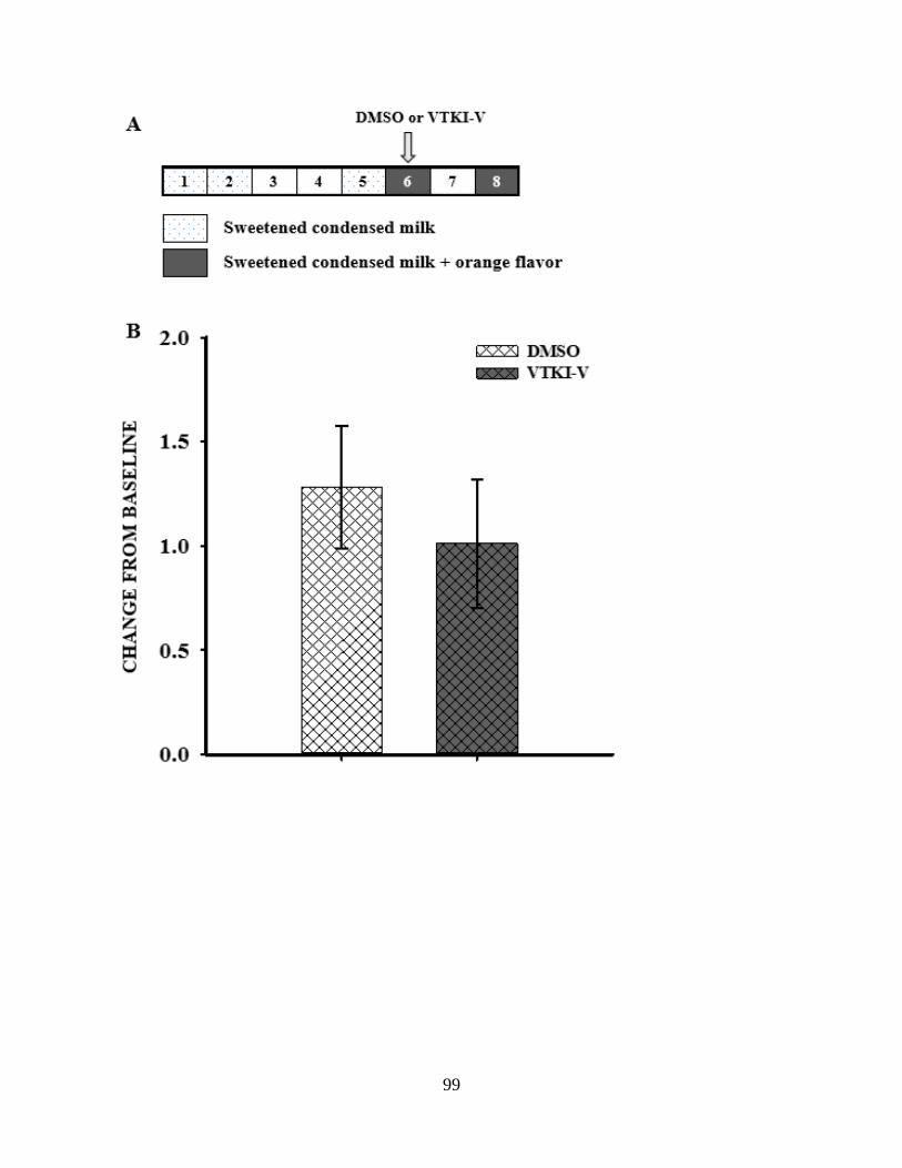
99
100
Figure 4.5. Conditioned taste aversion test. (A) Training paradigm: dots indicate days with one
hour training with sweetened condensed milk (SCM) alone, grey boxes indicate days with SCM
+ orange flavor, the day of the conditioning trial is indicated with an arrow. Animals did not
exhibit aversion to unconditioned stimulus (orange flavor) following conditioning trial with one
dose of DMSO or VTKI-V. Data are change from baseline means ± S.E.M. (n = 5 (vehicle) n = 7
(drug)). *p ≤ 0.05; **p ≤ 0.01; ***p ≤ 0.001.
101
CHAPTER 5:
DISCUSSION AND GENERAL CONCLUSIONS
Integration of Nutrient Detection and Satiety State
Feeding behaviors are dynamically tuned according to sensory determinants of palatability and
physiological need. A state of internal energy depletion dictates that an animal must activate
foraging relevant behavioral programs to maximize food intake while suppressing all other
extraneous activities such as reproduction or avoidance of predators. While in this state,
metabolic processes minimized, foraging relevant behaviors are activated or promoted, sensory
perceptions of food related stimuli are heightened, and mechanisms of memory formation are
potentiated. Once an energetically appropriate amount of food has been obtained and ingested,
internal energy sensing mechanisms activate negative feedback signals which act to suppress
ingestion and foraging behaviors, allowing the fully satiated animal to return to other behaviors.
While the states of “starvation” and “satiation” are the maximal endpoints of the energetic scale,
“hunger” and “satiety” are non-absolute states positioned somewhere between these opposite
extremes. Where an animal falls within the satiety spectrum determines how likely it is to
execute or terminate feeding-relevant behavioral programs. The predisposition of an individual
to overconsumption of calories and weight gain can develop from disruption or maladaptation of
these basic biological processes. The mechanisms by which consumption is controlled by
external sensory cues and internal metabolic status therefore remain a critical topic of
investigation in efforts to prevent weight gain and obesity. Satiety-state dependent mechanisms

102
which block overconsumption of high energy foods are of particular interest, as they offer
potential targets for preventative treatments against weight gain.
Studies of food intake in model organisms have demonstrated that feeding behavior plasticity is
achieved in part by alterations of feeding thresholds in response to internal energy needs and
external food availability (40, 68). However, the mechanisms coupling satiety state to sensory
perception and coding are continuing to be elucidated. The primary driving force for feeding
initiation and ingestion in Drosophila is the detection of gustatory cues signaling the availability
of nutritive foods (64, 65). The character of the behavioral responses elicited by such sensory
cues are dynamically modulated based on internal metabolic status via circulating hormones
from neuroendocrine cells and fat body tissue such as the Drosophila insulin-like peptides (16,
61), adipokinetic hormone (58), and leptin homolog Unpaired 2 act as coding mechanisms for
the availability of carbohydrate and lipid stores (57). Post-ingestive feedback from the gut
following a meal may also inhibit feeding via the recurrent nerve or medial abdominal nerve
(68). This behavioral plasticity is evident in the responses of larvae to nutrient detection and
satiety state. The baseline activities of well-fed larvae are optimized to promote the ingestion of
nutritive foods and minimize energy expenditure. If provided with sweet nutritive foods, they
tend to eat at a steady baseline rate, but decline foods that are difficult to masticate or have
aversive taste (15, 16). The character of baseline behaviors are modified in response to food
deprivation, which enhances the rate of feeding (16) and food preferences (15) in order to restore
energy balance. Similarly in the adult fly, gustatory information is obtained by gustatory receptor
neurons in the periphery respond to bitter and sweet compounds and promote either acceptance
or rejection behaviors (64, 65) by regulating motor neurons subprograms for proboscis extension
and ingestion (14, 216-218). The thresholds for gustatory gating of acceptance or rejection is

103
dependent on satiety state, as food deprived flies display alterations in responses to both sweet
and aversive gustatory inputs (219) . However, how the detection of peripheral signals of
metabolic state is translated into alteration of feeding thresholds in adult and larval stages
remains a topic of investigation (220), and whether this reflects a satiety state-dependent neural
mechanism or results from the failure to ingest enough sugar is unclear (88). In order to
determine how the expression of different behavioral characteristics is constrained by satiety or
promoted by hunger, we must first identify the neuronal circuits whose activity is necessary for
the maintenance of basal feeding behavior qualities.
Evidence for satiety-state dependent mechanism for feeding rate suppression
Taken together, several aspects of the feeding behavior demonstrated larvae (16, 66) provide
evidence for the existence of satiety-state dependent processes regulating their rate of ingestion.
First, satiated larvae steadily feed at a rate of roughly one bite per second when provided with
nutritive food that is easily masticated, and this baseline rate is maintained over long periods of
time. However, they are capable of feeding at a maximum rate of 2 bites per second when
deprived of food for 150 minutes or more. Given that the maximum feeding rate is only utilized
when larvae are experiencing a deficit of internal energy, this maximum rate is likely to be
energetically unfavorable. Furthermore, when larvae that have been deprived of food are placed
on nutritive media, the initial maximum rate of feeding gradually declines over time, and returns
to a steady baseline. The selective execution maximum feeding capacity indicates that it is
expendable for the maintenance of internal energy stores. The observation that re-feeding results
in time dependent decay of feeding rate suggests that ingestive output is modulated by post-
ingestive inhibitory mechanisms, and argues against the presence of starvation dependent feeding

104
rate enhancement mechanisms. In addition, several studies have shown that larval and adult
feeding behaviors are dependent on factors such as Drosophila insulin-like peptides (16), the
leptin-like Unpaired-2 protein (221), and circulating metabolite levels (222), whose levels are
dependent on food intake, and exert an inhibitory effect on feeding rate.
Recent studies in larvae and adults have identified two separate central circuits in with roles in
satiety-state dependent feeding regulation. Following the characterization of pumpless and
klumpfuss mutants, which fail to appropriately move food from the pharynx into the esophagus
and display reduced food intake (223), it was shown that these mutants also display increased
expression of a neuropeptide called hugin, a putative homologue of mammalian neuromedin U
(224), whose expression is found in roughly 20 neurons located in the larval SOG. Hugin
expression is suppressed during following starvation and amino acid deprivation, and its up-
regulation is correlated with decreased food intake and food seeking behavior, suggesting that
hugin levels act to encode satiety status. Circuit analysis revealed that hugin neurons receive
input from gustatory sensory neurons and chemosensory neurons of the pharynx, and extend
axonal processes to the ring glad (a major endocrine organ) and pharyngeal muscles (required for
mouth hook movement and ingestion), indicating that hugin coordinates sensory inputs with
growth, metabolism and food intake (223). In contrast to the hugin circuit, which is nutrient
responsive and satiety dependent, Scott et. al recently identified a novel circuit which exerts a
tonic inhibitory tone to suppress meal initiation and consumption (220) by selectively gating
motor neuron output in adult flies. Mediated by four gabaergic neurons in the ventral region of
the SOG, which receive input from gustatory receptor neurons and project to motor neurons
regulating food intake, this circuit is unique in that its activity is not dependent a homeostatic
pathway or taste detection, but rather it establishes a central threshold on multiple feeding motor

105
neurons which must be overcome in order to initiate feeding (220). Together, these findings
indicate that 1) excessive food ingestion is suppressed by nutrient ingestion, and 2) both
homeostatic and taste dependent pathways modulate ingestive behavior rate by overcoming
feeding inhibition circuits. This suggests that the maximum capacity of larval ingestive motor
circuits is likely to be directly or indirectly inhibited in satiated larvae, and the maximum feeding
rate demonstrated by starved larvae is likely to be due to dis-inhibition of these circuits by
nutrient and gustatory dependent signals.
The Overlapping Roles of Octopamine and Dopamine in Appetitive Learning
The vast majority of investigations into the role of OA signaling and behavior in flies and other
insects have centered on its relationship to appetitive learning and reward driven behaviors. In
Drosophila, the prevailing paradigm regarding the contributions of dopamine (DA) and OA to
learning and behavior has been that DA is required for aversive learning but not reward learning,
and OA is required for reward learning but not aversive learning (78-81). Studies of the
appetitive behaviors of bees demonstrate that OA treatment effects behavioral responses to
sucrose reward (85, 225-227) and acts as a taste driven reward signal to enhance food seeking
(228), food intake (229), and appetitive learning (78, 79), as well as circadian rhythms (230,
231). In particular, studies in bees show that OA treatment effects behavioral responses to
sucrose reward (85, 225-227) and OA injection into the MB or AL can substitute for sucrose
presentation or pairing in conditioned learning paradigms (85). Also, flies lacking OA due to a
mutation in the tyramine-beta-hydroxylase (tβh) enzyme showed no impairment in aversive tests
involving the pairing of a novel odor with a shock, but did not learn to associate a sugar reward
with odor (78). Conversely, the acute inhibition of dopaminergic neurons was demonstrated to

106
block aversive learning without affecting sugar learning (78). Furthermore, light-induced
activation of dopaminergic neurons via channel rhodopsin expression was shown to substitute for
shock pairing to elicit aversive memory formation, while OA neuron stimulation was shown to
substitute for sugar reward (79). The requirement for dopamine signaling was mapped to three
clusters of cells in the MB, each with distinctive roles in the phases of aversive memory
encoding (232, 233).
More recently, however, the advent of a wide array of acutely inducible constructs for the
manipulation of neural activity, coupled with tools allowing fine spatial control of their
expression, has provided more insight into the relative contributions of OA and DA to learning.
In particular, recent studies manipulating small sets of dopamine neurons demonstrate that the
dichotomy of OA/food reward and DA/punishment is an oversimplification. For example, small
subsets of DA neurons and DA receptor neurons in the mushroom bodies (MB) have shown that
some dopamine signals may also modulate reward responses (82, 88, 234). In the insect brain,
the MBs comprise a protocerebral higher brain center known for roles in olfactory processing
and learning and memory (86), and in particular is required for associative learning (78, 235-
238). The dopamine receptor DA1, which acts by increasing adenylyl cyclase activity, is highly
expressed in adult MB (234). Studies using DA1 mutants demonstrated that lack of DA1
signaling in the adult MB blocked learning association of an odor stimulus with an electric
shock, which is supportive of a role for DA in aversive learning (239). However, the same
mutants also showed impairment of learning an odor associated with sucrose reward (239). In
larvae, DA1 mutants also showed defects in aversive and appetitive learning assays. Another
major finding came with the demonstration that satiety levels influence performance of flies in
appetitive memory tests via overlapping DA and NPF/NPFR1 signaling mechanisms (88). It was

107
demonstrated that the lower appetitive memory performance observed in satiated flies was due to
tonic inhibition of MB neurons medial lobe/pedunculus DA neurons (88). Furthermore,
stimulation of neurons expressing NPF promoted appetitive memory in satiated flies by
suppressing the inhibition of MB neurons by DA, enabling the expression of enhanced food
associated conditioned responses (88). It has since been discovered that the driver lines used in
earlier reward/aversion learning studies omitted a subset of dopaminergic neurons in the PAM
region of the MB, which are required for reward learning (89, 240). Activation of PAM DA
neurons paired with odor presentation stimulates robust appetitive memory formation, even in
flies lacking OA, and blocking these neurons disrupts appetitive learning with both
sweet/nutritive and sweet/non–nutritive sugars (89, 240). Also, pairing OA neuron activation
with odor presentation led to only short-term memory formation, which is rendered dispensable
by downstream PAM DA neuron activation (89). Moreover, it was demonstrated that part of the
OA-dependent short-term memory encoding is mediated via activation of its Oamb receptor on a
subset of DA neurons in the MB, as activation of DA/Oamb neurons was shown to bypass the
need for sugar in appetitive memory formation (89). It therefore appears that subsets of DA
neurons in the MB represent short-term and long-term reinforcing effects of nutritious sugar in
learning and act downstream of OA signals (89).
From these studies, a general picture of fly pathways regulating appetitive motivation is
emerging which involves parallel and overlapping contributions from DA, NPF/NPFR1, and OA.
DA appears to provide a tonic inhibitory signal which can be modulated by selective
NPF/NPFR1 gating mechanisms to promote execution of either appetitive or aversive behavior
(88), and also by OA/Oamb gated signaling which provides gustatory signals regarding food
quality (89). With this model in mind, DA neurons in the MB can be thought of as a central

108
behavioral subprogram selection circuit, in which subprogram selection is gated by both nutrient
detection and satiety state.
The Role of OA in Sweet Taste Encoding
While it is clear that OA signaling mediates the short-term reinforcing properties of sugar in
appetitive behavior, it remains to be determined which OA neurons are responsible for this
process, how they do so, and how OA-dependent sweetness encoding mechanisms interact with
other appetitive motivation pathways. One consistent observation is that both tβh mutants and
OA receptor mutants display inappropriate detection of, and responses to, sugar. Given the
ability of OA injection and tdc2-gal4 neuron activation to substitute for sugar in appetitive
learning paradigms, it seems clear that OA signaling is at least one component coupling
detection of external nutrient cues to appetitive responses. More recent work has demonstrated
that sweet taste and nutrient value represent parallel appetitive reinforcement pathways in flies
(90). OA-dependent sugar learning appears to provide the transient reinforcing properties of the
detection of sweet taste, but the need for OA reinforcement is bypassed by the nutrient value of
sugar. For example, acute inhibition of OA neurons blocked appetitive learning with arabinose, a
sweet but non-nutritive compound, but had no effect when sweet and nutritious sucrose was used
(89), which implicates OA in encoding the gustatory detection of “sweetness”. The presence of
OA neurons in the SOG, which is known to be an important site for integration of gustatory
inputs, provides anatomical support for this idea (119), and it may be that OA neurons act as
second order transducers of olfactory receptor neuron signals.

109
Overlapping functions of VUM1 and VUM2 OA neurons in feeding regulation
OA signaling is required for the establishment of basal feeding rate, as well as for starvation
dependent feeding rate enhancement. These regulatory roles can be mapped to two small clusters
of OA neurons in the SOG. Targeted lesioning of only the VUM1 group resulted in a significant
increase in the feeding activity of fed larvae. However, this increased activity was blocked when
the VUM2 group was ablated in tandem. In fasted larvae, lesioning of the VUM2 group reduced
hunger-dependent feeding rate enhancement, and this reduction was compounded by joint
ablation of both the VUM1 and VUM2 groups. This suggests that the proper control of feeding
rate in fed and fasted conditions requires negative regulatory input from VUM1 neurons, and
positive input from the VUM2 group. These findings suggest that components of the OA circuit
are selectively responsive to satiety state. As tdc2-gal4 neuron activation selectively promotes
the ingestion of sweet foods, it is likely that OA signaling is a component of a system that guides
larvae toward nutritive substances.
While we have shown that OA signaling is required for both starvation dependent feeding rate
enhancement and the suppression of food intake during satiety, further work will be necessary to
determine the relative contributions of OA neurons in the SOG in these processes, and what
downstream pathways mediate their effects. Lesioning of VUM2 OA neurons was sufficient to
block enhancement of food intake due to tdc2-Gal4 neuron excitation, while both VUM1 and
VUM2 lesion is necessary to reduce starvation dependent feeding rate to the same level as is
observed following inhibition of tdc2-Gal4 neurons. This suggests that both VUM1 and VUM2
groups contribute to starvation dependent feeding rate increases. The unique role of the VUM1
subset of OA SOG neurons in this behavioral response suggests that OA dependent feeding rate
enhancement is blocked under conditions of satiety and operates in a similar pathway as

110
Oamb/NPF, and it is possible that a VUM1 or VUM2 dependent mechanism directly or
indirectly links OA signaling to NPF.
A Novel OA/Oamb/NPF Pathway Maintains Basal Feeding Rate
While many studies have demonstrated that NPF/NPFR1 signaling is required for starvation
dependent alterations in motivational state and appetitive behavior (14, 66, 69, 88), targeted
manipulation of subsets of NPFR1 neurons reveals that NPF neurons mediate spatially and
functional distinct regulatory roles (69, 89). The selective manipulation of NPF SOG neurons has
yet to be investigated, and our experiments provide the first indication that NPF may also play an
important role in determining the character of basal feeding activity. Targeted lesioning of NPF
SOG neurons reveals an enhancement of palatable food ingestion, suggesting that these neurons
may act as a satiety level dependent braking system to hold the feeding rate of satiated larvae at a
baseline level. This is reminiscent of findings from mammals which demonstrate that
hypothalamic NPY expressing neurons are inhibited by high levels of leptin and insulin
(Figlewicz, 2009), and some are directly inhibited by glucose (Levin 2006). When animals are
deprived of food, the levels of leptin, insulin, and glucose fall, which results in a loss of NPY
neuron inhibition, and a subsequent enhancement of food intake. Currently, little is known
regarding how the activity levels of Drosophila NPF neurons are regulated. NPF-expressing
neurons innervate broad regions of the brain and may simultaneously modulate distinct neural
circuits to promote food seeking (88). NPF neurons in the SOG have extensive dendritic arbors
which are likely to receive inputs from multiple upstream pathways, and their proximity to the
esophagus makes them likely targets for directly or indirect targeting by circulating satiety
factors, sugars, or other nutrients.

111
The unique roles of subesophogeal ganglion neuropeptide F (NPF) and octopamine (OA)
neurons in feeding rate regulation provide support for the presence of novel circuit involved in
satiety-state dependent inhibition of sugar overconsumption. Laser lesion analysis indicates that
the presence of two NPF neurons in the SOG is necessary for the expression of basal feeding rate
characteristics on sweet, palatable food, as lack of these neurons enhances basal feeding rate.
Our findings demonstrate a phenotypic and functional overlap between NPF SOG neurons and
OA SOG neurons, as loss of the VUM1 OA cell group also enhances basal feeding rate.
Furthermore, manipulation of the expression of the Oamb OA receptor indicates that NPF
expression of Oamb is required for the inhibitory role of OA on basal feeding rate. Global CNS
knockdown of Oamb resulted in an enhancement of feeding rate, which was phenocopied by
targeting of the knockdown to NPF expressing neurons, as well as in an enhancer-trap based
Oamb driver line which colocalizes with NPF neurons in the SOG. In addition to the well
characterized role of OA in coding for gustatory detection of sugar (78, 81, 85, 89, 228), these
findings indicate that OA may act upstream of NPF to enhance their activity when sugar is
ingested. This is corroborated by previous experiments which demonstrate that NPF
immunoreactivity is selectively enhanced in the SOG region following prefeeding with both
nutritive sugar and non-nutritive sweeteners (186), suggesting that the activity of these neurons is
dependent on the detection of sweet taste, though they have not been demonstrated to directly
connect with gustatory receptor neurons.
The anatomical proximity of SOG NPF and OA neurons suggest that they may exert their
regulatory roles via parallel and overlapping downstream pathways in the SOG. Artificial
excitation of tdc2-Gal4 neurons or loss of OA neurons in the SOG results in the expression of
increased ingestive rate in satiated larvae, indicating the presence of an OA dependent

112
mechanism in the SOG for promotion of sweet food intake which is functionally silent at when
internal energy stores are at baseline. The finding that the Oamb is required in NPF neurons for
appropriate basal food ingestion suggests that OA and NPF work in a concerted fashion to
suppress excessive feeding of sugar during satiety. However, inhibition of OA signaling in tdc2-
Gal4>UAS-shibereTS larvae does not affect basal feeding rate, indicates that overlapping
mechanisms exist to regulate food intake during satiety.
NPF/NPFR1 signaling acts to inhibit downstream neurons, therefore lesioning of SOG NPF
neurons is likely to reduce an NPF/NPFR1 signal which exerts an inhibitory effect on local
neurons in the SOG in satiated larvae. Loss of Oamb in these neurons may also reduce this
inhibitory effect. It has been suggested that satiated flies receive maximum inhibitory feedback
on behavioral output such that sensory input is behaviorally ineffective, but this inhibition may
be decreased in a dose dependent fashion as food deprivation is prolonged, allowing sensory
input to become increasingly effective in initiating feeding (88). With such a mechanism, the
likelihood that an appetitive behavior is triggered by a conditioned odorant or gustatory cue
would therefore be determined by competition between inhibitory systems in the brain. If this is
the case, appetitive behaviors may be given their baseline characteristics via active tonic
inhibition by circuits sensitive to circulating nutrients.
Our findings provide the first evidence for an upstream regulatory pathway that specifically
targets NPF SOG neurons. Given the role of OA in gustatory encoding of sweet taste, local OA
signaling in the SOG may link NPF neuron activity indirectly with the detection of sweet tasting
compounds, which may provide the input to link a NPF/NPFR1 dependent braking system to
upstream gustatory inputs. It will be illuminating to determine how the basal activity level of
these neurons may be altered in different satiety states, and their responsiveness to known

113
regulators of food intake and satiety such as dILPs. It remains unclear how the activity of NPF
SOG neurons exerts an effect on feeding rate, which must be determined by functional
characterization of local SOG NPFR1 expressing neurons. Anatomical analysis and application
of the GRASP technique for mapping synaptic connections may reveal what direct connectivity
may exist between the NPF and OA cell groups in the SOG, and how OA neuron activity levels
correlate with NPF neuron activity in different satiety states.
A VEGFR Pathway for Behavioral Plasticity
Animal growth is determined by environmental, genetic, hormonal, and nutritional factors, and is
critically mediated by appropriate expression of growth factors. Despite important links between
growth factors and nutrition, direct roles for these proteins in the regulation of feeding behaviors
has been underexplored. Intriguingly, the levels of various growth factors have been found to be
elevated in the CSF of rats who were fed or injected with glucose relative to starved rats, and
intracerebroventricular administration of inhibitors selective for growth factor receptors have
been shown to transiently decrease food intake (141). We provide evidence of a novel regulatory
role for a PDGFR/VEGFR-related receptor (Pvr) required for OA-dependent homeostatic control
of feeding behavior and growth in flies. Pvr has been identified as sharing structural homology
with receptors for both platelet derived growth factor (PDGFR) and vesicular endothelial growth
factor receptor (VEGFR), though it shares greater similarity with the latter (132). Our findings in
the fly model of feeding behavior suggest that the neuronal effects of VEGFR signaling
pathways have relevance to the regulation of behavioral output. Furthermore, the extension of
these findings into a rodent model system of food intake and diet-induced hyperphagia indicate

114
that a conserved role exists for the central regulation of food intake by a mammalian VEGFR
pathway.
When given free access to a palatable, energy dense diet, humans and other animals tend to
consume more calories, and this results in an increase in the amount of stored fat they maintain
and defend (241-243). Many studies have implicated the “rewarding” nature of tasty foods in the
drive to overeat (77, 198). Palatable foods are more likely to be eaten in excess relative to bland
foods, and the pleasurable effects of consuming them may override homeostatic signals (77, 198-
200, 210, 244). Food that have tastes which provide cues of palatability activate reward
pathways (77), and it is speculated that obese individuals may be more susceptible to the
temptations of food. In support of this, obese adults display heightened activity in neural reward
circuits in response to palatable food or food-associated cues (201, 202, 245). Alternately, the
taste of energy dense food may contribute to overeating by suppressing satiety. For example, in
the presence of palatable food, endocannabinoids increase appetite by stimulating the desire to
eat while also blocking signals for meal termination (203).
However, it may be that high energy density foods bypass satiety pathways based on the mass of
food ingested. Many studies find that when rodent models raised on balanced diets are given free
access to diets that contain high levels of sugar and fat, animals continue to eat the same mass of
food (246) and similar findings have been shown in humans (247). If maintained on an energy
diet for long periods, rodents will consume excessive calories and become obese (248, 249), and
will maintain an elevated body weight (241, 242). While much is known regarding the initiation
and consumption phases of feeding behavior, much less is known of the complex processes
regulating the termination and postconsumption phases of feeding, the dysregulation which are
likely to contribute to the hyperphagia seen in diet dependent obesity (249).

115
In our study, young male rats were given ad libitum access to standard rodent CHOW, or were
acclimated to an energy dense palatable HFHS diet prior to treatment with VTKI-V. Animals
given long term access to the diet used in this study have been shown to exhibit rapid alterations
in meal pattern, including increased meal size and duration, which correlate with the
development of diet induced obesity over time (206, 213, 250). Our results show a similar trend.
DMSO injected control animals maintained on CHOW and HFHS diet displayed similar patterns
of food intake in terms of meal number, mass, duration, and number of bouts. Given the higher
energy density of the diet presented to the HFHS control group, consumption of the same mass
of food containing more calories resulted larger caloric intake overall, and larger average meal
size. After VTKI-V administration, CHOW fed animals reduced their intake slightly in terms of
both mass and calories, but the effect was only significant when overall cumulative intake was
assessed, and no detectable alteration was seen in other meal pattern variables. However, drug
treatment resulted an acute effect on the mass of food ingested by the HFHS diet group. Animals
decreased the mass of their meals by roughly 30% due to a reduction in the number of bouts per
meal and meal duration. This brought their average caloric meal size down to a similar level as
CHOW fed animals.
Our findings suggest that VTKI-V treatment selectively alters the mass of food ingested, but
only when the energy density of that food exceeds caloric need. Rats predisposed to diet induced
obesity display larger meal sizes and decreased satiety ratios on high energy palatable diets,
relative to diet induced obesity resistant rats (206). Rodent models of diet induced obesity have
central resistance to leptin (251), which has been correlated with reduction in meal size, not
number (56). Further work will be needed to determine if VTKI-V targets similar systems
involved in the regulation of food intake.

116
Additionally, we find that VTKI-V treatment results in a reduction in the rate of weight gain.
This effect was immediate and persisted over the five days following treatment. Interestingly,
reduction in weight gain was seen in both CHOW and HFHS fed groups, despite the fact that
CHOW fed animals displayed very little alteration in their pattern of food intake following drug
treatment. This suggests that there is some component of the variance in the rate of weight gain
between control and drug treated animals is not fully accounted for by alterations in food intake.
It is possible that VTKI-V interacts with other neural systems involved in metabolism. Given
that obesity is correlated with alterations in sympathetic nervous system output (252, 253) and
fat metabolism (27) there may be an effect on peripheral metabolism of stored fat or regulation
of energy expenditure. Rats acclimated to energy dense diets achieve and maintain a higher body
weight set point (207, 241), which is correlated with alterations in energy expenditure and
metabolism (212, 243). Furthermore, this is accompanied by alterations in the hypothalamic and
peripheral activity of noradrenergic systems, including differences in neural sympathetic output
and responsiveness (209, 252), ventromedial hypothalamus morphology (253), and neural
monoamine metabolism (252). Further work will be needed to determine if VTKI-V targets
VEGFR2 on norepinephrine neurons, sympathetic nervous system output, or metabolism.
117
REFERENCES
1. Kopelman PG (2000) Obesity as a medical problem. Nature 404(6778):635-643.
2. Finkelstein EA, Ruhm CJ, & Kosa KM (2005) Economic causes and consequences of
obesity. Annual review of public health 26:239-257.
3. Zhang T, Branch A, & Shen P (2013) Octopamine-mediated circuit mechanism
underlying controlled appetite for palatable food in Drosophila. Proc Natl Acad Sci U S A
110(38):15431-15436.
4. Zheng H & Berthoud HR (2007) Eating for pleasure or calories. Current opinion in
pharmacology 7(6):607-612.
5. Weiner IB (2003) The handbook of humanistic psychology: leading edges in theory,
research, and practice. Psychotherapy research : journal of the Society for Psychotherapy
Research 13(1):119-121.
6. Seward JP (1956) Drive, incentive, and reinforcement. Psychological review 63(3):195-
203.
7. Irving B. Weiner RJN, Sheri Mizumori (2012) Handbook of Psychology, Behavioral
Neuroscience (John Wiley & Sons, Hoboken, New Jersey) 2nd Ed p 739.
8. Watts AG (2000) Understanding the neural control of ingestive behaviors: helping to
separate cause from effect with dehydration-associated anorexia. Hormones and behavior
37(4):261-283.
9. J. L (1992) Neurobiology of feeding and nutrition (Academic Press, London).

118
10. Gibney MJ & Wolever TM (1997) Periodicity of eating and human health: present
perspective and future directions. The British journal of nutrition 77 Suppl 1:S3-5.
11. Blundell J (1991) Pharmacological approaches to appetite suppression. Trends in
pharmacological sciences 12(4):147-157.
12. Blundell JE, Lawton CL, Cotton JR, & Macdiarmid JI (1996) Control of human appetite:
implications for the intake of dietary fat. Annual review of nutrition 16:285-319.
13. de Graaf C, Blom WA, Smeets PA, Stafleu A, & Hendriks HF (2004) Biomarkers of
satiation and satiety. The American journal of clinical nutrition 79(6):946-961.
14. Gordon MD & Scott K (2009) Motor control in a Drosophila taste circuit. Neuron
61(3):373-384.
15. Wu QW, T.; Lee, G.; Park, J.H.; Cai, H.N.; Shen, P. (2003) Developmental control of
foraging and social behavior by the Drosophila neuropeptide Y-like system. Neuron
39(1):147-161.
16. Wu QZ, Y.; Xu, J.; Shen, P. (2005) Regulation of hunger-driven behaviors by neural
ribosomal S6 kinase in Drosophila. Proc Natl Acad Sci U S A 107(37):13289-13294.
17. Kennedy GC (1953) The role of depot fat in the hypothalamic control of food intake in
the rat. Proceedings of the Royal Society of London. Series B, Containing papers of a
Biological character. Royal Society 140(901):578-596.
18. Berthoud HRM, C. (2008) The brain, appetite, and obesity. Annual Review of Psychology
59:55-93.
19. Leibowitz SF & Brown LL (1980) Histochemical and pharmacological analysis of
noradrenergic projections to the paraventricular hypothalamus in relation to feeding
stimulation. Brain research 201(2):289-314.

119
20. Levin BEP, B. (1993) Defective glucoregulation of brain a2-adrenoceptors in obesity
prone rats. Am. J. Physiol. Regul. Integr. Comp. Physiol. 264:R305-R311.
21. Wellman PJD, B.T., Marien, A.; McMahon, L. (1993) Modulation of feeding by
hypothalamic paraventricular nucleus alpha1 and alpha2-adrenergic receptors. Life
Sciences 53:669-679.
22. Levin BE (1990) Obesity-prone and -resistant rats differ in their brain [3H]
paraminoclonidine binding. Brain research 512(1):54-59.
23. Oltmans GA, Lorden JF, Callahan MF, Beales M, & Fields JZ (1981) Increases in alpha-
adrenergic receptors in the hypothalamus of the genetically obese mouse (ob/ob). Brain
research 222(2):411-416.
24. Friedman JJ (1989) Vascular sensitivity and reactivity to norepinephrine in diabetes
mellitus. The American journal of physiology 256(4 Pt 2):H1134-1138.
25. Karkanias GB, Morales JC, & Etgen AM (1998) Effects of diabetes and estradiol on
norepinephrine release in female rat hypothalamus, preoptic area and cortex.
Neuroendocrinology 68(1):30-36.
26. Ganguly PK, Dhalla KS, Innes IR, Beamish RE, & Dhalla NS (1986) Altered
norepinephrine turnover and metabolism in diabetic cardiomyopathy. Circulation
research 59(6):684-693.
27. Berthoud HR (2002) Multiple neural systems controlling food intake and body weight.
Neuroscience and biobehavioral reviews 26(4):393-428.
28. Martin JH (2003) Neuroanatomy : text and atlas (McGraw-Hill, New York, N.Y.) 3rd Ed
pp xxiv, 532 p.

120
29. Considine RV, et al. (1996) Serum immunoreactive-leptin concentrations in normal-
weight and obese humans. The New England journal of medicine 334(5):292-295.
30. Schwartz MW, Peskind E, Raskind M, Boyko EJ, & Porte D, Jr. (1996) Cerebrospinal
fluid leptin levels: relationship to plasma levels and to adiposity in humans. Nature
medicine 2(5):589-593.
31. Baskin DG, Breininger JF, & Schwartz MW (1999) Leptin receptor mRNA identifies a
subpopulation of neuropeptide Y neurons activated by fasting in rat hypothalamus.
Diabetes 48(4):828-833.
32. Elmquist JK, Bjorbaek C, Ahima RS, Flier JS, & Saper CB (1998) Distributions of leptin
receptor mRNA isoforms in the rat brain. The Journal of comparative neurology
395(4):535-547.
33. Bagdade JD, Bierman EL, & Porte D, Jr. (1967) The significance of basal insulin levels
in the evaluation of the insulin response to glucose in diabetic and nondiabetic subjects.
The Journal of clinical investigation 46(10):1549-1557.
34. Woods SC, Lotter EC, McKay LD, & Porte D, Jr. (1979) Chronic intracerebroventricular
infusion of insulin reduces food intake and body weight of baboons. Nature
282(5738):503-505.
35. Bruning JC, et al. (2000) Role of brain insulin receptor in control of body weight and
reproduction. Science 289(5487):2122-2125.
36. Morton GJ, Meek TH, & Schwartz MW (2014) Neurobiology of food intake in health
and disease. Nature reviews. Neuroscience 15(6):367-378.

121
37. Billington CJ, Briggs JE, Grace M, & Levine AS (1991) Effects of
intracerebroventricular injection of neuropeptide Y on energy metabolism. The American
journal of physiology 260(2 Pt 2):R321-327.
38. Ollmann MM, et al. (1997) Antagonism of central melanocortin receptors in vitro and in
vivo by agouti-related protein. Science 278(5335):135-138.
39. Butler AA, et al. (2001) Melanocortin-4 receptor is required for acute homeostatic
responses to increased dietary fat. Nature neuroscience 4(6):605-611.
40. Morton GJ, Cummings DE, Baskin DG, Barsh GS, & Schwartz MW (2006) Central
nervous system control of food intake and body weight. Nature 443(7109):289-295.
41. Campfield LA, Smith FJ, Guisez Y, Devos R, & Burn P (1995) Recombinant mouse OB
protein: evidence for a peripheral signal linking adiposity and central neural networks.
Science 269(5223):546-549.
42. Halaas JL, et al. (1997) Physiological response to long-term peripheral and central leptin
infusion in lean and obese mice. Proc Natl Acad Sci U S A 94(16):8878-8883.
43. Lee GH, et al. (1996) Abnormal splicing of the leptin receptor in diabetic mice. Nature
379(6566):632-635.
44. Zhang Y, et al. (1994) Positional cloning of the mouse obese gene and its human
homologue. Nature 372(6505):425-432.
45. Kristensen P, et al. (1998) Hypothalamic CART is a new anorectic peptide regulated by
leptin. Nature 393(6680):72-76.
46. Cowley MA, et al. (2003) The distribution and mechanism of action of ghrelin in the
CNS demonstrates a novel hypothalamic circuit regulating energy homeostasis. Neuron
37(4):649-661.

122
47. Cowley MA, et al. (1999) Integration of NPY, AGRP, and melanocortin signals in the
hypothalamic paraventricular nucleus: evidence of a cellular basis for the adipostat.
Neuron 24(1):155-163.
48. Cowley MA, et al. (2001) Leptin activates anorexigenic POMC neurons through a neural
network in the arcuate nucleus. Nature 411(6836):480-484.
49. Kanatani A, et al. (1996) Potent neuropeptide Y Y1 receptor antagonist, 1229U91:
blockade of neuropeptide Y-induced and physiological food intake. Endocrinology
137(8):3177-3182.
50. Elias CF, et al. (2001) Characterization of CART neurons in the rat and human
hypothalamus. The Journal of comparative neurology 432(1):1-19.
51. Kenny PJ (2011) Reward mechanisms in obesity: new insights and future directions.
Neuron 69(4):664-679.
52. Gibbs J, Young RC, & Smith GP (1973) Cholecystokinin decreases food intake in rats.
Journal of comparative and physiological psychology 84(3):488-495.
53. Strubbe JH & Woods SC (2004) The timing of meals. Psychological review 111(1):128-
141.
54. McMinn JE, Sindelar DK, Havel PJ, & Schwartz MW (2000) Leptin deficiency induced
by fasting impairs the satiety response to cholecystokinin. Endocrinology 141(12):4442-
4448.
55. Barrachina MD, Martinez V, Wang L, Wei JY, & Tache Y (1997) Synergistic interaction
between leptin and cholecystokinin to reduce short-term food intake in lean mice. Proc
Natl Acad Sci U S A 94(19):10455-10460.

123
56. Kahler A, et al. (1998) Chronic administration of OB protein decreases food intake by
selectively reducing meal size in male rats. The American journal of physiology 275(1 Pt
2):R180-185.
57. Rieder LE & Larschan EN (2014) Wisdom from the fly. Trends in genetics : TIG.
58. Kim SK & Rulifson EJ (2004) Conserved mechanisms of glucose sensing and regulation
by Drosophila corpora cardiaca cells. Nature 431(7006):316-320.
59. Gronke S, Clarke DF, Broughton S, Andrews TD, & Partridge L (2010) Molecular
evolution and functional characterization of Drosophila insulin-like peptides. PLoS
genetics 6(2):e1000857.
60. Buch S, Melcher C, Bauer M, Katzenberger J, & Pankratz MJ (2008) Opposing effects of
dietary protein and sugar regulate a transcriptional target of Drosophila insulin-like
peptide signaling. Cell metabolism 7(4):321-332.
61. Geminard C, Rulifson EJ, & Leopold P (2009) Remote control of insulin secretion by fat
cells in Drosophila. Cell metabolism 10(3):199-207.
62. Slaidina M, Delanoue R, Gronke S, Partridge L, & Leopold P (2009) A Drosophila
insulin-like peptide promotes growth during nonfeeding states. Developmental cell
17(6):874-884.
63. Mirth CK, et al. (2014) Juvenile hormone regulates body size and perturbs insulin
signaling in Drosophila. Proc Natl Acad Sci U S A 111(19):7018-7023.
64. Thorne N, Chromey C, Bray S, & Amrein H (2004) Taste perception and coding in
Drosophila. Current biology : CB 14(12):1065-1079.
65. Wang Z, Singhvi A, Kong P, & Scott K (2004) Taste representations in the Drosophila
brain. Cell 117(7):981-991.

124
66. Wu Q, Zhao Z, & Shen P (2005) Regulation of aversion to noxious food by Drosophila
neuropeptide Y- and insulin-like systems. Nature neuroscience 8(10):1350-1355.
67. Xu J, Li M, & Shen P (2010) A G-protein-coupled neuropeptide Y-like receptor
suppresses behavioral and sensory response to multiple stressful stimuli in Drosophila.
The Journal of neuroscience : the official journal of the Society for Neuroscience
30(7):2504-2512.
68. Dethier VG (1976) The Hungry Fly (Harvard University Press, Cambridge).
69. Wang Y, Pu Y, & Shen P (2013) Neuropeptide-gated perception of appetitive olfactory
inputs in Drosophila larvae. Cell reports 3(3):820-830.
70. Nestler EJ (2001) Molecular basis of long-term plasticity underlying addiction. Nature
reviews. Neuroscience 2(2):119-128.
71. Nestler EJ (2005) Is there a common molecular pathway for addiction? Nature
neuroscience 8(11):1445-1449.
72. Wise RA (2004) Dopamine, learning and motivation. Nature reviews. Neuroscience
5(6):483-494.
73. Wise RA & Rompre PP (1989) Brain dopamine and reward. Annu Rev Psychol 40:191-
225.
74. Barron AB, Sovik E, & Cornish JL (2010) The roles of dopamine and related compounds
in reward-seeking behavior across animal phyla. Frontiers in behavioral neuroscience
4:163.
75. Wise RA & Schwartz HV (1981) Pimozide attenuates acquisition of lever-pressing for
food in rats. Pharmacology, biochemistry, and behavior 15(4):655-656.

125
76. Rolls ET (2006) Brain mechanisms underlying flavour and appetite. Philosophical
transactions of the Royal Society of London. Series B, Biological sciences
361(1471):1123-1136.
77. Lutter M & Nestler EJ (2009) Homeostatic and hedonic signals interact in the regulation
of food intake. The Journal of nutrition 139(3):629-632.
78. Schwaerzel M, et al. (2003) Dopamine and octopamine differentiate between aversive
and appetitive olfactory memories in Drosophila. The Journal of neuroscience : the
official journal of the Society for Neuroscience 23(33):10495-10502.
79. Schroll C, et al. (2006) Light-induced activation of distinct modulatory neurons triggers
appetitive or aversive learning in Drosophila larvae. Current biology : CB 16(17):1741-
1747.
80. Claridge-Chang A, et al. (2009) Writing memories with light-addressable reinforcement
circuitry. Cell 139(2):405-415.
81. Honjo K & Furukubo-Tokunaga K (2009) Distinctive neuronal networks and biochemical
pathways for appetitive and aversive memory in Drosophila larvae. The Journal of
neuroscience : the official journal of the Society for Neuroscience 29(3):852-862.
82. Selcho M, Pauls D, Han KA, Stocker RF, & Thum AS (2009) The role of dopamine in
Drosophila larval classical olfactory conditioning. PloS one 4(6):e5897.
83. Tempel BL, Bonini N, Dawson DR, & Quinn WG (1983) Reward learning in normal and
mutant Drosophila. Proc Natl Acad Sci U S A 80(5):1482-1486.
84. McBride SM, et al. (1999) Mushroom body ablation impairs short-term memory and
long-term memory of courtship conditioning in Drosophila melanogaster. Neuron
24(4):967-977.

126
85. Hammer M & Menzel R (1998) Multiple sites of associative odor learning as revealed by
local brain microinjections of octopamine in honeybees. Learning & memory 5(1-2):146-
156.
86. Farris SM (2008) Structural, functional and developmental convergence of the insect
mushroom bodies with higher brain centers of vertebrates. Brain, behavior and evolution
72(1):1-15.
87. Zhang K, Guo JZ, Peng Y, Xi W, & Guo A (2007) Dopamine-mushroom body circuit
regulates saliency-based decision-making in Drosophila. Science 316(5833):1901-1904.
88. Krashes MJ, et al. (2009) A neural circuit mechanism integrating motivational state with
memory expression in Drosophila. Cell 139(2):416-427.
89. Burke CJ, et al. (2012) Layered reward signalling through octopamine and dopamine in
Drosophila. Nature 492(7429):433-437.
90. Burke CJ & Waddell S (2011) Remembering nutrient quality of sugar in Drosophila.
Current biology : CB 21(9):746-750.
91. Alquist RP (1948) A study of the adrotropic receptors. The American journal of
physiology 153:586-600.
92. Ordway GAS, M.A.; Frazer, A. ed (2007) Brain Norepinephrine: Neurobiology and
Therapeutics (Cambridge University Press), 1 Ed.
93. Goldman CK, Marino, L., and Leibowitz (1985) Postsynaptic alpha2-noradrenergic
receptors mediate feeding induced by paraventricular nucleus injection of norepinephrine
and clonidien. European Journal of Pharmacology 115:11-19.

127
94. Leibowitz SF (1988) Hypothalamic Paraventricular Nucleus: Interaction Between c 2-
Noradrenergic System and Circulating Hormones and Nutrients in Relation to Energy
Balance. Neuroscience & Biobehavioral Reviews 12:101-109.
95. Fukuda A, Minami, T. Nabekura, J., and Oomura, Y. (1987) The effects of noradrenaline
on neurons in the rat dorsal motor nucleus of the vagus, in vitro. J. Physiol. 393:213-231.
96. Levin BE, Dunn-Meynell AA, & Routh VH (2002) CNS sensing and regulation of
peripheral glucose levels. International review of neurobiology 51:219-258.
97. Veneman T, Mitrakou A, Mokan M, Cryer P, & Gerich J (1993) Induction of
hypoglycemia unawareness by asymptomatic nocturnal hypoglycemia. Diabetes
42(9):1233-1237.
98. Mitrakou A, et al. (1991) Hierarchy of glycemic thresholds for counterregulatory
hormone secretion, symptoms, and cerebral dysfunction. The American journal of
physiology 260(1 Pt 1):E67-74.
99. Amoroso S, Schmid-Antomarchi H, Fosset M, & Lazdunski M (1990) Glucose,
sulfonylureas, and neurotransmitter release: role of ATP-sensitive K+ channels. Science
247(4944):852-854.
100. Roeper J, Hainsworth AH, & Ashcroft FM (1990) Tolbutamide reverses membrane
hyperpolarisation induced by activation of D2 receptors and GABAB receptors in
isolated substantia nigra neurones. Pflugers Archiv : European journal of physiology
416(4):473-475.
101. Mizuno Y & Oomura Y (1984) Glucose responding neurons in the nucleus tractus
solitarius of the rat: in vitro study. Brain research 307(1-2):109-116.

128
102. Adachi A, Shimizu N, Oomura Y, & Kobashi M (1984) Convergence of hepatoportal
glucose-sensitive afferent signals to glucose-sensitive units within the nucleus of the
solitary tract. Neuroscience letters 46(2):215-218.
103. Karschin A, Brockhaus J, & Ballanyi K (1998) KATP channel formation by the
sulphonylurea receptors SUR1 with Kir6.2 subunits in rat dorsal vagal neurons in situ.
The Journal of physiology 509 ( Pt 2):339-346.
104. Adachi A, Kobashi M, Miyoshi N, & Tsukamoto G (1991) Chemosensitive neurons in
the area postrema of the rat and their possible functions. Brain research bulletin
26(1):137-140.
105. Illes P, et al. (1994) Modulation of locus coeruleus neurons by extra- and intracellular
adenosine 5'-triphosphate. Brain research bulletin 35(5-6):513-519.
106. Finta EP, Harms L, Sevcik J, Fischer HD, & Illes P (1993) Effects of potassium channel
openers and their antagonists on rat locus coeruleus neurones. British journal of
pharmacology 109(2):308-315.
107. Swanson LW & Sawchenko PE (1983) Hypothalamic integration: organization of the
paraventricular and supraoptic nuclei. Annual review of neuroscience 6:269-324.
108. McCabe JRD, M.; Leibowitz, S.F. (1984) Clonidine-induced feeding: analysis of central
sites of action and fiber projections mediating this response. Brain research 309:85-104.
109. Jhanwar-Uniyal M & Leibowitz SF (1986) Impact of food deprivation on alpha 1- and
alpha 2-noradrenergic receptors in the paraventricular nucleus and other hypothalamic
areas. Brain research bulletin 17(6):889-896.
129
110. Leibowitz SF, Jhanwar-Uniyal M, Dvorkin B, & Makman MH (1982) Distribution of
alpha-adrenergic, beta-adrenergic and dopaminergic receptors in discrete hypothalamic
areas of rat. Brain research 233(1):97-114.
111. Penicaud L, Larue-Achagiotis C, & Le Magnen J (1983) Endocrine basis for weight gain
after fasting or VMH lesion in rats. The American journal of physiology 245(3):E246-
252.
112. Cincotta AH, et al. (2000) Chronic infusion of norepinephrine into the VMH of normal
rats induces the obese glucose-intolerant state. American journal of physiology.
Regulatory, integrative and comparative physiology 278(2):R435-444.
113. Monastirioti M, Linn CE, Jr., & White K (1996) Characterization of Drosophila tyramine
beta-hydroxylase gene and isolation of mutant flies lacking octopamine. The Journal of
neuroscience : the official journal of the Society for Neuroscience 16(12):3900-3911.
114. Han KA, Millar NS, & Davis RL (1998) A novel octopamine receptor with preferential
expression in Drosophila mushroom bodies. The Journal of neuroscience : the official
journal of the Society for Neuroscience 18(10):3650-3658.
115. Evans PD & Maqueira B (2005) Insect octopamine receptors: a new classification
scheme based on studies of cloned Drosophila G-protein coupled receptors. Invertebrate
neuroscience : IN 5(3-4):111-118.
116. Maqueira B, Chatwin H, & Evans PD (2005) Identification and characterization of a
novel family of Drosophila beta-adrenergic-like octopamine G-protein coupled receptors.
Journal of neurochemistry 94(2):547-560.
117. Roeder T (2005) Tyramine and octopamine: ruling behavior and metabolism. Annual
review of entomology 50:447-477.
130
118. Vomel M & Wegener C (2008) Neuroarchitecture of aminergic systems in the larval
ventral ganglion of Drosophila melanogaster. PloS one 3(3):e1848.
119. Busch S, Selcho M, Ito K, & Tanimoto H (2009) A map of octopaminergic neurons in the
Drosophila brain. The Journal of comparative neurology 513(6):643-667.
120. Melcher C, Bader R, & Pankratz MJ (2007) Amino acids, taste circuits, and feeding
behavior in Drosophila: towards understanding the psychology of feeding in flies and
man. The Journal of endocrinology 192(3):467-472.
121. Ferrara N (2002) Role of vascular endothelial growth factor in physiologic and
pathologic angiogenesis: therapeutic implications. Seminars in oncology 29(6 Suppl
16):10-14.
122. Warner-Schmidt JL & Duman RS (2007) VEGF is an essential mediator of the
neurogenic and behavioral actions of antidepressants. Proc Natl Acad Sci U S A
104(11):4647-4652.
123. Ferrara N, Gerber HP, & LeCouter J (2003) The biology of VEGF and its receptors.
Nature medicine 9(6):669-676.
124. Dunn IF, Heese O, & Black PM (2000) Growth factors in glioma angiogenesis: FGFs,
PDGF, EGF, and TGFs. Journal of neuro-oncology 50(1-2):121-137.
125. Shirakawa K, et al. (2002) Tumor-infiltrating endothelial cells and endothelial precursor
cells in inflammatory breast cancer. International journal of cancer. Journal
international du cancer 99(3):344-351.
126. Marshall CJ (1995) Specificity of receptor tyrosine kinase signaling: transient versus
sustained extracellular signal-regulated kinase activation. Cell 80(2):179-185.
131
127. Olsson AK, Dimberg A, Kreuger J, & Claesson-Welsh L (2006) VEGF receptor
signalling - in control of vascular function. Nature reviews. Molecular cell biology
7(5):359-371.
128. Reigstad LJ, Varhaug JE, & Lillehaug JR (2005) Structural and functional specificities of
PDGF-C and PDGF-D, the novel members of the platelet-derived growth factors family.
The FEBS journal 272(22):5723-5741.
129. Hiratsuka S, Minowa O, Kuno J, Noda T, & Shibuya M (1998) Flt-1 lacking the tyrosine
kinase domain is sufficient for normal development and angiogenesis in mice. Proc Natl
Acad Sci U S A 95(16):9349-9354.
130. Makinen T, et al. (2001) Inhibition of lymphangiogenesis with resulting lymphedema in
transgenic mice expressing soluble VEGF receptor-3. Nature medicine 7(2):199-205.
131. Duchek P, Somogyi K, Jekely G, Beccari S, & Rorth P (2001) Guidance of cell migration
by the Drosophila PDGF/VEGF receptor. Cell 107(1):17-26.
132. Munier AI, et al. (2002) PVF2, a PDGF/VEGF-like growth factor, induces hemocyte
proliferation in Drosophila larvae. EMBO reports 3(12):1195-1200.
133. Learte AR, Forero MG, & Hidalgo A (2008) Gliatrophic and gliatropic roles of
PVF/PVR signaling during axon guidance. Glia 56(2):164-176.
134. Cho NK, et al. (2002) Developmental control of blood cell migration by the Drosophila
VEGF pathway. Cell 108(6):865-876.
135. Heino TI, et al. (2001) The Drosophila VEGF receptor homolog is expressed in
hemocytes. Mechanisms of development 109(1):69-77.
136. Fredriksson L, Li H, & Eriksson U (2004) The PDGF family: four gene products form
five dimeric isoforms. Cytokine & growth factor reviews 15(4):197-204.

132
137. Sondell M, Lundborg G, & Kanje M (1999) Vascular endothelial growth factor
stimulates Schwann cell invasion and neovascularization of acellular nerve grafts. Brain
research 846(2):219-228.
138. Sondell M, Lundborg G, & Kanje M (1999) Vascular endothelial growth factor has
neurotrophic activity and stimulates axonal outgrowth, enhancing cell survival and
Schwann cell proliferation in the peripheral nervous system. The Journal of neuroscience
: the official journal of the Society for Neuroscience 19(14):5731-5740.
139. Matsuzaki H, et al. (2001) Vascular endothelial growth factor rescues hippocampal
neurons from glutamate-induced toxicity: signal transduction cascades. FASEB journal :
official publication of the Federation of American Societies for Experimental Biology
15(7):1218-1220.
140. Greene J, Banasr M, Lee B, Warner-Schmidt J, & Duman RS (2009) Vascular
endothelial growth factor signaling is required for the behavioral actions of
antidepressant treatment: pharmacological and cellular characterization.
Neuropsychopharmacology : official publication of the American College of
Neuropsychopharmacology 34(11):2459-2468.
141. Plata-Salaman CR (1988) Food intake suppression by growth factors and platelet
peptides by direct action in the central nervous system. Neuroscience letters 94(1-2):161-
166.
142. Oomura Y, et al. (1992) A new brain glucosensor and its physiological significance. The
American journal of clinical nutrition 55(1 Suppl):278S-282S.
143. Sasaki K, et al. (1991) Effects of fibroblast growth factors and platelet-derived growth
factor on food intake in rats. Brain research bulletin 27(3-4):327-332.

133
144. Cordeira JW, Frank L, Sena-Esteves M, Pothos EN, & Rios M (2010) Brain-derived
neurotrophic factor regulates hedonic feeding by acting on the mesolimbic dopamine
system. The Journal of neuroscience : the official journal of the Society for Neuroscience
30(7):2533-2541.
145. Ekstrand AJ, et al. (2003) Deletion of neuropeptide Y (NPY) 2 receptor in mice results in
blockage of NPY-induced angiogenesis and delayed wound healing. Proc Natl Acad Sci
U S A 100(10):6033-6038.
146. Lee EW, et al. (2003) Neuropeptide Y induces ischemic angiogenesis and restores
function of ischemic skeletal muscles. The Journal of clinical investigation
111(12):1853-1862.
147. Yeh WL, Lin CJ, & Fu WM (2008) Enhancement of glucose transporter expression of
brain endothelial cells by vascular endothelial growth factor derived from glioma
exposed to hypoxia. Molecular pharmacology 73(1):170-177.
148. Dantz D, et al. (2002) Vascular endothelial growth factor: a novel endocrine defensive
response to hypoglycemia. The Journal of clinical endocrinology and metabolism
87(2):835-840.
149. Wada H, et al. (2011) Distinct characteristics of circulating vascular endothelial growth
factor-a and C levels in human subjects. PloS one 6(12):e29351.
150. Rezaeian F, et al. (2012) Ghrelin protects musculocutaneous tissue from ischemic
necrosis by improving microvascular perfusion. American journal of physiology. Heart
and circulatory physiology 302(3):H603-610.
151. Hubold C, et al. (2006) High plasma VEGF relates to low carbohydrate intake in patients
with type 2 diabetes. Int J Obes (Lond) 30(9):1356-1361.
134
152. Berridge KC (2009) 'Liking' and 'wanting' food rewards: brain substrates and roles in
eating disorders. Physiology & behavior 97(5):537-550.
153. Root CM, Ko KI, Jafari A, & Wang JW (2011) Presynaptic facilitation by neuropeptide
signaling mediates odor-driven food search. Cell 145(1):133-144.
154. Verhagen LA, Luijendijk MC, Korte-Bouws GA, Korte SM, & Adan RA (2009)
Dopamine and serotonin release in the nucleus accumbens during starvation-induced
hyperactivity. European neuropsychopharmacology : the journal of the European
College of Neuropsychopharmacology 19(5):309-316.
155. Wu Q, et al. (2003) Developmental control of foraging and social behavior by the
Drosophila neuropeptide Y-like system. Neuron 39(1):147-161.
156. Barron AB, Maleszka R, Vander Meer RK, & Robinson GE (2007) Octopamine
modulates honey bee dance behavior. Proceedings of the National Academy of Sciences
of the United States of America 104(5):1703-1707.
157. Cole SH, et al. (2005) Two functional but noncomplementing Drosophila tyrosine
decarboxylase genes: distinct roles for neural tyramine and octopamine in female fertility.
The Journal of biological chemistry 280(15):14948-14955.
158. Baines RA, Uhler JP, Thompson A, Sweeney ST, & Bate M (2001) Altered electrical
properties in Drosophila neurons developing without synaptic transmission. The Journal
of neuroscience : the official journal of the Society for Neuroscience 21(5):1523-1531.
159. Kitamoto T (2001) Conditional modification of behavior in Drosophila by targeted
expression of a temperature-sensitive shibire allele in defined neurons. Journal of
neurobiology 47(2):81-92.

135
160. Nitabach MN, et al. (2006) Electrical hyperexcitation of lateral ventral pacemaker
neurons desynchronizes downstream circadian oscillators in the fly circadian circuit and
induces multiple behavioral periods. The Journal of neuroscience : the official journal of
the Society for Neuroscience 26(2):479-489.
161. Scott K, et al. (2001) A chemosensory gene family encoding candidate gustatory and
olfactory receptors in Drosophila. Cell 104(5):661-673.
162. Busch S & Tanimoto H (2010) Cellular configuration of single octopamine neurons in
Drosophila. The Journal of comparative neurology 518(12):2355-2364.
163. Yu JY, Kanai MI, Demir E, Jefferis GS, & Dickson BJ (2010) Cellular organization of
the neural circuit that drives Drosophila courtship behavior. Current biology : CB
20(18):1602-1614.
164. Gerber B & Stocker RF (2007) The Drosophila larva as a model for studying
chemosensation and chemosensory learning: a review. Chemical senses 32(1):65-89.
165. Xu J, Sornborger AT, Lee JK, & Shen P (2008) Drosophila TRPA channel modulates
sugar-stimulated neural excitation, avoidance and social response. Nature neuroscience
11(6):676-682.
166. Stern MJ, et al. (1993) The human GRB2 and Drosophila Drk genes can functionally
replace the Caenorhabditis elegans cell signaling gene sem-5. Molecular biology of the
cell 4(11):1175-1188.
167. Olivier JP, et al. (1993) A Drosophila SH2-SH3 adaptor protein implicated in coupling
the sevenless tyrosine kinase to an activator of Ras guanine nucleotide exchange, Sos.
Cell 73(1):179-191.

136
168. Simon MA, Dodson GS, & Rubin GM (1993) An SH3-SH2-SH3 protein is required for
p21Ras1 activation and binds to sevenless and Sos proteins in vitro. Cell 73(1):169-177.
169. Beiman M, Shilo BZ, & Volk T (1996) Heartless, a Drosophila FGF receptor homolog, is
essential for cell migration and establishment of several mesodermal lineages. Genes &
development 10(23):2993-3002.
170. Dura JM, Taillebourg E, & Preat T (1995) The Drosophila learning and memory gene
linotte encodes a putative receptor tyrosine kinase homologous to the human RYK gene
product. FEBS letters 370(3):250-254.
171. Loren CE, et al. (2001) Identification and characterization of DAlk: a novel Drosophila
melanogaster RTK which drives ERK activation in vivo. Genes to cells : devoted to
molecular & cellular mechanisms 6(6):531-544.
172. Oates AC, Wollberg P, Achen MG, & Wilks AF (1998) Sampling the genomic pool of
protein tyrosine kinase genes using the polymerase chain reaction with genomic DNA.
Biochemical and biophysical research communications 249(3):660-667.
173. Pulido D, Campuzano S, Koda T, Modolell J, & Barbacid M (1992) Dtrk, a Drosophila
gene related to the trk family of neurotrophin receptors, encodes a novel class of neural
cell adhesion molecule. The EMBO journal 11(2):391-404.
174. Shilo BZ (1992) Roles of receptor tyrosine kinases in Drosophila development. FASEB
journal : official publication of the Federation of American Societies for Experimental
Biology 6(11):2915-2922.
175. Raabe T, et al. (1995) Biochemical and genetic analysis of the Drk SH2/SH3 adaptor
protein of Drosophila. The EMBO journal 14(11):2509-2518.
137
176. Lee T, Feig L, & Montell DJ (1996) Two distinct roles for Ras in a developmentally
regulated cell migration. Development 122(2):409-418.
177. Leibowitz SF, Weiss GF, Yee F, & Tretter JB (1985) Noradrenergic innervation of the
paraventricular nucleus: specific role in control of carbohydrate ingestion. Brain research
bulletin 14(6):561-567.
178. Stanley BG, Schwartz DH, Hernandez L, Hoebel BG, & Leibowitz SF (1989) Patterns of
extracellular norepinephrine in the paraventricular hypothalamus: relationship to
circadian rhythm and deprivation-induced eating behavior. Life sciences 45(4):275-282.
179. Ramos EJ, Meguid MM, Campos AC, & Coelho JC (2005) Neuropeptide Y, alpha-
melanocyte-stimulating hormone, and monoamines in food intake regulation. Nutrition
21(2):269-279.
180. Morien A, McMahon L, & Wellman PJ (1993) Effects on food and water intake of the
alpha 1-adrenoceptor agonists amidephrine and SK&F-89748. Life sciences 53(2):169-
174.
181. Leibowitz SF (1988) Hypothalamic paraventricular nucleus: interaction between alpha 2-
noradrenergic system and circulating hormones and nutrients in relation to energy
balance. Neuroscience and biobehavioral reviews 12(2):101-109.
182. Leibowitz SF & Alexander JT (1998) Hypothalamic serotonin in control of eating
behavior, meal size, and body weight. Biological psychiatry 44(9):851-864.
183. Neckameyer WS (2010) A trophic role for serotonin in the development of a simple
feeding circuit. Developmental neuroscience 32(3):217-237.
184. Hammer M (1993) An identified neuron mediates the unconditioned stimulus in
associative olfactory learning in honeybees. Nature 366:59-63.

138
185. Schroter U, Malun D, & Menzel R (2007) Innervation pattern of suboesophageal ventral
unpaired median neurones in the honeybee brain. Cell and tissue research 327(3):647-
667.
186. Shen P & Cai HN (2001) Drosophila neuropeptide F mediates integration of
chemosensory stimulation and conditioning of the nervous system by food. Journal of
neurobiology 47(1):16-25.
187. Moressis A, Friedrich AR, Pavlopoulos E, Davis RL, & Skoulakis EM (2009) A dual role
for the adaptor protein DRK in Drosophila olfactory learning and memory. The Journal
of neuroscience : the official journal of the Society for Neuroscience 29(8):2611-2625.
188. Osterwalder T, Yoon KS, White BH, & Keshishian H (2001) A conditional tissue-
specific transgene expression system using inducible GAL4. Proceedings of the National
Academy of Sciences of the United States of America 98(22):12596-12601.
189. Hamada FN, et al. (2008) An internal thermal sensor controlling temperature preference
in Drosophila. Nature 454(7201):217-220.
190. Zhang B. FM, Waddell S (2010) Drosophila Neurobiology: A Laboratory Manual (Cold
Spring Harbor Lab Press, Cold Spring Harbor, New York).
191. Flood TF, et al. (2013) A single pair of interneurons commands the Drosophila feeding
motor program. Nature 499(7456):83-87.
192. Metaxakis A, Oehler S, Klinakis A, & Savakis C (2005) Minos as a genetic and genomic
tool in Drosophila melanogaster. Genetics 171(2):571-581.
193. Bellen HJ, et al. (2004) The BDGP gene disruption project: single transposon insertions
associated with 40% of Drosophila genes. Genetics 167(2):761-781.

139
194. Zhou C, et al. (2012) Molecular genetic analysis of sexual rejection: roles of octopamine
and its receptor OAMB in Drosophila courtship conditioning. The Journal of
neuroscience : the official journal of the Society for Neuroscience 32(41):14281-14287.
195. Lee HG, Seong CS, Kim YC, Davis RL, & Han KA (2003) Octopamine receptor OAMB
is required for ovulation in Drosophila melanogaster. Developmental biology 264(1):179-
190.
196. Kim YC, Lee HG, Lim J, & Han KA (2013) Appetitive learning requires the alpha1-like
octopamine receptor OAMB in the Drosophila mushroom body neurons. The Journal of
neuroscience : the official journal of the Society for Neuroscience 33(4):1672-1677.
197. Dietzl G, et al. (2007) A genome-wide transgenic RNAi library for conditional gene
inactivation in Drosophila. Nature 448(7150):151-156.
198. Saper CB, Chou TC, & Elmquist JK (2002) The need to feed: homeostatic and hedonic
control of eating. Neuron 36(2):199-211.
199. Shomaker LB, et al. (2010) Eating in the absence of hunger in adolescents: intake after a
large-array meal compared with that after a standardized meal. The American journal of
clinical nutrition 92(4):697-703.
200. Sunday SR, Sanders SA, & Collier G (1983) Palatability and meal patterns. Physiology &
behavior 30(6):915-918.
201. Gautier JF, et al. (2000) Differential brain responses to satiation in obese and lean men.
Diabetes 49(5):838-846.
202. Karhunen LJ, Lappalainen RI, Vanninen EJ, Kuikka JT, & Uusitupa MI (1997) Regional
cerebral blood flow during food exposure in obese and normal-weight women. Brain : a
journal of neurology 120 ( Pt 9):1675-1684.

140
203. Woods SC (2007) The endocannabinoid system: mechanisms behind metabolic
homeostasis and imbalance. The American journal of medicine 120(2 Suppl 1):S9-17;
discussion S29-32.
204. Harmange JC, et al. (2008) Naphthamides as novel and potent vascular endothelial
growth factor receptor tyrosine kinase inhibitors: design, synthesis, and evaluation. J Med
Chem 51(6):1649-1667.
205. Weiss MM, et al. (2008) Evaluation of a series of naphthamides as potent, orally active
vascular endothelial growth factor receptor-2 tyrosine kinase inhibitors. J Med Chem
51(6):1668-1680.
206. Farley C, Cook JA, Spar BD, Austin TM, & Kowalski TJ (2003) Meal pattern analysis of
diet-induced obesity in susceptible and resistant rats. Obesity research 11(7):845-851.
207. Levin BE & Dunn-Meynell AA (2000) Defense of body weight against chronic caloric
restriction in obesity-prone and -resistant rats. American journal of physiology.
Regulatory, integrative and comparative physiology 278(1):R231-237.
208. Yang J, Yao Y, Chen T, & Zhang T (2014) VEGF ameliorates cognitive impairment in in
vivo and in vitro ischemia via improving neuronal viability and function. Neuromolecular
medicine 16(2):376-388.
209. Levin BE, Triscari J, Hogan S, & Sullivan AC (1987) Resistance to diet-induced obesity:
food intake, pancreatic sympathetic tone, and insulin. The American journal of
physiology 252(3 Pt 2):R471-478.
210. Zheng H, Patterson LM, & Berthoud HR (2007) Orexin signaling in the ventral tegmental
area is required for high-fat appetite induced by opioid stimulation of the nucleus

141
accumbens. The Journal of neuroscience : the official journal of the Society for
Neuroscience 27(41):11075-11082.
211. Stanley BG, Anderson KC, Grayson MH, & Leibowitz SF (1989) Repeated hypothalamic
stimulation with neuropeptide Y increases daily carbohydrate and fat intake and body
weight gain in female rats. Physiology & behavior 46(2):173-177.
212. Berner LA, Avena NM, & Hoebel BG (2008) Bingeing, self-restriction, and increased
body weight in rats with limited access to a sweet-fat diet. Obesity 16(9):1998-2002.
213. Levin BE & Dunn-Meynell AA (1997) Dysregulation of arcuate nucleus
preproneuropeptide Y mRNA in diet-induced obese rats. The American journal of
physiology 272(5 Pt 2):R1365-1370.
214. Edmonds BK & Edwards GL (1996) The area postrema is involved in paraquat-induced
conditioned aversion behavior and neuroendocrine activation of the hypothalamic-
pituitary-adrenal axis. Brain research 712(1):127-133.
215. Boyle CN, Lorenzen SM, Compton D, & Watts AG (2012) Dehydration-anorexia derives
from a reduction in meal size, but not meal number. Physiology & behavior 105(2):305-
314.
216. Manzo A, Silies M, Gohl DM, & Scott K (2012) Motor neurons controlling fluid
ingestion in Drosophila. Proc Natl Acad Sci U S A 109(16):6307-6312.
217. Rajashekhar KP, and Singh, R.N. (1994) Organization of motor neurons innervating the
proboscis musculature in Drosophila melanogaster Meigen (Diptera: Drosophilidae). Int.
J. Insect Morphol. Embryol. 23:225-242.

142
218. Tissot M, Gendre N, & Stocker RF (1998) Drosophila P[Gal4] lines reveal that motor
neurons involved in feeding persist through metamorphosis. Journal of neurobiology
37(2):237-250.
219. Hidehiko Inagaki KP, David Anderson (2014) Independent, reciprocal neuromodulatory
control of sweet and bitter taste sensitivity during starvation in drosophila. Neuron, In
Press.
220. Pool AH, et al. (2014) Four GABAergic interneurons impose feeding restraint in
Drosophila. Neuron 83(1):164-177.
221. Rajan A & Perrimon N (2012) Drosophila cytokine unpaired 2 regulates physiological
homeostasis by remotely controlling insulin secretion. Cell 151(1):123-137.
222. Gruber F, et al. (2013) Suppression of conditioned odor approach by feeding is
independent of taste and nutritional value in Drosophila. Current biology : CB 23(6):507-
514.
223. Melcher C & Pankratz MJ (2005) Candidate gustatory interneurons modulating feeding
behavior in the Drosophila brain. PLoS biology 3(9):e305.
224. Melcher C, Bader R, Walther S, Simakov O, & Pankratz MJ (2006) Neuromedin U and
its putative Drosophila homolog hugin. PLoS biology 4(3):e68.
225. Mercer AR, and Menzel, R. (1982) The effects of biogenic amines on conditioned and
unconditioned responses to olfactory stimuli in the honeybee Apis mellifera. J. Comp.
Physiol. A 145:363-368.
226. Scheiner R, Pluckhahn S, Oney B, Blenau W, & Erber J (2002) Behavioural
pharmacology of octopamine, tyramine and dopamine in honey bees. Behavioural brain
research 136(2):545-553.

143
227. Schulz DJ, Barron AB, & Robinson GE (2002) A role for octopamine in honey bee
division of labor. Brain, behavior and evolution 60(6):350-359.
228. Barron AB, Schulz DJ, & Robinson GE (2002) Octopamine modulates responsiveness to
foraging-related stimuli in honey bees (Apis mellifera). Journal of comparative
physiology. A, Neuroethology, sensory, neural, and behavioral physiology 188(8):603-
610.
229. Long TF & Murdock LL (1983) Stimulation of blowfly feeding behavior by
octopaminergic drugs. Proc Natl Acad Sci U S A 80(13):4159-4163.
230. Crocker A & Sehgal A (2008) Octopamine regulates sleep in drosophila through protein
kinase A-dependent mechanisms. The Journal of neuroscience : the official journal of the
Society for Neuroscience 28(38):9377-9385.
231. Crocker A, Shahidullah M, Levitan IB, & Sehgal A (2010) Identification of a neural
circuit that underlies the effects of octopamine on sleep:wake behavior. Neuron
65(5):670-681.
232. Aso Y, et al. (2012) Three dopamine pathways induce aversive odor memories with
different stability. PLoS genetics 8(7):e1002768.
233. Aso Y, et al. (2010) Specific dopaminergic neurons for the formation of labile aversive
memory. Current biology : CB 20(16):1445-1451.
234. Kim YC, Lee HG, & Han KA (2007) D1 dopamine receptor dDA1 is required in the
mushroom body neurons for aversive and appetitive learning in Drosophila. The Journal
of neuroscience : the official journal of the Society for Neuroscience 27(29):7640-7647.
235. de Belle JS & Heisenberg M (1994) Associative odor learning in Drosophila abolished by
chemical ablation of mushroom bodies. Science 263(5147):692-695.

144
236. Hitier R, Heisenberg M, & Preat T (1998) Abnormal mushroom body plasticity in the
Drosophila memory mutant amnesiac. Neuroreport 9(12):2717-2719.
237. Margulies C, Tully T, & Dubnau J (2005) Deconstructing memory in Drosophila.
Current biology : CB 15(17):R700-713.
238. Krashes MJ, Keene AC, Leung B, Armstrong JD, & Waddell S (2007) Sequential use of
mushroom body neuron subsets during drosophila odor memory processing. Neuron
53(1):103-115.
239. Kim YC, Lee HG, Seong CS, & Han KA (2003) Expression of a D1 dopamine receptor
dDA1/DmDOP1 in the central nervous system of Drosophila melanogaster. Gene
expression patterns : GEP 3(2):237-245.
240. Liu C, et al. (2012) A subset of dopamine neurons signals reward for odour memory in
Drosophila. Nature 488(7412):512-516.
241. Levin BE & Dunn-Meynell AA (2002) Defense of body weight depends on dietary
composition and palatability in rats with diet-induced obesity. American journal of
physiology. Regulatory, integrative and comparative physiology 282(1):R46-54.
242. Levin BE & Keesey RE (1998) Defense of differing body weight set points in diet-
induced obese and resistant rats. The American journal of physiology 274(2 Pt 2):R412-
419.
243. Astrup A (1993) Dietary composition, substrate balances and body fat in subjects with a
predisposition to obesity. International journal of obesity and related metabolic disorders
: journal of the International Association for the Study of Obesity 17 Suppl 3:S32-36;
discussion S41-32.

145
244. Sorensen LB, Moller P, Flint A, Martens M, & Raben A (2003) Effect of sensory
perception of foods on appetite and food intake: a review of studies on humans.
International journal of obesity and related metabolic disorders : journal of the
International Association for the Study of Obesity 27(10):1152-1166.
245. Rothemund Y, et al. (2007) Differential activation of the dorsal striatum by high-calorie
visual food stimuli in obese individuals. NeuroImage 37(2):410-421.
246. Ramirez I & Friedman MI (1990) Dietary hyperphagia in rats: role of fat, carbohydrate,
and energy content. Physiology & behavior 47(6):1157-1163.
247. Rolls BJ, et al. (1999) Energy density but not fat content of foods affected energy intake
in lean and obese women. The American journal of clinical nutrition 69(5):863-871.
248. Rolls BJ, Rowe EA, & Turner RC (1980) Persistent obesity in rats following a period of
consumption of a mixed, high energy diet. The Journal of physiology 298:415-427.
249. Berthoud HR, Lenard NR, & Shin AC (2011) Food reward, hyperphagia, and obesity.
American journal of physiology. Regulatory, integrative and comparative physiology
300(6):R1266-1277.
250. Sclafani A, Lucas F, & Ackroff K (1996) The importance of taste and palatability in
carbohydrate-induced overeating in rats. The American journal of physiology 270(6 Pt
2):R1197-1202.
251. Levin BE & Dunn-Meynell AA (2002) Reduced central leptin sensitivity in rats with
diet-induced obesity. American journal of physiology. Regulatory, integrative and
comparative physiology 283(4):R941-948.
252. Levin BE (1995) Reduced norepinephrine turnover in organs and brains of obesity-prone
rats. The American journal of physiology 268(2 Pt 2):R389-394.

146
253. Levin BE (1996) Reduced paraventricular nucleus norepinephrine responsiveness in
obesity-prone rats. The American journal of physiology 270(2 Pt 2):R456-461.