copy of contents, certificate & acknowledgement-a...
TRANSCRIPT

Coimbatore Medical College Coimbatore - 641 014
CERTIFICATE
Certified that this is the bonafide dissertation done by
Dr. V. CHANDRAMOHAN
and
submitted in partial fulfillment of the requirements for the
Degree of M.S., General Surgery, Branch I of
The Tamilnadu Dr. M.G.R. Medical University, Chennai
Date : Unit Chief Date : Professor & Head Department of Surgery Date : Dean Coimbatore Medical College Coimbatore - 641 014

DECLARATION
I solemnly declare that the dissertation titled “A Study of Diagnostic
Accuracy in Benign Breast Disease with Special reference to Recent
Diagnostic Tools” was done by me from September 2003 onwards under the
guidance and supervision of Professor Dr. R. PERUMAL RAJAN M.S.
This dissertation is submitted to the Tamilnadu Dr. MGR Medical
University towards the partial fulfillment of the requirement for the award of MS
Degree in General Surgery (Branch I).
Place : Dr. V. CHANDRAMOHAN
Date :
ACKNOWLEDGEMENT
I wish to thank our Dean Dr.T.P. KALANITI M.D, for having permitted
me to use the resources and conduct the study in this hospital.
I am ever grateful to Professor and Head of the Department of Surgery
Prof. Dr. K.P. ARUNKUMAR.M.S, for his excellent expert advice, and help in
preparing this dissertation
I am greatly indebted to my unit chief Prof. Dr. R. PERUMAL RAJAN
M.S for his excellent guidance and generous help in the preparation of this
dissertation. Without his guidance and encouragement this work would not have
been completed successfully.
I thank all the surgical unit chiefs Prof.Dr.B.EASWARAN M.S,
Prof.Dr.PREM THAMARAI SELVI M.S, Prof.Dr.A.RAMAMOORTHY,
Prof.Dr.G.S.RAMACHANDRAN M.S for their constant encouragement and
support to carry out this study.
I also thank all the Assistant Professors of the Department of Surgery and
Department of Radiology for their guidance and help rendered throughout my
study.
I express my sincere gratitude to all the patients who co-operated in this
study.

CONTENTS
Page No.
1. INTRODUCTION 1
2. AIM OF THE STUDY 3
3. REVIEW OF LITERATURE 4
4. MATERIALS AND METHODS & PROFORMA 36
4. OBSERVATION AND RESULTS 41
5. ILLUSTRATIVE EXAMPLES 47
6. DISCUSSION 56
7. CONCLUSION 60
ANNEXUR
BIBLIOGRAPHY 61
MASTER CHART
1
INTRODUCTION
Benign breast disease is a common disorder. It is atleast 10 times more
common than breast cancer in hospital clinics (1). The histological changes of benign
breast disease are in reality part of the spectrum of changes that occur in the life time
of breast tissue. These histological charges do not proceed as a smooth continuum; the
individual elements often occur simultaneously and can give rise to anatomical
(palpable) abnormalities such as nodularity (or) cystic change, which may initiate
referral to hospital, but are not disease in the true sense of word.
However an increasing interest in histology of the normal human breast with
studies of autopsy and biopsy material, is providing a background which allowed a
better understanding of what is normal and what is abnormal, thus helping to correct
the tendency to overrate the malignant potential of benign Breast disease.
The term benign breast disease encompasses a wide range of clinical and
pathological entities. Up to 30% of women may suffer from a benign breast disorder
requiring treatment at sometime in their life. In general population on examination of
breast grossly evident cystic changes were found in 20% but histological evidence of
cystic changes were found in 59% of women(2). In patients attending breast clinic for
various breast problems, 40% of patients were found to be having fibrocystic changes
and about 7% having fibroadenoma.
Hence benign breast disease requires imaging studies for evaluations.
Mammography and ultrasound are the most useful tools for this purpose.
Mammography is used as a primary tool in benign breast disease and also as a
screening tool to detect early breast cancer.

2
Ultrasound is used to differentiate cystic lesions from solid lesions and
particularly useful in dense breast seen in young women. Both of these tools are also
useful in localizing to lesion and in guiding biopsy.
Hence both ultrasound and Mammography are the pillars on which the edifice of
this study is built.

3
AIM OF THE STUDY
• To compare the utility of mammography and sonography in the diagnosis of
benign breast diseases.
• To study the utility of 3D Ultrasound in the evaluation of Benign Breast lesions.

4
REVIEW OF LITERATURE
DEVELOPMENT OF THE BREAST
The first indication of mammary glands is found in the form of a band like
thickening of the epidermis as the mammary line (or) mammary ridge (3). In a seven
week embryo this line extends on each side of the body from the base of the forelimb to
the region of the hind limb. Although the major part of the mammary line disappears
shortly after it forms, a small portion in the thoracic region persists and penetrates the
underlying mesenchyme. Here it forms 16 to 24 sprouts, which in turn give rise to small
solid buds. By the end of prenatal life the epithelial sprouts are canalized and form to
lactiferous ducts and the buds form small ducts and alveoli of the gland.
Initially the lactiferous ducts open in to small epithelial pit. Shortly after birth this
pit is transformed in to the nipple by proliferation of the underlying mesenchyme.
Figure shows the Anatomy of the duct system of the breast. 1. Collecting duct 2. Lactiferous sinus. 3. Segmental duct. 4. Subsegmental duct. 5. Terminal duct lobular units (TDLU) arising from subsegmental duct. (Recent advances 11 – Taylor)(4)

5
The TDLU 1. Subsegmental duct. 2. Extralobular terminal duct. 3. Intra lobular terminal duct. 4. Ductule
(Recent advances 11 – Taylor)(4)
ANATOMY OF THE BREAST
The breasts consist of mammary and associated skin and connective tissues.
The mammary glands are modified sweat glands which consist of a series of ducts and
associated secretory lobules. These converge to form 15 to 20 lactiferous ducts,
opening independently on to the nipple. The nipple is surrounded by a circular
pigmented area of skin termed the areola.
A well developed connective tissue stroma surrounds the ducts and lobules of
the mammary gland. In certain regions, these condense to form well defined ligaments
(the suspensory ligaments of Astley cooper) which are continuous with the dermis of
the skin and support the breast.

6
In non-lactating women the predominant component of the breast is fat, while
glandular tissue is more abundant in lactating women. The breast lies on deep fascia
related to the pectoralis major muscle and other surrounding muscles.
A layer of the loose connective tissue (the retro mammary space) separates the
breast from the deep fascia and provides some degree of movement over underlying
structures. The base (or) attached surface of each breast extends vertically from Ribs II
to VI and transversely from the sternum to as far laterally as the mid axillary line. A
small extension called the axillary tail of Spence pierces the deep fascia and lies in the
axilla. Breast in the male is rudimentary and consists only of small ducts often
composed of cords of cells that normally do not extend beyond the areola.
BLOOD SUPPLY OF THE BREAST (5):
This is derived from
1. The lateral thoracic artery, from the 2nd part of the axillary artery.
2. The perforating cutaneous branches of the internal mammary artery to the
2nd, 3rd and 4th spaces.
3. The lateral branches of the 2nd, 3rd and 4th intercostal arteries.
VENOUS DRAINAGE
The superficial veins radiate from the breast and are characterized by their
proximity to the skin. They are accompanied by lymphatics and drain to axillary internal
mammary and intercostals vessels.
LYMPHATIC DRAINAGE (6):
The surgeons customarily describe 6 groups of nodes in relation to the axillary vein:
1) Axillary vein group (lateral) that consists of 4-6 lymph nodes, which lie medial or

7
posterior to the vein and receive most of the lymph drainage from the upper extremity.
2) External mammary group (anterior or pectoral group) that consists of 5-6 lymph
nodes which lie along the lower border of the pectoralis minor muscle contiguous with
the lateral thoracic vessels and receive most of the lymph drainage from the lateral
aspect of the breast; 3) the scapular group (posterior or subscapular) that consists of 5
to 7 lymph nodes, which lie along the posterior wall of the axilla at the lateral border of
the scapula contiguous with the subscapular vessels and receive lymph drainage
principally from the lower posterior neck, the posterior trunk, and the posterior shoulder;
4) the central group that consists of 3 or 4 sets of lymph nodes, which are embedded in
the fat of the axilla lying immediately posterior to the pectoralis minor muscle and
receive lymph drainage both from the axillary vein, extern, mammary, and scapular
groups of lymph nodes and directly fro the breast; 5) the subclavicular group (apical)
that consists of 6 12 sets of lymph nodes, which lie posterior and superior to the upper
border of the pectoralis minor muscle and receive lymph drainage from all of the other
groups of axillary lymph nodes; and 6) the interpectoral group (Rotter's) that consists of
1 to 4 lymph node which are interposed between the pectoralis major and pectoralis
minor muscles and receive lymph drainage directly from the breast. The lymph fluid that
passes through the interpectoral group of lymph nodes passes directly into the central
and subclavicular groups.
As indicated in Figure, the lymph node groups are assigned levels according to
their relationship to the pectoralis minor muscle. Lymph nodes located lateral to or
below the lower border o the pectoralis minor muscle are referred to as level I lymph
nodes; which include the axillary vein, external mammary, and scapular groups. Lymph
nodes located superficial or deep to the pectoralis minor muscle are referred to as level
II lymph nodes, which include the central and interpectoral groups. Lymph nodes
located medial to or above the upper border of the pectoralis minor muscle are referred
to as level III lymph nodes, which consist of the subclavicular group.

8
The plexus of lymph vessels in the breast arises in the interlobular connective
tissue and in the walls of the lactiferous duct and communicates with the subareolar
plexus of lymph vessels. Efferent lymph vessels from the breast pass around the lateral
edge of the pectoralis major muscle and pierce the clavipectoral fascia ending in the
external mammary (anterior, pectoral) group of lymph nodes. Some lymph vessels may
travel directly to the subscapular (posterior, scapular) group of lymph nodes. From the
upper part the breast, a few lymph vessels pass directly to the subclavicular (apical)
group of lymph nodes. The axillary lymph nodes usually receive more than 75% of the
lymph drainage from the breast. The rest is derived primarily from the medial aspect of
the breast, flows through the lymph vessels that accompany the perforating branch of
the internal mammary artery, and enters the parasternal (internal mammary) group of
lymph nodes.
BREAST IMAGING
Mammography has proved to be the single most important technique for
symptomatic and asymptomatic women. Symptomatic women with a known palpable
mass or a suspicious area of the breast require diagnostic problem solving
mammography. The first dedicated mammography machine was developed in 1966.
Until this time, mammographic images had been produced by simply using a
Conventional Radiography machine. In 1967, a research team designed a basic unit,
incorporating a more specific X-ray spectrum and tube to better focus on the breast
tissue and chest cavity. Through a dedicated design and the implementation of
molybdenum, a strong metal component, this machine (a tube and a lens on a three-
legged stand) produced better quality images than make-shift mammograms from a
conventional radiography equipment of that era. The first commercial model of the
“Senographe” (French for “picture of the breast”) as it was called, became available in
1967. In the early 80’s, the first motorized compression device was born. Today women
can expect state-of-the-art results from machines that use Rhodium, a metal element in
9
the X-ray tube that enables better penetration of the breast tissue with less radiation
exposure to the patient. Rhodium technology is especially helpful for women with dense
breasts (up to one third of the female population) who were not benefiting from
mammography before Rhodium was applied to breast imaging equipment.
Rhodium Name: rhodium Symbol: Rh Atomic number: 45 Atomic weight: 102.90550 (2) Group number: 9 Group name: Precious metal or Platinum group metal Period number: 5 Block: d-block
The use of rhodium filtration over others in thick breasts is because of the
lower administered dose and of shorter exposure time with direct magnification .
(Radiol Med (Torino). 1994 Sep; 88(3):295-300.) (7).
Remarkably ultrasound of the breast has been performed both in vitro and
clinically for 53 years. The first clinical application of breast ultrasound was reported in
1954 by Wild and Reid. The focus was however clearly on the goal of distinguishing
benign and malignant lesions and the results were remarkably accurate in this regard.
A major improvement occurred in 1969 with the introduction of grey scale imaging. In
the late 1960’s Kelly-fry et al (USA) attempted characterization of known masses with
an effort toward early detection of sub clinical lesions. Theirs was the first attempt to
identify the different structural elements of the mammary gland.
The early 1980's brought digital technology to the field of ultrasound in general,
and breast ultrasound in particular. Later, in the early 1990’s digital beam formers and
broad band width capabilities led to developments such as tissue harmonics and real
time spatial compounding. More recently, the use of color Doppler and power Doppler
analysis of the blood supply to breast tumors has clearly increased the specificity of

10
clinical breast ultrasound but still falls short of the goal of 100% specificity in
differentiating benign and malignant entities.
THREE DIMENSIONAL ULTRASOUND OF BREAST MASS
In patients with benign cystic masses the pericystic breast parenchyma is
compressed and pushed and hence shows a compressive pattern, however the
margins are smooth (8).
In patients with duct ectasia 3D ultrasound helps us by giving a cube of volume.
When the cube is sliced through the region of interest and rotated the ducts will be
seen coursing from the nipple to the deeper tissues.
In the case of fibroadenomas 3 D ultrasound is useful in seeing the margins of
the lesion. Measurements are best taken in the coronal view as this gives the widest
measurement.
Malignant lesions have the characteristic ‘Retraction pattern’ surrounding the
lesion due to traction on the surrounding tissues by the intense desmoplastic reaction
and infiltration. 3D vascular reconstructions can sometimes aid in the diagnosis of
malignancy by showing a distorted vascular branching pattern.

11
LOCALISATION OF LESION
Lesions on mammography are localized as medial or lateral with respect to its
location on the cephalo-caudal view and superior or inferior with respect to the lateral or
medio-lateral oblique view. It is then assigned to that particular quadrant of the breast
as in supero-lateral, infero-lateral, supero-medial or infero-medial.
Lesions on sonography are localized with respect to their clock position, plane
of the lesion and distance from the nipple. They are marked as 1, 2 or 3 depending on
their proximity to the nipple with lesions within the circumference of 1 being closer to
the nipple. Lesions are marked as A, B or C depending on the plane of the lesion,
A) being in the skin or subcutaneous plane, B) in the mammary layer and C) in the retro
mammary layer. For example, a lesion at 12 2 C is at 12 o clock position mid-way from
the nipple and in the retro-mammary layer.
SONOGRAPHIC LOCATION OF A MAMMOGRAPHIC LESION
Lesions which are lateral on the CC view will lie lower in the breast than
suggested by its location on the MLO view. Lesions which are medial will be located
more superiorly than suggested by the MLO projection. Mammograms are performed in
12
only 2 or 3 projections with the breast pulled away from the chest wall while
sonography is generally performed with the patient supine and the gland flattened
against the chest wall. Therefore the distance of the lesion from the chest wall cannot
be estimated correctly. Lesions deep within the breast tissue on the mammogram may
be superficial on sonography.
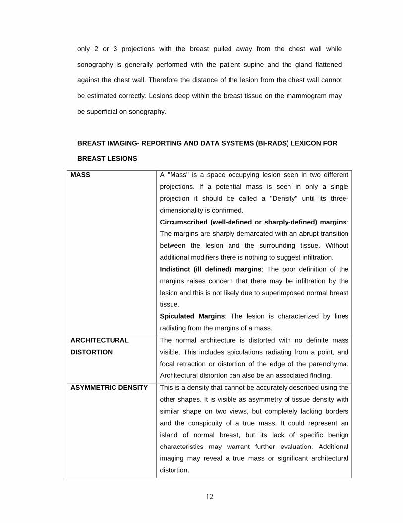
BREAST IMAGING- REPORTING AND DATA SYSTEMS (BI-RADS) LEXICON FOR
BREAST LESIONS
MASS
A "Mass" is a space occupying lesion seen in two different
projections. If a potential mass is seen in only a single
projection it should be called a "Density" until its three-
dimensionality is confirmed.
Circumscribed (well-defined or sharply-defined) margins:
The margins are sharply demarcated with an abrupt transition
between the lesion and the surrounding tissue. Without
additional modifiers there is nothing to suggest infiltration.
Indistinct (ill defined) margins: The poor definition of the
margins raises concern that there may be infiltration by the
lesion and this is not likely due to superimposed normal breast
tissue.
Spiculated Margins: The lesion is characterized by lines
radiating from the margins of a mass.
ARCHITECTURAL DISTORTION
The normal architecture is distorted with no definite mass
visible. This includes spiculations radiating from a point, and
focal retraction or distortion of the edge of the parenchyma.
Architectural distortion can also be an associated finding.
ASYMMETRIC DENSITY This is a density that cannot be accurately described using the
other shapes. It is visible as asymmetry of tissue density with
similar shape on two views, but completely lacking borders
and the conspicuity of a true mass. It could represent an
island of normal breast, but its lack of specific benign
characteristics may warrant further evaluation. Additional
imaging may reveal a true mass or significant architectural
distortion.

13
CALCIFICATION Amorphous or Indistinct Calcifications: These are often
round or "flake" shaped calcifications that are sufficiently small
or hazy in appearance that a more specific morphologic
classification cannot be determined.
Pleomorphic or Heterogeneous Calcifications: These are
usually more conspicuous than the amorphic forms and are
neither typically benign nor typically malignant irregular
calcifications with varying sizes and shapes that are usually
less than 0.5 mm in diameter.
Fine, Linear or Fine, Linear, Branching (Casting) Calcifications: These are thin, irregular calcifications that
appear linear, but are discontinuous and under 0.5 mm in
width. Their appearance suggests filling of the lumen of a duct
involved irregularly by breast cancer.
Benign Calcifications: Benign calcifications are usually
larger than calcifications associated with malignancy. They
are usually coarser, often round with smooth margins and are
much more easily seen.
ASSESSMENT CATEGORIES
Category 1 / Negative There is nothing to comment on. The breasts are
symmetrical and no masses, architectural disturbances or
suspicious calcifications present
Category 2 / Benign Finding
This is also a negative mammogram, but the interpreter may
wish to describe a finding. Involuting, calcified
fibroadenomas, multiple secretory calcifications, fat
containing lesions such as oil cysts, lipomas, galactoceles,
and mixed density hamartomas all have characteristic
appearances, and may be labeled with confidence. The
interpreter might wish to describe intramammary lymph
nodes, implants, etc. while still concluding that there is no
mammographic evidence of malignancy.
Category 3 / Probably Benign Finding - Short Interval Follow-Up
A finding placed in this category should have a very high
probability of being benign. It is not expected to change over
the follow-up interval, but the radiologist would prefer to

14
Suggested establish its stability. Follow – up is done at 6 month
intervals U/L and 1 year interval B/L for 2-3 years.
Category 4 / Suspicious Abnormality - Biopsy Should Be Considered
These are lesions that do not have the characteristic
morphologies of breast cancer but have a definite probability
of being malignant. The radiologist has sufficient concern to
urge a biopsy. If possible, the relevant probabilities should
be cited so that the patient and her physician can make the
decision on the ultimate course of action.
Category 5 / Highly Suggestive of Malignancy
These lesions have a high probability of being cancer.
Appropriate Action Should Be Taken
15
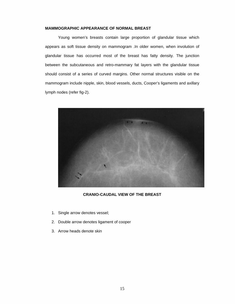
MAMMOGRAPHIC APPEARANCE OF NORMAL BREAST
Young women’s breasts contain large proportion of glandular tissue which
appears as soft tissue density on mammogram .In older women, when involution of
glandular tissue has occurred most of the breast has fatty density. The junction
between the subcutaneous and retro-mammary fat layers with the glandular tissue
should consist of a series of curved margins. Other normal structures visible on the
mammogram include nipple, skin, blood vessels, ducts, Cooper’s ligaments and axillary
lymph nodes (refer fig-2).
CRANIO-CAUDAL VIEW OF THE BREAST
1. Single arrow denotes vessel;
2. Double arrow denotes ligament of cooper
3. Arrow heads denote skin
16
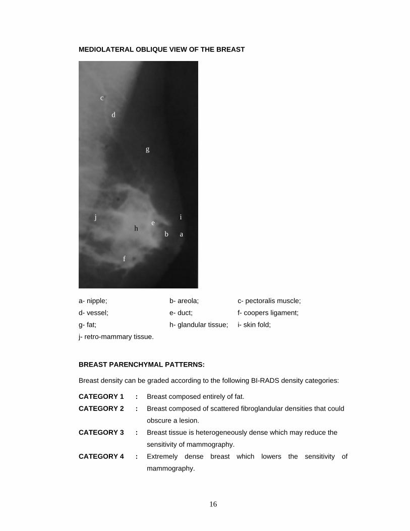
MEDIOLATERAL OBLIQUE VIEW OF THE BREAST
a- nipple; b- areola; c- pectoralis muscle;
d- vessel; e- duct; f- coopers ligament;
g- fat; h- glandular tissue; i- skin fold;
j- retro-mammary tissue.
BREAST PARENCHYMAL PATTERNS:
Breast density can be graded according to the following BI-RADS density categories:
CATEGORY 1 : Breast composed entirely of fat.
CATEGORY 2 : Breast composed of scattered fibroglandular densities that could
obscure a lesion.
CATEGORY 3 : Breast tissue is heterogeneously dense which may reduce the
sensitivity of mammography.
CATEGORY 4 : Extremely dense breast which lowers the sensitivity of
mammography.
a
j
d
i e
f
h b
g
c
17
Wolfe has described 4 parenchymal patterns of the breast based on the relative
amounts of fat, epithelial and connective tissue densities as seen mammographically.
N1 : Normal. The breast parenchyma is of low density and has a large proportion of
fat. No ducts are visible.
P1: Parenchyma is composed chiefly of fat with a prominent duct pattern in the
anterior portion of the breast but involving less than quarter of breast volume.
P2: Prominent duct pattern which involves more than quarter of breast volume and
with which there is often an associated nodular component.
DY: Increased density of breast parenchyma with or without areas of nodularity. The
density often obscures the underlying duct pattern.
P2 and DY patterns have an increased risk of breast cancer as reported by
Wolfe. But more recent studies have failed to confirm this association. Mammographic
sensitivity decreases with increasing density of breast parenchyma.
Developing breast has dense parenchymal pattern. With maturity, parenchyma
assumes a transparent appearance. Breasts of nulliparous women involute more slowly
than multiparous women. Hormone Replacement Therapy (HRT) may also be
associated with symmetric or asymmetric increase in breast density (8) and it inhibits
involutional processes causing persistence of dense P2 and DY patterns.
ULTRASONOGRAPHIC ANATOMY
On ultrasound, breast is a multilayered structure. The skin and fibro glandular
plate are relatively echogenic while the subcutaneous and retromammary fat layers are
echopoor. Normal skin thickness measures around 0.5-2 mm except at the
inframammary crease, cleavage and periareolar region where it is thicker. The
subcutaneous fat layer is separated from glandular tissue by a well defined scalloped
margin. The chest wall muscles are also echopoor in young patients but become more

18
echogenic with fatty infiltration of age. Ultrasound appearance of breast tissue depends
on how much involution of glandular tissue has taken place. Young glandular breast
contains a well defined layer of glandular tissue within which round or oval well
defined echo poor fat lobules may be present. As involution takes place, the breast
tissue shows fat lobules separated by fine curvilinear septa of increased echogenicity.
Normal ducts are often visible particularly in the subareolar region as anechoic tubular
structures.
SONOGRAPHIC ANATOMY OF THE BREAST
19
BENIGN DISEASES OF THE BREAST
CLASSIFICATION OF BENING BREAST DISORDERS (Schwartz’s Principles of Surgery, 8th Edition) (6)
Non – Proliferative disorders of the breast - Cysts and apocrine metaplasia
- Duct ectasia
- Calcification
- Fibro adenoma and related lesions
Proliferative disorders without atypia - Sclerosing adenosis
- Radial and complex sclerosing lesion
- Ductal epithelial hyperplasia
- Intraductal papillomas
Atypical proliferative lesions - Atypical lobular hyperplasia
- Atypical ductal hyperplasia
Relative Risk of invasive breast cancer (Recent Advances 21 I. Taylor) (1)
No Increased risk Adenosis – sclerosing or florid
Hyperplasia (mild 2-4 epithelia cells in depth)
Apocrine metaplasia
Cysts – macro and / or micro
Duct ectasia and periductal mastitis
Mastitis (inflammation)
Fibroadenoma
Squamous metaplasia
Fibrosis

20
Slightly increased risk (1.5-2 times)
Hyperplasia, moderate or florid, solid or papillary
Papilloma with fibrovascular core
Moderately increased risk (5 times)
A typical hyperplasia
Ductal Lobular
Lobular
Insufficient data to assign a risk
Solitary papilloma of lactiferous duct
Radial scar lesion
NOMENCLATURE
The basic principles underlying the aberration of normal development and
involvement (ANDI) classification of benign breast conditions are 1) Benign breast
disorders and diseases are related to the normal processes of reproductive life and to
involution 2) There is a spectrum of breast condition that ranges from normal to
disorder to disease; and 3) the ANDI classification encompasses all aspects of the
breast condition, including pathogenesis and the degree of abnormality (6). The
horizontal component of the following table defines ANDI along a spectrum from
normal, to mild abnormality (disorder), to severe abnormality (disease). The vertical
component defines the period during which the condition develops (6).

21
Normal Disorder Diseases
Early reproductive
years age (15-25)
Lobular development
Stromal Development
Nipple eversion
Fibroadenoma
Adolescent hypertrophy
Nipple inversion
Giant Fibroadenoma
Gigantomastia
Subareolar abscess
Mammary duct fistula
Later reproductive
years (age 25-40)
Cyclical changes of
menstruation
Epithelial hyperplasia
of pregnancy
Cyclical mastalgia
Nodularity
Bloody nipple discharge
Incapacitating
mastalgia
Involution
(age 35-55)
Lobular involution
Duct involution
Dilatation
Scelerosis
Epithelial turnover
Macrocysts
Sclerosing lesions
Duct ectasia
Nipple retraction
Epithelial hyperplasia
Periductal mastitis
Epithelial hyperplasia
with atypia
GENERAL FEATURES
Mammographically these lesions are well defined, oval or round, and may show
a complete radiolucent halo around the periphery due to Mach effect or compressed fat
tissue around the mass. When the lesion is mostly of fat density it is usually benign.
They may show few gentle lobulations (< 3).Some lesions may regress in size over a
period of time especially in post menopausal women. Skin thickening and architectural

22
distortion may or may not be present. Some lesions show benign forms of calcifications
( coarse, discrete, smooth monomorphic with central lucency, tea cup, punctuate or
scattered).
On ultrasound they are oval or ellipsoid wider than deep (depth: width < 0.7)
and aligned parallel to the skin. Smooth thin echogenic capsule with 2-3 gentle
lobulations may be seen. They have variable echogenicity and are usually
homogeneous with lateral edge shadowing and posterior enhancement. On color
Doppler, vessels are seen to go around the mass.
MASTALGIA
Mastalgia – pain in breast – is the commonest breast symptom seen in both
general practice and specialist referral clinics. Mastalgia as any other symptom,
deserves full assessment and careful evaluation; it doest no imply any particular
pathological process nor is it a diagnosis.
HISTORICAL ASPECT
Painful breasts are often regarded as part of a psychosomatic disorder (12). Sir
Astley Cooper (1829) was aware of the syndrome of painful breasts but considered
such complaints were of psychological origin and undeserving of further consideration:
`The disease is met within persons of an irritable and nervous temperament, in whom
there is excessive excitability of the system, accompanied by diminished power'.
Fitzwilliam (1924) in his book on breast disease hardly mentions breast pain but
devotes a whole chapter to hysterical breasts. Sir Hedley Atkins (1938) in his review of
breast disease, recognized endocrine, neuralgic and psychological components of the
problem of breast pain(12). He undertook a trial of oestrin in these patients but failed to
demonstrate a beneficial effect. There are, of course, examples of patients with
psychosomatic problems referred to the breast clinic but the work of Preece et al (1978)
23
has firmly established the organic nature of most episodes of breast pain. Preece et al
(1978) compared patients with breast pain with a group of women (referred for
consideration of a surgical procedure) with varicose veins. The psychological
assessment was performed using the well validated Middlesex Hospital questionnaire
(Crown et al, 1970) and showed that there was no increased psychological morbidity in
the group of women complaining of painful breasts (12).
The development of severe pain and nodularity which may last for the most part
of the menstrual cycle is considered as abnormal and may interfer significantly with the
patient's everyday activities. In most cases, aberrant physiological or pathological
processes underlie the symptom rather than neuroticism. Premenstrual breast pain is
normal, but 10-15% of patients referred to breast clinics required treatment.
Recently, mastalgia has been classified into three distinct clinical syndromes (1)
(Preece et al 1976, Bishop & Blarney 1979, Maddox et al 1989). Cyclical mastalgia
accounts for two-thirds of cases. Cyclical pain, which is usually bilateral (some times it
may be unilateral), varies during the menstrual cycle and is typically worse in the luteal
phase and relieved by the onset of the menopause. Non-cyclical pain accounts for the
remaining one-third, being further divided into true non-cyclical pain and chest wall pain
which is either felt along the costo-chondral junction (Tietze's syndrome) or the lateral
chest wall. This classification will identify those patients likely to respond to hormonal
treatment (cyclical and true non-cyclical) and those who will not (chest wall pain).

24
MASTALGIA
Cyclical (Two thirds of Patients)
Non Cyclical (One third of patients)
True
Chest wall pain
Mammography is often utilized and is of potential value to rule out carcinoma
and show signs of benign breast disease.
FIBROCYSTIC DISEASE
There is a miscellany of morphological alterations in the female breast are often
grouped under the term fibrocystic changes. Fibrocystic changes represent the single
most common disorder of the breast commonly presenting between 20 and 40 years of
age with peak incidence just before menopause. One in every fourteen women develop
palpable cyst. There are three principle patterns of morphological change 1) Cyst
formation, often with apocrine metaplasia 2) Fibrosis 3) Adenosis. Cysts of the breast
are usually round and well delimited. Often referred as blue dome cysts (Haagensen’s
diseases of the breast) (9).
Cysts develop when the lumina of the ducts and acini become dilated and lined
by atrophic epithelium. Micro cysts are < 3mm in diameter and gross cysts are > 3mm
in diameter. Gross cysts originate from blunt ducts and micro cysts.
Mammographically, they are indistinguishable from non calcified solid masses.
Shape is variable according to the amount of fluid present within it and the amount of
compression applied. Calcification occurs mostly in cysts that are greater than 10mm in
diameter. It is present as a peripheral rim. Micro cysts may contain granules of calcified
debris (milk of calcium). Ultrasonographically simple cysts are anechoic with posterior
enhancement. Hemorrhage and infection may cause internal echoes or debris to be
present. They are only partially mobile as they are attached to the glandular tissue.
25
FIBROADENOMA
Fibroadenoma (adenofibroma (9)) is a benign tumour composed of stromal and
epithelial elements. It is the most common tumour of the breast in women younger than
30 years of age. They are firm, solitary tumours, and well encapsulated with smooth
borders which may be lobulated.
Fibroadenomas appear mammographically as well circumscribed round or oval
masses frequently, with gentle lobulations and rarely exceeding 3cm in size. Myxoid
degeneration results in coarse nodular calcification. On ultrasound, they appear
homogeneous and hypoechoic with posterior enhancement and edge shadowing.
There is no reaction in the surrounding tissue and it is mobile. They may show posterior
shadowing if calcification or hyaline degeneration is present. They are only slightly
compressible and have their long axis parallel to the skin as they accommodate
themselves to the local anatomy defined by beast nodules and cooper’s ligaments with
blood vessels passing around them.
GIANT FIBROADENOMA
Giant fibroadenoma is a descriptive term that applies to a fibroadenoma that
attains an unusually large size, typically greater than 5 cm. Haagensen (9) calls these
lesions massive adenofibromas in youth to denote their common occurrence in
adolescent women.
PHYLLODES TUMOR
Phylloids tumours arise from intra lobular stroma. They can occur at any age
mostly in the sixth decade. They constitute 1% of all primary tumours of the breast (10).
A group of circumscribed biphasic tumours, basically analogous to fibroadenomas,
characterized by a double layered epithelial component arranged in cleft surrounded by

26
an over growing hyper cellular mesenchymal component typically organized in leaf -
like structures.
Mammographically, it resembles a large lobulated fibroadenoma with high soft
tissue density. Some margins may be irregular suggesting local invasion. They may
develop plaque like calcification. Ultrasonographically they are similar to fibroadenomas
with well defined margins and posterior enhancement. The otherwise uniform echoes
are usually broken up by fluid spaces. They are very vascular on color Doppler.
PAPILLOMA
Overall less than 10% of benign breast neoplasms correspond to papllomas,
presenting mainly during 4th and 5th decade (10). A proliferation of epithelial and
myoepithelial cells overlaying fibrovascular stalks creating an arborescent structure
within the lumen of a duct. Intra ductal papilloma of the breast is divided into central
(large duct) papilloma located in the sub areolar region and peripheral papilloma arising
in the Terminal Duct Lobular Unit (TDLU). They are normally solitary and central and
produce a serous or bloody discharge . Peripheral papillomas may occur in contiguous
branches of the ductal system.
Mammographically, a lobulated fairly well circumscribed mass usually in the
subareolar region is seen. Calcification may be punctate or similar to fibroadenoma.
Ultrasound will show a solid mass within a dilated duct or cyst. Multiple papillomas tend
to occur in younger women of less than 30 yrs and appear like fibroadenomas both
clinically and on imaging.
FIBROMATOSIS (DESMOID TUMOUR)
Fibromatosis accounts for less than 0.2% of all breast lesions, more common in
child bearing age(10). This is a locally aggressive lesion without metastatic potential

27
originates from fibroblasts and myofibroblasts within the breast parenchyma, excluding
mammary involvement by extension of a fibromatosis arising from the pectoral fascia.
These have the same characteristics of abdominal desmoid tumors and also
appear as round or oval circumscribed benign appearing masses. Rarely it may appear
as a spiculate mass on mammography. Ultrasound may reveal a solid mass
indistinguishable from a fibroadenoma.
SCLEROSING ADENOSIS
Sclerosing adenosis is characterized by a compact proliferation of acini with
preservation of the luminal epithelial and the peripheral myoepithelial cell layers along
with a surrounding basement membrane. Micro calcifications are common within the
glands.
RADIAL SCARS
A benign lesion on imaging resembles invasive carcinoma. Grossly and at low
power microscopy it resembles invasive carcinoma because the lobular architecture is
distorted by the sclerosing process (11). The term radial scar has been applied to small
lesions and complex sclerosing lesions to large ones that contain a variety of ductal
epithelial hyperplasia along with sclerosis. They are often multiple and bilateral.
Radial scars are common in post Mastectomy and after large excision biopsies.
Most are microscopic but some form larger (greater than 1 cm) palpable masses. They
are benign lesions of unknown etiology that mimic malignancy on imaging.
Mammogram shows spicules originating from a central nidus. But unlike Carcinoma, it
does not have a central mass but only a central area of architectural distortion.
Sonogram shows a hypo echoic mass with dense posterior acoustic shadowing.
28
GALACTOCELE
These are cysts containing inspissated milk, occurring in lactating women and in
late pregnancy. Usual site is the central breast. On mammogram, they may have a
mottled appearance with fat-fluid level on erect lateral view. On sonogram they appear
solid sometimes.
LYMPH NODES
Axillary lymph nodes are oval masses with a fatty hilum and are usually less
than 2 cms in size. Intra mammary lymph nodes are seen in the upper outer quadrant,
posteriorly, as well circumscribed lobulated nodules of less than 1cm in size with a hilar
notch and lucent center if fatty replacement has occurred. They may not be seen on
ultrasound. They are seen in diseases like rheumatoid arthritis, sarcoidosis and
infections like tuberculosis and other bacterial infections. Fine punctate calcifications
maybe seen in sarcoidosis and are mimicked in patients with rheumatoid arthritis on
gold therapy.
HEMATOMA
Trauma to the breast may result in hematoma formation which may present
clinically and mammographically as an ill-defined mass mimicking a carcinoma.
Occasionally, the late sequelae of a hematoma may resemble a carcinoma with micro
calcification and architectural distortion. Ultrasound shows it to be hypo or hyper echoic
depending on the duration. Shaggy walled cavities with echogenic debris and fluid that
move with posture may be seen.
GRANULAR CELL (MYOBLASTOMA) TUMOR
This is an extremely rare benign lesion probably of Schwann cell origin with a
6 % rate of occurrence in the breast and most frequently seen in the 3rd to 5th
decade(10). The tumor has a fibrous stroma with infiltrative margin mimicking a
29
scirrhous carcinoma clinically and mammographically. Sonographically, a hypo echoic
mass with post acoustic shadowing and echogenic interface anterior to the hypo echoic
lesion will be seen.
INFECTIONS:
ABSCESS:
Breast abscess is mostly encountered during lactation or in non-lactating
women in relation to duct ectasia. Usually occurs in a central or subareolar location
producing an irregular density with associated trabecular, skin or nipple distortion.
Ultrasound shows a cyst with internal echoes, shaggy walls, intracavitary ehogenic gas
bubbles and intense color signals at the hyperemic periphery.
TUBERCULOSIS:
Tuberculosis of the breast was first described by Cooper in 1829 as the
‘scrofulous swelling of the bosom'. Only a few clinical reports have been reported from
the Indian subcontinent in spite of the high prevalence of other forms of tuberculosis in
this region. The disease has been reported in the 20-50 years age group. Breast
tuberculosis may be primary where the breast lesion is the only manifestation of the
disease or it may be secondary in which a focus of tuberculosis has already been
diagnosed elsewhere in the body and the disease appears in the breast at a later
stage. The method of spread to the breast is by the hematogenous or lymphatic routes,
or by direct extension from adjacent tissues.
The disease presents in three forms: nodular, diffuse and sclerosing. The
nodular form is characterized by a slow growing caseating lesion and
mammographically presents as a dense round area with indistinct margins. The diffuse
form consists of multiple, intercommunicating foci of tuberculosis within the breast,
which may caseate leading to ulceration and numerous discharging sinuses. The skin

30
may be thickened, with a tense and tender breast. In addition to the breast lesion, the
axillary lymph nodes are frequently affected.
Radiographs show a dense breast and thickened skin. In the sclerosing form,
excessive fibrosis rather than caseation is the dominating feature. Progress is slow and
suppuration is rarely seen. The entire breast becomes hard because of the dense
fibrous tissue and the nipple gets retracted. Increased density of the gland is seen on
mammography. All the three forms of the disease are indistinguishable from breast
cancer clinically as well as on mammography. The nodular form of the disease is more
common and the lesions are hypo echoic sonographically with ill defined margins or
complex cystic masses may be seen. In cases of diffuse tuberculosis ill-defined
hypoechoic masses are seen. In patients with sclerosing tuberculosis, increased
echogenicity of the breast parenchyma is seen with no definite mass.
FILARIASIS:
As with tuberculosis, a solitary inflammatory mass surrounding the filarial worms
may mimic a carcinoma radiologically.
LIPOMA
This is a fatty tumor which on mammography appears as a lucent or less radio
dense lesion with a well defined capsule and smooth rounded margin. Sonographically,
it has the same echogenicity as surrounding fat.
FAT NECROSIS / OIL CYST
These are single or multiple, 2-3 cms in diameter and result from trauma which
is usually surgical. They maybe asymptomatic or may present as a palpable mass.
Mammogaphically, an irregular spiculated mass with or without calcifications or oil cyst
or only calcifications may be seen. They may have curvilinear calcifications.

31
Sonographically, hypoechoic mass with posterior acoustic shadowing may be seen.
Complex mass with mixed echotexture intra cystic soft tissue mass may also be a
feature. An oil cyst shows echogenic oil floating over watery component.
ADENOLIPOMA / HAMARTOMA
These are often large when diagnosed, measuring greater than 6 cm, and are
soft on clinical examination. Mammographic appearance depends on the fat and
glandular tissue content. Thin soft tissue density capsule is usually present.
SKIN LESIONS
Sebaceous Cysts, warts, neurofibromas, keloid scars can be seen in the region
of the skin. Mammographic opacity is usually very well defined because it is outlined by
air.
STUDIES RELATED TO BENIGN BREAST DISEASES
Several studies have been conducted to evaluate the role of combined
sonomammographic imaging in patients with palpable abnormalities of the breast.
In the ACR BI-RADS (14&15) a mass is defined as a space occupying lesion
seen in at least 2 projections. If seen in only one projection then it is called as a density.
Lawrence W Basset (15) has described that when the margins of a lesion are
not uniform throughout, then, the descriptor indicating the portion of greatest concern
should be used.
Stavros et al (16) has reported that a mass to be characterized as benign on
ultrasound should have 3 or less gentle lobulations.

32
Color Doppler is an adjunct to ultrasound in the differential diagnosis of benign
tumors. Malignant tumors are characterized by more than 1 vascular pole (17,18).
Wendie A Berg et al (19) have studied cystic lesions of the breast with
sonographic and pathological correlation and concluded that all clustered microcysts
are benign but cysts with thick walls or septations (>=0.5mm), intracystic masses, or
predominantly solid masses with eccentric cystic foci should be examined at biopsy as
23% of them had proved to be malignant. Also they have stated that size concordance,
mammographic, sonographic, pathologic concordances are critical with papillary
lesions as 2 of 12 lesions excised were malignant.
Ultrasound with high frequency transducer is essential for accurate non invasive
diagnosis of breast cysts and has showed promise in the differentiation of benign from
malignant solid masses (20). However it requires state of the art equipment with
appropriate technical settings to create an optimal image (21).
As reported by Manju Bala Popli (22) tuberculosis of the breast occurs in the
upper outer quadrant of the breast and can appear nodular or diffuse with
intercommunicating foci and discharging sinuses or sclerosed with increased density of
the gland on mammography. Multiple chronic discharging sinuses suggest
Tuberculosis.
The measured sonographic size of a mass may differ slightly from the
mammographic size but significant differences cannot be reconciled readily by a
change in imaging modalities. This principle is important because the majority of breast
cysts are not visible on mammography (23). Lesions in the axillary region may also be
more difficult to detect because of the overlying pectoralis muscle causing decreased
contrast in this region (23).
33
Breast carcinoma is diagnosed in only 4 % of patients with breast symptoms
indicating that appropriate management of breast physical findings is important for the
primary physician (24).
There is considerable documentation that mammography is less useful in
younger women due to greater likelihood of dense breast tissue composition (15, 25).
In women younger than 40 years, the normal glandular nodularity may be
mistaken for dominant masses (27). Women with negative mammograms and
ultrasound scans are at low risk for cancer but should be followed up at short term
intervals with clinical examination and imaging if biopsy is not elected by their surgeon /
clinician (24). Breast thickening is a particularly vague descriptor of a physical finding at
clinical breast examination that encompasses a wide range of descriptions including
breast nodularity; diffuse cystic change fibrocystic change and breast fullness (24). In
this study 78% of the cases with breast thickening were normal when breast ultrasound
was normal.
One of the various factors leading to false negative findings on mammography
is the effect of breast density. In the study conducted by Pavel Crystal et al (28) out of 7
sonographically detected cancers 2 were in patients with BI-RADS category 4 and 5
were in patients with BIRADS category 3 parenchymal density. No carcinomas were
detected with BIRADS category 2 density. No statistical difference was found in the
number of cancers among a range of breast densities. Dense glandular tissue has a
hyperechoic appearance on sonography, so hypoechoic cancers are easily detected.
According to Stephen H Taplin et al (29) BIRADS assessments and
management recommendations are consistent for negative and benign assessments
34
and there is evidence of how well terminology consistent with BI-RADS was
implemented in practice by 1997.
Ultrasound is not only helpful in the depiction of lymph nodes but also
involvement of skin and pectoral muscle. In posterior lesions, especially those close to
the chest wall musculature accurate measurement of the size and extension may not
be possible with mammography prior to surgery (30).
In the study done by Mahesh K Shetty et al (13), palpable abnormality has a
variety of descriptors – lump, thickening, knot and cord. In 2002 Mahesh K Shetty et al
(13) have showed the value of combined sonomammographic assessment in patients
with palpable breast abnormalities and have reported a sensitivity and negative
predictive value of 100% each with a specificity of 80%. 40% of the lesions were
categorized as benign after a combined evaluation showing the value of imaging in
helping avoid unnecessary biopsies.
False negative rates of 0-2.6% have been reported in multiple studies for
combined sonomammographic evaluation in patients with palpable abnormalities of the
breast (15, 31, 32).
Moss et al (33) reported a sensitivity of 94.2% and specificity of 67.9% in 368
patients in who combined sonomammographic evaluation had been performed for
palpable breast abnormalities and who underwent surgical biopsy.
Rotten D et al have studied the usefulness of 3D ultrasound in breast
diseases (34,35). Benign tumors are surrounded by a continuous hyperechogenic rim,
i.e. the rim is present irrespective of the section plane orientation. In case of

35
fibroadenomas, complete wall continuity of the mass was readily apparent. The
hyperechoic bands of fibrous tissue peripheral to the masses appeared either as
distinct from the central image (compressive pattern). Three-dimensional ultrasound
mammography had higher specificity, but lower sensitivity, than two-dimensional
ultrasound mammography.
36
MATERIALS AND METHODS
Female patients in the pre, peri and post menopausal age groups with benign
breast disorders who underwent breast ultrasound and mammography from September
2003 were included in this study. Patients with features of malignancy were excluded
from the study. Standard nomenclature for characterization of the lesions on
mammography and ultrasound were used. Subsequently all the patients underwent
histopathological examinations of lesions in the form of excision biopsy or true cut
biopsy.
Patients in whom histopathology could not be done due to practical reasons
Fine Needle Aspiration Cytology was done. In correlation with the clinical diagnosis and
FNAC final diagnosis was arrived and patients were treated accordingly.
Methods:
Cranio-caudal and Medio-Lateral Oblique views of mammographs of both
breasts were taken –24-30 KV; 100mAs. Ultrasound examination was done with high
frequency linear transducer of both breasts.
3D volume probe was used to acquire coronal plane which was simply used to
classify the margins of the lesion into three types of patterns- Retraction, Compression
and Indeterminate.
We have compared the sensitivity USG with the mammography using Z test. P
value less than 0.05 was considered as statistically significant. HPE was considered as
gold standard. Where HPE is not done due to practical reasons, Fine Needle Aspiration
Cytology was considered as gold standard. Statistically analysis were done using
SPSS 11.5 version. (Statistical Package for Social Sciences)

37
COIMBATORE MEDICAL COLLEGE HOSPITAL, COIMBATORE
A STUDY OF DIAGNOSTIC ACCURACY IN BENIGN BREAST DISEASE WITH SPECIAL REFERENCE TO
RECENT DIAGNOSTIC TOOLS
Patient Name: Age: Sex:
Serial No. OP/IP No. :
Ward / Unit :
Complaints & History :
1. History of breast lump-duration and site.
2. H/o pain and whether related to menstrual cycle
3. H/o nipple discharge-serous or serosanguinous
4. H/o fever
5. H/o intake of oral contraceptive pills or surgeries for the breast
6. Menstrual history
7. Marital history
8. Obstetric history-last child birth and breast feeding
9. Family h/o of breast diseases
CLINICAL FINDINGS :
• Palpable lump, thickening, knot, cord
• Location, size and number
• Margins, consistency and mobility
• Associated findings: skin changes, nipple retraction, axillary lymph
nodes.

38
CLINICAL DIAGNOSIS :
FINE NEEDLE ASPIRATION CYTOLOGY : No.
Date done: Reported on:
FINDINGS :
BIOPSY : No.
Date done: Reported on:
FINDINGS :

39
MAMMOGRAPHIC FINDINGS:
• Number, location and density of the lesion(s)
• Margins- well-defined, ill-defined, spiculated, obscured
• Shape - round, oval, irregular
• Architectural distortion and tenting
• Calcification
• Halo sign.
• Skin thickening
• Nipple retraction
• Axillary lymph nodes with benign or malignant features
Impression
40
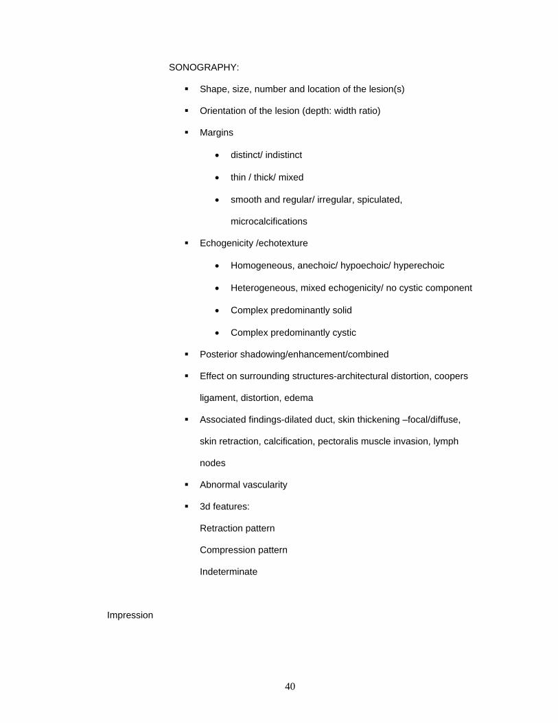
SONOGRAPHY:
Shape, size, number and location of the lesion(s)
Orientation of the lesion (depth: width ratio)
Margins
• distinct/ indistinct
• thin / thick/ mixed
• smooth and regular/ irregular, spiculated,
microcalcifications
Echogenicity /echotexture
• Homogeneous, anechoic/ hypoechoic/ hyperechoic
• Heterogeneous, mixed echogenicity/ no cystic component
• Complex predominantly solid
• Complex predominantly cystic
Posterior shadowing/enhancement/combined
Effect on surrounding structures-architectural distortion, coopers
ligament, distortion, edema
Associated findings-dilated duct, skin thickening –focal/diffuse,
skin retraction, calcification, pectoralis muscle invasion, lymph
nodes
Abnormal vascularity
3d features:
Retraction pattern
Compression pattern
Indeterminate
Impression

41
OBSERVATION AND RESULTS
Sixty nine patients were included in this study. All patients underwent ultrasound
of the breast and mammography. All the 69 patients underwent FNAC.
Histopathological examinations were done for 49 patients.
In Our study out of 69 patients histopathological confirmation was possible in 49
patients. Of the remaining patients, 10 patients were not willing for surgery because of
the small size of the lesions and for cosmetic objections and another 10 patients did not
require biopsy and were conservatively treated. Out of the 49 patients in whom
histopathological confirmation and FNAC were done, result of the FNAC did not
correlate with the HPE in 5 patients. Because of difficulty in finding out location of the
lesion in 3 patients FNAC was negative. In 2 cases of phylloids tumour FNAC was
unable to diagnose correctly. All other cases FNAC was consistent with
histopathological examination.
Out of the 49 patients Ultrasound showed positive diagnosis for 46 patients with
a sensitivity of 93.9%. Whereas mammography was positive was only in 33 patients
with a sensitivity of 67.3%. Statistical test of proportion showed that Z value is 3.3 with
the corresponding p value less than 0.001. Hence it is concluded that ultrasound
produces statistically significant higher sensitivity compared to mammography. We
compared sensitivity of ultrasound and mammography keeping gold standard as
histopathological examination. It is found that ultrasound in general shows the
sensitivity of 92.8% (positive results for 64 patients out of 69 examined), whereas
mammography showed the sensitivity of 66.7% (Positive results for 46 out of 69
patients examined). This difference is statistically significant because the z value is 3.8.
Hence the corresponding p value is less than 0.001.

42
TABLE 1
AGE GROUP WISE DISTRIBUTION OF BENIGN BREAST DISEASES
AGE GROUP (YRS)
NO. OF PATIENTS
%
< 20 15 21.74
21- 30 26 37.68
31- 40 21 30.43
> 41 7 10.14
0 5 10 15 20 25 30
No. of Patients
< 20
21- 30
31- 40
> 41
Age
Gro
up
AGE GROUP WISE DISTRIBUTION OF BENIGN BREAST DISEASES

43
TABLE 2
ULTRASOUND AND MAMMOGRAPHIC COMPARISION
Mammography Ultrasound
46 64
-19
192939495969
No. o
f Pat
ient
s
Mammographic Ultrasound
Sensitivity
ULTRASOUND AND MAMMOGRAPHIC COMPARISION
44
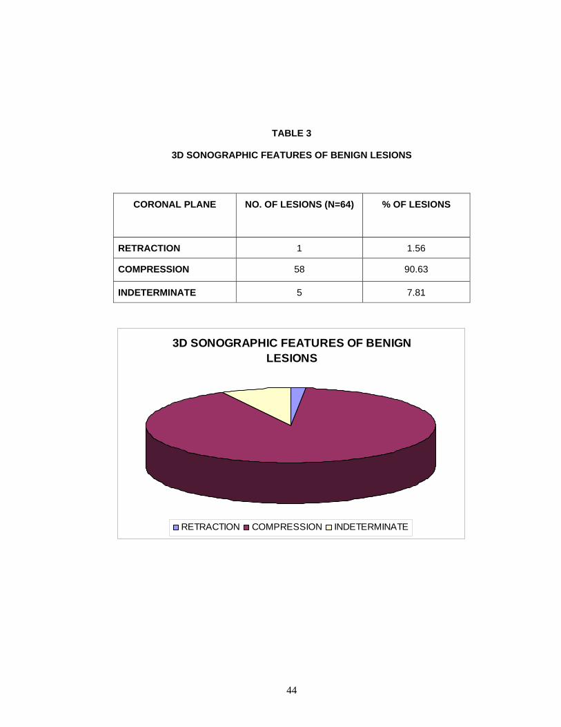
TABLE 3
3D SONOGRAPHIC FEATURES OF BENIGN LESIONS
CORONAL PLANE NO. OF LESIONS (N=64) % OF LESIONS
RETRACTION 1 1.56
COMPRESSION 58 90.63
INDETERMINATE 5 7.81
3D SONOGRAPHIC FEATURES OF BENIGN LESIONS
RETRACTION COMPRESSION INDETERMINATE

45
TABLE 4
BENIGN BREAST DISEASE PATTERN
Name of the Disease No. of Patients
(n=69)
%
Fibroadenoma 50 72.46
Fibrocystic Disease 10 14.49
Breast Abscess 5 7.25
Fibroadenosis 2 2.90
Phylloides tumour 2 2.90
-1 9 19 29 39 49 59 69
No. of Cases
Fibroadenoma
Fibrocystic Disease
Breast Abscess
Fibroadenosis
Phylloides tumour
Type
of D
isea
se
BENIGN BREAST DISEASE PATTERN

46
TABLE 5
MAMMOGRAPHICALLY DENSE BREAST
AGE WISE DISTRIBUTION
AGE GROUP Dense Breast
< 20 7
21- 30 7
31- 40 3
> 41 1
-2 2 6 10 14 18
No. of Patients
< 20
21- 30
31- 40
> 41
Age
Gro
up
AGE WISE DISTRIBUTION OF DENSE BREAST IN MAMMOGRAM

47
ILLUSTRATIVE EXAMPLES
CASE 1 - FIBROCYSTIC DISEASE
CASE 2 - FIBROCYSTIC DISEASE
CASE 3 - BREAST ABSCESS
CASE 4 - TUBERCULAR BREAST ABSCESS
CASE 5 - FIBROADENOMA

48
CASE 1 - FIBROCYSTIC DISEASE
39 year old patient came with H/O left breast lumps. Mammogram showed Category IV BI-RADS parenchymal density with obscuration of lesions, not able to comment on nature of lesion on the mammography. U/S screening showed dilated ducts with multiple cysts suggestive of BI-RADS Grade II lesions – FIBROCYSTIC DISEASE.
Cranio-caudal and Medio-lateral oblique view Mammograms of the left breast
Ultrasound of the left breast showing cysts and dilated ducts
Contd..
49
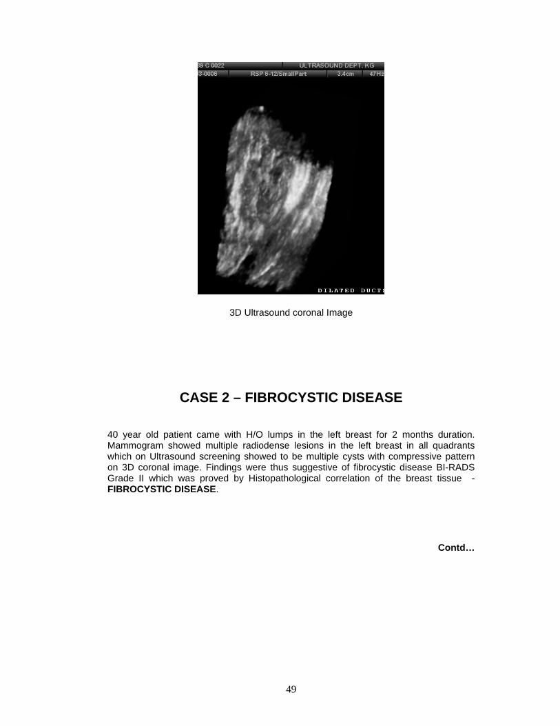
3D Ultrasound coronal Image
CASE 2 – FIBROCYSTIC DISEASE
40 year old patient came with H/O lumps in the left breast for 2 months duration. Mammogram showed multiple radiodense lesions in the left breast in all quadrants which on Ultrasound screening showed to be multiple cysts with compressive pattern on 3D coronal image. Findings were thus suggestive of fibrocystic disease BI-RADS Grade II which was proved by Histopathological correlation of the breast tissue - FIBROCYSTIC DISEASE.
Contd…

50
Cranio-caudal and Medio-lateral oblique view Mammograms of the left breast showing multiple radiodense lesions (arrows)
Ultrasound screening of left breast with 3D coronal reconstruction showed the cyst having well defined margins in the coronal planes with no irregularity of surrounding
suggestive of compressive pattern. Contd..
51
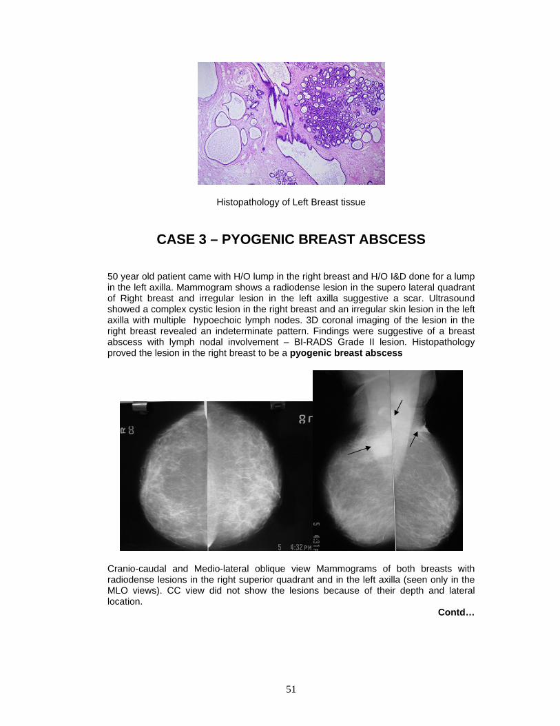
Histopathology of Left Breast tissue
CASE 3 – PYOGENIC BREAST ABSCESS
50 year old patient came with H/O lump in the right breast and H/O I&D done for a lump in the left axilla. Mammogram shows a radiodense lesion in the supero lateral quadrant of Right breast and irregular lesion in the left axilla suggestive a scar. Ultrasound showed a complex cystic lesion in the right breast and an irregular skin lesion in the left axilla with multiple hypoechoic lymph nodes. 3D coronal imaging of the lesion in the right breast revealed an indeterminate pattern. Findings were suggestive of a breast abscess with lymph nodal involvement – BI-RADS Grade II lesion. Histopathology proved the lesion in the right breast to be a pyogenic breast abscess
Cranio-caudal and Medio-lateral oblique view Mammograms of both breasts with radiodense lesions in the right superior quadrant and in the left axilla (seen only in the MLO views). CC view did not show the lesions because of their depth and lateral location.
Contd…

52
Ultrasound screening of left axilla showing Lymphnodes. The linear hypoechoic lesion in the left picture corresponds to the presence of a surgical scar.
3D coronal image of Right Breast lesion
Histopathology of Right Breast Lesion
53
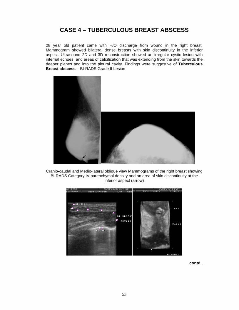
CASE 4 – TUBERCULOUS BREAST ABSCESS
28 year old patient came with H/O discharge from wound in the right breast. Mammogram showed bilateral dense breasts with skin discontinuity in the inferior aspect. Ultrasound 2D and 3D reconstruction showed an irregular cystic lesion with internal echoes and areas of calcification that was extending from the skin towards the deeper planes and into the pleural cavity. Findings were suggestive of Tuberculous Breast abscess – BI-RADS Grade II Lesion
Cranio-caudal and Medio-lateral oblique view Mammograms of the right breast showing
BI-RADS Category IV parenchymal density and an area of skin discontinuity at the inferior aspect (arrow)
contd..

54
2D Ultrasound and 3D Reconstruction of right breast lesion
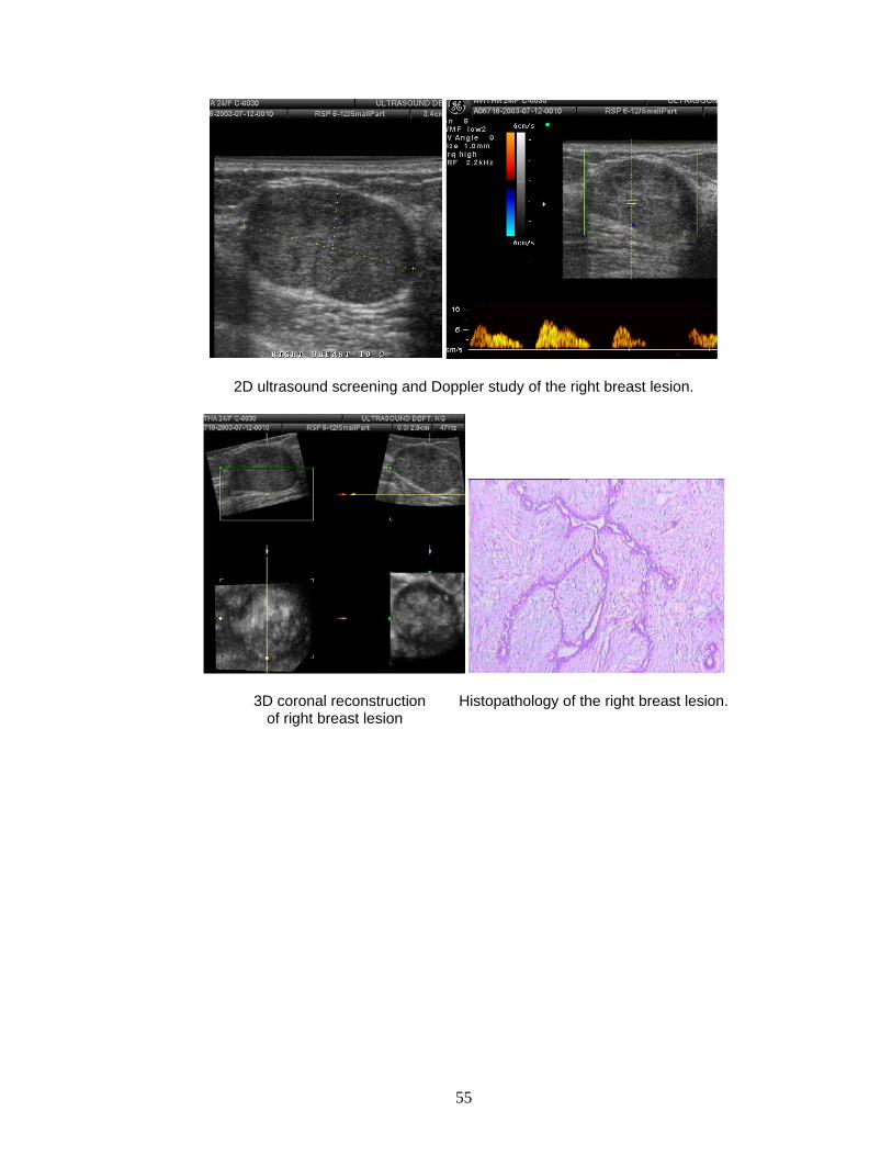
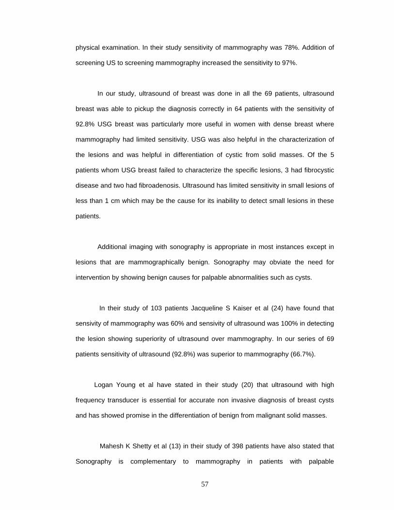
CASE 5 – FIBRO ADENOMA 24 year old patient came with complaints of palpable mass in the right breast. Mammogram shows a well defined radiodense lesion in the Right supero lateral quadrant with specks of calcification. Ultrasound screening showed a well defined hypoechoic lesion which showed a compressive pattern on 3D reconstruction and no abnormal vascularity on Doppler study. Findings were suggestive of a fibroadenoma – BI-RDAS Grade III lesion. Histopathology of the lesion showed it to be a Fibroadenoma.
Mammogram of the right breast showing well defined radiodense lesion with specks of calcification and halo sign in the supero lateral quadrant
Contd..
55
2D ultrasound screening and Doppler study of the right breast lesion.
3D coronal reconstruction Histopathology of the right breast lesion. of right breast lesion

56
DISCUSSION
The role of mammography in patients with benign breast diseases is to
delineate the lesion with its shape and location. Mammography is also helpful to rule
out malignant features in the involved breast and also to screen the opposite breast.
In our study of 69 patients mammography was useful in detecting 46 of the
lesions with the sensitivity of 66.7%. 18 patients had mammographically dense breast
hence lesions could not be made out. In another 5 patients mammography was unable
to give the features of specific diagnosis.
Linda Moy et al (32), in their study of 829 patients, have concluded that
mammography failed to detect lesions in patients with dense breasts. In our series of
69 patients mammography failed to detect lesions in 23 patients as 18 out of these 23
patients had mammographically dense breast.
Bennet C et al (25) and Lawrence W Bassett (15) have documented the
decreased sensitivity of mammography in dense breast mostly in young women. In our
study of 69 patients 18 patients had mammographically dense breast, of whom 14 were
less than 30 years of age.
According to Thomas M Kolb et al (26) who have studied 11130 women, have
documented that sensitivity of mammography to detect lesions was 77.6%.
Mammographic sensitivity for breast lesions declines significantly with increasing breast
density. Addition of screening US significantly increases detection of small lesions and
depicts significantly more lesions and at smaller size and lower stage than does
57
physical examination. In their study sensitivity of mammography was 78%. Addition of
screening US to screening mammography increased the sensitivity to 97%.
In our study, ultrasound of breast was done in all the 69 patients, ultrasound
breast was able to pickup the diagnosis correctly in 64 patients with the sensitivity of
92.8% USG breast was particularly more useful in women with dense breast where
mammography had limited sensitivity. USG was also helpful in the characterization of
the lesions and was helpful in differentiation of cystic from solid masses. Of the 5
patients whom USG breast failed to characterize the specific lesions, 3 had fibrocystic
disease and two had fibroadenosis. Ultrasound has limited sensitivity in small lesions of
less than 1 cm which may be the cause for its inability to detect small lesions in these
patients.
Additional imaging with sonography is appropriate in most instances except in
lesions that are mammographically benign. Sonography may obviate the need for
intervention by showing benign causes for palpable abnormalities such as cysts.
In their study of 103 patients Jacqueline S Kaiser et al (24) have found that
sensivity of mammography was 60% and sensivity of ultrasound was 100% in detecting
the lesion showing superiority of ultrasound over mammography. In our series of 69
patients sensitivity of ultrasound (92.8%) was superior to mammography (66.7%).
Logan Young et al have stated in their study (20) that ultrasound with high
frequency transducer is essential for accurate non invasive diagnosis of breast cysts
and has showed promise in the differentiation of benign from malignant solid masses.
Mahesh K Shetty et al (13) in their study of 398 patients have also stated that
Sonography is complementary to mammography in patients with palpable

58
abnormalities, its superiority over mammography is in being able to show lesions
obscured by dense breast tissue and in characterizing palpable lesions that are
mamographically visible or occult. Mammography is complementary to sonography
because of its ability to screen the remainder of the ipsilateral and contralateral breast
for clinically occult lesions. It has been reported that the accuracy of sonography is
comparable with that of mammography as a screening modality for breast lesion.
In their study of 1517 women Pavel Crystal et al(28) have found that breast
screening with sonography in the population of women with dense breast tissue is
useful in detecting lesions not seen in mammography.
Isabelle le conte et al,(37) in their study of 4236 ptients, have found that
sonograpy is an useful adjunct after mammography for the detection of lesions
particularly in the dense breast. In their study sensitivity of ultrasound was 88% and
that of mammography was 56%.
In one of our patients, extension of the cystic lesion into the pleural cavity was
detected only on ultrasound and helped us to come to a diagnosis of tuberculous
abscess.
Current surgical therapy by John. L.Cameron says that Addition of screening
sonography to screening mammography could increase by 42% the number of
nonpalpable lesions diagnosed. The benefits were greatest in women with dense
breasts, as mammograms have the lowest sensitivity in this population (39).
3D ultrasound was helpful in additionally characterizing most of the lesions.
Compressive pattern was seen in 3D ultrasound almost all benign lesions (58 out of 64
benign lesions) and in one patient retraction pattern was seen and further investigation
59
delineated atypical cells suspicious for malignancy. Five Patients showed indeterminate
pattern on 3D ultrasound with subsequent diagnosis of breast abscess in 4 patients and
in one patient with a benign lesion, which on further investigation delineated fibrocystic
disease. In these 5 patients 3D ultrasound did not provide any better information than
was available with 2D ultrasound. Hence 3D ultrasound was not helpful in lesions
causing lesser degrees of architectural distortions. (Lesions which had irregular or
lobulated margins on 2D ultrasound could not be grouped into either of these patterns
where therefore indeterminate for benign or malignant lesions on 3D ultrasound).
Rotten D et al (34, 35) also found in their study of the usefulness of 3D
ultrasound in breast diseases and these 2 patterns of compression & retraction were
preferentially associated with benign and malignant lesions respectively. Three
dimensional ultrasound mammography had a higher specificity but lower sensitivity
than two dimensional mammography in their study.
In Our study, out of 69 patients histopathological confirmation was possible in
49 patients. Of the remaining patients, 10 patients were not willing for surgery because
of the small size of the lesions and for cosmetic objections and another 10 patients did
not require biopsy and were conservatively treated. Out of the 49 patients in whom
histopathological confirmation and FNAC were done, result of the FNAC did not
correlate with the HPE in 5 patients because of difficulty in finding out location of the
lesion in 3 patients and in 2 cases phylloids tumour FNAC was unable to diagnose. All
other cases FNAC was consistent with histopathological examination.

60
CONCLUSION
MAMMOGRAPHY IS SUPERIOR TO ULTRASOUND IN THE DETECTION OF
MICROCALCIFICATION.
SONOGRAPHY IS COMPLEMENTARY TO MAMMOGRAPHY IN PATIENTS
WITH PALPABLE ABNORMALITIES OF THE BREAST.
SONOGRAPHY’S SUPERIORITY OVER MAMMOGRAPHY IS IN IT’S ABILITY
TO SHOW THE PRESENCE AND EXTENT OF LESIONS THAT ARE OBSCURED BY
DENSE BREAST TISSUE AND IN CHARACTERISING PALPABLE LESIONS THAT
ARE MAMMOGRAPHICALLY NOT VISIBLE OR OCCULT.
ULTRASONOGRAM IS MOST HELPFUL IN CHARACTERISING CYSTIC
LESIONS AND STUDYING THE INTERNAL COMPONENT OF THESE LESIONS.
THREE DIMENSIONAL ULTRASOUND IS HELPFUL IN ADDITIONALLY
CHARACTERISING MOST OF THE LESIONS THAT CAUSE GREATER DEGREES
OF ARCHITECTURAL DISTORTION.
COMPRESSION PATTERN PROVES TO BE MORE SPECIFIC FOR BENIGN
LESIONS.
THREE DIMENSIONAL ULTRASOUND IS NOT VERY SPECIFIC FOR
LESIONS CAUSING LESSER DEGREES OF ARCHITECTURAL DISTORTION.

61
BIBLIOGRAPHY 1. Recent advances in surgery 21 by I.Taylor 1998 pg 71-83
2. Robins Pathologic basis of disease 6th ed 1999 ch25 pg 1093-1120
3. Longman’s Medical embryology 9th ed 2004 pg 431
4. Recent advances in surgery 11 by R.C.G.Russel 1982 pg101-128
5. Lee McGregor’s Synopsis of surgical anatomy 12th ed 1986 ch13 161-171
6 Schwartz’s Principles of surgery 8th ed ch16 pg 453-499
7 Pub med ( National library of medicine, USA ). Radio l Med (Torino). 1994
Sep; 88(3):295-300
8 Text book of Radiology and Imaging by David Sutton 7th ed pg 1451-1488
9 Haagensen Diseases of the Breast 3rd ed 1986
10. World Health Organization. Tumours of the breast Histological typing 2ndedition.
11. Sabiston Text book of Surgery 17th ed ch32 pg 867-944 12. Progress in surgery vol I by I.Taylor 1985 pg 27-36 13. Mahesh K. Shetty, Yogesh P. Shah et al. Prospective evaluation of the value of
combined mammographic and sonographic assessment in patients with
palpable abnormalities of the breast. journal of ultrasound in medicine 2003;
22:263-268
14. American College of Radiology: Breast Imaging Reporting and Data System (BI-
RADS) Ed Reston, VA, American College of Radiology 1998.
15. Lawrence W Basset. Imaging of Breast Masses. RCNA July 2000 Volume 38.
16. Stavros A.T, Thickman D et al. Solid breast nodules: use of sonography to
distinguish between benign and malignant lesions. Radiology 1995; 196: 123-
134
17. DO Cosgrove, RP Kedar, JC Bamber et al. Breast diseases: color Doppler
ultrasound in differential diagnosis. Radiology 1993; 189: 99-104.
18. S Raza and JK Baum. Solid breast lesions: evaluation with power Doppler
ultrasound. Radiology 1997; 203: 164-168.
19. Wendie A Berg, Cristina I Compassi et al. Cystic lesions of the Breast-
Sonographic- pathological correlation. Radiology 2003;227: 183-191.
20. WW Logan-Young, NY Hoffman, and JA Janus. FNAC in the detection of breast
cancer in non suspicious lesions. Radiology 1992; 184: 49-53
62
21. Jay A. Baker, Mary Scott Soo. Breast ultrasound: Assessment of technical
quality and image interpretation. Radiology April 2002; 223: 229-238.
22. Manju Bala Polpli. Tuberculosis of the Breast. IJRI 1999; 9: 3: 127-132.
23. R. James Brenner. False negative mammograms - RCNA July 2000 Volume 38,
Number 4:741-757
24. Jacqueline S. Kaiser, Mark A Helvie et al. Palpable breast thickening – Role of
mammography and ultrasound in cancer detection. Radiology June 2002;
223:839-844.
25. Diagnosis of breast cancer in younger women. Aust NZ Surg 1991; 61: 284-
289.
26. Thomas M Kolb, Jacob Lichy et al. Comparison of the performance of screening
mammography, physical examination and breast ultrasound and evaluation of
the factors that influences them. An analysis of 27,825 patient evaluations.
Radiology Oct 2002; 225:165-175.
27. F M Hall. Sonography of the breast. Controversies and opinions. AJR 1997;
169: 1635-1636.
28. Pavel Crystal, Selwyn D Strano et al. Using sonography to screen women with
mammographically dense breasts –AJR 2003; 181:177-182
29. Stephen H Taplin, Laura E Ichikawa et al. Concordance of Breast Imaging
Reporting and Data System Assessments and Management recommendations
in screening mammography. Radiology 2002; 222: 529-535.
30. Isil Gunhan Bilgen, Esin Emin Ustun et al. Inflammatory Breast Carcinoma:
Mammographic, ultrasonographic, clinical and pathological findings in 142
cases. Radiology 2002; 223:829-838
31. Mark A Dennis, Steve H. Parker Anita J. Klaus et al. Breast biopsy avoidance:
The value of normal mammograms and normal sonograms in the setting of a
palpable lump. Radiology April 2001; 219: 186-191.
32. Linda Moy, Priscilla J Slanetz, Richard Moore et al. Specificity of mammography
ultrasound in the evaluation of a palpable abnormality: Retrospective Review.
Radiology 2002; 225: 176-181.
33. Moss HA, Britton PD, Flower CD, Freeman AH, Lomas DJ, Warren RM.How
reliable is modern breast imaging in differentiating benign from malignant breast
lesions in the symptomatic population? Clinical Radiology 1999; 54: 676-682.
63
34. Rotten D, Levaillant JM, Constancis E, Collet Billon A et al. Three-dimensional
imaging of solid breast tumors with ultrasound: preliminary data and analysis of
its possible contribution to the understanding of the standard two-dimensional
sonographic images. Ultrasound Obstet Gynecol. 1991 Nov 1;1(6):384-90.
35. Rotten D, Levaillant JM, Zerat L.Analysis of normal breast tissue and of solid
breast masses using 3D ultrasound mammography. Ultrsound Obstet Gynecol
1999; 14: 114-124
36. Use of 3D ultrasound mammography to analyse normal breast tissue and solid
breast masses. In Merz E, ed 3D ultrasonogrphy in Obstetrics and Gynecology.
Philadelphia: Lipponcott Williams and Wilkin, 1998: 73-78.
37. Isabelle Leconet, Chantal Feger, Christine Galant, et al., Mammography and
Subsequent Whole – Breast Sonography of Non palpable Breast Cancers : The
important of Radilologic Breast Density. AJR : 180,. June 2003, 1675-1679.
38. Atlas of clinical Anatomy by Frank H.Netter 1997 plates 167-169 39. Current surgical therapy by John.L.Cameron 8th ed 2004 pg 615-625

64
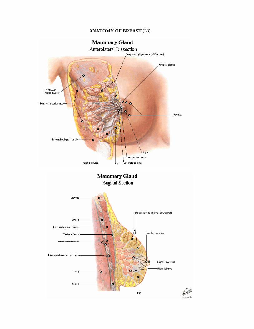
ANATOMY OF BREAST (38)
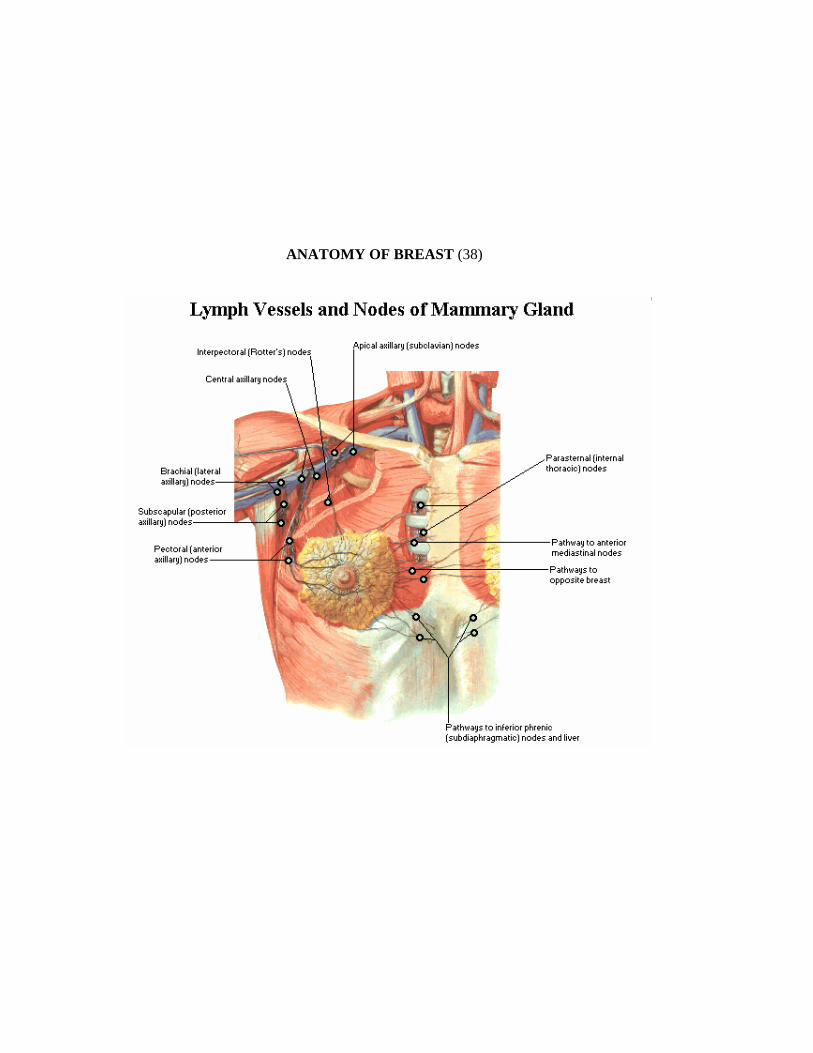
ANATOMY OF BREAST (38)

ANATOMY OF BREAST (38)

S.No. Name AgeIP.
/OP.No.Clinical Diagnosis FNAC Mammography Ultrasound 3D USG
BIRADS Probable Diagnosis HPE Follow-up
1 Parvathy 45 OP 8140Lt. Fibro Adenoma
Fibrocystic Disease Dense Breast
Round Shape, Well defined B/L Fibrocystic disease, Fibroadenoma Lt. Comp II
Fibrocystic Disease
Conservative treatment
2 Natchammal 37 IP 17348
Lt. Fibro cystic disease
Fibrocystic Disease
Dense Breast Fibroadenoma Lt. Brem RD Round Shape, Well Defined Comp II
Fibroscystic Disease
Conservative treatment
3 Rajalakshmi 19 IP18068
Rt. Fibroadenoma
Fibroadenoma Dense Breast
Fibroadenoma Rt. Breast Round Shape, Well Defined Comp III
Excision Fibroadenoma Rt. Excision done
4 Krishnaveni 3 OP9954Lt. Fibro Adenoma
Fibroadenoma Fibroadenoma CT Multiple fibroadenoma Lt. Rt. Comp II
Excision Fibroadenoma Rt. Excision Done
5 Karuppathal 27 IP32368
Rt. Fibroadenoma
Fibroadenoma Dense Breast Rt. Fibroadenoma Comp III
Fibroadenoma Rt. Excision Done
6 Thilagamani 28 OP36416Lt. Fibro Adenoma
Fibroadenoma Lt. Round Fibro adenoma
Lt. Fibroadenoma Rt. Small lesions Comp IV
Fibroadenoma Lt. Excision Done
7 Janaki 35 OP 6883Lump Rt. Breast
Fibrocystic Disease
Benign Lesion Lt. Side Fibrocystic Disease B/L Multiple Fibroadenoma Comp III
Floride Adenosis Excision Done
8 Saroja 39 IP36916Lump Rt. Breast
Fibrocystic Disease
Multiple Benign Lesion fibrocystic disease Giant Fibroadenoma Rt. Comp IV
Fibrocystic Disease
Incision biopsy conservative management
9 Soujath 17 IP 51391Fibroadenoma
Fibroadenoma Dense Breast Lt. Lt. Fibroadenoma Comp III
Fibroadenoma Excision done
10 Kalyani 31 IP17347
Lt. Fibroadenoma
Lt.Fibro adenoma Fibroadenoma Lt Lt. Fibroadenoma Comp III
Fibroadenoma Excision done
11 Revathi 24 OP 6992Lump Rt. Breast
Breast Abcess Lt. Radio Dense Lesion Lt. Breast Abscess
indeterminate II
Breast Abcess Drained
12 Indira 34 IP 32247
Lt. Fibroadenoma
Fibroadenoma Lt. Fibro adenoma Lt. Fibroadenoma Comp III
Fibroadenoma Excision done
13 Kaliyammal 55 IP 66568Lump Rt. Breast Abscess Rt. Radio Dense lesion Rt. Abscess
indeterminate II
Breast Abcess Drained
14 Shanthi 34 IP 65174
Lt. Fibroadenoma
Fibroadenoma Lt. Fibro adenoma Lt. Fibroadenoma Comp III
Fibroadenoma Excision done
15 Prabhavathy 29 IP 52969
Lt. Fibroadenoma
Lt.Fibro adenoma Lt. Fibro adenoma Lt. Fibroadenoma Comp III
Fibroadenoma Excision done
16 Musthirabanu 14 OP 4450
Lt. Fibroadenoma
Lt.Fibro adenoma Dense Breast Lt. Fibroadenoma Comp III
Fibroadenoma Excision done
17 Nagamani 20 OP 8631Lump Lt. Breast
Fibroadenoma Lt. Fibro adenoma Lt. Fibroadenoma Comp III
Fibroadenoma Excision done
18 Vijaya 37 OP 4217
Fibroadenoma Lt. Breast
Fibroadenoma Lt. Fibro adenoma Fibroadadenoma Lt. Comp III
Fibroadenoma Excision biopsy
19 Sundarammal 27 IP 49437Lump Lt. Breast
Breast Abcess
Radiodense Lesion with ill defined margins Breast abscess
Intermediate III
Breast Abcess Drained
20 Kamalam 23 OP 553Fibroadenoma Rt.
Fibroadenoma Dense Breast Fibroadenoma Rt. Breast R WD Comp III
Fibroadenoma Excision done
21 Angathal 55 IP 13888Lump Rt. Breast
Fibro cystic disease
Rt. Giant Fibroadenoma and phyllaids tumour Phylloids tumour Rt Comp III
Phylloides Tumour Excision biopsy
22 Preethamary 23OP 20347
Lt. Fibro Adenoma
Fibroadenoma Lt. Fibro adenoma Lt. Fibroadenoma Comp IV
Fibroadenoma Excision done
23 Rajeswari 18OP 48157
Lt. Fibro Adenoma
Fibroadenoma Lt. Fibro adenoma Lt. Fibroadenoma Comp III
Fibroadenoma Excision done
24 Shakila 27 IP 40141Lt. Fibro Adenoma
Fibroadenoma Rt. Fibroadenoma Rt. Fibroadenoma Comp III
Fibroadenoma Excision
25 Sumithra 21 IP 1889
Rt. Fibroadenoma
Fibroadenoma Rt. Fibroadenoma Rt. Fibroadenoma Comp III
Fibroadenoma Excision done
26 Kamala 20OP 12087
Lump Rt. Breast
Fibroadenoma Rt. Fibroadenoma Rt. Fibroadenoma Comp III
Fibroadenoma Excision done
27 Amaravathy 24 IP 65758
Rt. Fibroadenoma
Fibroadenoma Rt. Fibroadenoma Rt. Fibroadenoma Comp III
Fibroadenoma Excision done
28 Naseera 19 OP 2716
Rt. Fibroadenoma
Fibroadenoma Rt. Fibroadenoma Rt. Fibroadenoma Comp III
Fibroadenoma Excision done
29 Remja 32 OP 9194
Lt. Fibroadenoma
Fibroadenoma Lt. Fibroadenoma Fibrocystic Disease Lt. Comp II
Fibrocystic disease Excision done
30 Dhanalakshmi 37 IP 5525
Lt. Fibroadenoma
Fibroadenoma matoid hyperplasia Lt. Fibroadenoma Lt. Fibroadenoma Comp III
Fibroadenoma Excision
31 Amsabegam 22 OP7567
Rt. Fibroadenoma
Fibroadenoma matoid hyperplasia Dense Breast
Rt. Fibroadenoma with fibrocystic changes Comp III
Fibroadenoma Excision
32 Lakshmi 39 IP5608Lump Rt. Breast
Fibrocystic Diseases
Dense Breast BD, Rt Fibroadenoma
Fibrocystic Diseasese Rt. Fibroadenoma
indeterminate III
Fibroadenoma Excision
33 Kavitha 27 IP 35387 Lumpt LtFibroadenoma Dense Breast Fibroadenoma Comp III
Fibroadenoma Excision
34 Kaliammal 47 IP 55337Fibroadenoma Lt.
Fibrocystic Diseases Giant Fibroadenoma Phylloids tumour Benign Comp III
Phylloides Tumour Excision
35 Kamarinisha 21 OP1475Lt. Abscess
Breast Abcess
B/L Dense Breasts, Dense lesion Lt. Breast abscess Lt
Intermediate II
Tuberculous breast abcess Drained
36 Thulasimani 45 OP4334Lt. Abscess
Breast Abcess Bilateral Dense Breast Breast Abcess
Intermediate II
Breast Abscess Aspirated
37 Fathima 19 IP43375Fibroadenoma Rt.
Fibroadenoma Dense Breast Fibroadenoma Rt. Comp III
Fibroadenoma Excision
38 Suganthi 35 IP40960Lump Lt. Breast
Fibrocystic Disease Dense Breast B/L Fibrocystic Diseases - II
Fibrocystic Disease
lt. Excision of Lump
39 Maheswari 19 IP55952 FAD RtFibroadenoma Fibroadenoma Fibroadenoma Rt. Comp III
Fibroadenoma Excision
40 Prabha 25 IP56625 Rt FADFibroadenoma Dense Breast Rt. Fibroadenoma Comp III
Fibroadenoma Excision
41 Kalaimagal 32 IP 54167 Lt FADFibroadenoma Lt. Fibroadenoma Lt. Fibroadenoma Comp III
Fibroadenoma Excision
42 Vijayalakshmi 43 IP 37970
Lt. Fibroadenoma
Fibroadenoma Lt. Fibroadenoma Lt. Fibroadenoma Comp III
Fibroadenoma Excision

43 Parvathy 50 IP 36743 Rt. FAADFibroadenoma Rt. Fibroadenoma Rt. Fibroadenoma Comp III
Fibroadenoma Excision Done
44 Radha 23 IP 62651 Rt. FADFibroadenoma Dense Breast Rt. Fibroadenoma Comp III
Fibroadenoma Excision Rt.
45 Muthulakshmi 16 IP 62719 Rt. FADFibroadenoma Dense Breast Rt. Fibroadenoma Comp III
Fibroadenoma Excision
46 Selvi 19 IP 51048Fibroadenoma Lt.
Fibroadenoma Dense Breasts Lt. Fibroadenoma Comp III
Fibroadenoma Excision
47 Sabeena 19 IP11763 Lt. FADFibroadenoma Dense Breasts Lt. Fibroadenoma Comp II
Fibroadenoma Excision
48 Leena 23 IP27809 Rt. FADFibroadenoma
Dense Breasts & Benign Lesion Rt. Rt. Fibroadenoma Comp III
Fibroadenoma Excision
49 Parvathy 32 IP52861 FAD Rt.Fibroadenoma Rt. Fibroadenoma Rt. Fibroadenoma Comp III
Fibroadenoma Excision
50 Krishnaveni 20 OP2917
Lt. Fibroadenoma
Fibrosystic disease Lt. Benign lesions
Rt. Fibroadenoma Lt. Fibroadenoma Comp III
Conservative treatment
51 Kalyani 31OP 12347
lt. Breast Multiple Fibroadenoma
Fibroadenoma Fibroadenoma Lt. Fibroadenoma Lt. Comp III
Conservative treatment
52 Vidhya 35 OP 8451
Lt. Fibroadenoma
Lt. Fibroadenoma Lt. Fibroadenoma Lt. Fibroadenoma Comp III
Conservative Management
53 Rathinam 23 OP6827
Rt. Fibroadenoma
Fibroadenoma Fibroadenoma Rt. Rt. Fibroadenoma Comp III
Conservative treatment
54 Omana 28 OP3011
Lt. Fibroadenoma
Fibro adenoma Lt. Fibroadenoma Lt. Fibroadenoma Comp III
Conservative Management
55 Jagadeswari 19 OP4603
Rt. Fibroadenoma
Fibro adenoma Rt. Fibroadenoma Rt. Fibroadenoma Comp III
Conservative Management
56 Kavitha 20 OP17153
Lt. Fibroadenoma
Fibro adenoma Lt. Fibroadenoma Lt. Fibroadenoma Comp III
Conservative Management
57 Vijayalakshmi 22 OP484
Lt. Fibroadenoma
Fibro adenoma Lt. Fibroadenoma Lt. Fibroadenoma Comp III Conservative
58 Valliyammal 21 OP3902
Rt. Fibroadenoma
Fibro adenoma Rt. Fibroadenoma Lt. Fibroadenoma BRITOS III Comp III Conservative
59 Easwari 25 OP1240Lump Rt. Breast
Fibro adenoma Lt. Fibroadenoma Lt. Fibroadenoma Comp III Conservative
60 Valarmathi 30 OP5806Fibrocystic Disease
Fibrocystic Disease Dense Breast Fibrocystic Disease Comp III Conservative
61 Parvathy 35 OP7891Lump Lt. Breast
Fibro adenoma Lt. Fibroadenoma Lt. Fibroadenoma Comp III Conservative
62 Ranjitha 26 OP232 Lt. LumpFibro adenoma Lt. Fibroadenoma Lt. Fibroadenoma Comp III Conservative
63 Valli 37 OP636 Lt. LumpFibrocystic Disease Dense Breast Lt. Fibroadenoma Comp II Conservative
64 Renganayaki 21 OP2343
Rt. Fibroadenoma
Fibro adenoma Rt. Fibroadenoma Rt. Fibrocystic Disease Comp II Conservative

65 Vasuki 30 OP9568 Lump Rt.Fibro adenosis Rt. Benign Lesion Rt. Fibrocystic Disease Comp II Conservative
66 Jayamani 37 OP39774 Lump Rt.Fibrocystic diseases Rt. Benign Lesion Rt. Fibrocystic Disease Comp II Conservative
67 Chellammal 35 OP3684
Rt. Fibroadenoma
Fibroadenoma Rt. Fibroadenoma Rt. Fibroadenoma Comp III Conservative
68 Roja 35 OP2748
Rt. Fibroadenoma
Rt.Atypical cells Multiple Benign Masses
Rt. 12B B IV Fibroadenoma, Fibroadenosis, Smart Cyst Lt. RE IV, III
Conservative Treatment
69 Geetha 24 IP25822
Lt. Fibroadenoma
Fibrocystic Disease Normal Focal Fibrocystic Champs Comp II Conservative