corynebacterium diphtheriae - usu opencourseware
TRANSCRIPT
12/3/2010
1
Diphtheria
Corynebacterium diphtheriae
Corynebacteria are Gram-positive, aerobic,nonmotile, rod-shaped bacteria classified asActinobacteria. Corynebacteria are relatedphylogenetically to mycobacteria andactinomycetes. They do not form spores or branchas do the actinomycetes, but they have thecharacteristic of forming irregular, club-shaped or V-shaped arrangements in normal growth. Theyundergo snapping movements just after cell division,which brings them into characteristic formsresembling Chinese letters or palisades.
12/3/2010
2
The genus Corynebacterium consists of a
diverse group of bacteria including animal
and plant pathogens, as well as saprophytes.
Some corynebacteria are part of the normal
flora of humans, finding a suitable niche in
virtually every anatomic site, especially the
skin and nares. The best known and most
widely studied species is Corynebacterium
diphtheriae, the causal agent of the disease
diphtheria.
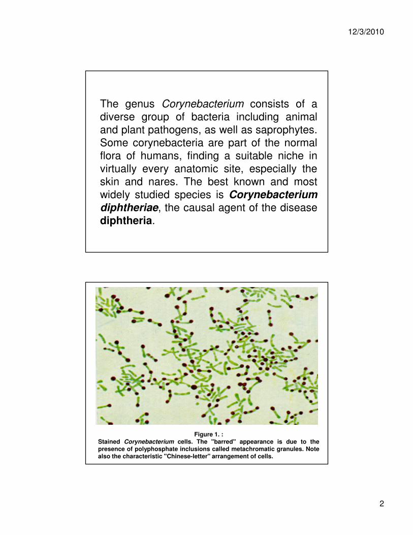
Figure 1. :Stained Corynebacterium cells. The "barred" appearance is due to thepresence of polyphosphate inclusions called metachromatic granules. Notealso the characteristic "Chinese-letter" arrangement of cells.
12/3/2010
3
Diphtheria is an upper respiratory tract illness characterized
by sore throat, low fever, and an adherent membrane (called a
pseudomembrane) on the tonsils, pharynx, and/or nasal
cavity. Diphtheria toxin produced by C. diphtheriae, can cause
myocarditis, polyneuritis, and other systemic toxic effects. A
milder form of diphtheria can be restricted to the skin.
Diphtheria is a contagious disease spread by direct physical
contact or breathing aerosolized secretions of infected
individuals. Once quite common, diphtheria has largely been
eradicated in developed nations through wide-spread use of
the DPT vaccine. For example, in the U.S., between 1980 and
2004 there were 57 reported cases of diphtheria. However, it
remains somewhat of a problem worldwide (3,978 reported
cases to WHO in 2006) in the face of efforts to achieve global
vaccination coverage.
Diphtheria is a serious disease, with fatality rates
between 5% and 10%. In children under 5 years
and adults over 40 years, the fatality rate may be
as much as 20%. Outbreaks, although very rare,
still occur worldwide, even in developed nations.
Following the breakup of the former Soviet Union
in the late 1980s, vaccination rates in the
constituent countries fell so low that there was a
surge in diphtheria cases. In 1991 there were
2,000 cases of diphtheria in the USSR. By 1998,
according to Red Cross estimates, there were as
many as 200,000 cases in the Commonwealth of
Independent States, with 5,000 deaths.
12/3/2010
4
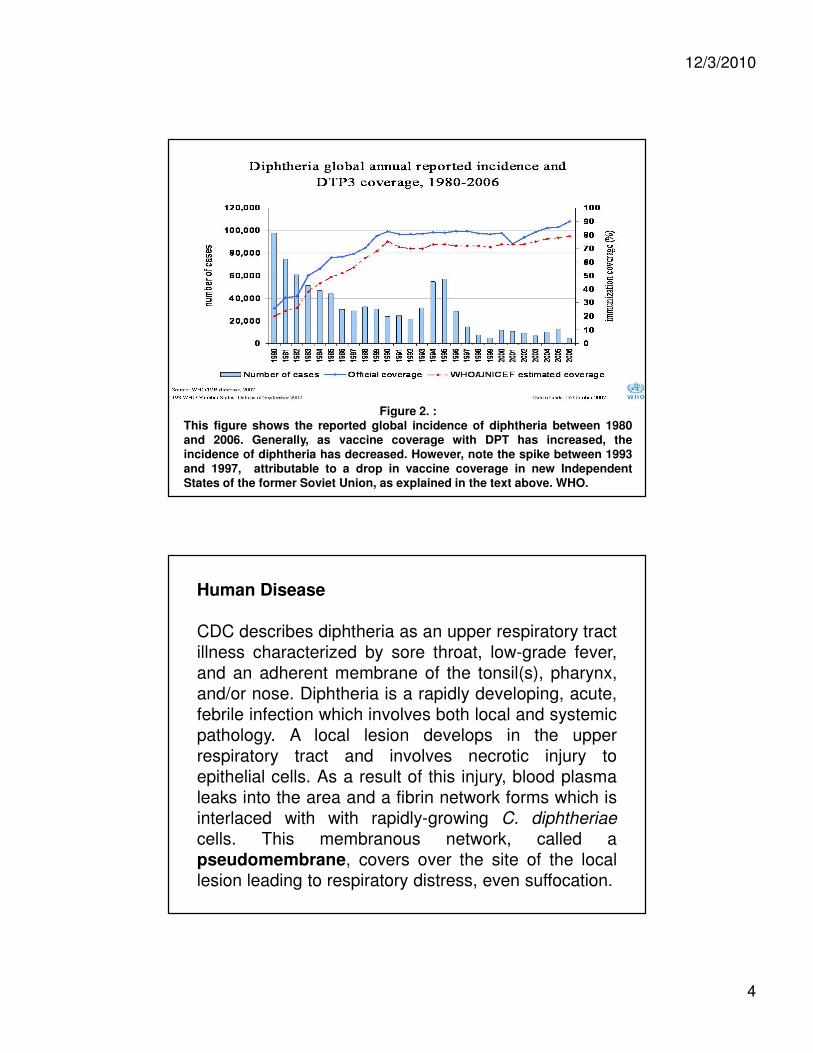
Figure 2. :This figure shows the reported global incidence of diphtheria between 1980and 2006. Generally, as vaccine coverage with DPT has increased, theincidence of diphtheria has decreased. However, note the spike between 1993and 1997, attributable to a drop in vaccine coverage in new IndependentStates of the former Soviet Union, as explained in the text above. WHO.
Human Disease
CDC describes diphtheria as an upper respiratory tract
illness characterized by sore throat, low-grade fever,
and an adherent membrane of the tonsil(s), pharynx,
and/or nose. Diphtheria is a rapidly developing, acute,
febrile infection which involves both local and systemic
pathology. A local lesion develops in the upper
respiratory tract and involves necrotic injury to
epithelial cells. As a result of this injury, blood plasma
leaks into the area and a fibrin network forms which is
interlaced with with rapidly-growing C. diphtheriae
cells. This membranous network, called a
pseudomembrane, covers over the site of the local
lesion leading to respiratory distress, even suffocation.
12/3/2010
5
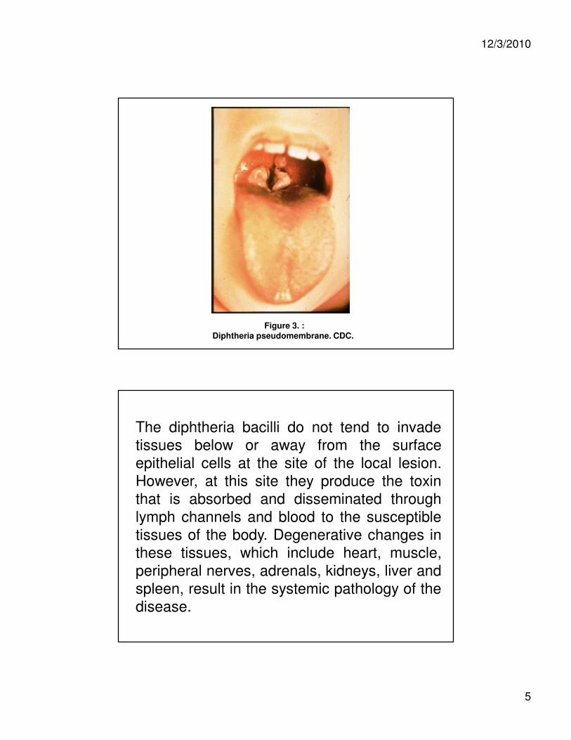
Figure 3. :Diphtheria pseudomembrane. CDC.
The diphtheria bacilli do not tend to invade
tissues below or away from the surface
epithelial cells at the site of the local lesion.
However, at this site they produce the toxin
that is absorbed and disseminated through
lymph channels and blood to the susceptible
tissues of the body. Degenerative changes in
these tissues, which include heart, muscle,
peripheral nerves, adrenals, kidneys, liver and
spleen, result in the systemic pathology of the
disease.
12/3/2010
6
Pathogenicity
The pathogenicity of Corynebacterium diphtheriae
includes two distinct phenomena:
1.Invasion of the local tissues of the throat,
which requires colonization and subsequent
bacterial proliferation. Little is known about the
adherence mechanisms of C. diphtheriae, but
the bacteria produce several types of pili. The
diphtheria toxin, as well, may be involved in
colonization of the throat.
2.Toxigenesis: bacterial production of the
toxin. The diphtheria toxin causes the death
eucaryotic cells and tissues by inhibition
protein synthesis in the cells. Although the
toxin is responsible for the lethal symptoms of
the disease, the virulence of C. diphtheriae
cannot be attributed to toxigenicity alone,
since a distinct invasive phase apparently
precedes toxigenesis. However, it has not
been ruled out that the diphtheria toxin plays
an essential role in the colonization process
due to short-range effects at the colonization
site.
12/3/2010
7
Three strains of Corynebacterium diphtheriaeare recognized, gravis, intermedius and mitis.
They are listed here by falling order of the
severity of the disease that they produce in
humans. All strains produce the identical toxin
and are capable of colonizing the throat. The
differences in virulence between the three strains
can be explained by their differing abilities to
produce the toxin in rate and quantity, and by
their differing growth rates.
The gravis strain has a generation time (in vitro) of60 minutes; the intermedius strain has a generationtime of about 100 minutes; and the mitis stain has ageneration time of about 180 minutes. The fastergrowing strains typically produce a larger colony onmost growth media. In the throat (in vivo), a fastergrowth rate may allow the organism to deplete thelocal iron supply more rapidly in the invaded tissues,thereby allowing earlier or greater production of thediphtheria toxin. Also, if the kinetics of toxinproduction follow the kinetics of bacterial growth, thefaster growing variety would achieve an effectivelevel of toxin before the slow growing varieties.
12/3/2010
8
Toxigenicity
Two factors have great influence on the ability ofCorynebacterium diphtheriae to produce thediphtheria toxin: (1) low extracellularconcentrations of iron and (2) the presence of alysogenic prophage in the bacterial chromosome.The gene for toxin production occurs on thechromosome of the prophage, but a bacterialrepressor protein controls the expression of thisgene. The repressor is activated by iron, and it is inthis way that iron influences toxin production. Highyields of toxin are synthesized only by lysogenicbacteria under conditions of iron deficiency.
The role of iron. In artificial culture the most important factorcontrolling yield of the toxin is the concentration of inorganic iron(Fe++ or Fe+++) present in the culture medium. Toxin is synthesizedin high yield only after the exogenous supply of iron has becomeexhausted (This has practical importance for the industrial productionof toxin to make toxoid. Under the appropriate conditions of ironstarvation, C. diphtheriae will synthesize diphtheria toxin as 5% of itstotal protein). Presumably, this phenomenon takes place in vivo aswell. The bacterium may not produce maximal amounts of toxin untilthe iron supply in tissues of the upper respiratory tract has becomedepleted. It is the regulation of toxin production in the bacterium thatis partially controlled by iron. The tox gene is regulated by amechanism of negative control wherein a repressor molecule,product of the DtxR gene, is activated by iron. The active repressorbinds to the tox gene operator and prevents transcription. When ironis removed from the repressor (under growth conditions of ironlimitation), derepression occurs, the repressor is inactivated andtranscription of the tox genes can occur. Iron is referred to as acorepressor since it is required for repression of the toxin gene.
12/3/2010
9
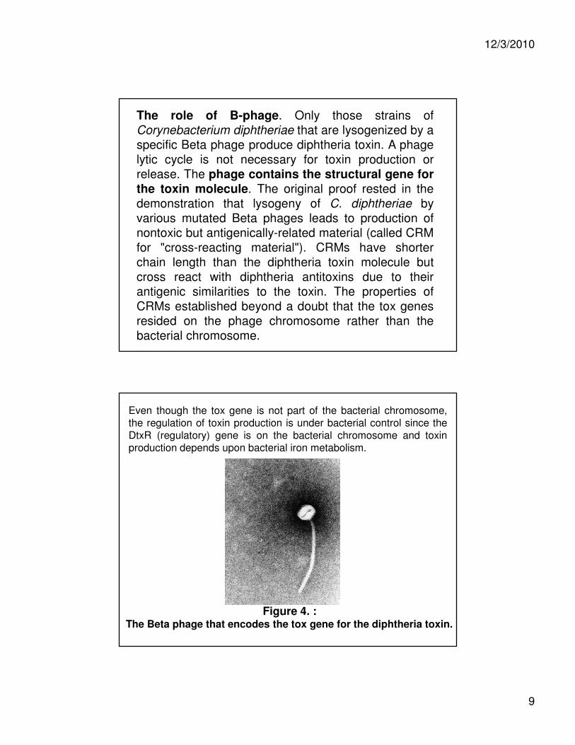
The role of B-phage. Only those strains of
Corynebacterium diphtheriae that are lysogenized by a
specific Beta phage produce diphtheria toxin. A phage
lytic cycle is not necessary for toxin production or
release. The phage contains the structural gene for
the toxin molecule. The original proof rested in the
demonstration that lysogeny of C. diphtheriae by
various mutated Beta phages leads to production of
nontoxic but antigenically-related material (called CRM
for "cross-reacting material"). CRMs have shorter
chain length than the diphtheria toxin molecule but
cross react with diphtheria antitoxins due to their
antigenic similarities to the toxin. The properties of
CRMs established beyond a doubt that the tox genes
resided on the phage chromosome rather than the
bacterial chromosome.
Figure 4. :The Beta phage that encodes the tox gene for the diphtheria toxin.
Even though the tox gene is not part of the bacterial chromosome,the regulation of toxin production is under bacterial control since theDtxR (regulatory) gene is on the bacterial chromosome and toxinproduction depends upon bacterial iron metabolism.
12/3/2010
10
It is of some interest to speculate on the role of the diphtheria
toxin in the natural history of the bacterium. Of what value
should it be to an organism to synthesize up to 5% of its total
protein as a toxin that specifically inhibits protein synthesis in
eucaryotes and archaea? Possibly the toxin assists
colonization of the throat (or skin) by killing epithelial cells or
neutrophils. There is no evidence to suggest a key role of the
toxin in the life cycle of the organism. Since mass
immunization against diphtheria has been practiced, the
disease has virtually disappeared, and C. diphtheriae is no
longer a component of the normal flora of the human throat
and pharynx. It may be that the toxin played a key role in the
colonization of the throat in nonimmune individuals and, as a
consequence of exhaustive immunization, toxigenic strains
have become virtually extinct.
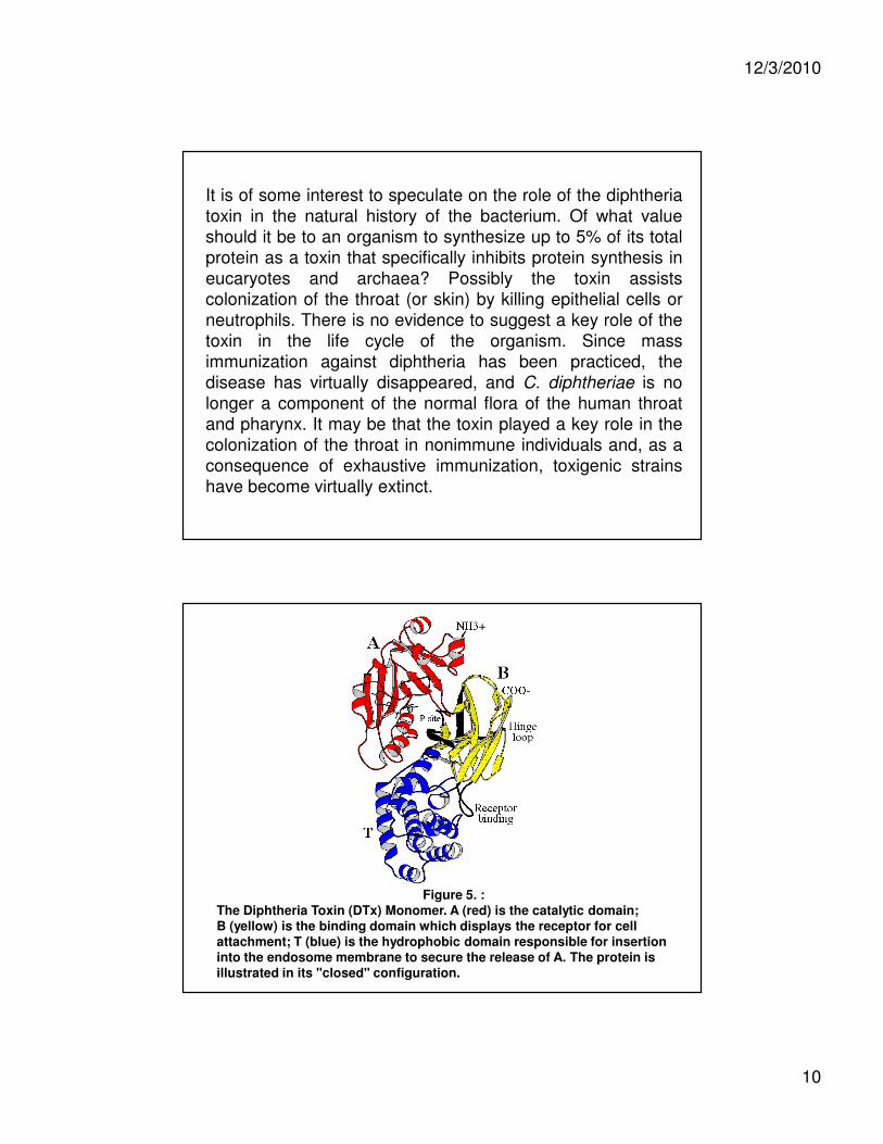
Figure 5. :The Diphtheria Toxin (DTx) Monomer. A (red) is the catalytic domain; B (yellow) is the binding domain which displays the receptor for cell
attachment; T (blue) is the hydrophobic domain responsible for insertion into the endosome membrane to secure the release of A. The protein is illustrated in its "closed" configuration.
12/3/2010
11
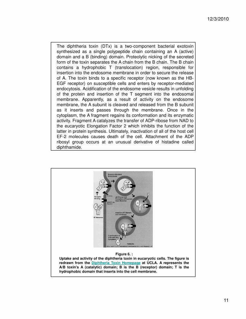
The diphtheria toxin (DTx) is a two-component bacterial exotoxinsynthesized as a single polypeptide chain containing an A (active)domain and a B (binding) domain. Proteolytic nicking of the secretedform of the toxin separates the A chain from the B chain. The B chaincontains a hydrophobic T (translocation) region, responsible forinsertion into the endosome membrane in order to secure the releaseof A. The toxin binds to a specific receptor (now known as the HB-EGF receptor) on susceptible cells and enters by receptor-mediatedendocytosis. Acidification of the endosome vesicle results in unfoldingof the protein and insertion of the T segment into the endosomalmembrane. Apparently, as a result of activity on the endosomemembrane, the A subunit is cleaved and released from the B subunitas it inserts and passes through the membrane. Once in thecytoplasm, the A fragment regains its conformation and its enzymaticactivity. Fragment A catalyzes the transfer of ADP-ribose from NAD tothe eucaryotic Elongation Factor 2 which inhibits the function of thelatter in protein synthesis. Ultimately, inactivation of all of the host cellEF-2 molecules causes death of the cell. Attachment of the ADPribosyl group occurs at an unusual derivative of histadine calleddiphthamide.
Figure 6. :Uptake and activity of the diphtheria toxin in eucaryotic cells. The figure isredrawn from the Diphtheria Toxin Homepage at UCLA. A represents theA/B toxin's A (catalytic) domain; B is the B (receptor) domain; T is thehydrophobic domain that inserts into the cell membrane.
12/3/2010
12
In vitro, the native diphtheria toxin is inactive
and can be activated by trypsin in the
presence of thiol. The enzymatic activity of
fragment A is masked in the intact toxin.
Fragment B is required to bind the native toxin
to its cognate receptor and to permit the
escape of fragment A from the endosome. The
C terminal end of Fragment B contains the
peptide region that attaches to the HB-EGF
receptor on the sensitive cell membrane, and
the N-terminal end is a strongly hydrophobic
region which will insert into a membrane lipid
bilayer.
The specific membrane receptor, heparin-bindingepidermal growth factor (HB-EGF) precursor is aprotein on the surface of many types of cells. Theoccurrence and distribution of the HB-EGF receptoron cells determines the susceptibility of an animalspecies, and certain cells of an animal species, tothe diphtheria toxin. Normally, the HB-EGFprecursor releases a peptide hormone thatinfluences normal cell growth and differentiation.One hypothesis is that the HB-EGF receptor itself isthe protease that nicks the A fragment and reducesthe disulfide bridge between it and the B fragmentwhen the A fragment makes its way through theendosomal membrane into the cytoplasm.
12/3/2010
13
Immunity to Diphtheria
Acquired immunity to diphtheria is due primarily
to toxin-neutralizing antibody (antitoxin). Passive
immunity in utero is acquired transplacentally
and can last at most 1 or 2 years after birth. In
areas where diphtheria is endemic and mass
immunization is not practiced, most young
children are highly susceptible to infection.
Probably, active immunity can be produced by a
mild or inapparent infection in infants who retain
some maternal immunity, and in adults infected
with strains of low virulence (inapparent
infections).
Individuals that have fully recovered from diphtheria may
continue to harbor the organisms in the throat or nose for
weeks or even months. In the past, it was mainly through
such healthy carriers that the disease was spread, and
toxigenic bacteria were maintained in the population. Before
mass immunization of children, carrier rates of C. diphtheriae
of 5% or higher were observed.
Because of the high degree of susceptibility of children,
artificial immunization at an early age is universally
advocated. Toxoid is given in 2 or 3 doses (1 month apart) for
primary immunization at an age of 3 - 4 months. A booster
injection should be given about a year later, and it is
advisable to administer several booster injections during
childhood. Usually, infants in the United States are
immunized with a trivalent vaccine containing diphtheria
toxoid, pertussis vaccine, and tetanus toxoid (DPT or DTaP
vaccine).
12/3/2010
14
The relative absence of diphtheria in the UnitedStates is due primarily to the high level ofappropriate immunization in children, and to anapparent reduction in toxin-producing strains of thebacterium. However, the increasing percentage ofdiphtheria cases in adults suggests that manyadults may not be protected against diphtheria,because they have not received boosterimmunizations within the past ten years. A similarsituation exists with tetanus.
Sex but not loveSex but not love
Bed but not sleepBed but not sleep
Computer but not brainComputer but not brain
Food but not appetiteFood but not appetite
Finery but not beautyFinery but not beauty
House but not homeHouse but not home
Medicine but not healthMedicine but not health
Luxuries but not cultureLuxuries but not culture
Amusements but not happinessAmusements but not happiness

12/3/2010
15