development of a coronary-perfused interventricular septal preparation as a model for studying the...
TRANSCRIPT

Available online at www.sciencedirect.com
40 (2007) S142–S144www.jecgonline.com
Journal of Electrocardiology
Development of a coronary-perfused interventricular septal preparation asa model for studying the role of the septum in arrhythmogenesis☆
Aaron Glass, BS, Serge Sicouri, MD,⁎ Charles Antzelevitch, PhDMasonic Medical Research Laboratory, Utica, NY, USA
Received 29 March 2007; accepted 14 May 2007
Abstract Background: Coronary-perfused ventricular wedge preparations have proven valuable in the
☆ Supported by GInstitute (CA) and NSupport for AG was pand Oneida Counties
⁎ Corresponding5648.
E-mail address: si
0022-0736/$ – see frodoi:10.1016/j.jelectroc
elucidation of the mechanisms of arrhythmias. This study was undertaken to develop an arteriallyperfused model of the interventricular (IV) septum.Methods: A canine septal preparation was developed via cannulation of the septal artery. Actionpotentials were recorded from ventricular endocardial surfaces and locations within the septum usingfloating microelectrodes; a transseptal electrocardiogram was simultaneously recorded. In someexperiments, the calcium agonist BayK 8644 was used to enhance transseptal heterogeneity of actionpotential (AP) duration.Results: Distinctive electrocardiographic waveforms and dissimilar AP morphologies and durationswere observed across the IV septum. The range of AP durations observed exceeded that found in theleft ventricular wedge. BayK 8644 further accentuated these differences and induced torsades depointes arrhythmias.Conclusions: The arterially perfused septal preparation is a sensitive model for the study ofarrhythmias that may arise from the IV septum.© 2007 Elsevier Inc. All rights reserved.
Keywords: Electrophysiology; Dispersion of repolarization; Arrhythmias; Pharmacology
Introduction
The development of the arterially perfused ventricularwedge heralded a new paradigm in cardiac electrophysiol-ogy, allowing the measurement of action potentials (APs)from transmural regions containing endocardial, epicardialand midmyocardial cells simultaneously. The characteriza-tion of these 3 electrophysiologically distinct cell typeswithin the ventricular myocardium led to the understandingthat disparities in the times of repolarization of cardiac cellscan contribute to the development of arrhythmias.
Electrophysiologic heterogeneity, the presence of APswith different durations and morphologies within the sametissue, has been previously identified within the canine
rant HL47678 from National Heart, Lung, and BloodYS and Florida Grand Lodges F & AM. Financialrovided by The Community Foundation of Herkimer(Utica, NY).author. Tel.: +1 315 735 2217x157; fax: +1 315 735
nt matter © 2007 Elsevier Inc. All rights reserved.ard.2007.05.030
interventricular (IV) septum using superfused tissue slices1
and dissociated septal cells.2
This study was undertaken to develop an arteriallyperfused preparation derived from the IV septum to confirmthe presence of heterogeneity within the intact septum,quantitate the transseptal dispersion of repolarization (TDR),and delineate its potential role in arrhythmogenesis.
Materials and methods
In compliance with guidelines established by the Institu-tional Animal Care and Use Committee (Utica, NY), canineswith a mass between 20 and 35 kg were anticoagulated withheparin (180 IU/kg) and anesthetized with sodium pento-barbital (35 mg/kg) intravenously. A left thoracotomyprocedure was used to excise the animal's heart, whichwas immediately immersed in an ice-cold cardioplegicsolution ([K+]0 = 8 mmol/L).
Tyrode solution, composed of (in mmol/L) NaCl (129),KCl (4), NaH2PO4 (0.9), NaHCO3 (20), CaCl2 (1.8),MgSO4 (0.5), and D-glucose (5.5), was used as a perfusate
FPLa
S143A. Glass et al. / Journal of Electrocardiology 40 (2007) S142–S144
throughout all experiments. Several different approaches todeliver the perfusate to the septal preparation were attemptedbefore a stable model was developed.
All approaches to a functional septal preparation beganwith the removal of the right and left ventricular (LV) freewalls approximately 1 cm from the IV septum andremoval of the atria and any tissues superior to theatrioventricular valves. Portions of the heart not used inthe septal preparation were distributed among otherinvestigators at our institution, ensuring the organ'scomplete use for research.
The double-cannulated preparation
Our initial attempt at a septal preparation usedcannulation of the left coronary artery (LCA) and thedorsal IV coronary artery (commonly known as posteriorIV artery [PIVA]). To sustain perfusion pressure between 40and 50 mm Hg, the circumflex artery, any severed branchesof the ventral IV artery (commonly referred as left anteriordescending [LAD]), and the PIVA were ligated. To evaluatethe quality of perfusion in the double-cannulated prepara-tion, Evans blue dye (Sigma-Aldrich, St Louis, MO) wasdissolved in the perfusate and pumped through bothcannulas concurrently for 20 minutes. Upon sectioningthe tissue transversely, scattered regions of dye-coloredmyocardium were observed to comprise less than half ofthe preparation. The less-than-optimal perfusion in thedouble-cannulated preparation prompted us to explore adifferent approach.
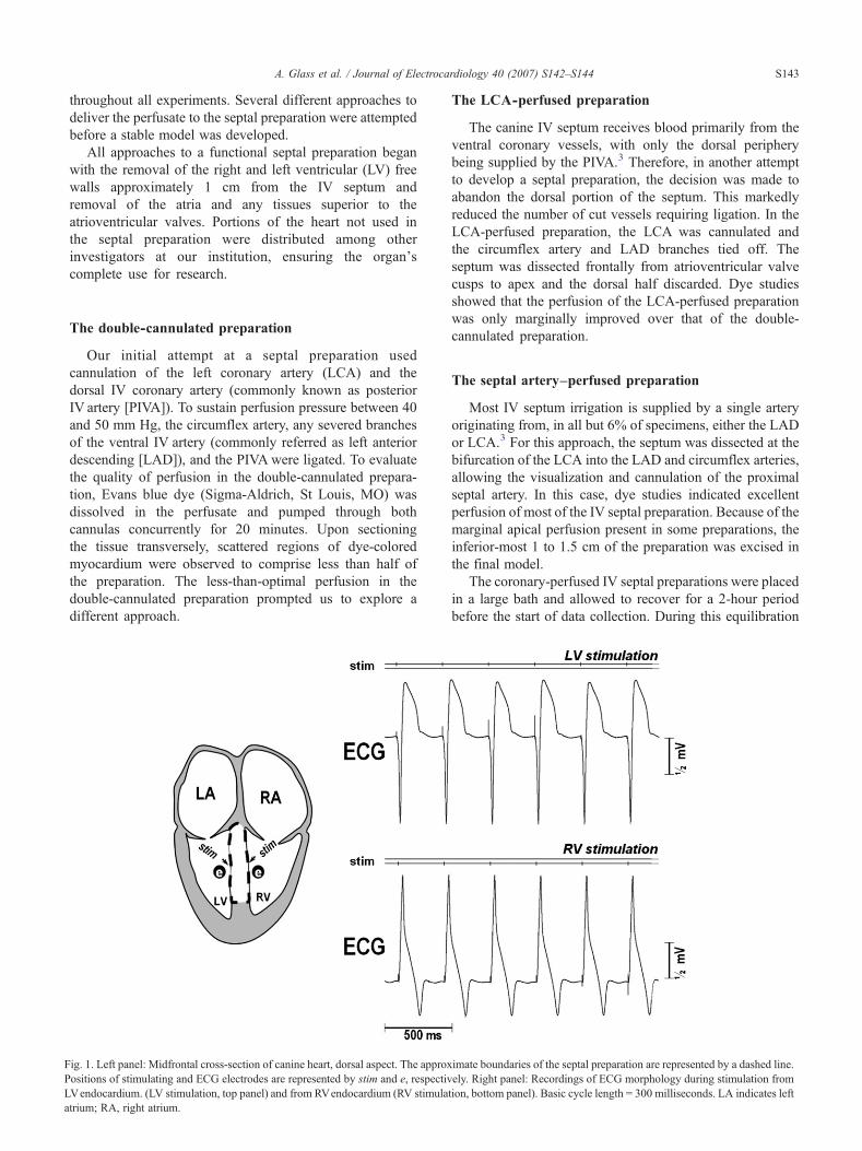
ig. 1. Left panel: Midfrontal cross-section of canine heart, dorsal aspect. The approxositions of stimulating and ECG electrodes are represented by stim and e, respectivVendocardium. (LV stimulation, top panel) and from RVendocardium (RV stimulatrium; RA, right atrium.
The LCA-perfused preparation
The canine IV septum receives blood primarily from theventral coronary vessels, with only the dorsal peripherybeing supplied by the PIVA.3 Therefore, in another attemptto develop a septal preparation, the decision was made toabandon the dorsal portion of the septum. This markedlyreduced the number of cut vessels requiring ligation. In theLCA-perfused preparation, the LCA was cannulated andthe circumflex artery and LAD branches tied off. Theseptum was dissected frontally from atrioventricular valvecusps to apex and the dorsal half discarded. Dye studiesshowed that the perfusion of the LCA-perfused preparationwas only marginally improved over that of the double-cannulated preparation.
The septal artery–perfused preparation
Most IV septum irrigation is supplied by a single arteryoriginating from, in all but 6% of specimens, either the LADor LCA.3 For this approach, the septum was dissected at thebifurcation of the LCA into the LAD and circumflex arteries,allowing the visualization and cannulation of the proximalseptal artery. In this case, dye studies indicated excellentperfusion of most of the IV septal preparation. Because of themarginal apical perfusion present in some preparations, theinferior-most 1 to 1.5 cm of the preparation was excised inthe final model.
The coronary-perfused IV septal preparations were placedin a large bath and allowed to recover for a 2-hour periodbefore the start of data collection. During this equilibration
imate boundaries of the septal preparation are represented by a dashed lineely. Right panel: Recordings of ECG morphology during stimulation fromtion, bottom panel). Basic cycle length = 300 milliseconds. LA indicates lef
.
t
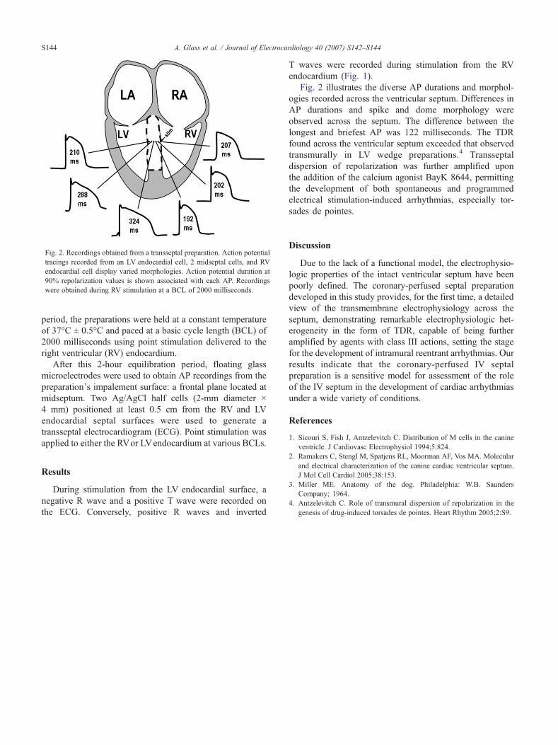
Fig. 2. Recordings obtained from a transseptal preparation. Action potentialtracings recorded from an LV endocardial cell, 2 midseptal cells, and RVendocardial cell display varied morphologies. Action potential duration at90% repolarization values is shown associated with each AP. Recordingswere obtained during RV stimulation at a BCL of 2000 milliseconds.
S144 A. Glass et al. / Journal of Electrocardiology 40 (2007) S142–S144
period, the preparations were held at a constant temperatureof 37°C ± 0.5°C and paced at a basic cycle length (BCL) of2000 milliseconds using point stimulation delivered to theright ventricular (RV) endocardium.
After this 2-hour equilibration period, floating glassmicroelectrodes were used to obtain AP recordings from thepreparation's impalement surface: a frontal plane located atmidseptum. Two Ag/AgCl half cells (2-mm diameter ×4 mm) positioned at least 0.5 cm from the RV and LVendocardial septal surfaces were used to generate atransseptal electrocardiogram (ECG). Point stimulation wasapplied to either the RVor LVendocardium at various BCLs.
Results
During stimulation from the LV endocardial surface, anegative R wave and a positive T wave were recorded onthe ECG. Conversely, positive R waves and inverted
T waves were recorded during stimulation from the RVendocardium (Fig. 1).
Fig. 2 illustrates the diverse AP durations and morphol-ogies recorded across the ventricular septum. Differences inAP durations and spike and dome morphology wereobserved across the septum. The difference between thelongest and briefest AP was 122 milliseconds. The TDRfound across the ventricular septum exceeded that observedtransmurally in LV wedge preparations.4 Transseptaldispersion of repolarization was further amplified uponthe addition of the calcium agonist BayK 8644, permittingthe development of both spontaneous and programmedelectrical stimulation-induced arrhythmias, especially tor-sades de pointes.
Discussion
Due to the lack of a functional model, the electrophysio-logic properties of the intact ventricular septum have beenpoorly defined. The coronary-perfused septal preparationdeveloped in this study provides, for the first time, a detailedview of the transmembrane electrophysiology across theseptum, demonstrating remarkable electrophysiologic het-erogeneity in the form of TDR, capable of being furtheramplified by agents with class III actions, setting the stagefor the development of intramural reentrant arrhythmias. Ourresults indicate that the coronary-perfused IV septalpreparation is a sensitive model for assessment of the roleof the IV septum in the development of cardiac arrhythmiasunder a wide variety of conditions.
References
1. Sicouri S, Fish J, Antzelevitch C. Distribution of M cells in the canineventricle. J Cardiovasc Electrophysiol 1994;5:824.
2. Ramakers C, Stengl M, Spatjens RL, Moorman AF, Vos MA. Molecularand electrical characterization of the canine cardiac ventricular septum.J Mol Cell Cardiol 2005;38:153.
3. Miller ME. Anatomy of the dog. Philadelphia: W.B. SaundersCompany; 1964.
4. Antzelevitch C. Role of transmural dispersion of repolarization in thegenesis of drug-induced torsades de pointes. Heart Rhythm 2005;2:S9.