diagnostic recommendations alz pre clinical
TRANSCRIPT
8/4/2019 Diagnostic Recommendations Alz Pre Clinical
http://slidepdf.com/reader/full/diagnostic-recommendations-alz-pre-clinical 1/13
Toward defining the preclinical stages of Alzheimer’s disease:Recommendations from the National Institute on Aging and the
Alzheimer’s Association workgroup
Reisa A. Sperlinga,*, Paul S. Aisenb, Laurel A. Beckettc, David A. Bennettd, Suzanne Craf te,Anne M. Faganf , Takeshi Iwatsubog, Clifford R. Jack h, Jef frey Kayei, Thomas J. Montine j,Denise C. Park k , Eric M. Reimanl, Christopher C. Rowem, Eric Siemersn, Yaakov Sterno,
Kristine Yaffep, Maria C. Carrilloq, Bill Thiesq, Marcelle Morrison-Bogoradr, Molly V. Wagsterr,Creighton H. Phelpsr
aCenter for Alzheimer Research and Treatment, Department of Neurology, Brigham and Women’s Hospital, Massachusetts General Hospital,
Harvard Medical School, Boston, MA, USAb Department of Neurosciences, University of California San Diego, San Diego, CA, USAc Division of Biostatistics, School of Medicine, University of California, Davis, CA, USAd Rush Alzheimer’s Disease Center, Rush University Medical Center, Chicago, IL, USA
eGeriatric Research, Education, and Clinical Center, Veterans Affairs Puget Sound; Department of Psychiatry and Behavioral Sciences,
University of Washington School of Medicine, Seattle, WA, USA f Department of Neurology, Washington University School of Medicine, St. Louis, MO, USA
g Department of Neuropathology, Graduate School of Medicine, University of Tokyo, Tokyo, Japanh Department of Radiology, Mayo Clinic Minnesota, Rochester, MN, USA
i Departments of Neurology and Biomedical Engineering, Layton Aging & Alzheimer’s Disease Center, Oregon Center for Aging & Technology,
Oregon Health & Science University and Portland Veteran’s Affairs Medical Center, Portland, OR, USA j Department of Pathology, University of Washington, Seattle, WA, USA
k Center for Vital Longevity, University of Texas at Dallas, Dallas, TX, USA
l Banner Alzheimer’s Institute, Phoenix, AZ, USAm Austin Health, University of Melbourne, Melbourne, Australia
nEli Lilly and Company, Indianapolis, IN, USA
oCognitive Neuroscience Division, Taub Institute, Columbia University College of Physicians and Surgeons, New York, NY, USA
p Departments of Psychiatry, Neurology, and Epidemiology and Biostatistics, University of California San Francisco, San Francisco VA Medical Center,
San Francisco, CA, USAq Alzheimer’s Association, Chicago, IL, USA
r Division of Neuroscience, National Institute on Aging, Bethesda, MD, USA
Abstract The pathophysiological process of Alzheimer’s disease (AD) is thought to begin many years
before the diagnosis of AD dementia. This long “preclinical” phase of AD would provide a critical
opportunity for therapeutic intervention; however, we need to further elucidate the link between the
pathological cascade of AD and the emergence of clinical symptoms. The National Institute onAging and the Alzheimer’s Association convened an international workgroup to review the bio-
marker, epidemiological, and neuropsychological evidence, and to develop recommendations to
determine the factors which best predict the risk of progression from “normal” cognition to
mild cognitive impairment and AD dementia. We propose a conceptual framework and operational
research criteria, based on the prevailing scientific evidence to date, to test and refine these models
with longitudinal clinical research studies. These recommendations are solely intended for research
purposes and do not have any clinical implications at this time. It is hoped that these recommen-
*Corresponding author. Tel.: 1 1-617-732-8085; Fax: 11-617-264-5212.
E-mail address: [email protected]
1552-5260/$ - see front matter Ó 2011 The Alzheimer’s Association. All rights reserved.doi:10.1016/j.jalz.2011.03.003
FLA 5.1.0 DTD � JALZ1250_proof � 16 April 2011 � 12:04 pm � ce
Alzheimer’s & Dementia- (2011) 1–13
8/4/2019 Diagnostic Recommendations Alz Pre Clinical
http://slidepdf.com/reader/full/diagnostic-recommendations-alz-pre-clinical 2/13
dations will provide a common rubric to advance the study of preclinical AD, and ultimately, aid
the field in moving toward earlier intervention at a stage of AD when some disease-modifying ther-
apies may be most efficacious.
Ó 2011 The Alzheimer’s Association. All rights reserved.
Keywords: Preclinical Alzheimer’s disease; Biomarker; Amyloid; Neurodegeneration; Prevention
1. Introduction
Converging evidence from both genetic at-risk cohorts
and clinically normal older individuals suggests that the
pathophysiological process of Alzheimer’s disease (AD)
begins years, if not decades, before the diagnosis of clinical
dementia [1]. Recent advances in neuroimaging, cerebrospi-
nal fluid (CSF) assays, and other biomarkers now provide the
ability to detect evidence of the AD pathophysiological pro-
cess in vivo. Emerging data in clinically normal older indi-
viduals suggest that biomarker evidence of amyloid beta(Ab) accumulation is associated with functional and struc-
tural brain alterations, consistent with the patterns of abnor-
mality seen in patients with mild cognitive impairment
(MCI) and AD dementia. Furthermore, clinical cohort
studies suggest that there may be very subtle cognitive
alterations that are detectable years before meeting criteria
for MCI, and that predict progression to AD dementia. It is
also clear, however, that some older individuals with the
pathophysiological process of AD may not become symp-
tomatic during their lifetime. Thus, it is critical to better de-
fine the biomarker and/or cognitive profile that best predicts
progression from the preclinical to the clinical stages of MCIand AD dementia. The long preclinical phase of AD pro-
vides a critical opportunity for potential intervention with
disease-modifying therapy, if we are able to elucidate the
link between the pathophysiological process of AD and
the emergence of the clinical syndrome.
A recent report on the economic implications of the im-
pending epidemic of AD, as the “baby boomer” generation
ages, suggests that more than 13.5 million individuals just
in the United States will manifest AD dementia by the year
2050 (http://www.alz.org/alzheimers_disease_trajectory.
asp). A hypothetical intervention that delayed the onset of
AD dementia by 5 years would result in a 57% reductionin the number of patients with AD dementia, and reduce
the projected Medicare costs of AD from $627 to $344
billion dollars. Screening and treatment programs instituted
for other diseases, such as cholesterol screening for cardio-
vascular and cerebrovascular disease and colonoscopy for
colorectal cancer, have already been associated with a de-
crease in mortality because of these conditions. The current
lifetime risk of AD dementia for a 65-year-old is estimated to
be at 10.5%. Recent statistical models suggest that a screen-
ing instrument for markers of the pathophysiological process
of AD (with 90% sensitivity and specificity) and a treatment
that slows down progression by 50% would reduce that risk to 5.7%.
Both laboratory work and recent disappointing clinical
trial results raise the possibility that therapeutic interven-
tions applied earlier in the course of AD would be more
likely to achieve disease modification. Studies with trans-
genic mouse models suggest that Ab-modifying therapies
may have limited effect after neuronal degeneration has be-
gun. Several recent clinical trials involving the stages of mild
to moderate dementia have failed to demonstrate clinical
benefit, even in the setting of biomarker or autopsy evidence
of decreased Ab burden. Although the field is already mov-
ing to earlier clinical trials at the stage of MCI, it is possiblethat similar to cardiac disease and cancer treatment, AD
would be optimally treated before significant cognitive
impairment, in the “presymptomatic” or “preclinical” stages
of AD. Secondary prevention studies, which would treat
“normal” or asymptomatic individuals or those with subtle
evidence of impairment due to AD so as to delay the onset
of full-blown clinical symptoms, are already in the planning
stages. The overarching therapeutic objective of these
preclinical studies would be to treat early pathological pro-
cesses (e.g., lower Ab burden or decrease neurofibrillary tan-
gle pathology) to prevent subsequent neurodegeneration and
eventual cognitive decline.For these reasons, our working group sought to examine
the evidence for a definable preclinical stage of AD, and to
review the biomarker, epidemiological, and neuropsycho-
logical factors that best predict the risk of progression from
asymptomatic to MCI and AD dementia. To narrow the scope
of our task, we chose to specifically focus on predictors of
cognitive decline thought to be due to the pathophysiological
process of AD. We did not address cognitive aging in the
absence of recognized pathological changes in the brain, or
cognitive decline because of other common age-related brain
diseases; however, we readily acknowledge that these brain
diseases, in particular, cerebrovascular disease, Lewy bodydisease, and other neurodegenerative processes, may signif-
icantly influence clinical manifestations of AD and possibly
its pathophysiology. Although there are likely lifelong
characteristics and midlife risk factors that influence the
likelihood of developing cognitive impairment late in life,
for feasibility in current studies, we chose to focus on
the 10-year period before the emergence of cognitive
impairment.
Furthermore, we propose a research framework to provide
a common language to advance the scientific understanding
of the preclinical stages of AD and a foundation for the eval-
uation of preclinical AD treatments. These criteria areintended purely for research purposes, and have no clinical
FLA 5.1.0 DTD � JALZ1250_proof � 16 April 2011 � 12:04 pm � ce
R.A. Sperling et al. / Alzheimer’s & Dementia- (2011) 1–132
8/4/2019 Diagnostic Recommendations Alz Pre Clinical
http://slidepdf.com/reader/full/diagnostic-recommendations-alz-pre-clinical 3/13
or diagnostic utility at the present time. We hope these crite-
ria will enable researchers to characterize further the
sequence of biological events over the course of preclinical
AD, refine biomarker criteria that will best predict clinical
outcome, and ultimately aid in selecting appropriate popula-
tions for preclinical therapeutic intervention.
2. Redefining the earliest stages of AD
The term “Alzheimer’s disease” has referred in some con-
texts to the neuropathological criteria for AD and in other
contexts to the clinical syndrome of progressive cognitive
and behavioral impairment, typically at the stage of AD
dementia. As we move toward defining the earliest stages
of AD, the dissociation between these two connotations of
the term “Alzheimer’s disease” becomes particularly salient.
It has become increasingly clear that both the underlying
pathophysiological process of AD and its clinical symptom-
atology are best conceptualized as a continuum or a trajec-tory, and that these processes may evolve in parallel but
temporally offset trajectories.
To facilitate the possibility of future presymptomatic/pre-
clinical treatment of AD, our working group, as well as the
other two groups, felt it was important to define AD as
encompassing the underlying pathophysiological disease
process, as opposed to having “AD” connote only the clini-
cal stages of the disease [2]. To disambiguate the term “AD,”
it may be useful to refer to evidence of the underlying brain
disease process as AD-pathophysiological process (abbrevi-
ated as AD-P) and the clinical phases of the illness as “AD-
Clinical” (abbreviated as AD-C), which would include notonly AD dementia but also individuals with MCI due to
AD-P. AD-P is thought to begin years before the emergence
of AD-C. In particular, emerging evidence from both genetic
at-risk and aging cohorts suggests that there may be a time
lag of a decade or more between the beginning of the path-
ological cascade of AD and the onset of clinically evident
impairment. We postulate that AD begins with a long
asymptomatic period during which the pathophysiological
process is progressing, and that individuals with biomarker
evidence of early AD-P are at increased risk for developing
cognitive and behavioral impairment and progression to AD
dementia (AD-C). The extent to which biomarkers of AD-Ppredict a cognitively normal individual’s subsequent clinical
course remains to be clarified, and we acknowledge that
some of these individuals will never manifest clinical symp-
toms in their lifetime. Thus, it is critical to better define the
preclinical stage of AD, to determine the factors that best
predict the emergence of clinical impairment and progres-
sion to eventual AD dementia, and to reveal the biomarker
profile that will identify individuals most likely to benefit
from early intervention.
The concept of a preclinical phase of disease should not
be too foreign because medical professionals readily
acknowledge that cancer can be detected at the stage of “car-cinoma in situ” and that hypercholesterolemia and athero-
sclerosis can result in narrowing of coronary arteries that
is detectable before myocardial infarction. It is widely
acknowledged that symptoms are not necessary to diagnose
human disease. Type II diabetes, hypertension, renal insuffi-
ciency, and osteoporosis are frequently detected through lab-
oratory tests (i.e., biomarkers), and effective treatment can
prevent the emergence of symptoms. Thus, we should beopen to the idea that AD could one day be diagnosed pre-
clinically by the presence of biomarker evidence of AD-P,
which may eventually guide therapy before the onset of
symptoms.
The difficulty in the field of AD is that we have not yet
established a firm link between the appearance of any
specific biomarker in asymptomatic individuals and the sub-
sequent emergence of clinical symptomatology. If we can,
however, definitively determine the risk of developing AD
dementia and the temporal course of clinical progression as-
sociated with AD-P in individuals without dementia or MCI,
we will open a crucial window of opportunity to intervenewith disease-modifying therapy. Although we hypothesize
that the current earliest detectable pathological change will
be in the form of Ab accumulation, it is possible that Ab
accumulation is necessary but not sufficient to produce the
clinical manifestations of AD. It is likely that cognitive
decline would occur only in the setting of Ab accumulation
plus synaptic dysfunction and/or neurodegeneration, includ-
ing paired helical filament tau formation and neuronal loss. It
also remains unknown whether there is a specific threshold
or regional distribution of AD pathology, and/or a specific
combination of biomarker abnormalities that will best pre-
dict the emergence of clinical symptoms. Evidence also sug-gests that additional factors, such as brain and cognitive
reserve, and conversely, the presence of other age-related
brain diseases, may modulate the relationship between
AD-P and AD-C. We also recognize that some individuals
can evidence all of the diagnostic neuropathological features
of AD at autopsy but never express dementia during their
life; it remains unknown whether these individuals would
have manifested clinical symptoms should they have lived
longer. It is also possible that some individuals are relatively
resistant to AD-P because of cognitive or brain reserve, pro-
tective genetic factors, or environmental influences. Recent
advances in antemortem biomarkers now allow us to testthe hypothesis that many individuals with laboratory evi-
dence of AD-P are indeed in the preclinical stages of AD,
and determine which biomarker and cognitive profiles are
most predictive of subsequent clinical decline and emer-
gence of AD-C.
3. The continuum of AD
The other two working groups established by the National
Institute on Aging/Alzheimer’s Association are focused on
developing diagnostic criteria for the clinical stages of
MCI and dementia due to underlying AD-P [3–5]. Ourgroup focused on developing research recommendations
FLA 5.1.0 DTD � JALZ1250_proof � 16 April 2011 � 12:04 pm � ce
R.A. Sperling et al. / Alzheimer’s & Dementia- (2011) 1–13 3
8/4/2019 Diagnostic Recommendations Alz Pre Clinical
http://slidepdf.com/reader/full/diagnostic-recommendations-alz-pre-clinical 4/13
for the study of individuals who have evidence of early AD
pathological changes but do not meet clinical criteria for
MCI or dementia. It is likely that even this preclinical
stage of the disease represents a continuum from
completely asymptomatic individuals with biomarker
evidence suggestive of AD-P at risk for progression to AD
dementia to biomarker-positive individuals who are alreadydemonstrating very subtle decline but not yet meeting stan-
dardized criteria for MCI (refer to accompanying MCI work-
group recommendations by Albert et al). This latter group of
individuals might be classified as “Not normal, not MCI” but
would be included under the rubric of preclinical AD
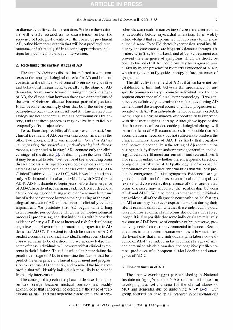
(Fig. 1). Importantly, this continuum of preclinical AD
would also encompass (1) individuals who carry one or
more apolipoprotein E ( APOE ) 34 alleles who are known
to have an increased risk of developing AD dementia, at
the point they are AD-P biomarker-positive, and (2) carriers
of autosomal dominant mutations, who are in the presymp-
tomatic biomarker-positive stage of their illness, and whowill almost certainly manifest clinical symptoms and prog-
ress to dementia.
Our group carefully considered several monikers to best
capture this stage of the disease, including “asymptomatic,”
“presymptomatic,” “latent,” “premanifest,” and “preclini-
cal.” The term “preclinical” was felt to best encompass
this conceptual phase of the disease process but is not meant
to imply that all individuals who have evidence of early AD
pathology will necessarily progress to clinical AD dementia.
Individuals who are biomarker positive but cognitively nor-
mal might currently be defined as “asymptomatic at risk for
AD dementia.” Indeed, our goal is to better define the factorswhich best predict cognitive decline in biomarker-positive
individuals, so as to move toward an accurate profile of pre-
clinical AD.
4. Models of the pathophysiological sequence of AD
To facilitate the discussion of the concept of a preclinical
stage of AD, we propose a theoretical model of the patho-
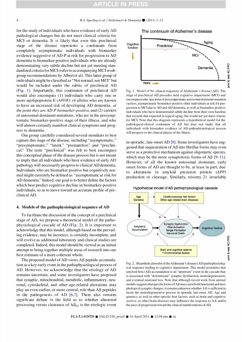
physiological cascade of AD (Fig. 2). It is important to
acknowledge that this model, although based on the prevail-
ing evidence, may be incorrect, is certainly incomplete, and
will evolve as additional laboratory and clinical studies arecompleted. Indeed, this model should be viewed as an initial
attempt to bring together multiple areas of research into our
best estimate of a more coherent whole.
The proposed model of AD views Ab peptide accumula-
tion as a key early event in the pathophysiological process of
AD. However, we acknowledge that the etiology of AD
remains uncertain, and some investigators have proposed
that synaptic, mitochondrial, metabolic, inflammatory, neu-
ronal, cytoskeletal, and other age-related alterations may
play an even earlier, or more central, role than Ab peptides
in the pathogenesis of AD [6,7]. There also remains
significant debate in the field as to whether abnormalprocessing versus clearance of Ab42 is the etiologic event
in sporadic, late-onset AD [8]. Some investigators have sug-
gested that sequestration of Ab into fibrillar forms may even
serve as a protective mechanism against oligomeric species,
which may be the more synaptotoxic forms of Ab [9–11].
However, of all the known autosomal dominant, early
onset forms of AD are thought to be, at least in part, due
to alterations in amyloid precursor protein ( APP)
production or cleavage. Similarly, trisomy 21 invariably
p r i n t &
w e b 4 C = F P O
p r i n t &
w e b 4 C = F P O
Fig. 1. Model of the clinical trajectory of Alzheimer’s disease (AD). The
stage of preclinical AD precedes mild cognitive impairment (MCI) and
encompassesthe spectrum of presymptomatic autosomal dominant mutation
carriers, asymptomatic biomarker-positive older individuals at risk for pro-
gression to MCI due to AD and AD dementia, as well as biomarker-positive
individuals who have demonstrated subtle decline from their own baseline
that exceeds that expected in typical aging, but would not yet meet criteria
for MCI. Note that this diagram represents a hypothetical model for thepathological-clinical continuum of AD but does not imply that all
individuals with biomarker evidence of AD-pathophysiological process
will progress to the clinical phases of the illness.
p r i n t &
w e b 4 C = F P O
p r i n t &
w e b 4 C = F P O
Fig. 2. Hypotheticalmodel of the Alzheimer’s disease (AD)pathophysiolog-
ical sequence leading to cognitive impairment. This model postulates that
amyloid beta (Ab) accumulation is an “upstream” event in the cascade that
is associated with “downstream” synaptic dysfunction, neurodegeneration,
and eventual neuronal loss. Note that although recent work from animal
models suggests thatspecificforms of Abmay causeboth functional and mor-
phological synaptic changes, it remains unknown whether Ab is sufficient to
incite the neurodegenerative process in sporadic late-onset AD. Age and
genetics, as well as other specific host factors, such as brain and cognitive
reserve, or other brain diseases may influence the response to Ab and/orthe pace of progression toward the clinical manifestations of AD.
FLA 5.1.0 DTD � JALZ1250_proof � 16 April 2011 � 12:04 pm � ce
R.A. Sperling et al. / Alzheimer’s & Dementia- (2011) 1–134
8/4/2019 Diagnostic Recommendations Alz Pre Clinical
http://slidepdf.com/reader/full/diagnostic-recommendations-alz-pre-clinical 5/13
results in AD-P in individuals who have three intact copies
of the APP coding region located on chromosome 21.
Finally, APOE , the major genetic risk factor for late-onset
AD, has been implicated in amyloid trafficking and plaque
clearance. Both autopsy and biomarker studies (see later in
the text) similarly suggest that Ab42 accumulation increases
with advanced aging, the greatest risk factor for developingAD. At this point, it remains unclear whether it is meaning-
ful or feasible to make the distinction between Ab as a risk
factor for developing the clinical syndrome of AD versus Ab
accumulation as an early detectable stage of AD because
current evidence suggests that both concepts are plausible.
Also, it is clear that synaptic depletion, intracellular hy-
perphosphorylated forms of tau, and neuronal loss invariably
occur in AD, and at autopsy, these markers seem to correlate
better than plaque counts or total Ab load with clinical
impairment. Although we present evidence later that the
presence of markers of “upstream” Ab accumulation is asso-
ciated with markers of “downstream” pathological change,including abnormal tau, neural dysfunction, glial activation,
and neuronal loss and atrophy, it remains to be proven that
Ab accumulation is sufficient to incite the downstream path-
ological cascade of AD. It remains unknown whether this
neurodegenerative process could be related to direct synaptic
toxicity due to oligomeric forms of Ab, disruption of axonal
trajectories from fibrillar forms of Ab, or a “second hit” that
results in synaptic dysfunction, neurodegeneration, neurofi-
brillary tangle formation, and eventually neuronal loss.
Epidemiological data suggest there are significant modu-
lating factors that may alter the pace of the clinical expression
of AD-P, although evidence that these factors alter the under-lying pathophysiological process itself is less secure. Large
cohort studies have implicated multiple health factors that
may increase the risk for developing cognitive decline and de-
mentia thought to be caused by AD [12]. In particular, vascular
risk factors such as hypertension, hypercholesterolemia, and
diabetes have been associated with an increased risk of
dementia, and may contribute directly to the effect of AD
pathology on the aging brain [13,14]. Depressive sym-
ptomatology, apathy, and chronic psychological distress
have also been linked to increased risk of manifesting MCI
and dementia [15–17]. It also remains unclear whether there
are specific environmental exposures, such as head trauma,that may influence the progression of the pathophysiological
sequence or the clinical expression of the pathology. On the
positive side, there is some evidence that engagement in
specific activities, including cognitive, physical, leisure, and
social activity, may be associated with decreased risk of
MCI and AD dementia [18].
The temporal lag between the appearance of AD-P and the
emergence of AD-C also may be altered by factors such as
brain or cognitive reserve [19]. The concept of reserve was
originally invoked to provide an explanation for the observa-
tion that the extent of AD histopathological changes at
autopsy did not always align with the degree of clinicalimpairment, and can be thought of as the ability to tolerate
higher levels of brain injury without exhibiting clinical symp-
toms. “Brain reserve” refers to the capacity of the brain to
withstand pathological insult, perhaps because of greater syn-
aptic density or larger number of healthy neurons, such that
sufficient neural substrate remains to support normal func-
tion. In contrast, “cognitive reserve” is thought to represent
the ability to engage alternate brain networks or cognitivestrategies to cope with the effects of encroaching pathology.
It is not clear, however, that the data support a sharp demarca-
tion between these two constructs because many factors, such
as higher socioeconomic status or engagement in cognitively
stimulating activities, may contribute to both forms of
reserve. Higher education and socioeconomic status have
been associated with lower age-adjusted incidence of AD
diagnosis. Recent studies suggest that high reserve may pri-
marily influence the capability of individuals to tolerate their
AD-P for longer periods, but may also be associated with
rapid decline after a “tipping point” is reached and compen-
satory mechanisms begin to fail [20,21].
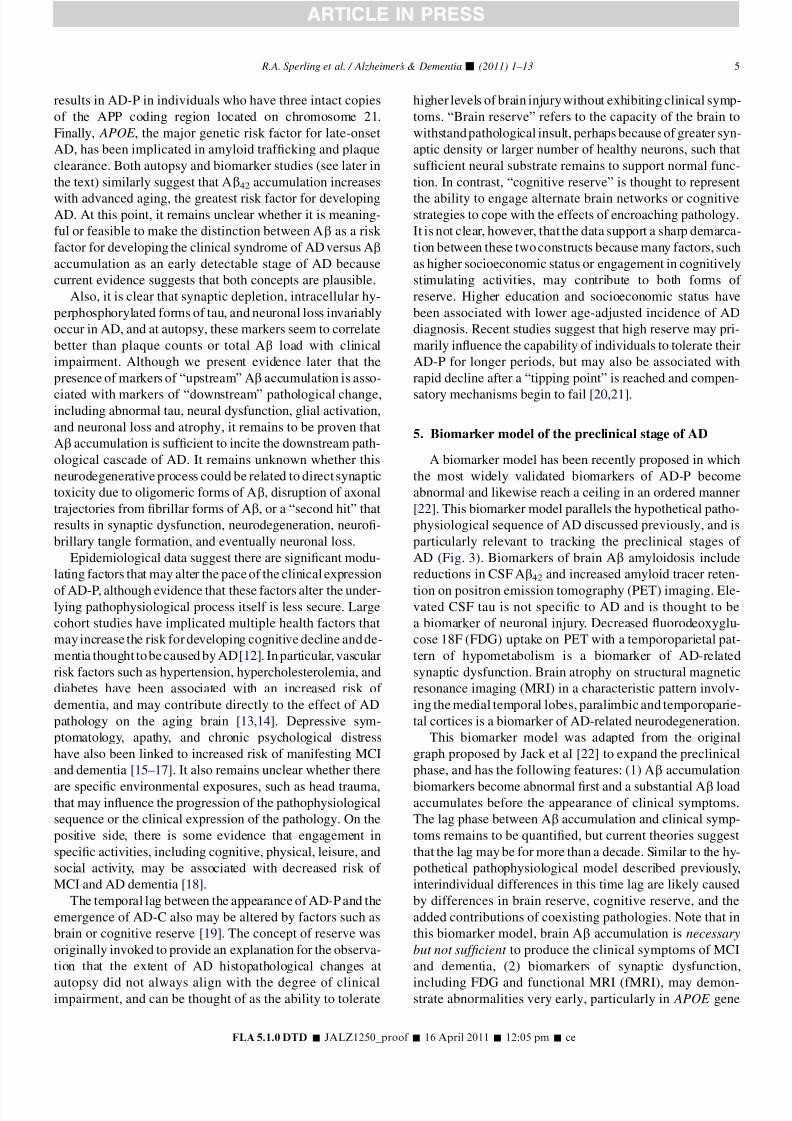
5. Biomarker model of the preclinical stage of AD
A biomarker model has been recently proposed in which
the most widely validated biomarkers of AD-P become
abnormal and likewise reach a ceiling in an ordered manner
[22]. This biomarker model parallels the hypothetical patho-
physiological sequence of AD discussed previously, and is
particularly relevant to tracking the preclinical stages of
AD (Fig. 3). Biomarkers of brain Ab amyloidosis include
reductions in CSF Ab42 and increased amyloid tracer reten-
tion on positron emission tomography (PET) imaging. Ele-vated CSF tau is not specific to AD and is thought to be
a biomarker of neuronal injury. Decreased fluorodeoxyglu-
cose 18F (FDG) uptake on PET with a temporoparietal pat-
tern of hypometabolism is a biomarker of AD-related
synaptic dysfunction. Brain atrophy on structural magnetic
resonance imaging (MRI) in a characteristic pattern involv-
ing the medial temporal lobes, paralimbic and temporoparie-
tal cortices is a biomarker of AD-related neurodegeneration.
This biomarker model was adapted from the original
graph proposed by Jack et al [22] to expand the preclinical
phase, and has the following features: (1) Ab accumulation
biomarkers become abnormal first and a substantial Ab loadaccumulates before the appearance of clinical symptoms.
The lag phase between Ab accumulation and clinical symp-
toms remains to be quantified, but current theories suggest
that the lag may be for more than a decade. Similar to the hy-
pothetical pathophysiological model described previously,
interindividual differences in this time lag are likely caused
by differences in brain reserve, cognitive reserve, and the
added contributions of coexisting pathologies. Note that in
this biomarker model, brain Ab accumulation is necessary
but not sufficient to produce the clinical symptoms of MCI
and dementia, (2) biomarkers of synaptic dysfunction,
including FDG and functional MRI (fMRI), may demon-strate abnormalities very early, particularly in APOE gene
FLA 5.1.0 DTD � JALZ1250_proof � 16 April 2011 � 12:05 pm � ce
R.A. Sperling et al. / Alzheimer’s & Dementia- (2011) 1–13 5
8/4/2019 Diagnostic Recommendations Alz Pre Clinical
http://slidepdf.com/reader/full/diagnostic-recommendations-alz-pre-clinical 6/13
34 allele carriers, who may manifest functional abnormali-
ties before detectable Ab deposition [23–25]. The severity
and change over time in these synaptic markers correlate
with clinical symptoms during MCI and AD dementia, (3)
structural MRI is thought to become abnormal a bit later,as a marker of neuronal loss, and MRI retains a close
relationship with cognitive performance through the
clinical phases of MCI and dementia [26], (4) none of the
biomarkers is static; rates of change in each biomarker
change over time and follow a nonlinear time course, which
is hypothesized to be sigmoid shaped, and (5) anatomic in-
formation from imaging biomarkers provides useful disease
staging information in that the topography of disease-related
imaging abnormalities changes in a characteristic manner
with disease progression.
6. Biomarker and autopsy evidence linking AD
pathology to early symptomatology
Several multicenter biomarker initiatives, including the
Alzheimer’s Disease Neuroimaging Initiative; the Australian
Imaging, Biomarkers and Lifestyle Flagship Study of Aging;
as well as major biomarker studies in preclinical populations
at several academic centers, are ongoing. These studies have
already provided preliminary evidence that biomarker abnor-
malities consistent with AD pathophysiological process are
detectable before the emergence of overt clinical symptom-
atology and are predictive of subsequent cognitive decline.
Many of the recent studies have focused on markers of Abusing either CSF assays of Ab42 or PET amyloid imaging
with radioactive tracers that bind to fibrillar forms of Ab.
Both CSFand PETamyloidimagingstudiessuggestthata sub-
stantial proportion of clinically normal older individuals dem-
onstrate evidence of Ab accumulation [27–32]. The exact
proportion of “amyloid-positive” normal individuals isdependent on the age and genetic background of the cohort,
but ranges from approximately 20% to 40% and is very
consonant with large postmortem series [33,34]. Furth-
ermore, there is evidence that the AD-P detected at autopsy
is related to episodic memory performance even within the
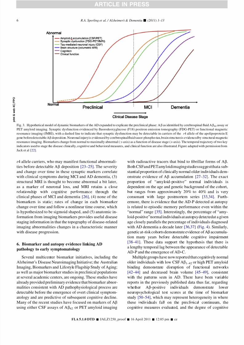
“normal” range [35]. Interestingly, the percentage of “amy-
loid-positive”normalindividualsat autopsydetectedat a given
age closely parallels the percentage of individuals diagnosed
with AD dementia a decade later [36,37] (Fig. 4). Similarly,
genetic at-risk cohorts demonstrate evidence of Ab accumula-
tion many years before detectable cognitive impairment
[38–41]. These data support the hypothesis that there isa lengthy temporal lag between the appearance of detectable
AD-P and the emergence of AD-C.
Multiple groups have now reported that cognitively normal
older individuals with low CSF Ab1–42 or high PET amyloid
binding demonstrate disruption of functional networks
[42–44] and decreased brain volume [45–49], consistent
with the patterns seen in AD. There have been variable
reports in the previously published data thus far, regarding
whether Ab-positive individuals demonstrate lower
neuropsychological test scores at the time of biomarker
study [50–54], which may represent heterogeneity in where
these individuals fall on the preclinical continuum, thecognitive measures evaluated, and the degree of cognitive
p r i n t &
w e b 4 C = F P O
p r i n t &
w e b 4 C = F P O
Fig. 3. Hypothetical model of dynamic biomarkers of the AD expanded to explicate the preclinical phase: Ab as identified by cerebrospinal fluid Ab42 assay or
PET amyloid imaging. Synaptic dysfunction evidenced by fluorodeoxyglucose (F18) positron emission tomography (FDG-PET) or functional magnetic
resonance imaging (fMRI), with a dashed line to indicate that synaptic dysfunction may be detectable in carriers of the 34 allele of the apolipoprotein E
gene beforedetectable Ab deposition. Neuronal injuryis evidenced by cerebrospinalfluid tauor phospho-tau,brain structureis evidencedby structural magnetic
resonance imaging. Biomarkers change from normal to maximally abnormal ( y-axis) as a function of disease stage ( x-axis). The temporal trajectory of two key
indicators used to stage the disease clinically, cognitive and behavioral measures, and clinical function are also illustrated. Figure adapted with permission from
Jack et al [22].
FLA 5.1.0 DTD � JALZ1250_proof � 16 April 2011 � 12:05 pm � ce
R.A. Sperling et al. / Alzheimer’s & Dementia- (2011) 1–136
8/4/2019 Diagnostic Recommendations Alz Pre Clinical
http://slidepdf.com/reader/full/diagnostic-recommendations-alz-pre-clinical 7/13
reserve in the cohorts. A few early studies have reported that
Ab positivity in clinically normal older individuals isassociated with an increased rate of atrophy [55] and an in-
creased risk of cognitive decline and progression to dementia
[56–62]. Multiple studies focused on other biomarkers,
including volumetric MRI, FDG-PET, or plasma biomarkers,
in cohorts of clinically normal older individuals have also re-
ported evidence that these markers are predictive of cognitive
decline (refer [63,64] for recent examples). Additional
longitudinal studies are clearly needed to confirm these
findings and to elucidate the combination of factors that best
predict likelihood and rate of decline, and to better
understand individual diff-erences in risk for decline.
As a complement to longitudinal studies in the populationat risk by virtue of age, researchers continue to detect and
track the biological and cognitive changes associated with
the predisposition to AD in cognitively normal people at dif-
ferential genetic risk for AD alone or in conjunction with
other risk factors (such as a person’s reported family history
of the disease). To date, the best established genetic risk fac-
tors for AD include common allelic variants of APOE ; the
major late-onset AD susceptibility gene; uncommon early-
onset AD-causing mutations in the presenilin 1, presenilin
2, and APP genes; and trisomy 21 (Down syndrome). Bio-
marker studies in presymptomatic carriers of these genetic
risk factors have revealed evidence of Ab accumulation on
CSF and PET amyloid imaging, as well as FDG-PET hypo-
metabolism, fMRI abnormalities, and brain atrophy that may
precede symptoms by more than a decade.
7. Cognitive studies
Despite the clear potential of biomarkers for detectingevidence of the AD pathophysiological process, it is impor-
tant not to lose sight of the potential that behavioral markers
hold for early identification. Tests developed by both
neuropsychological and cognitive aging researchers have
provided evidence that normal aging is accompanied by de-
clines in speed of information processing, executive function
(working memory, task switching, inhibitory function), and
reasoning. Studies that have conducted assessments of cog-
nitive function at multiple time points before dementia have
also shown consistently a long period of gradual cognitive
decline in episodic memory as well as nonmemory domains
progressing up to a decade before onset of dementia. Impor-tantly, in studies that have modeled the curve of cognitive
change versus time, the preclinical trajectory suggests not
only a long- and slow rate of presymptomatic change but
also a period of acceleration of performance decrement
that may begin several years before MCI onset [65]. Recent
studies also suggest that self-report of subtle cognitive
decline, even in the absence of significant objective impair-
ment on testing, may portend future decline in older individ-
uals. Despite the existence of multiple studies spanning
thousands of participants, the promise of both subjective
and objective cognitive measures for assessing risk of
progression to AD in individual elders has not yet been fullyrealized. It is likely that measured change in cognition over
time will be more sensitive than any one-time measure.
Additional longitudinal studies of older individuals, perhaps
combining biomarkers with measures sensitive to detecting
very subtle cognitive decline, are clearly needed.
8. Caveats
Although the aforementioned studies provide compelling
evidence that markers of Ab in “normal” older individuals
are associated with other brain alterations consistent those
seen in AD dementia, and that specific factors may
p r i n t &
w e b 4 C = F P O
p r i n t &
w e b 4 C = F P O
Fig. 4. Postulated temporal lag of approximately a decade between the de-
position of Ab (% of individuals with amyloid plaques in a large autopsy se-
ries [68]) and the clinical syndrome of AD dementia (estimated prevalence
from three epidemiological studies [69–71]). Figure courtesy of Mark
Mintun and John Morris, Washington University.
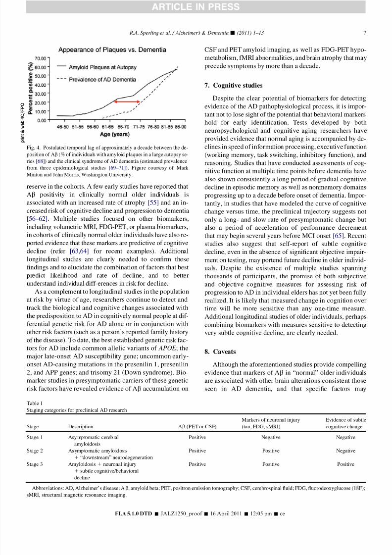
Table 1
Staging categories for preclinical AD research
Stage Description Ab (PET or CSF)
Markers of neuronal injury
(tau, FDG, sMRI)
Evidence of subtle
cognitive change
Stage 1 Asymptomatic cerebral
amyloidosis
Positive Negative Negative
Stage 2 Asymptomatic amyloidosis
1 “downstream” neurodegeneration
Positive Positive Negative
Stage 3 Amyloidosis 1 neuronal injury
1 subtle cognitive/behavioral
decline
Positive Positive Positive
Abbreviations: AD, Alzheimer’s disease; Ab, amyloid beta; PET, positron emission tomography; CSF, cerebrospinal fluid; FDG, fluorodeoxyglucose (18F);sMRI, structural magnetic resonance imaging.
FLA 5.1.0 DTD � JALZ1250_proof � 16 April 2011 � 12:05 pm � ce
R.A. Sperling et al. / Alzheimer’s & Dementia- (2011) 1–13 7
8/4/2019 Diagnostic Recommendations Alz Pre Clinical
http://slidepdf.com/reader/full/diagnostic-recommendations-alz-pre-clinical 8/13
accurately predict those individuals who are at a higher risk
of progression to AD-C, it is important to note several poten-
tial confounding issues in the majority of these studies. It is
likely that many of these studies suffer from cohort biases. In
particular, the biomarker and cognitive studies likely are not
representative of the general older population because they
are typically “samples of convenience,” that is, volunteer co-horts who tend to come from highly educated and socioeco-
nomic status backgrounds. These individuals may also be
less likely to harbor typical age-related comorbidities that
may influence the rate of cognitive decline. Older individ-
uals who are willing to participate in such intensive studies
may also represent the “volunteer gene,” and may be more
actively engaged than the typical aging population. Con-
versely, these cohorts may include individuals who self-
select for this research because of subjective concerns about
their own memory function or positive family history, as re-
flected by the high rate of APOE 34 carriers in some of these
cohorts.It is also important to note that although these biomarkers
have revolutionized the field of early AD, these markers are
merely “proxies” for the underlying disease and may not fully
reflect the biological processes in the living brain. For exam-
ple, both CSF and PET amyloid imaging markers seem to be
estimates of the deposition of fibrillar forms of Ab, and may
not provide information about oligomeric forms, which may
be the relevant species for synaptic toxicity. Similarly, our
proxy measurements for synaptic dysfunction, such as fMRI
or FDG-PET, are indirect measurements of neural function.
Other markers of neurodegeneration such as CSF tau and vol-
umetric MRI are not specific to the AD process. Finally, it isimportant to acknowledge that the relationship between bio-
markers and cognition may vary significantly across age and
genetic cohorts. In particular, the dissociation between the
presence or absence of AD-P and clinical symptomatology
in the oldest-old needs to be better understood.
Finally, it is important to re-emphasize that although Ab
deposition and neuritic plaque formation are required for the
diagnosis of definite AD, and that current evidence suggests
that Ab accumulation is an early detectable stage of the
pathological-clinical continuum of AD, the role of Ab as
the etiologic agent in sporadic late-onset AD remains to be
proven. There may be pathophysiological events that are “up-stream” of Ab accumulation yet to be discovered, and the re-
lationship between Ab and neurodegeneration is not yet clear.
In particular, the failure of biologically active Ab-lowering
therapies to demonstrate clinical benefit thus far is of concern.
Thus, it is important to continue research in alternative
pathophysiological pathways and therapeutic avenues.
9. Draft operational research framework for staging
preclinical AD
To facilitate future studies, we propose draft operational
research criteria to define study cohorts at risk for develop-ing AD dementia for use in (1) longitudinal natural history
studies to determine whether the presence of Ab markers,
either in isolation or in combination with additional markers
of neurodegeneration, is predictive of cognitive decline in
clinically normal older individuals, and (2) clinical trials
of potential disease-modifying agents to investigate effects
on biomarker progression and/or the emergence of clinical
symptoms.We emphasize again that this framework is not intended
to serve as diagnostic criteria for clinical purposes. Use of
these biomarkers in the clinical setting is currently unwar-
ranted because many individuals who satisfy the proposed
research criteria may not develop the clinical features of
AD in their lifetime. Inappropriate use of this information
in this context could be associated with unwarranted concern
because there is currently insufficient information to relate
preclinical biomarker evidence of AD to subsequent rates
of clinical progression with any certainty.
These research criteria are based on the postulate that AD
is characterized by a sequence of biological events thatbegins far in advance of clinical dementia. On the basis of
current evidence from both genetic at-risk and older cohort
studies, we put forth the hypothesis that Ab accumulation,
or the stage of cerebral amyloidosis, is currently one of the
earliest measurable stages of AD, and occurs before any
other evidence of cognitive symptomatology. We postulate
that the presence of biomarker “positivity” for Ab in clini-
cally normal older individuals, particularly in combination
with evidence of abnormality on other biomarkers of
AD-P, may have implications for the subsequent course of
AD-C and the responsiveness to treatments targeting AD-P.
Recognizing that the preclinical stages of AD representa continuum, including individuals who may never progress
beyond the stage of Ab accumulation, we further suggest the
following staging schema (see Table 1), which may prove
useful in defining research cohorts to test specific hypothe-
ses. Research cohorts could be selected on the basis of these
staging criteria, to optimize the ability to ascertain the spe-
cific outcomes important for a given type (e.g., natural his-
tory or treatment trial) and duration of the study. Evidence
of “downstream” biomarkers or subtle cognitive symptoms
in addition to evidence of Ab accumulation may increase
the likelihood of rapid emergence of cognitive symptomatol-
ogy and clinical decline to MCI within several years. Thepresence of one or more of these additional biomarkers
would indicate that individuals are already experiencing
early neurodegeneration, and as such, it is possible that
amyloid-modifying therapies may be less efficacious after
the downstream pathological process is set in motion. There
are specific circumstances, however, such as pharmaceutical
industry trials that may require a cognitive or clinical end-
point, rather than relying solely on biomarker outcomes. In
these cases, it may be advantageous to enrich the study pop-
ulation with individuals in late preclinical stages of AD with
evidence of very subtle cognitive change, who would be
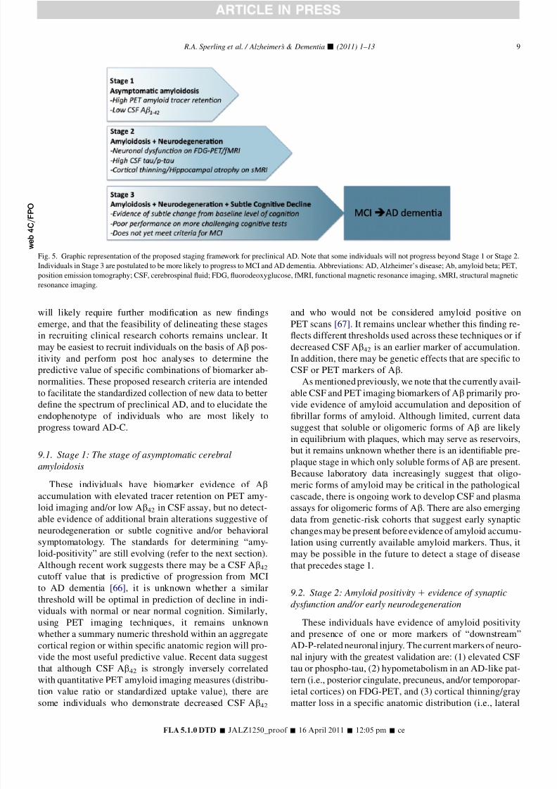
most likely to rapidly decline and manifest MCI withina short period (see Fig. 5). We recognize that these stages
FLA 5.1.0 DTD � JALZ1250_proof � 16 April 2011 � 12:05 pm � ce
R.A. Sperling et al. / Alzheimer’s & Dementia- (2011) 1–138
8/4/2019 Diagnostic Recommendations Alz Pre Clinical
http://slidepdf.com/reader/full/diagnostic-recommendations-alz-pre-clinical 9/13
will likely require further modification as new findings
emerge, and that the feasibility of delineating these stages
in recruiting clinical research cohorts remains unclear. It
may be easiest to recruit individuals on the basis of Ab pos-
itivity and perform post hoc analyses to determine the
predictive value of specific combinations of biomarker ab-
normalities. These proposed research criteria are intended
to facilitate the standardized collection of new data to better
define the spectrum of preclinical AD, and to elucidate the
endophenotype of individuals who are most likely toprogress toward AD-C.
9.1. Stage 1: The stage of asymptomatic cerebral
amyloidosis
These individuals have biomarker evidence of Ab
accumulation with elevated tracer retention on PET amy-
loid imaging and/or low Ab42 in CSF assay, but no detect-
able evidence of additional brain alterations suggestive of
neurodegeneration or subtle cognitive and/or behavioral
symptomatology. The standards for determining “amy-
loid-positivity” are still evolving (refer to the next section).Although recent work suggests there may be a CSF Ab42cutoff value that is predictive of progression from MCI
to AD dementia [66], it is unknown whether a similar
threshold will be optimal in prediction of decline in indi-
viduals with normal or near normal cognition. Similarly,
using PET imaging techniques, it remains unknown
whether a summary numeric threshold within an aggregate
cortical region or within specific anatomic region will pro-
vide the most useful predictive value. Recent data suggest
that although CSF Ab42 is strongly inversely correlated
with quantitative PET amyloid imaging measures (distribu-
tion value ratio or standardized uptake value), there aresome individuals who demonstrate decreased CSF Ab42
and who would not be considered amyloid positive on
PET scans [67]. It remains unclear whether this finding re-
flects different thresholds used across these techniques or if
decreased CSF Ab42 is an earlier marker of accumulation.
In addition, there may be genetic effects that are specific to
CSF or PET markers of Ab.
As mentioned previously, we note that the currently avail-
able CSF and PET imaging biomarkers of Ab primarily pro-
vide evidence of amyloid accumulation and deposition of
fibrillar forms of amyloid. Although limited, current datasuggest that soluble or oligomeric forms of Ab are likely
in equilibrium with plaques, which may serve as reservoirs,
but it remains unknown whether there is an identifiable pre-
plaque stage in which only soluble forms of Ab are present.
Because laboratory data increasingly suggest that oligo-
meric forms of amyloid may be critical in the pathological
cascade, there is ongoing work to develop CSF and plasma
assays for oligomeric forms of Ab. There are also emerging
data from genetic-risk cohorts that suggest early synaptic
changes may be present before evidence of amyloid accumu-
lation using currently available amyloid markers. Thus, it
may be possible in the future to detect a stage of diseasethat precedes stage 1.
9.2. Stage 2: Amyloid positivity 1 evidence of synaptic
dysfunction and/or early neurodegeneration
These individuals have evidence of amyloid positivity
and presence of one or more markers of “downstream”
AD-P-related neuronal injury. The current markers of neuro-
nal injury with the greatest validation are: (1) elevated CSF
tau or phospho-tau, (2) hypometabolism in an AD-like pat-
tern (i.e., posterior cingulate, precuneus, and/or temporopar-
ietal cortices) on FDG-PET, and (3) cortical thinning/graymatter loss in a specific anatomic distribution (i.e., lateral
w e b 4 C = F P O
w e b 4 C = F P O
Fig. 5. Graphic representation of the proposed staging framework for preclinical AD. Note that some individuals will not progress beyond Stage 1 or Stage 2.
Individuals in Stage 3 are postulated to be more likely to progress to MCI and AD dementia. Abbreviations: AD, Alzheimer’s disease; Ab, amyloid beta; PET,
position emission tomography; CSF, cerebrospinal fluid; FDG, fluorodeoxyglucose, fMRI, functional magnetic resonance imaging, sMRI, structural magneticresonance imaging.
FLA 5.1.0 DTD � JALZ1250_proof � 16 April 2011 � 12:05 pm � ce
R.A. Sperling et al. / Alzheimer’s & Dementia- (2011) 1–13 9

8/4/2019 Diagnostic Recommendations Alz Pre Clinical
http://slidepdf.com/reader/full/diagnostic-recommendations-alz-pre-clinical 10/13
and medial parietal, posterior cingulate, and lateral temporal
cortices) and/or hippocampal atrophy on volumetric MRI.
Future markers may also include fMRI measures of default
network connectivity. Although previous studies have dem-
onstrated that, on average, amyloid-positive individuals
demonstrate significantly greater abnormalities on these
markers as compared with amyloid-negative individuals,there is significant interindividual variability. We hypothe-
size that amyloid-positive individuals with evidence of early
neurodegeneration may be farther down the trajectory (i.e.,
in later stages of preclinical AD). It remains unclear whether
it will be feasible to detect differences among these other
biomarkers of AD-P, but there is some evidence that early
synaptic dysfunction, as assessed by functional imaging
techniques such as FDG-PET and fMRI, may be detectable
before volumetric loss.
9.3. Stage 3: Amyloid positivity 1 evidence of
neurodegeneration1 subtle cognitive decline
We postulate that individuals with biomarker evidence of
amyloidaccumulation, early neurodegeneration, and evidence
of subtle cognitive decline are in the last stage of preclinical
AD, and are approaching the border zone with the proposed
clinical criteria for MCI. These individuals may demonstrate
evidence of decline from their own baseline (particularly if
proxies of cognitive reserve are taken into consideration),
even if they still perform within the “normal” range on stan-
dard cognitive measures. There is emerging evidence that
more sensitive cognitive measures, particularly with challeng-
ing episodic memory measures, may detect very subtle cogni-tive impairment in amyloid-positive individuals. It remains
unclear whether self-complaint of memory decline or other
subtle neurobehavioral changes will be a useful predictor of
progression, but it is possible that the combination of bio-
markers and subjective assessment of subtle change will prove
to be useful.
10. Need for additional study
We propose a general framework with biomarker criteria
for the study of the preclinical phase of AD; however, more
work is needed to clarify the optimal CSF assays, PET orMRI analytic techniques, and in particular, the specific
thresholds needed to meet these criteria. There are signifi-
cant challenges in implementing standardized biomarker
“cut-off” values across centers, studies, and countries.
Work to standardize and validate both fluid-based and imag-
ing biomarker thresholds is ongoing in multiple academic
and pharmaceutical industry laboratories, as well as in
several multicenter initiatives. These criteria will need to
be validated in large multicenter natural history studies, or
as provisional criteria for the planning of preventative clini-
cal trials. For instance, it will be important to establish the
test–retest and cross-center reliability of biomarker measure-ments, further characterize the sequence of biomarker
changes, and the extent to which these biomarkers predict
subsequent clinical decline or clinical benefit. In particular,
there is an important need to evaluate methods for determin-
ing “amyloid-positivity” because it remains unclear whether
there is a biologically relevant continuum of Ab accumula-
tion, or whether there is a clear threshold or “cut-off” value
that could be defined on the basis of predictive value forsubsequent clinical decline, as has been suggested in several
CSF studies [28,66]. It also remains unknown whether these
thresholds should be adjusted for age or genotype. After
these thresholds are established, it may be most feasible to
select research cohorts for large studies solely on the basis
of “amyloid-positivity” on CSF or PET amyloid imaging,
and to use additional biomarker and cognitive measures for
post hoc analyses to determine additional predictive value.
Although recent advances in biomarkers have revolution-
ized our ability to detect evidence of early AD-P there is still
a need for novel biomarker development. In particular, al-
though the current biomarkers provide evidence of Abdeposition, an in vivo marker of oligomeric forms of Ab
would be of great value. Imaging markers of intraneuronal
pathology, including specific markers of specific forms of
tau/tangles and alpha-synuclein, are also needed. In addi-
tion, more sensitive imaging biomarkers that can detect early
synaptic dysfunction and functional and structural discon-
nection, such as fMRI and diffusion tensor imaging, may
one day prove to be useful to track early response to
amyloid-lowering therapies. Finally, we may be able to use
the currently available biomarkers as a new “gold standard”
to re-evaluate simple blood and urine markers that were dis-
carded on the basis of excessive overlap between clinicallynormal and AD patients. The significant proportion of clin-
ically normal individuals who are “amyloid-positive” on
both CSF and PET imaging may have confounded previous
studies attempting to differentiate “normal” controls from
patients with AD.
Similarly, additional work is required to identify and
validate neuropsychological and neurobehavioral measures
to detect the earliest clinical manifestations of AD. We need
to develop sensitive measures in multiple cognitive and be-
havioral domains that will reveal evidence of early synaptic
dysfunction in neural networks vulnerable to AD pathology.
We also need to develop measures of very early functionalchanges in other domains, including social interaction,
mood, psychomotor aspects of function, and decision making.
These measures would allow us to link better the pathological
processes to the emergence of clinical symptoms, and may be
particularly useful to monitor response to potential disease-
modifying therapies in these very early stages.
The proposed criteria apply primarily to individuals at risk
by virtue of advanced age because inclusion criteria for trials
in autosomal dominant mutation carriers and homozygous
APOE 34 carriers will be likely defined primarily on genetic
status. Trials in genetic-risk populations might use these crite-
ria to stage individuals within the preclinical phase of AD. Ingenetic-risk cohorts, it may even be possible to detect an even
FLA 5.1.0 DTD � JALZ1250_proof � 16 April 2011 � 12:05 pm � ce
R.A. Sperling et al. / Alzheimer’s & Dementia- (2011) 1–1310
8/4/2019 Diagnostic Recommendations Alz Pre Clinical
http://slidepdf.com/reader/full/diagnostic-recommendations-alz-pre-clinical 11/13
earlier stage of presymptomatic AD, before the point when
there is already detectable cerebral amyloidosis. Several
FDG-PET and fMRI studies have suggested that evidence
of synaptic dysfunction may be present in young and
middle-aged APOE 34 carriers (see Fig. 3), and there may
be other biological alterations that are present before signifi-
cant deposition of fibrillar forms of amyloid that would bepreferentially responsive to presymptomatic intervention.
The emerging concept of preclinical AD and the role of
biomarkers in the detection and tracking in this stage of
the disease have important implications for the development
of effective treatments. Therapies for preclinical AD would
be intended to postpone, reduce the risk of, or completely
prevent the clinical stages of the disorder. As recently noted,
the use of clinical endpoints in clinical trials of such treat-
ments would require large numbers of healthy volunteers,
large amounts of money, and many years of study.
Researchers have raised the possibility of evaluating bio-
marker endpoints for these treatments in cognitively normalpeople at increased risk for AD because these studies might
be performed more rapidly than otherwise possible. Subjects
enrolled in these studies could include individuals with auto-
somal dominant mutation carriers (with essentially a 100%
chance of developing clinical AD) or those at increased
risk of developing sporadic AD (e.g., APOE 34 carriers or
subjects with biomarker evidence of preclinical AD pathol-
ogy). The use of biomarkers rather than clinical outcomes
could accelerate progress in these trials; however, regulatory
agencies must be assured that a given biomarker is “reason-
ably likely” to predict a clinically meaningful outcome be-
fore they would grant approval for treatments tested intrials using biomarkers as surrogate endpoints. Research
strategies have been proposed to provide this evidence by
embedding the most promising biomarkers in preclinical
AD trials of people at the highest imminent risk of clinical
onset to establish a link between a biomarker effect and
the onset of clinical symptoms of AD. We envision the
time when the scientific means and accelerated regulatory
approval pathway support multiple preclinical AD trials us-
ing biomarkers to identify subjects and provide shorter term
outcomes, such that demonstrably effective treatments to
ward off the clinical stages of AD are found as quickly as
possible. There are several burgeoning efforts to designand conduct clinical trials in both genetic at-risk and
amyloid-positive older individuals, including the Domi-
nantly Inherited Alzheimer Network (study of familial
AD), the Alzheimer Prevention Initiative, and Anti-
Amyloid Treatment in Asymptomatic AD (A4) trial being
considered by the Alzheimer’s Disease Cooperative Study.
Finally, the ethical and practical implications surrounding
the issues of future implementation of making a “diagnosis”
of AD at a preclinical stage need to be studied, should the
postulates put forth previously prove to be correct. Although
at this point our recommendations are strictly for research
purposes only, the public controversy surrounding the iden-tification of asymptomatic individuals with evidence of AD-
P raised several important points that the field must consider.
In particular, the poignant question of “why would anyone
want to know they have AD a decade before they might de-
velop symptoms, if there is nothing they can do about it?”
should be carefully considered well before any results
from research is translated into clinical practice. First, there
may be important reasons, including social and financialplanning, why some individuals would want to know their
likelihood of developing AD dementia within the next
decade, even in the absence of an available disease-
modifying therapy. It is our hope, however, that the advances
in preclinical detection of AD-P will enable earlier, more
effective treatment, just as nearly all of therapeutic gains
in cancer, cardiovascular disease, osteoporosis, and diabetes
involve treatment before significant clinical symptoms are
present. It is entirely possible that promising drugs, particu-
larly amyloid-modifying agents, will fail to affect the clini-
cal course of AD at the stage of dementia or even MCI, when
the neurodegenerative process is well entrenched, but maybe efficacious at the earliest stages of the AD-P, before the
onset of symptoms.
The definitive studies to determine whether the majority
of asymptomatic individuals with evidence of AD-P are
indeed destined to develop AD dementia, to elucidate the
biomarker and/or cognitive endophenotype that is most
predictive of cognitive decline, and to determine whether
intervention with potential disease-modifying therapies in
the preclinical stages of AD will prevent dementia are
likely to take more than a decade to fully accomplish.
Thus, we must move quickly to test the postulates put forth
previously, and adjust our models and study designs as newdata become available. Because potential biologically ac-
tive treatments may be associated with small but significant
risk of adverse side effects, we will need to determine
whether we can predict the emergence of cognitive symp-
toms with sufficient certainty to appropriately weigh the
risk/benefit ratios to begin treatment in asymptomatic indi-
viduals. It is clear that many questions remain to be
answered, and that there may be additional factors which
will influence the probability of developing clinical
AD. However, the considerable progress made over the
past two decades now enables a strategic path forward to
test these hypotheses, move the field toward earlier inter-vention, and ultimately, toward the prevention of AD
dementia.
Acknowledgments
The chair (Reisa Sperling) acknowledges the invaluable
assistance of Dr. Cerise Elliott at National Institute on Ag-
ing, as well as thoughtful input solicited from several indi-
viduals, in particular, Drs. Keith Johnson, Dorene Rentz,
Peter Davies, Deborah Blacker, Steve Salloway, Sanjay As-
thana, and Dennis Selkoe, as well as the helpful public com-mentary provided by our colleagues in the field.
FLA 5.1.0 DTD � JALZ1250_proof � 16 April 2011 � 12:05 pm � ce
R.A. Sperling et al. / Alzheimer’s & Dementia- (2011) 1–13 11
8/4/2019 Diagnostic Recommendations Alz Pre Clinical
http://slidepdf.com/reader/full/diagnostic-recommendations-alz-pre-clinical 12/13
Reisa Sperling has served as a site investigator and/or con-
sultant to several companies developing imaging biomarkers
and pharmacological treatments for early AD, including
Avid, Bayer, Bristol-Myers-Squibb, Elan, Eisai, Janssen,
Pfizer, and Wyeth. Paul Aisen serves on a scientific advisory
board for NeuroPhage; serves as a consultant to Elan Corpo-
ration, Wyeth, Eisai Inc., Bristol-Myers Squibb, Eli Lilly andCompany, NeuroPhage, Merck & Co., Roche, Amgen, Ab-
bott, Pfizer Inc., Novartis, Bayer, Astellas, Dainippon, Bio-
marin, Solvay, Otsuka, Daiichi, AstraZeneca, Janssen, and
Medivation Inc.; receives research support from Pfizer Inc.
and Baxter International Inc.; and has received stock options
from Medivation Inc. and NeuroPhage. Clifford Jack serves
as a consultant for Eli Lilly, Eisai, and Elan; is an investigator
in clinical trials sponsored by Baxter and Pfizer Inc.; and
owns stock in Johnson and Johnson. Denise Park has re-
ceived research support from Avid Pharmaceuticals. Eric
Siemers is an employee of Eli Lilly and Company, which ac-
quired Avid Pharmaceuticals. Yaakov Stern has consulted toBayer Pharmaceuticals and has received research support
from Bayer, Janssen, Eli Lilly, and Elan; Maria Carrillo is
an employee of the Alzheimer’s Association and reports no
conflicts; Bill Thies is an employee of the Alzheimer’s Asso-
ciation and reports no conflicts; Creighton Phelps is an em-
ployee of the U.S. Government and reports no conflicts.
References
[1] Morris JC. Early-stage and preclinical Alzheimer disease. Alzheimer
Dis Assoc Disord 2005;19:163–5.
[2] Dubois B, Feldman HH, Jacova C, Cummings JL, Dekosky ST, Bar-berger-Gateau P, et al. Revising the definition of Alzheimer’s disease:
a new lexicon. Lancet Neurol 2010;9:1118–27.
[3] Jack CR Jr, AlbertM, Knopman DS,McKhann GM,Sperling RA,Car-
rillo M, et al. Introduction to revised criteria for the diagnosis of Alz-
heimer’s disease: National Institute on Aging and the Alzheimer’s
Association workgroup. Alzheimers Dement 2011;7:in press.
[4] AlbertMS, DeKosky ST, Dickson D, Dubois B, FeldmanHH, Fox NC,et
al. The diagnosis of mild cognitive impairment due to Alzheimer’s
disease: Recommendations from the National Institute on Agingand Alz-
heimer’s Association workgroup. Alzheimers Dement 2011;7:in press.
[5] McKhann GM,Knopman DS,Chertkow H, Hyman BT, Jack Jr CR, Ka-
was CH, et al. The diagnosis of dementia due to Alzheimer’s disease:
Recommendations from the National Institute on Aging and the Alz-
heimer’s Association workgroup. Alzheimers Dement 2011;7:in press.[6] Pimplikar SW, Nixon RA, Robakis NK, Shen J, Tsai LH. Amyloid-
independent mechanisms in Alzheimer’s disease pathogenesis.
J Neurosci 2010;30:14946–54.
[7] Herrup K. Reimagining Alzheimer’s disease–an age-based hypothesis.
J Neurosci 2010;30:16755–62.
[8] Mawuenyega KG, Sigurdson W, Ovod V, Munsell L, Kasten T,
Morris JC, et al. Decreased Clearance of CNS {beta}-Amyloid in Alz-
heimer’s Disease. Science 2010;330:1774.
[9] Selkoe DJ. Alzheimer’s disease is a synaptic failure. Science 2002;
298:789–91.
[10] Lee HG, Casadesus G, Zhu X, Takeda A, Perry G, Smith MA.
Challenging the amyloid cascade hypothesis: senile plaques and
amyloid-beta as protective adaptations to Alzheimer disease. Ann N
Y Acad Sci 2004;1019:1–4.
[11] Shankar GM, Li S, Mehta TH, Garcia-Munoz A, Shepardson NE,Smith I, et al. Amyloid-beta protein dimers isolated directly from Alz-
heimer’s brains impair synaptic plasticity and memory. Nat Med 2008;
14:837–42.
[12] Yaffe K, Fiocco AJ, Lindquist K, Vittinghoff E, Simonsick EM,
Newman AB, et al. Predictors of maintaining cognitive function
in older adults: the Health ABC study. Neurology 2009;
72:2029–35.
[13] Arvanitakis Z, Wilson RS, Bienias JL, Evans DA, Bennett DA. Diabe-
tes mellitus and risk of Alzheimer disease and decline in cognitivefunction. Arch Neurol 2004;61:661–6.
[14] Craft S. The role of metabolic disorders in Alzheimer disease and
vascular dementia: two roads converged. Arch Neurol 2009;66:300–5.
[15] Ganguli M, Du Y, Dodge HH, Ratcliff GG, Chang CC. Depressive
symptoms and cognitive decline in late life: a prospective epidemio-
logical study. Arch Gen Psychiatry 2006;63:153–60.
[16] Wilson RS, Arnold SE, Schneider JA, Kelly JF, Tang Y, Bennett DA.
Chronic psychological distress and risk of Alzheimer’s disease in old
age. Neuroepidemiology 2006;27:143–53.
[17] Onyike CU, Sheppard JM, Tschanz JT, Norton MC, Green RC,
Steinberg M, et al. Epidemiology of apathy in older adults: the Cache
County Study. Am J Geriatr Psychiatry 2007;15:365–75.
[18] Wilson RS, Scherr PA, Schneider JA, Tang Y, Bennett DA. Relation of
cognitive activity to risk of developing Alzheimer disease. Neurology
2007;69:1911–20.
[19] Stern Y. Cognitive reserve. Neuropsychologia 2009;47:2015–28.
[20] Fotenos AF, Mintun MA, Snyder AZ, Morris JC, Buckner RL. Brain
volume decline in aging: evidence for a relation between socioeco-
nomic status, preclinical Alzheimer disease, and reserve. Arch Neurol
2008;65:113–20.
[21] Wilson RS, Barnes LL, Aggarwal NT, Boyle PA, Hebert LE, Mendes
de Leon CF, et al. Cognitive activity and the cognitive morbidity of
Alzheimer disease. Neurology 2010;75:990–6.
[22] Jack CR Jr, Knopman DS,Jagust WJ,ShawLM, Aisen PS,WeinerMW,
et al. Hypothetical model of dynamic biomarkers of the Alzheimer’s
pathological cascade. Lancet Neurol 2010;9:119–28.
[23] Reiman EM, Chen K, Alexander GE, Caselli RJ,Bandy D, Osborne D,
et al. Functional brain abnormalities in young adults at genetic risk for
late-onset Alzheimer’s dementia. Proc Natl Acad Sci U S A 2004;101:284–9.
[24] Filippini N, MacIntosh BJ, Hough MG, Goodwin GM, Frisoni GB,
Smith SM, et al. Distinct patterns of brain activity in young carriers
of the APOE-epsilon4 allele. Proc Natl Acad Sci U S A 2003;
106:7209–14.
[25] Sheline YI, Morris JC, Snyder AZ, Price JL, Yan Z, D’Angelo G, et al.
APOE4 allele disrupts resting state fMRI connectivity in the absence
of amyloid plaques or decreased CSF Abeta42. J Neurosci 2010;
30:17035–40.
[26] Vemuri P, Wiste HJ, Weigand SD, Knopman DS, Trojanowski JQ,
Shaw LM, et al. Serial MRI and CSF biomarkers in normal aging,
MCI, and AD. Neurology 2010;75:143–51.
[27] Rowe CC, Ellis KA, Rimajova M, Bourgeat P, Pike KE, Jones G, et al.
Amyloid imaging results from the Australian Imaging, Biomarkersand Lifestyle (AIBL) study of aging. Neurobiol Aging 2010;
31:1275–83.
[28] Mintun MA, Larossa GN, Sheline YI, Dence CS, Lee SY, Mach RH,
et al. [11C]PIB in a nondemented population: potential antecedent
marker of Alzheimer disease. Neurology 2006;67:446–52.
[29] Jack CR Jr, Lowe VJ, Senjem ML, Weigand SD, Kemp BJ,
Shiung MM, et al. 11C PiB and structural MRI provide complemen-
tary information in imaging of Alzheimer’s disease and amnestic
mild cognitive impairment. Brain 2008;131(Pt 3):665–80.
[30] Gomperts SN, Rentz DM, Moran E, Becker JA, Locascio JJ,
Klunk WE, et al. Imaging amyloid deposition in Lewy body diseases.
Neurology 2008;71:903–10.
[31] De Meyer G, Shapiro F, Vanderstichele H, Vanmechelen E,
Engelborghs S, De Deyn PP, et al. Diagnosis-independent Alzheimer
disease biomarker signature in cognitively normal elderly people.Arch Neurol 2010;67:949–56.
FLA 5.1.0 DTD � JALZ1250_proof � 16 April 2011 � 12:05 pm � ce
R.A. Sperling et al. / Alzheimer’s & Dementia- (2011) 1–1312
8/4/2019 Diagnostic Recommendations Alz Pre Clinical
http://slidepdf.com/reader/full/diagnostic-recommendations-alz-pre-clinical 13/13
[32] Montine TJ, Peskind ER, Quinn JF, Wilson AM, Montine KS,
Galasko D. Increased cerebrospinal fluid F(2)-isoprostanes are associ-
ated with aging and latent Alzheimer’s disease as identified by bio-
markers. Neuromolecular Med 2011;13:37–43.
[33] Arriagada PV, Marzloff K, Hyman BT. Distribution of Alzheimer-type
pathologic changes in nondemented elderly individuals matches the
pattern in Alzheimer’s disease. Neurology 1992;42:1681–8.
[34] Morris JC, Storandt M, McKeel DWJr,Rubin EH,Price JL, Grant EA,et al. Cerebral amyloid deposition and diffuse plaques in “normal”
aging: evidence for presymptomatic and very mild Alzheimer’s
disease. Neurology 1996;46:707–19.
[35] Bennett D, Schneider J, Arvanitakis Z, Kelly J, Aggarwal N, Shah R,
et al. Neuropathology of older persons without cognitive impairment
from two community-based studies. Neurology 2006;66:1837–44.
[36] Brookmeyer R, Gray S, Kawas C. Projections of Alzheimer’s disease
in the United States and the public health impact of delaying disease
onset. Am J Public Health 1998;88:1337–42.
[37] Association As. 2009 Alzheimer’s disease facts and figures. Alz-
heimers Dement 2009;5:234–70.
[38] Moonis M, Swearer JM, Dayaw MP, St George-Hyslop P, Rogaeva E,
Kawarai T, et al. Familial Alzheimer disease: decreases in CSF
Abeta42 levels precede cognitive decline. Neurology 2005;65:323–5.
[39] Klunk WE, Price JC, Mathis CA, Tsopelas ND, Lopresti BJ,
Ziolko SK, et al. Amyloid deposition begins in the striatum of
presenilin-1 mutation carriers from two unrelated pedigrees. J Neuro-
sci 2007;27:6174–84.
[40] Ringman JM, Younkin SG, Pratico D, Seltzer W, Cole GM,
Geschwind DH, et al. Biochemical markers in persons with preclinical
familial Alzheimer disease. Neurology 2008;71:85–92.
[41] Reiman EM, Chen K, Liu X, Bandy D, Yu M, Lee W, et al. Fibrillar
amyloid-beta burden in cognitively normal people at 3 levels of ge-
netic risk for Alzheimer’s disease. Proc Natl Acad Sci U S A 2009;
106:6820–5.
[42] Sperling RA, Laviolette PS, O’Keefe K, O’Brien J, Rentz DM,
Pihlajamaki M, et al. Amyloid deposition is associated with impaired
default network function in older persons without dementia. Neuron
2009;63:178–88.[43] Hedden T, Van Dijk KR, Becker JA, Mehta A, Sperling RA,
Johnson KA, et al. Disruption of functional connectivity in clinically
normal older adults harboring amyloid burden. J Neurosci 2009;
29:12686–94.
[44] Sheline YI, Raichle ME,Snyder AZ, Morris JC, HeadD, Wang S, etal.
Amyloid plaques disrupt resting state default mode network connec-
tivity in cognitively normal elderly. Biol Psychiatry 2010;67:584–7.
[45] Fjell AM, Walhovd KB, Fennema-Notestine C, McEvoy LK,
Hagler DJ, Holland D, et al. Brain atrophy in healthy aging is related
to CSF levels of Abeta1-42. Cereb Cortex 2010;20:2069–79.
[46] Dickerson BC, Bakkour A, Salat DH, Feczko E, Pacheco J, Greve DN,
et al. The cortical signature of Alzheimer’s disease: regionally specific
cortical thinning relates to symptom severity in very mild to mild AD
dementia and is detectable in asymptomatic amyloid-positive individ-uals. Cereb Cortex 2009;19:497–510.
[47] Desikan RS, Sabuncu MR, Schmansky NJ, Reuter M, Cabral HJ,
Hess CP, et al. Selective disruption of the cerebral neocortex in Alz-
heimer’s disease. PLoS One 2010;5:e12853.
[48] Becker JA, Rentz D, Carmasin JS, Hedden T, Hamdi I, Buckner RL,
et al. Amyloid deposition and brain volume across the continuum of
aging and AD. Ann Neurol (in press).
[49] Oh H, Mormino EC, Madison C, Hayenga A, Smiljic A, Jagust WJ.
beta-Amyloid affects frontal and posterior brain networks in normal
aging. Neuroimage 2011;54:1887–95.
[50] Pike KE, Savage G, Villemagne VL, Ng S, Moss SA, Maruff P, et al.
Beta-amyloid imaging and memory in non-demented individuals:
evidence for preclinical Alzheimer’s disease. Brain 2007;130(Pt
11):2837–44.
[51] Mormino EC, Kluth JT, Madison CM, Rabinovici GD, Baker SL,Miller BL, et al. Episodic memory loss is related to hippocampal-
mediated {beta}-amyloid deposition in elderly subjects. Brain 2009;
132:1310–23.
[52] Aizenstein HJ, Nebes RD, Saxton JA, Price JC, Mathis CA,
TsopelasND, et al.Frequent amyloiddeposition withoutsignificantcog-
nitive impairment among the elderly. Arch Neurol 2008;65:1509–17.
[53] Rentz DM, Locascio JJ, Becker JA, Moran EK, Eng E, Buckner RL,
et al. Cognition, reserve, and amyloid deposition in normal aging.
Ann Neurol 2010;67:353–64.[54] Villemagne VL, Pike KE, Chetelat G, Ellis KA, Mulligan RS,
Bourgeat P, et al. Longitudinal assessment of Abeta and cognition in
aging and Alzheimer disease. Ann Neurol 2011;69:181–92.
[55] Schott JM, Bartlett JW, Fox NC, Barnes J; Investigators ftAsDNI. In-
creased brain atrophy rates in cognitively normal older adults with low
cerebrospinal fluid Ab1-42. Ann Neurol (in press).
[56] Fagan AM, Roe CM, Xiong C,Mintun MA, MorrisJC, Holtzman DM.
Cerebrospinal fluid tau/beta-amyloid(42) ratio as a prediction of cog-
nitive decline in nondemented older adults. Arch Neurol 2007;
64:343–9.
[57] Li G, Sokal I, Quinn JF, Leverenz JB, Brodey M, Schellenberg GD,
et al. CSF tau/Abeta42 ratio for increased risk of mild cognitive im-
pairment: a follow-up study. Neurology 2007;69:631–9.
[58] Storandt M, Mintun MA, Head D, Morris JC. Cognitive decline and
brainvolume loss as signatures of cerebral amyloid-beta peptide depo-
sition identified with Pittsburgh compound B: cognitive decline asso-
ciated with Abeta deposition. Arch Neurol 2009;66:1476–81.
[59] Resnick SM, Sojkova J, Zhou Y, An Y, Ye W, Holt DP, et al. Longitu-
dinal cognitive decline is associated with fibrillar amyloid-beta mea-
sured by [11C]PiB. Neurology 2010;74:807–15.
[60] Morris JC, Roe CM, Grant EA, Head D, Storandt M, Goate AM, et al.
Pittsburgh Compound B imaging and prediction of progression from
cognitive normality to symptomatic Alzheimer disease. Arch Neurol
2009;66:1469–75.
[61] Villemagne VL, Pike KE, Darby D, Maruff P, Savage G, Ng S, et al.
Abeta deposits in older non-demented individuals with cognitive de-
cline are indicative of preclinical Alzheimer’s disease. Neuropsycho-
logia 2008;46:1688–97.
[62] Chetelat G, Villemagne VL, Pike K,Ellis K, Bourgeat P, Jones G, etal.Independent contribution of temporal b-amyloid deposition to mem-
ory decline in non-demented elderly. Brain (in press).
[63] Vemuri P, Wiste HJ, Weigand SD, Shaw LM, Trojanowski JQ,
Weiner MW, et al. MRI and CSF biomarkers in normal, MCI, and
AD subjects: predicting future clinical change. Neurology 2009;
73:294–301.
[64] Yaffe K, Weston A, Graff-Radford NR, Satterfield S, Simonsick EM,
Younkin SG, et al. Association of plasma beta-amyloid level and cogni-
tive reserve with subsequent cognitive decline. JAMA 2011;305:261–6.
[65] Howieson DB, Carlson NE, Moore MM, Wasserman D, Abendroth CD,
Payne-Murphy J, et al. Trajectory of mild cognitive impairment onset.
J Int Neuropsychol Soc 2008;14:192–8.
[66] Shaw LM, Vanderstichele H, Knapik-Czajka M, Clark CM, Aisen PS,
Petersen RC, et al. Cerebrospinal fluid biomarker signature in Alz-heimer’s disease neuroimaging initiative subjects. Ann Neurol 2009;
65:403–13.
[67] Fagan AM, Mintun MA, Mach RH,Lee SY, Dence CS, Shah AR, et al.
Inverse relation between in vivo amyloid imaging load and cerebrospi-
nal fluid Abeta42 in humans. Ann Neurol 2006;59:512–9.
[68] Braak H, Braak E. Neuropathological staging of Alzheimer-related
changes. Acta Neuropathol 1991;82:239–59.
[69] Hebert LE, Scherr PA, Beckett LA, Albert MS, Pilgrim DM,
Chown MJ, et al. Age-specific incidence of Alzheimer’s disease in
a community population. JAMA 1995;273:1354–9.
[70] Ganguli M, Dodge HH, Chen P, Belle S, DeKosky ST. Ten-year inci-
dence of dementia in a rural elderly US community population: the
MoVIES Project. Neurology 2000;54:1109–16.
[71] Kukull WA, Higdon R, Bowen JD, McCormick WC, Teri L,
Schellenberg GD, et al. Dementia and Alzheimer disease incidence:a prospective cohort study. Arch Neurol 2002;59:1737–46.
R.A. Sperling et al. / Alzheimer’s & Dementia- (2011) 1–13 13