direct bacterial cell detection in a microfluidic chip
TRANSCRIPT

Direct bacterial cell detection in a microfluidic chip
Inês Pereira Domingos¹
Thesis to obtain the Master of Science Degree in Biomedical Engineering
Intituto Superior Técnico, Lisboa, Portugal
November 2019
Abstract
Direct capture and detection of bacterial cells is one of the aimed goals in the therapeutics field. One of
the reasons behind this search is the increasingly incidence of Antibiotic Resistance, occurring due to
an uncontrolled antibiotic prescription and incorrect use of this drug. Conventional diagnostic methods
are cost and time consuming since, in the majority of the cases, rely on cultures and amplification
approaches. In microfluidics there is the opportunity to miniaturize and mimic biological processes,
promising the development of systems low-cost, with high sensitivity, ease of transport and time saving.
In the development of this project, a microfluidic chip to perform direct capture and detection of
Staphylococcus aureus was to be achieved using a porous bead-based system.
PDMS microchannels were the basis of the microfluidic system, fabricated using a soft-lithography
technique and sealed against a PDMS membrane. Aptamers were acquired to be used as probes in
order to perform a capture-detection sandwich, resourcing a biotin to allow immobilization and a
fluorophore to detect. Different bead types were tested and one selected to assess the aptamers binding
efficacy. Distinct dyes were used in the matter of evaluating if cellular viability was somehow
compromised. A final assessment was established regarding the sensibility of physically capturing cells
using solely the microfluidic chip packed with beads.
Keywords: Microfluidic system, beads, Staphyloccus aureus, aptamers, detection, capture.
Introduction
Antibiotics are considered one of the biggest
discoveries that came across Medicine’s
history. In the majority of human existence,
infectious diseases threatened its health and
well-being [1]. Antibiotics arrived to change that
path of the history’s course.
Between 1950 and 1970, the majority of
antibiotic classes were discovered and after this
golden era, there were not others to be found,
solely modifications of already existent ones,
which gave space to bacteria resistance
emergence and rise.[2][3].
Antibiotic Resistance (AR) is the designation
attributed to the phenomenon of infectious
pathogens to overcome the effect of drugs that
were design to kill them in first instance,
developing the ability to resist antibiotic’s action.
As all organisms, also bacteria are prone to
evolve in order to adapt to the surrounding
environment, gaining different mechanisms to
sustain life and allow the species proliferation.
[3][4][5].

2
The problem is antibiotic resistance has been
having an increasingly higher incidence the past
few years, existing more and more species that
persist the drugs usage, on a rate that
overcomes the natural evolutionary adaptability.
This speeded up raising is converting antibiotic
resistance in a true crisis, which can lead to the
re-emergence of harmful bacterium, that were
considered as trivialized upon antibiotic use,
gaining ability to be successful in their goal.
[3][6]. The reasons behind this are the overuse
and misconception of antibiotics, which played
and still play the alarming role on the crisis
emergence.
It is natural to assume that the use of antibiotics
contributes to its resistance, by eliminating
certain bacteria and those who survive
proliferate as result of natural selection, but
when this use is uncontrolled, the resistance
reaches critical levels [3][4].
There are various organisms characterized as
multi-drug resistant, having the ability to survive
to antibiotics or other antibacterial drugs action.
Thus, a higher importance is given in trying to
identify these organisms upon a infection or
infectious disease, to set the correct course of
action, avoiding ineffective treatments that can
be time and cost consuming.
Nowadays, routine cultures are performed, from
blood, sputum and urine. The samples are
collected and cultured, allowing bacteria to
growth and expressing their featured proteins.
From that, a possible profile can be obtained in
order to identify the pathogen/s present in the
sample. Nevertheless, the need to culture the
cells is a time burdening, considered an
essential counter back in the patient’s course
treatment. The use of polymerase chain
reaction (PCR) in order to amplify the samples
DNA can also be used to identify a bacteria type
and genus. Although it overcomes the time
consumption issue from the cultured
approaches, its sensitivity is not as ideal.
[7][8][9]
Current approaches focus on phenotypical and
genomically-based tests, used to achieve the
goal of bacteria identification, such as
Biochemical testing, Fluorescence In Situ
Hybridization, PCR Techniques as already
mentioned, Microarrays, among other.
The idea of having a mechanism able of
avoiding the negative aspects of nowadays
approaches is enticing and is also the main
reason behind a vast range of studies.
The Microfluidic field has the potential to
miniaturize several procedures avoiding large
consumption of reagents and other materials
involved on these processes. Even the more
complex approaches can be scaled down.
Microfluidics brings advantages, like cost and
time efficiency adding the fact of be possible to
use as a device, bringing ease to the user and
to the means of transportation. This field can
bring another view, perspective and innovative
features to Antibiotic Resistance thematic and
somehow be the tool to overcome some of the
disadvantages of the current practices. [10]
Being multifaceted, there was the need of
Microtechnology to be qualified according to its
aim. It was in the 1980s that the field of
Microelectro-mechanical systems (MEMS)
began to emerge, but only later, in the 1990s,
medical, biological and even chemical
applications took their place in the field.
Through fluidic manipulation under artificial
conditions, where there is a scaled down to the
range of micrometers. Microfluidics studies “…
flows that are simple or complex, mono- or
multiphasic...”, according to Patrick Tabeling,
on the “Introduction to Microfluidics” published
by Oxford Press. [11][12][13]
Microfluidics present innumerous advantages
that make its use suitable for a vast range of
applications. Besides MEMS, there are also
perks in the fields of electrical sensors and Lab
3
on a chip technology. Through its use it is
possible to have a restrict control in the
consumption of reagents, samples and
materials needed on those applications, adding
to low-cost fabrication, it should be easier for the
user in terms of employment and transportation.
For analyses and assays, adds the advantage
of low time consumption face other techniques
[10][11][14].
Current techniques in the field of microfluidics,
base their detection into two distinct methods.
Detection by immune-sensing, where a probe
acts to target a specific molecule characteristic
of a certain bacteria. In this case a probe can be
selected and can be used to perform the bound
antibody-antigen or aptamer-target. Another
method is based on the detection resourcing
nucleic-acid identification, other specific type of
targets.
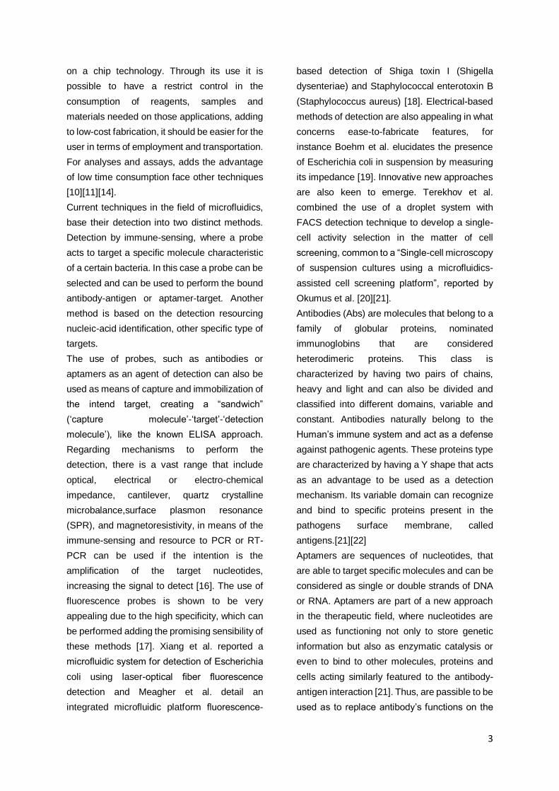
The use of probes, such as antibodies or
aptamers as an agent of detection can also be
used as means of capture and immobilization of
the intend target, creating a “sandwich”
(‘capture molecule’-‘target’-‘detection
molecule’), like the known ELISA approach.
Regarding mechanisms to perform the
detection, there is a vast range that include
optical, electrical or electro-chemical
impedance, cantilever, quartz crystalline
microbalance,surface plasmon resonance
(SPR), and magnetoresistivity, in means of the
immune-sensing and resource to PCR or RT-
PCR can be used if the intention is the
amplification of the target nucleotides,
increasing the signal to detect [16]. The use of
fluorescence probes is shown to be very
appealing due to the high specificity, which can
be performed adding the promising sensibility of
these methods [17]. Xiang et al. reported a
microfluidic system for detection of Escherichia
coli using laser-optical fiber fluorescence
detection and Meagher et al. detail an
integrated microfluidic platform fluorescence-
based detection of Shiga toxin I (Shigella
dysenteriae) and Staphylococcal enterotoxin B
(Staphylococcus aureus) [18]. Electrical-based
methods of detection are also appealing in what
concerns ease-to-fabricate features, for
instance Boehm et al. elucidates the presence
of Escherichia coli in suspension by measuring
its impedance [19]. Innovative new approaches
are also keen to emerge. Terekhov et al.
combined the use of a droplet system with
FACS detection technique to develop a single-
cell activity selection in the matter of cell
screening, common to a “Single-cell microscopy
of suspension cultures using a microfluidics-
assisted cell screening platform”, reported by
Okumus et al. [20][21].
Antibodies (Abs) are molecules that belong to a
family of globular proteins, nominated
immunoglobins that are considered
heterodimeric proteins. This class is
characterized by having two pairs of chains,
heavy and light and can also be divided and
classified into different domains, variable and
constant. Antibodies naturally belong to the
Human’s immune system and act as a defense
against pathogenic agents. These proteins type
are characterized by having a Y shape that acts
as an advantage to be used as a detection
mechanism. Its variable domain can recognize
and bind to specific proteins present in the
pathogens surface membrane, called
antigens.[21][22]
Aptamers are sequences of nucleotides, that
are able to target specific molecules and can be
considered as single or double strands of DNA
or RNA. Aptamers are part of a new approach
in the therapeutic field, where nucleotides are
used as functioning not only to store genetic
information but also as enzymatic catalysis or
even to bind to other molecules, proteins and
cells acting similarly featured to the antibody-
antigen interaction [21]. Thus, are passible to be
used as to replace antibody’s functions on the
4
therapeutic field, overcoming some of the
challenges that are being faced, and also, used
as mean to capture and detect pathogenic
agents.
The objective of this project is the development
of a microfluidic device capable of direct capture
and detect bacterial cells from the bacterium
model Staphylococcus aureus (SA) resourcing
microbeads to increase the immobilization area.
The capture aims to function due to the
integration on the used beads, in the
microfluidic chip, of labeled aptamers that in
other approach are used as a detection tool
together with fluorescence and imaging
techniques.
Materials and Methods
Microfabrication: Soft Lithography
To fabricate Polydimethylsiloxane (PDMS)
structures by a soft lithography technique it was
necessary to use a mold with simple columns
engraved on it. The master mold (SU-8) was
already available for usage.
Regarding the elastomer fabrication, it was
necessary to mix the PDMS base and the curing
agent from the elastomer kit (Sylgard 184
silicon elastomer kit, Dow Corning), on a ratio of
10:1. The mixture should have an opaque white
color with a bubbly consistence. Ready to be
gazed-out, the mixture was left on the Vacuum
desiccator (Bel-Art Products , South Wayre,
NJ/USA) during 45 minutes. The pre-polymer
was then ready to cured and should be poured
in the Petry dish that had the SU8 mold, already
available, that contained the columns were the
assays were to be performed. An extra attention
should be taken into consideration, of not
leaving bubbles on the PDMS mixture, which
can appear due to the pouring action. Next, the
Petry dish was left in the oven (Memmert,
Schwabach, DE), during 90 minutes at an
average temperature of 70ºC. Cured, the PDMS
structure was cut down and the inlets and
outlets were open with different size needles.
For the inlet, 20Ga blunt needles (Instech
Laboratories, Inc., Plymouth Meeting, PA/USA),
were used and for the outlet, 17Ga blunt
needles (Instech Laboratories, Inc., Plymouth
Meeting, PA/USA) ones. With the performed
holes, it was possible to flow solutions inside the
column, so, the remaining step was to seal the
channels.
The structure was sealed against a 500 µm
PDMS membrane, where the pre-polymer
preparation was proceeded equally as above.
Once having it, the mixture was poured onto a
silicon wafer and spin coated at 250 rpm during
25 seconds. This is the suitable program to
achieve the desired thickness. The PDMS was
then ready to be cured and the wafer was left
on the oven, during 90 minutes at 70ºC,
previously identical. From the wafer, several
membranes could be cut down with shape and
size able to cover the fabricated structure.
To seal the structure against a membrane, both
needed to be oxidize, which was possible to
achieve with the use of a plasma cleaner
(Harrick Plasma, Ithaca, NY/USA) during 60
seconds at medium power. After the removal
from the equipment, the structure was placed
on the membrane and Si-O-Si covalent bonds
were created.
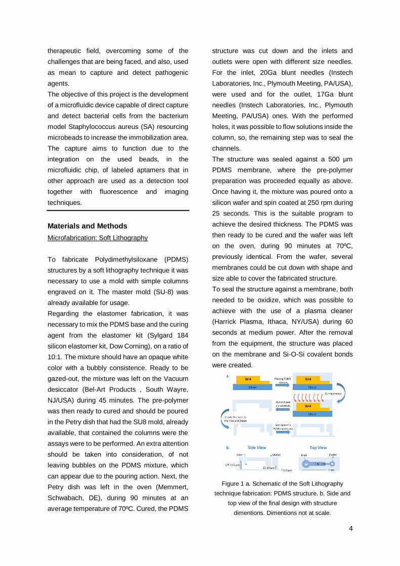
Figure 1 a. Schematic of the Soft Lithography
technique fabrication: PDMS structure. b. Side and
top view of the final design with structure
dimentions. Dimentions not at scale.
5
The plasma treatment allowed the sealing and
it marked the end of the fabrication process. It
is of relevance to point out that on this stage,
the remaining surface of the PDMS is yet
oxidized, and a rest period of 24 hours should
be regarded before the structure use. The
process is schematized on figure 1.
Bead Packing
Within the different beads, the packing protocol
had always the same features. The pumping
system was set to pull at 5.5 µL/min flow rate,
during approximately, 3 minutes, which gave a
volume margin to avoid air entering the column.
Regarding the washing step, 20 µL of
Phosphate buffered saline (PBS) (P4417,
Sigma-Aldrich, USA) were flowed with the same
features as the packing process.
Staphylococcus aureus assays
Within the different working concentrations, the
cells protocol had always the same features.
The pumping system was set to pull at 2.5
µL/min flow rate, during, 20 minutes.
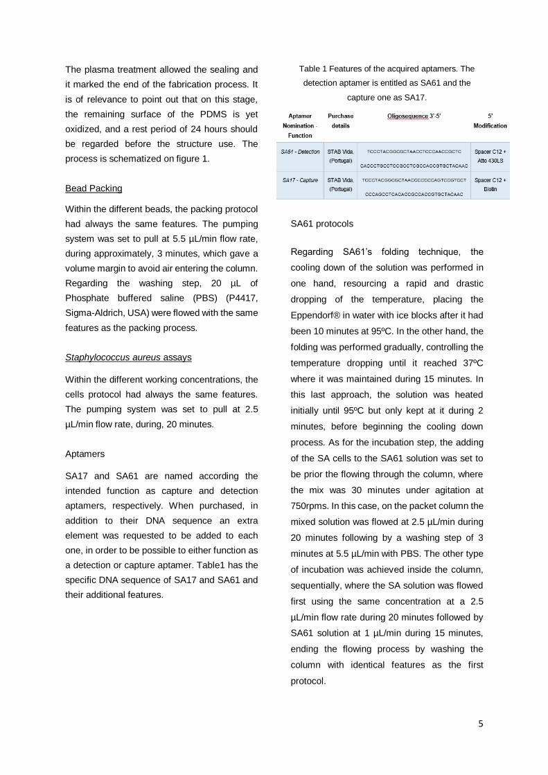
Aptamers
SA17 and SA61 are named according the
intended function as capture and detection
aptamers, respectively. When purchased, in
addition to their DNA sequence an extra
element was requested to be added to each
one, in order to be possible to either function as
a detection or capture aptamer. Table1 has the
specific DNA sequence of SA17 and SA61 and
their additional features.
Table 1 Features of the acquired aptamers. The
detection aptamer is entitled as SA61 and the
capture one as SA17.
SA61 protocols
Regarding SA61’s folding technique, the
cooling down of the solution was performed in
one hand, resourcing a rapid and drastic
dropping of the temperature, placing the
Eppendorf® in water with ice blocks after it had
been 10 minutes at 95ºC. In the other hand, the
folding was performed gradually, controlling the
temperature dropping until it reached 37ºC
where it was maintained during 15 minutes. In
this last approach, the solution was heated
initially until 95ºC but only kept at it during 2
minutes, before beginning the cooling down
process. As for the incubation step, the adding
of the SA cells to the SA61 solution was set to
be prior the flowing through the column, where
the mix was 30 minutes under agitation at
750rpms. In this case, on the packet column the
mixed solution was flowed at 2.5 µL/min during
20 minutes following by a washing step of 3
minutes at 5.5 µL/min with PBS. The other type
of incubation was achieved inside the column,
sequentially, where the SA solution was flowed
first using the same concentration at a 2.5
µL/min flow rate during 20 minutes followed by
SA61 solution at 1 µL/min during 15 minutes,
ending the flowing process by washing the
column with identical features as the first
protocol.

6
Dyes – Staphylococcus aureus viability assay
A specific viability test assay took
motion in order to assess the cellular integrity of
Staphylococcus aureus cells. With that aim, two
different dyes were selected to perform an
alive/dead cell test. EvaGreen™ (31000,
Biotium Inc., USA) and Hoechst 33342
(ThermoFisher, USA) gathered certain features
that shown to be useful in the mentioned
assessment. Hoechst 33342 is able to stain
every cell, regardless of its viability, having the
ability to cross an intact cellular membranes, by
other words can be used as an accountant for
the total number of cells in solution.
EvaGreen™, however, is not passible of
crossing a viable cell membrane. This dye
fluoresces when bound to ssDNA and its action
is only allowed upon the membrane disruption
thus, acts as disrupted cell stain. . Hoechst
33342 measurement acquisition is based on the
use of UV Olympus florescence microscope
filter, emitting on the blue color, on the other
hand EvaGreen™ needs the use of the Blue
filter to be excited which results visually on a
green staining.
To analyze the channel images
different approaches were taken into
consideration. Nevertheless, the entire analysis
set was performed resourcing ImageJ software
and for the results presentation, Origin 2020
software
Bright Field Measurements
Existing the need to perform an analysis across
different colored images, two types of ImageJ
tools were selected. Grayscale measurements
assess the change in color intensity translated
to shades of gray across a defined region. In
this case, a middle line crossing the inside’s
channel was defined to measure the changes
across the column. The second approach uses
ROI manager tool from the software to output a
mean RGB intensity value of certain sectional
area. This analsys consisted in selecting a oval
shape form inside the column and a squared
one outside (background). Afterwards, the
absolute value from inside the channel was
achieved by performing a subtraction between
the background average intensity and the
measurments acquired from the selected
channel region. Figure 2
Figure 2 Section used for signal measurements in
ImageJ software: a. Grayscale analysis; b. Mean
RGB signal intensity
Fluorescence Measurements
An identical aproach to figure 2.15b was carried
to perform fluorescence measurments. ImageJ
allows the splinter of the images into the three
color channels: red, green and blue. In the
majority of the assays, the fluorophores used,
when excited, colored on green, so, that was the
channel used to perform the measurments. This
way, the albsolut value for the mean
fluorescence intensity was achieved by, once
again subtracting the selected region inside the
column by the background signal. Figure 2.b
Results and Discussion
Beads assay
Four different bead types were tested, aiming to
understand if exists a particular characteristic
that could influence, positively, the capture of S.
aureus (SA) cells. Nevertheless, it was intended

7
to selected one of those options in order to
understand if the aptamers were binding to the
cells.
𝐶18 beads both grayscale and Mean bright field
intensity signal had dubious results. Besides the
non-coherence across the measurements, the
truth is, during the multiple attempts of
performing this experiment, several side backs
were encountered. Air entering the channel was
the main problem, due to the beads
hydrophobicity and the high cell concentration,
there were complications in flowing the solution
properly. To avoid this situation, 𝐶18 beads were
excluded from the selection.
The grayscale analysis from 𝑆𝑖𝑂𝟐 bead test had
quite positive results in which concerned the
physical trapping of staphylococcus aureus
cells. Although showing a progression of the
cellular content across the flowing time, the
truth is 𝑆𝑖𝑂𝟐 beads were able to sustain SA cells
in part of the column, slowing down its passage.
Apart from these, 𝑁𝐻2 beads also showed
pretension to sustain cellular content across the
column. Although having a positive outcome,
the trapping effect by 𝑁𝐻2 beads was less
accentuated when compared to the action of
𝑆𝑖𝑂𝟐 beads, tending to allow SA cells to flow
with less retention. Nevertheless, regarding the
signal analysis using the ROI tool, both of the
above bead types have shown good results
among the all set tested. Although 𝑁𝐻2 beads
presented a higher signal, 𝑆𝑖𝑂𝟐 beads were
selected to continue the development of this
project. Positively charged beads, like 𝑁𝐻2, can
present an advantage in the Staphylococcus
aureus attraction, due to the cell surface
properties, but they also would have influence
near the flow of the aptamer since is a DNA
sequence and this side back was to be avoided.
SA61 binding efficacy test
After choosing the best bead type to use and
according to the results obtained in the previous
section, it was possible to perform an essay
where SA61 binding capability was tested. The
idea of having a strategy capable of
concentrating the cells in the channel was an
optimal path to assess the detection aptamer
efficacy. SA61 was tested using two different
protocols of DNA folding and incubation.
The experimental image of all four protocols
have not showed any relevant fluorescence in
the entire set. By analyzing figure 3, the signal
values were not featured to be labeled as a
positive result, however, there was a
particularity that was interesting and should be
pointed out. Observing the signal difference
between the darker and lighter sites of the
beads in the channel, it was constant thought all
four protocols that the signal measured onto the
concentrated cell’s zone was slightly higher.
This could mean that a small portion of the
aptamers could have been being withheld, but
not at the cell’s concentration proportion.
Figure 3 Mean fluorescence intensity signal
measured on a section of the column after the
washing step. Values from protocols 1-4 on the
concentrated cell site (Cells) and outside that zone
(Beads). (Aquisition: Olympus Microscope CHX41,
exposure time: 500ms, 1s and 2s, gain: 0 dB,
magnification:10x.)
Considering the information mentioned above,
there was the need to perform a trouble
shooting of the situation and there was the need

8
to understand how the performed protocols
affect cell viability not allowing the binding
process.
Staphylococcus aureus viability assay
The performed protocol on the assay with SA61
aptamer, had different actions that could have
jeopardized SA viability and must be counted in,
such as high cell concentration, centrifugation
steps, pumping flow rates and the bead’s
physical characteristics.
To this end, two different dyes were used:
EvaGreen™ that colors the cell if there is a
disruption on the membrane, meaning that its
viability was compromised; and Hoechst 33342,
that can pass through the cell wall even if it is
intact, in other words, it will stain all cells (alive
or dead).
Taking into consideration the entire set of
results, what was intended to understand was if
the protocol followed on SA61 assay was
somehow compromising SA viability ending up
jeopardizing the aptamer binding potential.
From the experimental images and analytical
results was possible to ascertain that although
there was live cellular content posterior to 𝑆𝑖𝑂2
beads experiment, there was also
Staphylococcus aureus cells being affected due
to the protocol features. This could have
happened due to the fact that cells were highly
concentrated, which required centrifugation
action that end up jeopardizing the cells. In
addiction, 𝑆𝑖𝑂2 beads are heavily dense, and
the applied flow rate against them could also
play a role in the cells death. The following
approach was to reduce the concentrated SA
solution in order to skip centrifugation steps.
Adjusting SA cellular concentration, from
8*109cells/mL to 4*108cells/mL.
Analyzing the analytical outcome from figure 4
Nevertheless, with the new established SA
solution concentration, no death was occurring
and SA61 aptamer could once more be tested
in order to access its binding efficacy.
Figure 4 Mean flourescence intensity signal
measured on a section of the column after the
washing step. Values from a 4*108cells/mL SA
solution on Q-Sepharose® beads and Spherical
silica® beads; Hoechst 33342 and EvaGreen™
were used simultaneously on Q-Sepharose® and
𝑆𝑖𝑂2 nr.2 experiments and sequentially on 𝑆𝑖𝑂2 nr.1.
(Aquisition: Olympus Microscope CHX41, exposure
time: 2s, gain: 0 dB, magnification:10x.)
In spite of the new protocol features, SA61 was
still not binding to Staphylococcus aureus cells.
SA17 binding efficacy test
The failure of using SA61 as a detection
aptamer propel the search of an alternative, as
so, the strategy was to use SA17, the capture
aptamer, as a mean for both functions.
Converting this aptamer (suitable to function as
a detection tool) was proceeded by adding to it,
a molecule of streptavidin-FITC. SA17 aptamer
has a biotin incorporated in the end of the DNA
sequence and the bound biotin-streptavidin has
an extremely high efficacy rate.
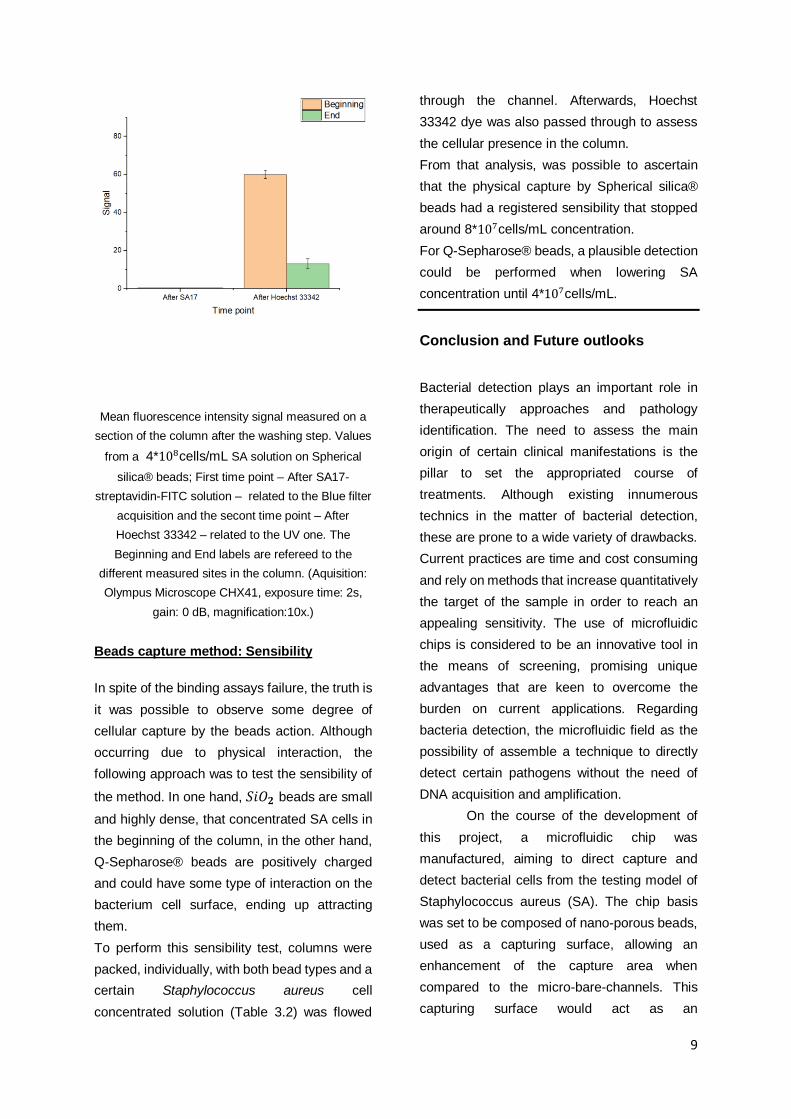
Figure 5 does not show any visible fluorescence
as should be expected if SA17 was bound to
Staphylococcus cells.
9
Mean fluorescence intensity signal measured on a
section of the column after the washing step. Values
from a 4*108cells/mL SA solution on Spherical
silica® beads; First time point – After SA17-
streptavidin-FITC solution – related to the Blue filter
acquisition and the secont time point – After
Hoechst 33342 – related to the UV one. The
Beginning and End labels are refereed to the
different measured sites in the column. (Aquisition:
Olympus Microscope CHX41, exposure time: 2s,
gain: 0 dB, magnification:10x.)
Beads capture method: Sensibility
In spite of the binding assays failure, the truth is
it was possible to observe some degree of
cellular capture by the beads action. Although
occurring due to physical interaction, the
following approach was to test the sensibility of
the method. In one hand, 𝑆𝑖𝑂𝟐 beads are small
and highly dense, that concentrated SA cells in
the beginning of the column, in the other hand,
Q-Sepharose® beads are positively charged
and could have some type of interaction on the
bacterium cell surface, ending up attracting
them.
To perform this sensibility test, columns were
packed, individually, with both bead types and a
certain Staphylococcus aureus cell
concentrated solution (Table 3.2) was flowed
through the channel. Afterwards, Hoechst
33342 dye was also passed through to assess
the cellular presence in the column.
From that analysis, was possible to ascertain
that the physical capture by Spherical silica®
beads had a registered sensibility that stopped
around 8*107cells/mL concentration.
For Q-Sepharose® beads, a plausible detection
could be performed when lowering SA
concentration until 4*107cells/mL.
Conclusion and Future outlooks
Bacterial detection plays an important role in
therapeutically approaches and pathology
identification. The need to assess the main
origin of certain clinical manifestations is the
pillar to set the appropriated course of
treatments. Although existing innumerous
technics in the matter of bacterial detection,
these are prone to a wide variety of drawbacks.
Current practices are time and cost consuming
and rely on methods that increase quantitatively
the target of the sample in order to reach an
appealing sensitivity. The use of microfluidic
chips is considered to be an innovative tool in
the means of screening, promising unique
advantages that are keen to overcome the
burden on current applications. Regarding
bacteria detection, the microfluidic field as the
possibility of assemble a technique to directly
detect certain pathogens without the need of
DNA acquisition and amplification.
On the course of the development of
this project, a microfluidic chip was
manufactured, aiming to direct capture and
detect bacterial cells from the testing model of
Staphylococcus aureus (SA). The chip basis
was set to be composed of nano-porous beads,
used as a capturing surface, allowing an
enhancement of the capture area when
compared to the micro-bare-channels. This
capturing surface would act as an
10
immobilization platform for a specific S. aureus
targeting molecule, creating a selection-
specific-approach that could be used among
other desired targets. Upon capturing, other
molecule target-specific, containing an
identification marker, would set to be flowed
through the beads to bind to the capture target,
creating a molecule-target-molecule sandwich-
type method.
To the present work, aptamers were chosen to
act as molecule-targeting probes. Two
aptamers were selected from the literature,
where is stated their optimal binding efficiency
to S.aureus cells [23] to attempt accomplishing
the described approach. SA17 was purchased
with a biotin molecule to allow the bead
immobilization and SA16 with Atto 430LS
molecule, a fluorophore to be able to be
detected.
From the results analysis, Spherical silica
beads® were selected, thus were prone to slow
down the cells flow through the column, acting
as a temporary capturing agent. The beads test
results, showed that this trapping-effect was set
time enough, that SA61 could be flowed and
detected. On this assessment assay, the
fluorescence measurements show no
significant signal, although cellular content was
clearly present in the channel. This gave rise to
multiple questions. To continue the
development of the project, one of those
questions gained the need to be clarified and
so, a viability assessment assay was performed
resourcing two different dyes as probes.
Hoechst 33342 and EvaGreen™, are able to
stain every cell (alive or dead) and only
compromised targets, respectively, creating an
Alive/Dead cell evaluation test. From the results
analysis, different conclusions could be
obtained. The signal acquisition among distinct
approaches of the dyes usage, led to the
assumption of a binding competition between
the dyes. Nevertheless, the initial question was
able to be assessed and in fact, the high
concentrations of S. aureus solutions and the
need to perform centrifugation steps to
achieved such concentration values, was
indeed bursting the cells
After the reach of this solution, the natural path
action was to re-try the binding efficacy tests on
SA61 detection aptamer, using this new
concentration, which showed to be enough to
still slow down the SA cells flow through the
microfluidic channel, using Hoechst’s dye to
confirm cellular presence upon the aptamers
passage. Unfortunately, the binding was not yet
successful and thus not related to viability
issues. The following approach was to perform
the same assessment for the capture aptamer
type, SA17, modified to act as a detection
molecule by the conjugation of a fluorescence
fluorophore, aiming to avoid the negative results
by using SA17 on both functions. Nevertheless,
also this aptamer showed no binding efficacy
against SA cells, which lead to the emergent
questioning about the correct folding of both
aptamers.
Threre was still a visible capturing method that
was followed across the entire project’s
development: physical capture by the bead-
packed columns. As a final approach, two
different bead-types were assessed in their
physical effects retaining bacterial cells in the
column, using Hoechst 33342 to confirm cellular
presence. Q-Sepharose® and Spherical silica®
beads were packed prior to specific SA cells
solutions concentrations to be flowed. Q-
Sepharose® beads showed an higher
sensitivity in comparison, where the lowest
detection was set on ~ 4*107cells/mL solution.
This number is extremely far from being
considered an ideal detection tool, as was
already expected, nevertheless it was a mean
of addressing the sensibility aspect of
techniques.
11
Due to the faced issues during the
course of the work hereby described, the need
to continue addressing the intended goal is
keen to be a path that follows what has been set
so far. The attempt of clarifying the encountered
binding issues and assess feasible features on
why aptamers were not binding, is somehow
appealing. For instance, assessing the folding
processes trying distinct protocols and buffers,
upon literature study, that differ either in
composition or physiological properties can be
an approach to continue the binding trouble
shooting. Other approach seen as future
work could be using the “sandwich” initial idea,
changing the targeting molecules from
aptamers to antibodies. Although aptamers
assemble a wide range of stated advantages
over antibodies, the truth is, these last are an
extremely sensitive approach in the matter of
targeting and bounding. Thus, antibodies can
be specific selected and also be keen to perform
an antibody-target-antibody interaction, since
can also be used as immobilization and
detection tools, resourcing feature addition
(such as a fluorophore).
References
[1] Davies J, Davies D. Origins and evolution of
antibiotic resistance. Microbiol Mol Biol Rev.
2010;74(3):417–433.
doi:10.1128/MMBR.00016-10
[2] Aminov RI. A brief history of the antibiotic
era: lessons learned and challenges for the
future. Front Microbiol. 2010;1:134. 2010.
doi:10.3389/fmicb.2010.00134
[3] Ventola CL. The antibiotic resistance crisis:
part 1: causes and threats. P T.
2015;40(4):277–283.
[4] Michael CA, Dominey-Howes D, Labbate M.
The antibiotic resistance crisis: causes,
consequences, and management. Front Public
Health. 2014; 2:145. doi:
10.3389/fpubh.2014.00145
[5] Peterson JW. Bacterial Pathogenesis. Baron
S, editor. Medical Microbiology. 4th edition.
Galveston (TX): University of Texas Medical
Branch at Galveston; 1996. Chapter 7.
https://www.ncbi.nlm.nih.gov/books/NBK8526/,
September 2019
[6] Fair RJ, Tor Y. Antibiotics and bacterial
resistance in the 21st century. Perspect Medicin
Chem. 2014;6:25–64. 2014;
doi:10.4137/PMC.S14459
[7] L. Zeng, L. Wang, J. Hu. Current and
Emerging Technologies for Rapid Detection of
Pathogens, Biosensing Technologies for the
Detection of Pathogens - A Prospective Way for
Rapid Analysis. IntechOpen. 2018 doi:
10.5772/intechopen.73178
[8] Franco-Duarte R, Černáková L, Kadam S, et
al. Advances in Chemical and Biological
Methods to Identify Microorganisms-From Past
to Present. Microorganisms. 2019;7(5):130.
Published 2019 May 13.
doi:10.3390/microorganisms7050130
[9] Linda V., Jia Li., David E. H., John D. P.,
Rosaleen J. A., Sylvain O., Paul W. G. Methods
for the detection and identification of pathogenic
bacteria: past, present, and future. Chemical
Society Reviews. Issue 16, 2017 doi:
10.1039/c6cs00693k
[10] Harikrishnan J., Himanshu S., Bruce K G.
Applications of Microfluidics for Molecular
Diagnostics. Humana Press. 2013; 305:334 doi:
10.1007/978-1-62703-134-9_20
[11] Ashraf MW, Tayyaba S, Afzulpurkar N.
Micro Electromechanical Systems (MEMS)
Based Microfluidic Devices for Biomedical
Applications. Int J Mol Sci. 2011;12(6):3648–
3704. doi:10.3390/ijms12063648
[12] Emily M. C. Microfluidic Visualisation and
Analysis of Multiphase Flow Phenomena at the
Pore Scale. Imperial College London. 2014
[13] Tabeling, Patrick & Troian, Sandra.
Introduction to Microfluidics. Physics Today.
2006; 59. 58-. 10.1063/1.2435685.

12
[14] Luka G, Ahmadi A, Najjaran H, et al.
Microfluidics Integrated Biosensors: A Leading
Technology towards Lab-on-a-Chip and
Sensing Applications. Sensors (Basel).
2015;15(12):30011–30031. Published 2015
Dec 1. doi:10.3390/s151229783
[15] Grieshaber D, MacKenzie R, Vörös J,
Reimhult E. Electrochemical Biosensors -
Sensor Principles and Architectures. Sensors
(Basel). 2008;8(3):1400–1458. Published 2008
Mar 7. doi:10.3390/s80314000
[16] Drake CR, Miller DC, Jones EF. Activatable
Optical Probes for the Detection of Enzymes.
Curr Org Synth. 2011;8(4):498–520.
doi:10.2174/157017911796117232
[17] Jackson MP. Detection of Shiga toxin-
producing Shigella dysenteriae type 1 and
Escherichia coli by using polymerase chain
reaction with incorporation of digoxigenin-11-
dUTP. J Clin Microbiol. 1991;29(9):1910–1914.
[18] Jayamohan, Harikrishnan & Sant,
Himanshu & Gale, Bruce. (2013). Applications
of Microfluidics for Molecular Diagnostics.
10.1007/978-1-62703-134-9_20.
[19] Terekhov SS, Smirnov IV, Stepanova AV,
et al. Microfluidic droplet platform for ultrahigh-
throughput single-cell screening of biodiversity.
Proc Natl Acad Sci U S A. 2017;114(10):2550–
2555. doi:10.1073/pnas.1621226114
[20] Okumus B, Baker CJ, Arias-Castro JC, et
al. Single-cell microscopy of suspension
cultures using a microfluidics-assisted cell
screening platform. Nat Protoc.
2018;13(1):170–194.
doi:10.1038/nprot.2017.127
[21] Jan H. A therapeutic battle: Antibodies vs.
Aptamers.Research paper. Nanoscience
master program. NS109.
[22] Janeway CA Jr, Travers P, Walport M, et
al. Immunobiology: The Immune System in
Health and Disease. 5th edition. New York:
Garland Science; 2001. The structure of a
typical antibody molecule.
https://www.ncbi.nlm.nih.gov/books/NBK27144
/, November 2019
[23] Chang YC, Yang CY, Sun RL, Cheng YF,
Kao WC, Yang PC. Rapid single cell detection
of Staphylococcus aureus by aptamer-
conjugated gold nanoparticles. Sci Rep.
2013;3:1863. doi:10.1038/srep01863