direct endovascular thrombolytic therapy for dural sinus
TRANSCRIPT

Direct Endovascular Thrombolytic Therapy for Dural SinusThrombosis: Infusion of Alteplase
Sun Yong Kim and Jung Ho Suh
PURPOSE: To evaluate the efficacy, safety, and results of direct thrombolytic therapy in intracra-nial dural sinus thrombosis by infusion of alteplase (recombinant tissue plasminogen activator).METHODS: Nine patients were treated during a 2-year period for intracranial dural sinus throm-bosis. A microcatheter was placed directly into the thrombus in the dural sinus via the transfemoralroute. Thrombolysis was initiated with a rapid injection of 10 mg of alteplase over 10 minutes,followed in 3 hours by a continuous infusion of 50 mg, then a continuous infusion at 5 mg per houruntil complete thrombolysis or a total dose of 100 mg per day had been reached. Repeatthrombolysis was tried the following day if complete recanalization did not occur at 100 mg perday. RESULTS: Successful recanalization with improvement of symptoms was achieved in allcases. Time required for complete thrombolysis was between 8 and 43 hours. The total dose ofalteplase ranged from 50 to 300 mg. Complications of a small intrapelvic hemorrhage and oozingat a femoral puncture site occurred in separate cases, but were not related to the amount of infusedalteplase. MR venograms obtained 1 to 4 weeks after the procedure showed no evidence ofreocclusion of the dural sinuses. CONCLUSION: Direct fibrinolytic therapy with alteplase is safe,fast, and effective in treating dural sinus thrombosis. However, to prevent hemorrhagic complica-tions, further studies are required to determine its optimal dose and proper rate of administration.
Index terms: Thrombosis, dural sinus; Thrombolysis; Interventional materials
AJNR Am J Neuroradiol 18:639–645, April 1997
Many causes and predisposing factors play arole in the development of dural sinus thrombo-sis (1). Because of the broad spectrum of non-specific clinical findings, the early diagnosis isoften missed, which results in a high rate ofmorbidity and mortality (2–4). Emergency in-tervention is inevitable in cases of dural sinusthrombosis, but methods of treatment still re-main controversial.
Thrombolytic agents, such as heparin, strep-tokinase, or urokinase, are used frequently;however, significant risks of cerebral hemor-rhage as well as systemic coagulopathy areencountered (2, 5). Recently, alteplase (recom-binant tissue plasminogen activator), a fibrin-
Received March 20, 1996; accepted after revision October 21.From the Department of Diagnostic Radiology, Ajou University School
of Medicine, San 5, Wonchon-dong, Paldal-gu, Suwon 442–749, Korea.Address reprint requests to Sun Yong Kim, MD.
AJNR 18:639–645, Apr 1997 0195-6108/97/1804–0639
© American Society of Neuroradiology
63
selective thrombolytic agent, has become avail-able and has been used successfully in thetreatment of thromboembolic disease of the pe-ripheral vasculature (6, 7), heart (8, 9), andbrain (10, 11). On the basis of these encourag-ing results and the recent development of ad-vanced catheterization techniques relating tothe dural sinus, this study was undertaken toevaluate the safety and efficacy of direct infu-sion of alteplase into the thrombosed dural si-nuses.
Patients and MethodsBetween August 1993 and September 1995, nine pa-
tients with intracranial dural sinus thrombosis were treatedwith direct infusion of alteplase into the thrombosed duralsinus. The study group consisted of six women and threemen, ranging in age from 24 to 52 years (mean, 33 years).Patient information is summarized in the Table. The meanduration of symptomatic disease was 4.2 weeks (range, 1to 16 weeks). Radiologic studies, including magnetic res-onance (MR) imaging and computed tomography (CT),were obtained in all patients. A cerebral angiogram and
9

640 YONG AJNR: 18, April 1997
Patients with dural sinus thrombosis and results of alteplase infusion
PatientAge,y/Sex
Causes ofThrombosis
Signs and SymptomsDuration,
wkLocation of Thrombosis
Totaldosage,
mg
TimeRequiredfor Lysis,
h
Complications
1 42/F Acute mastoiditis Headache, papilledema 3 R transverse sinus 50 9 None2 29/F Systemic lupus
erythematosusLethargy, L
hemiparesis4 Superior sagittal sinus,
R transverse sinus150 8 Oozing at puncture
site3 44/M Antiphospholipid
antibody syndromeHeadache, seizure 16 Superior sagittal sinus,
L transverse sinus200 31 Intraperitoneal
hematoma4 52/F . . . Headache, lethargy 4 L transverse sinus 100 12 None5 37/M Acute mastoiditis Headache 1.5 R transverse sinus 130 22 None6 24/F Dehydration Headache, seizure 2 Superior sagittal sinus,
R transverse sinus100 13 None
Straight sinus, vein ofGalen
7 27/F Postpartum Headache, seizure,palsy of fourthcranial nerve
1 L transverse sinus 75 8 None
8 42/M . . . Headache, Lhemiparesis
4 Superior sagittal sinus,R transverse sinus,jugular bulb
300 43 None
9 43/F Catheter induced Seizure 2 L transverse sinus,jugular vein
100 16 None
sinus venogram were initially obtained to assess the extentof sinus thrombosis. Four patients (patients 1 through 4)were treated initially with systemic heparinization but eachhad continued symptoms with increasingly severe head-aches, seizures, and lethargy. Direct thrombolysis wasperformed in four patients (patients 5 through 8) as aninitial treatment because of its effectiveness in the first fourpatients. In three of the nine patients (patients 1, 4, and 7),brain parenchyma was normal on imaging studies. Fivepatients had minimal brain swelling and sulcal effacementbut no signal change. Intracerebral hemorrhage was notseen except in one patient (patient 8; Fig 1), who had focalswelling in the right parietal lobe near the mid superiorsagittal sinus, which was considered a recent venous in-farction.
After confirming the presence of thrombus in the duralsinus and verifying its location and extent on the venousphase of carotid angiography, we introduced a 6F poly-urethane guiding catheter into the internal jugular vein viathe femoral vein. A 2.8F microcatheter (Target Therapeu-tics, San Jose, Calif) was advanced into the occluded duralsinus coaxially through the guiding catheter. The tip of thecatheter was placed through and proximal to the thrombusas far as possible. When complete thrombosis of the duralsinus prevented optimal placement of the catheter tip,mechanical disruption of the thrombus was tried via ma-nipulation of the guidewire and catheter (patient 6; Fig 2).When thrombi occluded the jugular vein, retrograde super-selection was made along the contralateral jugular veinand transverse sinus (patient 9; Fig 3).
The alteplase (Actilyse, Boehringer; Ingelheim, Ger-many) solution was prepared by mixing 100 mg of lyoph-ilized alteplase with 20 mL of distilled water, to which
normal saline was added until the volume reached 200 mL(with an approximate final concentration of 0.5 mg/mL).The dosage and rate of alteplase administration modifiedas a treatment for myocardial infarction have been de-scribed previously (6). Thrombolytic therapy was initiatedby bolus injection of alteplase, 10 mg over 10 minutes asa loading dose, within the initial thrombosed site in thesinus. Subsequent continuous infusion of 50 mg of alte-plase was given over 3 hours via a rate-regulated infusionpump followed by a slower continuous infusion (5 mg/h)until complete thrombolysis or a dose of 100 mg per dayhad been reached. In four patients, who had residualthrombus in their partially recanalized dural sinuses, thesame infusion regimen was used the following day. Duringthe infusion therapy, digital dural sinus venography wasperformed every 30 minutes, and the catheter tip wasadvanced more proximally as thrombolysis progressed.Patients were kept in either the neurologic intensive careunit or the angiography suite throughout infusion so that vi-tal signs and neurologic status could be assessed frequently.
All patients received heparin by means of continuousinfusion via an introducer catheter to prevent pericatheterthrombus formation during thrombolysis (partial thrombo-plastin time at 1.5 times control). After the thrombolytictherapy, patients were converted to oral anticoagulanttherapy (warfarin). Coagulation parameters (includingprothrombin time, partial thromboplastin time, fibrinogen,and fibrin split products) and hematologic parameters (in-cluding hemoglobin and hematocrit) were determined justbefore initiation of therapy and at 4-hour intervals. Sevenpatients were treated with warfarin for 3 months afterthrombolytic treatment. Patients were followed up closelywith MR venography and clinical examination.

AJNR: 18, April 1997 DURAL SINUS THROMBOSIS 641
Fig 1. Patient 8.A, Lateral, venous phase of carotid an-
giogram shows extensive filling defect dueto thrombi throughout entire superior sag-ittal sinus.
B, Anteroposterior digital venogram,after placement of microcatheter into theright transverse sinus from the right jugularvein, shows extensive thrombus within thedural sinus (arrows).
C, Selective superior sagittal sinusvenogram after partial thrombolysis of themid and posterior thirds of the superiorsagittal sinus. Microcatheter was advancedthrough the thrombus.
D, Venous phase of angiogram imme-diately after thrombolysis shows goodopacification of the entire superior sagittalsinus.
Results
Results are summarized in the Table. Angio-graphic verification of thrombolysis and pa-tency of the entire dural sinus system wereachieved in all nine patients. Clinical signs andsymptoms, including neurologic deficits, sei-zures, and headaches, were treated successfullyin all patients during the 3-month follow-up pe-riod.
Two patients (patients 4 and 6), who initiallyhad severe headaches, experienced a rapid re-duction of symptoms during the procedure.
Although a small amount of retained throm-bus was seen on the wall of the superior sagittalsinus and the adjacent cortical vein in one pa-tient (patient 2), no additional thrombolytictreatment was performed owing to marked im-provement of clinical symptoms and the pa-tient’s subsequent refusal of additional treat-ment. The mean time to complete the lysis was20 hours (range, 8 to 43 hours). The mean totaldose of alteplase was 135 mg (range, 50 to 300mg).
During the catheterization of the thrombosedsinus, some patients complained of pain, pre-sumably caused by friction on the wall of thedural sinus. The cause, extent of thrombosis,and intensity of symptoms did not correlate withthe dosage of infused alteplase or with the timetaken to achieve complete thrombolysis. In co-agulation parameter profiles, no significant dif-ferences were observed in the prothrombin timeand partial thromboplastin time at baseline andafter alteplase treatment (except the intentionalprolongation of partial thromboplastin time to1.5 times normal). The level of fibrin degrada-tion products in all but two patients was be-tween 10 and 30 mg/mL, which was subnormalbut of uncertain clinical significance. Complica-tions were seen in only two patients. In one(patient 2), minor bleeding occurred at the fem-oral puncture site but did not require transfusionor interruption of lytic therapy. In the other (pa-tient 3), a small intrapelvic hemorrhage devel-oped the day after initial therapy. This patientreceived blood products (2 U of fresh frozen

642 YONG AJNR: 18, April 1997
Fig 2. Patient 6.A, T1-weighted (500/12/1 [repetition time/echo
time/excitations]) MR image shows increased sig-nal intensity of superior sagittal sinus, straight si-nus, vein of Galen, and internal cerebral vein.
B, Selective venogram from the frontal aspect ofthe superior sagittal sinus, after mechanical disrup-tion of thrombi and thrombolysis, shows the par-tially patent lumen of the sinus.
C, Venous phase of follow-up angiogram 1month later shows patency of the entire dural sinus.
plasma) for the management of a precipitousdecline in fibrinogen (baseline was 302 mg/dL;18 hours after the procedure, it was 58 mg/dL).The fibrinogen level subsequently returned tonormal.
Discussion
The clinical manifestations of dural sinusthrombosis are variable and the radiologic find-ings have been well documented (3, 4, 12).Symptoms depend on the specific sinuses in-volved, the rate of evolution of thrombosis, theinvolvement of cerebral veins, and the forma-tion of collateral veins. Therefore, outcomes areunpredictable. This variability makes differenttreatment regimens difficult to compare.
Three different types of thrombus occur in thedural sinus, including a poorly organized redthrombus, a platelet-rich white thrombus, andan organized collagen-rich thrombus; all threehave a similar appearance at MR imaging. The
age of a thrombus is not clearly related to theresponse to therapy. Moreover, a chronic clotthat contains different stages of thrombi andthat may have existed for years may be lysed byfibrinolytics (13).
The criteria by which patients are selected fortreatment of dural sinus thrombosis with sys-temic thrombolysis are difficult to establish, be-cause published results have been variable.High doses of a systemic standard thrombolyticagent (heparin, streptokinase, and urokinase)have often been used to lyse dural sinus throm-bosis, but the results have not been satisfactoryowing to slow rates of thrombolysis and hemor-rhagic complications (14–16).
The impetus for our use of alteplase in thetreatment of dural sinus thrombosis was basedon the encouraging clinical results obtained inother sites of thromboembolic disease (4–9)and on the drug’s mechanism of action and itspharmacophysiological characteristics. First,alteplase only catalyzes the conversion of fibrin-
AJNR: 18, April 1997 DURAL SINUS THROMBOSIS 643
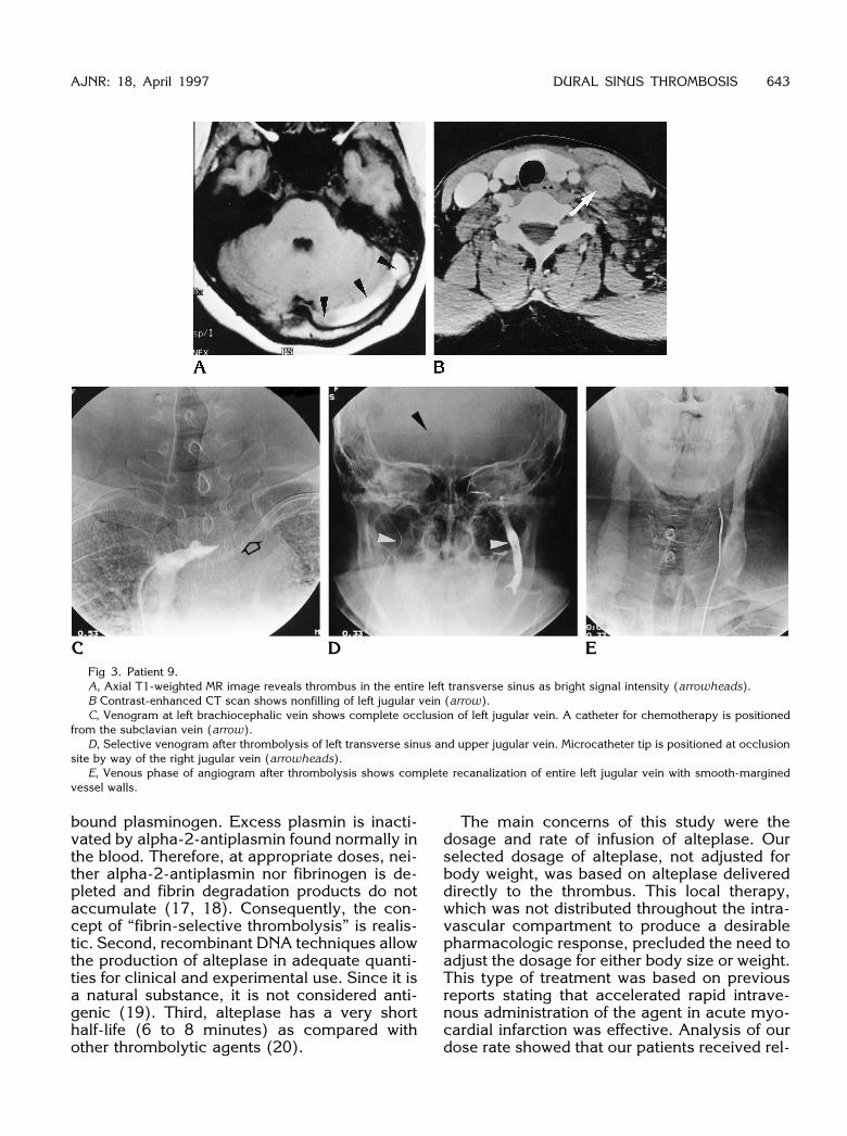
Fig 3. Patient 9.A, Axial T1-weighted MR image reveals thrombus in the entire left transverse sinus as bright signal intensity (arrowheads).B Contrast-enhanced CT scan shows nonfilling of left jugular vein (arrow).C, Venogram at left brachiocephalic vein shows complete occlusion of left jugular vein. A catheter for chemotherapy is positioned
from the subclavian vein (arrow).D, Selective venogram after thrombolysis of left transverse sinus and upper jugular vein. Microcatheter tip is positioned at occlusion
site by way of the right jugular vein (arrowheads).E, Venous phase of angiogram after thrombolysis shows complete recanalization of entire left jugular vein with smooth-margined
vessel walls.
bound plasminogen. Excess plasmin is inacti-vated by alpha-2-antiplasmin found normally inthe blood. Therefore, at appropriate doses, nei-ther alpha-2-antiplasmin nor fibrinogen is de-pleted and fibrin degradation products do notaccumulate (17, 18). Consequently, the con-cept of “fibrin-selective thrombolysis” is realis-tic. Second, recombinant DNA techniques allowthe production of alteplase in adequate quanti-ties for clinical and experimental use. Since it isa natural substance, it is not considered anti-genic (19). Third, alteplase has a very shorthalf-life (6 to 8 minutes) as compared withother thrombolytic agents (20).
The main concerns of this study were thedosage and rate of infusion of alteplase. Ourselected dosage of alteplase, not adjusted forbody weight, was based on alteplase delivereddirectly to the thrombus. This local therapy,which was not distributed throughout the intra-vascular compartment to produce a desirablepharmacologic response, precluded the need toadjust the dosage for either body size or weight.This type of treatment was based on previousreports stating that accelerated rapid intrave-nous administration of the agent in acute myo-cardial infarction was effective. Analysis of ourdose rate showed that our patients received rel-

644 YONG AJNR: 18, April 1997
atively concentrated doses during the first fewhours. As compared with the focal and smallthrombi seen in coronary or peripheral arteries,the majority of thrombi in dural sinus thrombo-sis are large and extensive. This modifiedmethod was fairly effective. The time requiredfor thrombolysis was relatively short (8 to 43hours; mean, 20 hours) compared with the timerequired to obtain results with urokinase (88 to238 hours) (14).
These advantages of alteplase seemed tocontribute to the favorable and rapid improve-ment of clinical symptoms in patients with duralsinus thrombosis, as seen in our study. Reportsin the literature have documented that throm-bolysis can be accelerated by mechanical dis-ruption of thrombus with transthrombus bolusinfusion of fibrinolytic agents and with a pulse-spray technique via a side-hole catheter (21,22). It was unfortunate that a side-hole catheterwas not available on the market at the time ofour study.
The age of the thrombus was not clearly re-lated to the response to direct thrombolytictherapy. Although older organized thrombi arethought to lyse far more slowly than fresh ones,our experience showed that thrombolysis pro-ceeds rapidly in most cases, suggesting thatthrombolysis augmented by increasing the sur-face area between clot and thrombolytic agentthrough fragmentation with the catheter andguidewire is helpful. However, manipulation of acatheter in a thrombosed dural sinus should beperformed with extreme care and without undueforce, because vigorous contact with the sinuswall can be very painful and perforation of adural sinus wall or a large cortical vein could becatastrophic.
In our series, minor hemorrhagic events inpatients were not proportional to the dose ofadministered alteplase, even though total dos-age greater than 0.9 mg/kg has been found tobe related to intracranial hemorrhage whengiven intravenously for acute ischemic stroke(23). In one study (6), 0.7% of patients withmyocardial infarction receiving 1.5 mg/kg ofalteplase intravenously to a maximum of 100mg over 90 minutes suffered hemorrhagicstrokes. However, it is likely that undeterminedand complex factors contribute to the develop-ment of hemorrhagic complications in patientsreceiving alteplase. It is also well known thatdiastolic hypertension (.100 mm Hg) is closely
related to hemorrhagic complications in pa-tients receiving fibrinolytics.
Although our preliminary results from a smallnumber of cases do not allow us to propose adefinitive treatment regimen for dural sinusthrombosis, alteplase nevertheless appears tobe the first choice among various thrombolyticagents in terms of safety and efficacy. The rapidtherapeutic response to alteplase in acutely de-teriorating cases makes it especially appealingas an initial regimen. Further study is requiredto determine the optimal dosage and adminis-tration technique to prevent hemorrhagic com-plications and reocclusion.
AcknowledgmentsWe thank Heun Young Yoon and Sang Joon Park for
valuable advice and critical review of the manuscript.
References1. Bousser MG, Chiras J, Bories J, Castaigne P. Cerebral venous
thrombosis: a review of 38 cases. Stroke 1985;16:199–2132. Gettelfinger DM, Kokmen E. Superior sagittal sinus thrombosis.
Arch Neurol 1977;34:2–63. Yuh WTC, Simonson TM, Wang AM, et al. Venous sinus occlusive
disease: MR findings. AJNR Am J Neuroradiol 1994;15:309–3164. Tsai FY, Wang AM, Matovich VB, et al. MR staging of acute dural
sinus thrombosis: correlation with venous pressure measurementsand implications for treatment and prognosis. AJNR Am J Neuro-radiol 1995;16:1021–1029
5. Agnelli G, Buchanan MR, Fernandez F, et al. A comparison of thethrombolytic and hemorrhagic effects of tissue-type plasminogenactivator and streptokinase in rabbits. Circulation1985;72:178–182
6. Bero CJ, Cardella JF, Reddy K, et al. Recombinant tissue plas-minogen activator for the treatment of lower extremity peripheralvascular occlusive disease. J Vasc Interv Radiol 1995;6:571–577
7. Koppensteiner R, Minar E, Ahmadi R, et al. Low doses of recom-binant human tissue-type plasminogen activator for local throm-bolysis in peripheral arteries. Radiology 1988;168:877–878
8. GUSTO Investigators. An international, randomized trial compar-ing four thrombolytic strategies for acute myocardiac infarction.N Engl J Med 1993;329:673–682
9. Verstraete M, Bory M, Collen D, et al. Randomized trial of intrave-nous recombinant tissue-type plasminogen activator versus intra-venous streptokinase in acute myocardial infarction. Lancet 1985;13:842–847
10. Brott T, Haley EC, Levy DE, et al. Very early therapy for cerebralinfarction with tissue plasminogen activator (tPA) (abstract).Stroke 1988;19:133
11. Kummer RV, Forsting M, Kacke W, Sartor K. Reperfusion ofintracranial arteries in acute stroke after treatment with intrave-nous plasminogen. Neuroradiology 1991;33(Suppl):381–383
12. Vogl TJ, Bergman C, Villringer A, Einhaupl K, Lissner J, Felix R.Dural sinus thrombosis: value of venous MR angiography fordiagnosis and follow-up. AJNR Am J Neuroradiol 1994;162:1191–1198
13. Becker GJ, Rabe FE, Richmond BD, et al. Low dose fibrinolytictherapy: results and new concepts. Radiology 1983;148:663–670

AJNR: 18, April 1997 DURAL SINUS THROMBOSIS 645
14. Smith TP, Higashida RT, Barnwell SL, et al. Treatment of duralsinus thrombosis by urokinase infusion. AJNR Am J Neuroradiol1994;15:801–807
15. Tsai FY, Higashida RT, Matovitch V, Alflet K. Acute thrombosis ofthe intracranial dural sinus: direct thrombolytic treatment. AJNRAm J Neuroradiol 1992;13;1137–1141
16. Higashida RT, Helmer E, Halbach VV, Hieshima GB. Direct throm-bolytic therapy for sagittal sinus thrombosis. AJNR Am J Neuro-radiol 1989;10(Suppl):S4–S6
17. Sobel BE, Gross RW, Robinson AK. Thrombolysis, clot selectivity,and kinetics. Circulation 1984;70:160–164
18. Kissel P, Cherhrazi B, Seibert J, Wagner FC. Digital angiographicquantification of blood flow dynamics in embolic stroke treatedwith tissue type plasminogen activator. J Neurosurg 1987;67:395–405
19. Jang IK, Vanhaechke J, De Geest, et al. Coronary thrombolysis
with recombinant tissue-type plasminogen activator: patency rateand regional wall motion after 3 months. J Am Coll Cardiol 1986;8:1455–1460
20. Eisenberg PR, Sherman LA, Tiefennbrunn AJ, et al. Sustainedfibrinolysis after administration of TPA despite its short half-life inthe circulation. Thromb Haemost 1987;57:35–40
21. Davis GB, Dowd CF, Bookstein JJ, Maroney TP, Lang EV, HalaszN. Thrombosed dialysis grafts: efficacy of intrathrombotic depo-sition of concentrated urokinase, clot maceration, and angio-plasty. AJR Am J Roentgenol 1987;149:177–181
22. Bookstein JJ, Fellmeth B, Roberts A, Valji K, Davis G, Machado T.Pulse-spray pharmacomechanical thrombolysis: preliminary clin-ical results. AJR Am J Roentgenol 1989;122:1097–1100
23. Levy DE, Brott TG, Haley C, et al. Factors related to intracranialhematoma formation in patients receiving tissue-type plasmino-gen activator for acute ischemic stroke. Stroke 1994;25:291–297