discrete nascent chain lengths are required for the insertion of
TRANSCRIPT

Discrete Nascent Chain Lengths are Required for the Insertion of Presecretory Proteins into Microsomal Membranes Sandra L. Wolin** and Peter Walter* * Department of Biochemistry and Biophysics, University of California Medical School, San Francisco, California 94143-0448; and ~ Howard Hughes Medical Institute and Department of Cell Biology, Yale University School of Medicine, New Haven, Connecticut 06510
Abstract. Ribosomes synthesizing nascent secretory proteins are targeted to the membrane by the signal recognition particle (SRP), a small ribonucleoprotein that binds to the signal peptide as it emerges from the ribosome. SRP arrests further elongation, causing ribosomes to stack behind the arrested ribosome. Upon interaction of SRP with its receptor on the ER membrane, the translation arrest is released and the ribosome becomes bound to the ER membrane. We have examined the distribution of unattached and membrane-bound ribosomes during the translation of mRNAs encoding two secretory proteins, bovine
preprolactin and rat preproinsulin I. We find that the enhancement of ribosome stacking that occurs when SRP arrests translation of these proteins is relaxed in the presence of microsomal membranes. We also dem- onstrate that two previously described populations of membrane-associated ribosomes, distinguished by their sensitivity to high salt or EDTA extraction, cor- respond to ribosomes that have synthesized differing lengths of the nascent polypeptide. This analysis has revealed that nascent chain insertion into the mem- brane begins at distinct points for different presecre- tory proteins.
T HE synthesis of secretory proteins in higher eu- karyotes is strictly coupled to their translocation across the endoplasmic reticulum. A common feature
of these proteins is a signal peptide, usually an amino- terminal extension of 15-30 amino acids. A small cyto- plasmic ribonucleoprotein, the signal recognition particle (SRP), j binds to the nascent chain as it emerges from the ribosome. SRP then arrests or slows translation in the cyto- plasmic space, by an as yet unknown mechanism (Walter and Blobel, 1981; Wolin and Walter, 1989). The translational ar- rest is released when the ribosome/SRP complex interacts with the SRP receptor on the ER membrane. This interaction results in the establishment of a ribosome-membrane junc- tion, and translation proceeds as the nascent chain is translo- cated across the endoplasmic reticulum membrane (re- viewed by Rapoport, 1992; Sanders and Schekman, 1992).
As the synthesis of secretory proteins takes place on ribo- somes bound to the ER membrane, it is unclear how attach- ment to membranes affects ribosome movement along the mRNA. In addition, membrane-bound ribosomes can be separated into "loosely" bound and "tightly" bound fractions, which can be distinguished by their sensitivity to high salt or EDTA extraction (Adelman et al., 1973; Harrison et al.,
Address correspondence to Dr. Wolin at the Howard Hughes Medical Insti- tute and Department of Cell Biology, Yale University School of Medicine, New Haven, CT 06510.
1. Abbreviation used in this paper: SRP, signal recognition particle.
1974; Gilmore and Blobel, 1985; Connolly and Gilmore, 1986). Loosely bound ribosomes are thought to represent those ribosomes which have been targeted to the ER, but whose nascent chains have not yet engaged the translocation machinery in the membrane. Upon nascent chain insertion, ribosomes become tightly bound and resistant to extraction with either KOAc or EDTA (Gilmore and Blobel, 1985; Con- nolly and Gilmore, 1986). It would thus be interesting to know if these two populations of ribosomes correspond to ribosomes that have migrated different distances along the mRNA. Such an analysis should also reveal the minimum length of nascent chain necessary for insertion into the ER membrane.
We previously developed a method that allows us to deter- mine the positions of paused ribosomes along an mRNA (Wolin and Walter, 1988). Using this method to monitor translation on a model mRNA, bovine preprolactin, we showed that SRP arrests translation at the first natural pause site after the signal peptide emerges from the ribosome and becomes available for binding. When the leading ribosome pauses, other ribosomes stack up behind the paused ribo- some. These ribosomes become very tightly stacked, such that the centers of the ribosomes are only 28 nucleotides apart. This "ribosome stacking" occurs whenever the leading ribosome pauses, and is enhanced when SRP arrests transla- tion (Wolin and Walter, 1988).
We have now used this method to probe the distribution of membrane-bound ribosomes along mRNAs encoding two
© The Rockefeller University Press, 0021-9525/93/06/1211/9 $2.00 The Journal of Cell Biology, Volume 121, Number 6, June 1993 1211-1219 1211
on March 17, 2006
www.jcb.orgDownloaded from

secretory proteins, preprolactin and preproinsulin. We re- port that the presence of membranes results in the relaxation of ribosome stacking, presumably because the translational arrest mediated by SRP is released. In addition, we show that nascent chains become competent to engage the transloca- tion machinery in the membrane at a distinct point in their synthesis. Interestingly, this point varies for different pro- teins, which may reflect differences in the length and/or structure of the signal peptide.
Materials and Methods
Reagents Wheat germ extract was prepared as described by Erickson and Blobel (1983). SRP was prepared as described by Walter and Blobel (1983b), ex- cept that the final sucrose gradient step was omitted. Salt-washed pancreatic microsomal vesicles (K-RM) were prepared as described (Waiter and Blobel, 1983a), except that the column washing step was omitted and the membranes were depleted of SRP and ribosomes as described previously (Wolin and Walter, 1989). T4 DNA polymerase, gene 45 and genes 44/62 proteins were obtained both from Drs. Jack Barry and Bruce Alberts (University of California, San Francisco) and from Drs. William Konigs- berg and Maureen Munn (Yale University, New Haven, CT).
Clones and In Vitro Transcription For transcription of rat preproinsulin I, we began by isolating a fuil-length eDNA clone by screening a plasmid eDNA library constructed from insuli- noma 38 cells (Murphy and Efstradiatis, 1987) with a partial rat preproinsu- lin eDNA clone, rpsub9 (both a kind gift of A. Efstradiatis, Columbia University, New York). DNA was isolated from positive colonies and the longest inserts were sequenced. These inserts were found to contain eDNA clones with an extended 5' untranslated region, encompassing an additional 71 nucleotides 5' of the previously reported mRNA cap site (Soares et al., 1985). We therefore used the PCR to construct a eDNA clone with a 5' end identical to the major rat insulin transcript (Soares et al., 1985). For this, we used the oligonucleotides CGC~CGAATTCTAAGTGACCAGCTACA- ATC and GACCGC~CGAAGCTTrTTTTI'TTTITI" in a PCR reaction with the parent eDNA clone. The amplified DNA was isolated, digested with EcoRI and Hind III, and cloned into pGEM2 (Promega Corp., Madi- son, WI). The resulting cloned DNA was linearized with Hind Ill and tran- scribed as described previously (Wolin and Walter, 1988).
For transcription of bovine preprolactin, we constructed a variant of the previously described pSPBP4 (Siegel and Walter, 1988a). In this new con- struct, pPLPCR, the poly G tail that follows the poly A tail in pSPBP4 has been removed. This was accomplished by using the oligonucleotides C G C ~ C G G A A T T C G C T r G T T C T T T ~ G and GACCGGCCGAA- GCTTTTTTTTrTTTTT in a PCR reaction with pSPBP4 DNA, digesting the amplified DNA with EcoRI and Hind HI, and cloning into pGEM2. The resulting construct was linearized with Hind HI and transcribed as de- scribed (Wolin and Walter, 1988).
The eDNA clone encoding the 19-kD subunit of SRP has been previously described (Lingelbach et al., 1988).
Isolation and Mapping of Ribosome-protected mRNA Fragments Translations of pPLPCR using wheat germ extract were performed as de- scribed for pSPBP4 (Wolin and Walter, 1988) except that 7.5 eq of mem- branes [as defined by Walter and Blobel, 1983a] were added to a 25 #1 trans- lation reaction. After a 15-rain incubation at 26°C, the reaction was placed on ice, and 0.5 pl of a 50 mM cycloheximide stock was added to final con- centration of 1 raM. Micrococcal nuclease was added to the desired concen- tration, and the reaction was supplemented with CaCI2 and Mg(OAc)2 to give final concentrations of 3 and 3.5 raM, respectively, in a final vol of 40 /zl. After a 3-min incubation on ice, the reaction was digested for 30 rain at 26°C. The nucleaso digestion was terminated by the addition of 10/~l of either 20 mM Hepes-KOH pH 7.5, 150 mM KOAc, 25 mM EGTA, 36 mM Mg(OAc)2, 2 mM DTT (150 mM KOAc buffer) or 20 mM Hepes pH 7.5, 1.9 M KOAc, 25 mM EGTA, 36 mM Mg(OAc)2, 2 mM DTT (to give a final concentration of 500 mM KOAc) and 20/~g of total yeast RNA. The
mixture was then overlaid on a 100/~1 cushion of 1.8 M sucrose in 20 mM Hepes, 10 mM Mg(OAc)2, 5 raM EGTA, 2 mM DTT (Buffer T) contain- ing either 150 mM or 500 mM KOAc and spun for 14 h at 55,000 rpm in a TLS55 rotor in a Beckman TLI00 ultracentrifuge. The top 120 ~1 (con- taining membrane-beund ribosomes) was removed. The bottom 30 td (free ribosomal "pellet") was mixed with 100 #1 of 50 mM NaC1, 50 ram Tris- HC1 pH 7.5, 5 mM EDTA, 0.5% SDS, 200/~g/ml proteinase K and in- cubated at 37°C for 30 min. The mixture was then removed, extracted with phenol/chloroform/isoamyl alcohol (50:50:1), and precipitated with ethanol in the presence of 10 ~g Escherichia coil tRNA. To the top 120/d, the deter- gent CHAPS was added to a final concentration of 1%. The mixture was overlaid on a cushion of 60/LI of 1.8 M sucrose in Buffer T and spun for 7 h at 55,000 rpm as before. The top 150/~1 (the supernatant) was removed, extracted with phenol/chloroform/isoamyl alcohol and precipitated with ethanol. The bottom 30 #1 (containing bound ribosomes) was treated with proteinase K, extracted with phenol/chloroform/isoamyl alcohol and precipitated with ethanol as described above.
Isolation of protected fragments of rat preproinsulin I mRNA was per- formed as described for bovine preprolactin, except that nuclease V1 (Phar- macia LKB Biotechnology, Inc., Piscataway, NJ) was added during diges- tion to a final concentration of 0.025 U/#l (Doohan and Samuel, 1992). This was necessary because the highly structured preproinsulin mRNA was found to be incompletely digested with micrococcal nuclease alone. For ex- periments in which membrane-bound and free ribosomes were not sepa- rated (such as those shown in Fig. 5), ribosome-protected fragments were isolated as described previously (Wolin and Walter, 1988), except that ribo- somes were pelleted at 100,000 rpm for 30 min in a TLA100 rotor in a Beck- man TLI00 ultracentrifuge.
Mapping of ribosome-protected fragments was performed as previously described (Wolin and Waiter, 1988).
~sS-Labeled In Vitro Translation and Translocation Assays Synthetic mRNAs encoding rat preproinsulin I and SRP19 were translated in wheat-germ extracts as described by Erickson and Blobel (1983) with the following modifications. The ionic conditions of the reactions were main- tained at 150 mM KOAc and 3.5 mM Mg(OAc)2, and each 10/~1 transla- tion reaction contained 15 #Ci of 35S-cysteine. After a 20-rain incubation at 26°C, proteins were precipitated with 20% trichloroacetic acid and ana- lyzed by SDS-PAGE followed by autoradiography.
Results
Preparation of Ribosome-protected mRNA Fragments from Free and Membrane-bound Ribosomes In our assay for determining the positions of paused ribo- somes on an mRNA, translation extracts are treated with mi- crococcal nuclease to obtain ribosome-protected mRNA fragments. The ribosome-protected fragments are then an- nealed to the antisense strand of a cDNA clone encoding the mRNA of interest. Positions of ribosome pausing along the mRNA can then be visualized by annealing a labeled primer upstream of the protected fragment, and extending the primer with T4 DNA polymerase. Because T4 DNA poly- merase will not displace the hybridized mRNA fragment, the primer extension reaction stops at the first nucleotide of the protected fragment. The gel bands generated in this assay thus correspond to the trailing edges of the stalled ribo- somes, and can be sized by using the same labeled oligonu- cleotide in a parallel sequencing reaction (Wolin and Walter, 1988).
Because we wished to determine how ribosome movement was affected by ribosome attachment to the ER membrane, it was necessary to separate mRNA fragments protected by membrane-bound ribosomes from those mRNA fragments protected by free ribosomes. To accomplish this, translation reactions containing microsomal membranes were first
The Journal of Cell Biology, Volume 121, 1993 1212
on March 17, 2006
www.jcb.orgDownloaded from

Figure 1. Strategy for isolating ribosome-protected fragments from free and membrane-bound ribosomes. Translation reactions per- formed in the presence of microsomal membranes are treated with nuclease to isolate ribosome-protected mRNA fragments. The translation reaction is then sedimented through a sucrose cushion, which is subsequently fractionated to isolate membrane-bound and free ribosomes. The membrane-bound ribosome fraction is subse- quently treated with detergent to disrupt membranes, and the ribo- somes sedimented through a second sucrose cushion (not shown).
treated with micrococcal nuclease to trim away fragments of mRNA that were not in contact with ribosomes and then overlaid on a 1.8 M sucrose cushion (diagrammed in Fig. 1). Upon centrifugation, membrane-bound ribosomes will collect in a band on top of the sucrose cushion, whereas free ribosomes will pellet. As the translation extract is treated with micrococcal nuclease before isolating membrane- bound ribosomes, our membrane-bound fraction will con- tain only those ribosomes that have actually been targeted to the membrane; upstream ribosomes that have begun transla- tion on the same mRNA, but have not yet been targeted to the membrane will be present in the free ribosome fraction. The membrane-bound ribosomes are then removed from the membranes by adding the detergent CHAPS to a final con- centration of 1% and pelleting the ribosomes through a sec- ond sucrose cushion.
We began by preparing a 32p-labeled synthetic mRNA en- coding bovine preprolactin and examining the protection of the labeled mRNA by membrane-bound ribosomes. We in- cubated this labeled mRNA in a wheat germ translation ex- tract in the presence or absence of SRP and salt-washed microsomal membranes (which lack SRP). After micrococ- cal nuclease digestion, the membrane-bound and free ribo-
somes were separated on sucrose gradients, and the frag- ments of RNA associated with each fraction were isolated and analyzed on denaturing gels. When translation was per- formed in the absence of membranes, all of the ribosome protected fragments were found in the free ribosome frac- tion, as expected (Fig. 2 A, compare lanes 2 and 3 with lanes 6 and 7). As found previously (Wolin and Walter, 1988), the ribosome protected fragments were between 24 and 32 nucleotides in length (data not shown). In contrast, when the translation was supplemented with salt-washed canine mi- crosomai membranes, ,025-30% of the ribosome-protected fragments were found in the bound ribosome fraction (Fig. 2 A, compare lane 4 with 8). Note that no ribosome pro- tected fragments were recovered when the translation reac- tion was inhibited with the cap analogue 7-methylguano- sine-5'-monophosphate (7mG).
Membrane-bound ribosomes can be separated into two populations by their resistance to extraction with high salt. Ribosomes that have been simply targeted to the membrane, but whose nascent chains have not yet undergone membrane insertion, are removed from the membrane upon treatment with high salt (0.5 M KOAc; Connolly and Gilmore, 1986). Once translocation of the nascent chain through the mem- brane has begun, the ribosome-membrane junction becomes resistant to dissociation by high salt. We therefore further fractionated the membrane-bound ribosomes into these two populations by performing the sucrose gradient centrifuga- tion in 0.5 M KOAc. As shown in Fig. 2 B, a slightly smaller fraction of the ribosome-protected fragments fractionates with the bound polysomes in 0.5 M KOAc than when the fractionation is performed in 150 mM KOAc (compare lanes 1 and 3 with lanes 2 and 4).
Only Ribosomes that Have Synthesized Approximately 75 Amino Acids of Preprolactin Are Tightly Bound to Microsomal Membranes To determine the positions of the membrane-bound ribo- somes along the mRNA, we mapped the 5' ends of the pro- tected RNA fragments using the primer extension assay detailed above. As described previously, during translation of preprolactin in a wheat germ extract, there are four major positions of ribosome pausing 0h'olin and Walter, 1988). Two of these pauses correspond to initiation and termination of translation. When translation is performed in the presence of signal recognition particle, translation is arrested at "075 amino acids, at the first SRP-independent pause site where the signal peptide is out of the ribosome and available for binding (pause region b in Fig. 3, lane 3; Siegel and Walter, 1988b; Wolin and Walter, 1988). Upon SRP arrest of transla- tion, other ribosomes stack behind the stalled ribosome, resulting in the enhancement of bands corresponding to be- tween 100 and 200 nucleotides of mRNA coding sequence (amino acids 38-70; Fig. 3, compare lane 2 with 3). When translation is performed in the presence of SRP and salt- washed canine pancreatic microsomal membranes (Fig. 3, lanes 4 and 5), ribosome pausing decreases to approximately that seen in the absence of SRP (compare lane 2 with lanes 4 and 5), and ribosomes continue past this point on the mRNA, as pausing is again evident at the region labeled c in Fig. 3 (roughly between nucleotides 380 and 480).
When the ribosomes are separated into free and mem-
Wolin and Walter Membrane-associated Ribosomes 1213
on March 17, 2006
www.jcb.orgDownloaded from

Figure 2. Preprolactin mRNA fragments protected by membrane-bound and free ribosomes from nuclease di- gestion. (A) 32P-labeled syn- thetic mRNA encoding bovine preprolactin was used to pro- gram 25/~1 wheat-germ trans- lation reactions in the pres- ence of either 10 mM 7-methylguanosine (lanes 1, 5, and 9), 30 nM SRP (lanes 3, 7, and 11), or 30 nM SRP and 7.5 eq K-RM (lanes 4, 8, and 12) or no additions (lanes 2, 6, and 10). After digestion with micrococcal nuclease, the mixtures were fractionated by sucrose gradient sedimenta- tion into free ribosomes (lanes 1-4), membrane-bound ribo- somes (lanes 5-8) and post- ribosomal supernatants (lanes 9--12). The RNA fragments associated with each fraction were extracted and analyzed by electrophoresis in a 8.3 M urea, 8 % polyacrylamide gel. (B) 32P-labeled preprolactin mRNA was added to 25 #1 translation reactions in the
presence of 30 nM SRP and 7.5 eq K-RM. After digestion with micrococcal nuclease, the KOAc concentration of the reaction was either maintained at 150 mM (lanes 1, 3, and 5) or raised to 500 mM (lanes 2, 4, and 6). The mixtures were then fractionated in sucrose gradients into free ribosomes (lanes 1-2), membrane-bound ribosomes (lanes 3-4) or post-ribosomal supernatants (lanes 5-6).
brane-bound pools in the presence of 150 mM potassium acetate, most of the ribosomes that pause over the initiation codon fractionate to the free pool, as expected (Fig. 3, com- pare lane 4 with 5, pause region a). Of the ribosomes that pause at position b, approximately half of these ribosomes are found in the membrane-bound pool, indicating that they represent ribosomes that have been targeted to the mem- brane. In addition, many of the pausing ribosomes that have only traversed between 100 and 200 nucleotides of coding sequence are also bound to the membranes under these con- ditions. As some of these ribosomes will have very short na- scent chains (in some cases shorter than the ~40 amino acids required to span the large ribosomal subunit), many of these ribosomes must be brought to the membrane because they are part of a polysome on which other ribosomes have syn- thesized longer nascent chains.
When the fractionation is performed in the presence of 500 mM KOAc (which distinguishes between those ribo- somes that have undergone nascent chain insertion from those that have merely been targeted to the membrane), only ribosomes whose nascent chains are greater than 72 amino acids long remain membrane-bound (Fig. 3; compare pause region b in lanes 6 and 7). Thus, although the ribosomes be- hind the leading ribosome have already been targeted to the membrane, and some of these nascent chains are as long as 67 amino acids in length, nascent chain insertion does not occur until at least 72 amino acids have been synthesized.
Translation of Rat Preproinsulin I Is Arrested 1Ightly by SRP and Translocation Across Membranes Requires SRP
The finding that nascent chains of preprolactin do not insert into the membrane until they reach 72 amino acids in length was surprising. As •40 amino acids are shielded by the ribo- some, nascent chains of 67 amino acids have the signal se- quence almost completely exposed outside the ribosome. In addition, Okun et al. (1990) have shown that nascent chains of angler fish preproinsulin of only 64 amino acids in length can be translocated across membranes in an SRP-dependent fashion. SRP can arrest translation of this protein after only 50 amino acids have been synthesized. We therefore wished to determine if the sharp cut-offof 72 amino acids in nascent chain length required for membrane insertion was unique to preprolactin, or whether it would be typical of other mem- brane proteins.
To this end, we chose to examine the synthesis of rat preproinsulin I. We began by determining if rat preproinsu- lin I, despite its small size (110 amino acids) was translocated across membranes in an SRP-dependent fashion. As shown in Fig. 4, in the absence of membranes (lanes 1-5), the trans- lation of insulin (but not the translation of a control protein, the 19-kD subunit of SRP) is arrested by SRP. Translation of preproinsulin is arrested by very low concentrations of SRP (<5 nM), indicating that SRP binds to the preproinsulin
The Journal of Cell Biology, Volume 121, 1993 1214
on March 17, 2006
www.jcb.orgDownloaded from

Figure 4. Rat preproinsulin I is arrested by SRP and requires SRP for translocation across microsomal membranes. Synthetic mRNAs encoding rat preproinsulin I and SRP19 were translated in a wheat germ extract in the absence (lanes 1-5) or presence (lanes 6-10) of salt-washed microsomal membranes (K-RM). Translation reactions were supplemented with SRP as indicated. The 35S-labeled trans- lation products were precipitated with TCA and fractionated in a 10-15 % SDS-polyacrylamide gel.
signal peptide with high affinity. When K-RMs are added to the translation reaction (lanes 6--10), the translational arrest is released, and translocation occurs, as measured by the ap- pearance of the signal peptidase cleavage product, proinsu- lin. Thus, rat preproinsulin I, as was previously demon- strated for anglerfish preproinsulin (Eskridge and Shields, 1983), is translocated via an SRP-dependent mechanism.
SRP-induced Stacking of Ribosomes on Preproinsulin mRNA is Relieved by Membranes We then prepared a 32P-labeled synthetic transcript encod- ing rat preproinsulin I and examined the protection from nuclease digestion of preproinsulin mRNA by translating ribosomes. As previously found for preprolactin mRNA
Figure 3. Distribution of membrane-bound and free ribosomes along preprolactin mRNA. Ribosome-protected fragments of bo- vine preprolaetin mRNA were isolated from free and membrane- bound ribosomes as described in Materials and Methods and an-
nealed to a single-stranded antisense preprolactin eDNA clone. A 5'-labeled oligonucleotide primer (the M13-40 primer GTTTTC- CCAGTCACGAC) was also annealed to the eDNA clone. The posi- tions of the annealed ribosome-protected fragments were then mapped by extending the primer using T4 DNA polymerase and the genes 44/62 and 45 accessory proteins. The labeled extension prod- ucts were then fractionated in a 8.3 M urea, 5 % polyaerylamide gel. To generate molecular size markers the labeled primer was also used in dideoxy sequencing reactions (lanes 8-11). In these marker lanes, the first nucleotide of the coding sequence is labeled +1. (Lanes 1-3) Translations performed in the absence of microsomal membranes, containing either 10 mM 7mG (lane 1 ), no added SRP (lane 2) or 30 nM SRP (lane 3). (Lanes 4-7) Translations performed in the presence of added SRP and K-RM were fractionated into free ribosomes (lanes 4 and 6) and membrane-associated ribosomes (lanes 5 and 7) in the presence of either 150 mM KOAc (lanes 4-5) or 500 mM KOAe (lanes 6-7). As the trailing edges of the ribo- somes in pause region b are at nucleotides 191, 203, and 216 of the preprolactin coding sequence, these ribosomes contain nascent chains of approximately 67, 72, and 76 amino acids, respectively (assuming 12 nueleotides from the trailing edge of the ribosome to the P site). In this particular experiment, ribosome pausing over the termination codon (pause region d in Wolin and Walter [1988]) was not prominent.
Wolin and Walter Membrane-associated Ribosomes 1215
on March 17, 2006
www.jcb.orgDownloaded from

Figure 5. Preproinsulin mRNA fragments protected by ribo- somes from micrococcal nu- clease digestion. (A) 32p_ labeled synthetic mRNA en- coding rat preproinsulin I was used to program a wheat germ translation reaction in the ab- sence (lanes 1-3) or presence (lanes 4-6) of 30 nM SRP. Af- ter 20 min of translation at 26°C, cycloheximide was added to block further elonga- tion and to freeze ribosomes on the mRNA. Micrococcal nuclease was added to a final concentration of either 0 (lanes 1 and 4), 0.7 (lanes 2 and 5), or 16 (lanes 3 and 6) U/#I of translation reac- tion. After nuclease digestion, ribosomes were pelleted and the RNA fragments associ- ated with the ribosomal pellets were analyzed by elec- trophoresis in a 8.3 M urea, 8% polyacrylamide gel. For optimal visualization, the amount of samples loaded in lanes 1 and 4 represents 1/5 the amount loaded in the other lanes. Markers were provided
from a Sau3A digest of plasmid pGEMI (Promega Corp.). (B) 3:p-labeled synthetic mRNA encoding preproinsulin I was used to program 10 #1 translation reactions in the presence of either 0 (lane/) or 30 nM SRP (lanes 2-6) and the indicated amounts of salt-washed canine pancreatic microsomal membranes. After a 30 min digestion with 0.7 U/#I micrococcal nuclease, ribosomes were pelleted and the RNA fragments associated with the ribosomal pellets were extracted and analyzed by electrophoresis in a 8.3 M urea, 8% polyacrylamide gel.
(Wolin and Walter, 1988), fragments of 24-32 nucleotides are protected from digestion when high amounts of nuclease are added (Fig. 5 A, lanes 3 and 6). When low amounts of nuclease are added, a ladder of bands is generated (lanes 2 and 5), which corresponds to multiple ribosomes stacking tightly on the RNA behind a paused ribosome (Wolin and Walter, 1988). This ladder of bands is present to some extent in the absence of SRP (Fig. 5 A, lane ,~), ~'" but becomes more prominent in the presence of SRP (lane 5), indicating that SRP significantly enhances ribosome pausing.
To determine whether the presence of membranes would affect the stacking of ribosomes, we added increasing amounts of membranes to the translation reaction, followed by low amounts of micrococcal nuclease. We found that as we increased the membrane concentration, the bands corre- sponding to ribosome stacking became less prominent, indi- cating that ribosome stacking was decreasing. In addition, we noticed that the individual bands corresponding to ribo- some protection of the mRNA became slightly larger (Fig. 5 B, compare lane 2 with 6), indicating that the ribosomes became less tightly stacked on the mRNA.
To determine where ribosomes pause during the transla- tion of preproinsulin mRNA, we then used the ribosome- protected fragments in the primer extension assay. During ongoing translation of preproinsulin, there are numerous po- sitions of ribosome pausing (Fig. 6 A, lane 3). The bands corresponding to the first position of ribosome pausing on
the mRNA consist of a triplet between 9-11 nucleotides be- fore the AUG initiation codon. Again, as the protected frag- ments of preproinsulin mRNA are ~24-32 nucleotides in length (and our assay detects the 5' end of protected frag- ments), these bands presumably correspond to ribosomes pausing over the initiation codon. After the triplet at nucleo- tides - 9 to -11, there are several additional ribosomal pauses that are spaced at roughly three nucleotide intervals (Fig. 6 A, pause region a). This suggests that ribosomes may pause during the decoding of each of the first four codons of rat preproinsulin I mRNA. Beyond pause region a, there are numerous other bands corresponding to ribosome paus- ing. The band corresponding to the last position of ribosome pausing is 12 nucleotides before the UAG stop codon (Fig. 6 B, lane 2, c), indicating that, as was previously observed for preprolactin, the last position of ribosome pausing on preprolactin mRNA consists of ribosomes positioned over the termination codon.
In the presence of SRP, there is an increase in ribosome pausing from 'x, nucleotide 200 (amino acid 71) extending back to the initiation codon (Fig. 6 A, compare lane 3 with 4; pause region b). This increased pausing that extends back to the initiation codon presumably represents the increase in ribosome stacking mediated by SRP observed when we ex- amined ribosome-protected fragments directly (Fig. 5 B, compare lane 1 with 2). All of the pause sites are accentuated in the presence of SRP and also occur when SRP is not added
The Journal of Cell Biology, Volume 121, 1993 1216
on March 17, 2006
www.jcb.orgDownloaded from
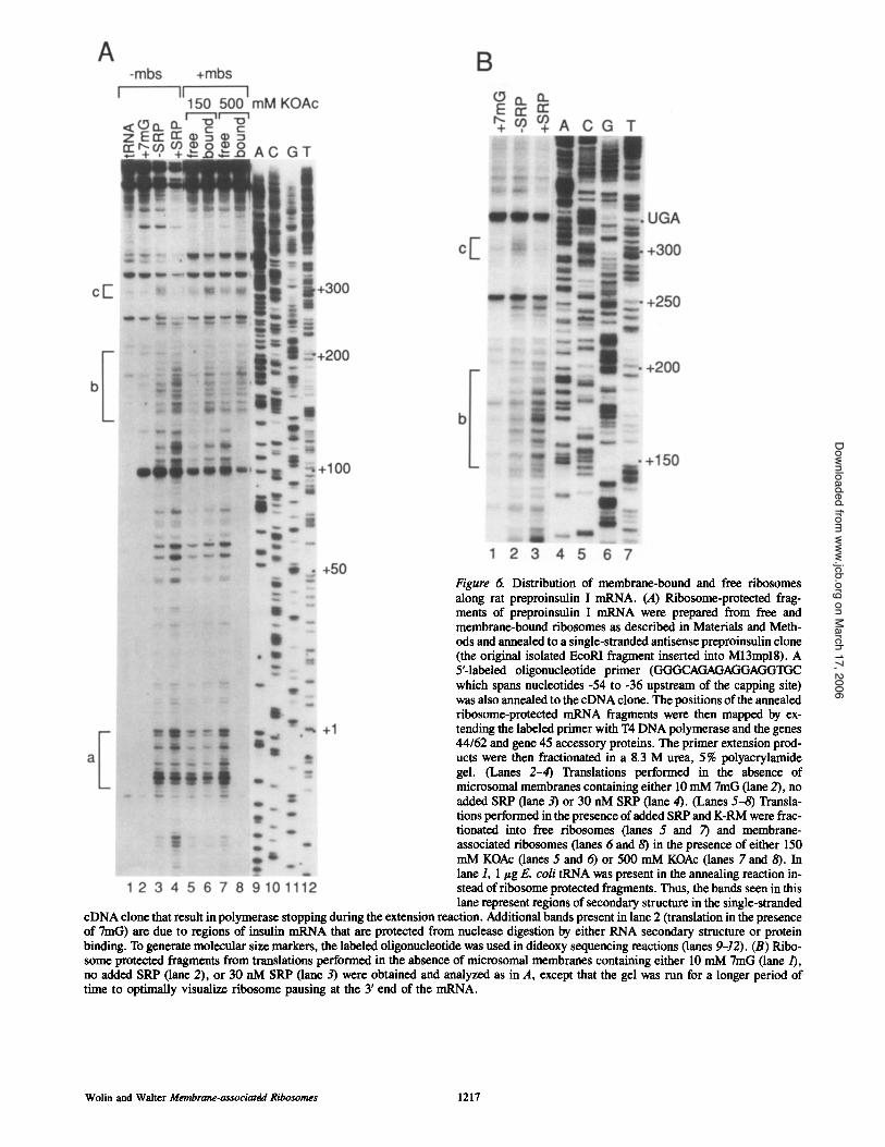
Figure 6. Distribution of membrane-bound and free ribosomes along rat preproinsulin I mRNA. (A) Ribosome-protected frag- ments of preproinsulin I mRNA were prepared from free and membrane-bound ribosomes as described in Materials and Meth- ods and annealed to a single-stranded antisense preproinsulin clone (the original isolated EcoRI fragment inserted into M13mpl8). A 5'-laboled oligonucleotide primer (GGCtCAGAGAGGAGG'IGC which spans nucleotides -54 to -36 upstream of the capping site) was also annealed to the cDNA clone. The positions of the annealed ribosome-protected mRNA fragments were then mapped by ex- tending the labeled primer with T4 DNA polymerase and the genes 44/62 and gene 45 accessory proteins. The primer extension prod- ucts were then fractionated in a 8.3 M urea, 5% polyacrylamide gel. (Lanes 2-4) Translations performed in the absence of microsomal membranes containing either 10 mM 7mG (lane 2), no added SRP (lane 3) or 30 nM SRP (lane 4). (Lanes 5-8) Transla- tions performed in the presence of added SRP and K-RM were frac- tionated into free ribosomes (lanes 5 and 7) and membrane- associated ribosomes (lanes 6 and 8) in the presence of either 150 mM KOAc (lanes 5 and 6) or 500 mM KOAc (lanes 7 and 8). In lane 1, 1 lzg E. coli tRNA was present in the annealing reaction in- stead of ribosome protected fragments. Thus, the bands seen in this lane represent regions of secondary structure in the single-stranded
cDNA clone that result in polymerase stopping during the extension reaction. Additional bands present in lane 2 (translation in the presence of 7mG) are due to regions of insulin mRNA that are protected from nuclease digestion by either RNA secondary structure or protein binding. To generate molecular size markers, the labeled oligonucleotide was used in dideoxy sequencing reactions (lanes 9-12). (B) Pdbo- some protected fragments from translations performed in the absence of microsomal membranes containing either 10 mM 7mG (lane/), no added SRP (lane 2), or 30 nM SRP (lane 3) were obtained and analyzed as in A, except that the gel was run for a longer period of time to optimally visualize ribosome pausing at the 3' end of the mRNA.
Wolin and Walter Membrane-associated Ribosomes 1217
on March 17, 2006
www.jcb.orgDownloaded from

(Fig. 6 A, compare lane 3 with 4), indicating that, as was pre- viously found for preprolactin, SRP arrests translation at the natural pause sites of ribosomes. This is also consistent with a mathematical model proposed by Rapoport et ai. (1987), in which they predicted that the major sites of interaction be- tween SRP and ribosomes would be at the natural pause sites of ribosomes.
When membranes are added to the translation reaction, and ribosomes bound to membranes are fractionated in 150 mM KOAc, approximately half of the ribosomes that pause over the initiation codon are retained on membranes, pre- sumably because they were targeted to the membrane as part of polysomes containing longer nascent chains (Figure 6 A, lanes 5 and 6, pause region a). In addition, ,o70% of the ribosomes that have advanced further down the mRNA re- main bound to the membrane (Fig. 6 A, compare lane 5 with 6, pause regions b and c). When the fractionation is per- formed at 500 mM KOAc, all initiating ribosomes are found in the free ribosome fraction, as expected (Fig. 6 A, lanes 7 and 8, pause region a). Only ribosomes pausing between nucleotide 144 and the termination codon at nucleotide 336 are retained on the membrane, indicating that the nascent chains larger than 52 amino acids have inserted into the membrane (Fig. 6 A, compare lane 7 with 8, pause regions b and c).
Discussion We have examined the distribution of membrane-bound ribo- somes during the translation of mRNAs encoding two secre- tory proteins, bovine preprolactin and rat preproinsulin I. This analysis has revealed that the previously described "loosely" and "tightly" bound populations of ribosomes cor- respond to ribosomes that have synthesized distinct lengths of the nascent polypeptide, and that nascent chain insertion into the membrane begins at distinct points for different presecretory proteins. In addition, we have shown that the enhancement of ribosome stacking that occurs when SRP ar- rests translation of these proteins is relaxed in the presence of microsomai membranes.
Insertion of Nascent Chains into Membranes Previously, membrane-bound ribosomes were divided into loosely and tightly bound fractions based on their sensitivity to extraction with either high salt or EDTA (Adelman et al., 1973; Harrison et ai., 1974; Gilmore and Blobel, 1985; Connolly and Gilmore, 1986). Connolly and Gilmore (1986) showed that the transition from loosely bound to tightly bound requires GTP, and that the loosely bound ribosomes probably represent an intermediate in the formation of a fight ribosome-membrane junction. This assertion is supported by our results which show that fight ribosome binding re- quires that the ribosome synthesize a nascent chain of a par- ticular length.
A number of laboratories have tried to define the shortest possible nascent chain that is capable of undergoing SRP- dependent membrane targeting and translocation. In all cases, the approach has been to prepare a series of synthetic mRNAs that produce truncated proteins of defined lengths, and then examine the ability of these short polypeptides to be targeted to or translocated across membranes in an SRP-
dependent fashion. Ibrahimi et al. (1986) determined that a 74 amino acid fragment of preprolysozyme was translo- cated, but that a 51 amino acid fragment failed to be translo- cated. Siegel and Waiter (1988b) concluded that nascent chains of bovine preprolactin of 87 amino acids in length were efficiently targeted to membranes, whereas ribosomes bearing nascent chains of 55 amino acids were not. Simi- larly, Okun et ai. (1990) found that ribosomes bearing na- scent chains of anglerfish preproinsulin containing 64 amino acids were translocated across membranes in an SRP- dependent manner, while ribosomes bearing nascent chains of 45 amino acids were not translocated. However, by sizing the nascent chains produced during SRP arrest of transla- tion, they concluded that SRP was able to arrest translation of anglerfish preproinsulin after only 50 amino acids had been synthesized. This result was in contrast to previous data regarding bovine preprolactin, in which the smallest SRP- arrested fragment is ,o70 amino acids in length, and trun- cated polypeptides containing only 55 amino acids are not targeted to or translocated across the membrane, implying that the portion of the preprolactin nascent chain emerging from the ribosome is not sufficient for recognition by SRP (Siegel and Waiter, 1988b).
Our results comparing bovine preprolactin and rat prepro- insulin I suggest that the shortest length of protein that can undergo SRP-dependent translocation and targeting will be different for different proteins. Some of the variability is likely due to differences in the length and structure of in- dividual signal peptides. Signal peptides can be divided into three distinct domains, an amino-terminai positively charged "11" region; a hydrophobic central region, and a more polar carboxyterminal domain (von Heijne, 1985). Preprolactin has one of the longest signal peptides known (31 amino acids) with much of the additional length occurring in the charged "n" region. Although the preproinsulin signal se- quence is shorter (24 amino acids), this length difference alone does not completely account for the difference in the size of inserted nascent chains, indicating that the structures formed by the individual signal peptides are likely to be a critical feature.
For a nascent chain to insert into the membrane, the poly- peptide must be sufficiently long to span the large ribosomal subunit (30-39 amino acids), and enough of the signal pep- tide must protrude to bind SRP. In addition, signal sequences are thought to insert into membranes as a loop, or helical hairpin, in which one of the two helices is formed by the sig- nal sequence (Engelman and Steitz, 1981; Kuhn, 1987; Shaw et al., 1988). Thus, enough of the nascent chain must pro- trude to allow the formation of both the first helix and the hairpin loop. If 30-39 amino acids span the ribosome large subunit, the 13-22 amino acids of preproinsulin that pro- trude from the ribosome will be barely long enough to form the first helix required to span a 30 A bilayer (15-20 amino acids) and begin a hairpin turn.
It was somewhat surprising that ribosomes with short or no nascent chains bind to the membrane in the presence of 150 mM KOAc (Fig. 3, lane 5; Fig. 6 A, lane 6), as these ribosomes cannot have been targeted to the membrane by SRP. As microsomai membranes contain binding sites for 80S ribosomes, it is possible that some of these ribosomes have not actually been targeted to the membrane, but bind nonspecificaily during the experimental manipulations. Al-
The Journal of Cell Biology, Volume 121, 1993 1218
on March 17, 2006
www.jcb.orgDownloaded from

ternatively, ribosomes that are targeted to the membrane as part of a larger polyribosome may normally bind "loosely" to the membrane. However, ribosome targeting to the mem- brane as part of a larger polyribosome is not sufficient to es- tablish a tight ribosome-membrane junction, as only those ribosomes that have translated a nascent chain of a particular length are resistant to extraction with 0.5 M KOAc.
SRP-induced Ribosome Pausing and Stacking We previously demonstrated that SRP-mediated transla- tional arrest of preprolactin results in increased ribosome pausing at what appear to be SRP-independent pause sites (Wolin and Walter, 1988). In contrast to preprolactin, which has only one strong position of ribosome pausing after the initiation codon, there are numerous natural pauses during the translation of rat preproinsulin I in the absence of SRP (compare Figs. 3 and 6). Thus, SRP does not arrest transla- tion of preproinsulin at a single site, but rather at multiple positions. This observation is likely to explain why a discrete arrested fragment has only been observed for a few secretory proteins (Walter and Blobel, 1981; Meyer et al., 1982). It seems likely that a discrete arrested fragment will only be observed when a single strong ribosomal pause site occurs immediately after the signal peptide is available for binding.
Because membrane-bound ribosomes appear evenly spaced along mRNA in most electron micrographs (Chris- tensen et al., 1987), it seemed likely that the SRP-induced stacking of ribosomes that we previously observed during translation of bovine preprolactin (Wolin and Walter, 1988) would be relaxed in the presence of membranes. We have found that this is the case when either rat preproinsulin I (Fig. 5 B) or bovine preprolactin (data not shown) is trans- lated in the presence of membranes. Ribosome pausing caused by SRP arrest of translation is reduced by the pres- ence of microsomal membranes, presumably because the ar- rest is released as SRP contacts the SRP receptor on the ER membrane.
We thank Jack Barry and Bruce Alberts (Department of Biochemistry, University of California, San Francisco), Bill Konigsberg (Department of Molecular Biophysics and Biochemistry, Yale University, New Haven, CT) and Maureen Munn (Department of Therapeutic Radiology, Yale University) for generously providing T4 DNA polymerase and polymerase accessory proteins. We also thank Argiris Efstradiatis (Columbia Univer- sity, NY) for providing the partial rat insulinoma eDNA and eDNA library. We are grateful to Mark Solomon and Carl Hashimoto for their comments on the manuscript.
When this work was initiated, S. L. Wolin was a LuciUe P. Markey scholar and this work was supported in part by a grant from the Lucille P. Markey Charitable Trust. This work was also supported by grants from the National Institutes of Health to P. Walter.
Received for publication 27 January 1993 and in revised form 31 March 1993.
References
Adelman, M. R., D. D. Sabatini, and G. Blobel. 1973. Ribosome-membrane interaction. Nondestructive disassembly of rat liver rough microsomes into ribosomal and membranous components. J. Cell Biol. 56:206-229.
Christensen, A. K., L. E. Kalm, and C. M. Bourne. 1987. Circular polysomes
predominate on the rough endoplasmic reticulum of somatotropes and mam- motropcs in the rat anterior pituitary. Am. J. Anat. 178:1-10.
Connolly, T., and R. Gilmore. 1986. Formation of a functional ribosome- membrane junction during translocation requires the participation of a GTP- binding protein. J. Cell Biol. 103:2253-2261.
Doohan, J. P., and C. E. Samuel. 1992. Biosynthesis of reovirus-specified poly- peptides: ribosome pausing during the translation of reovirus S1 mRNA. Virology. 186:409-425.
Engelman, D. M., and T. A. Steitz. 1981. The spontaneous insertion of proteins into and across membranes: the helical hairpin hypothesis. Cell. 23: 411-422.
Erickson, A. H., and G. Blobol. 1983. C e l l - ~ translation of messenger RNA in a wheat germ system. Methods Enzymol. 96:38-50.
Eskridge, E. M., and D. Shields. 1983. Cell-free processing and segregation of insulin precursors. J. Biol. Chem. 258:11487-11491.
Gilmore, R., and G. Blobel. 1985. Translecation of secretory proteins across the microsomal membrane occurs through an environment accessible to aqueous perturbants. Cell. 42:497-505.
Harrison, T. M., G. G. Browniee, and C. Milstein. 1974. Studies on polysome- membrane interactions in mouse myeloma cells. Eur. J. Biochem. 47: 613-620.
Ibrahimi, I., D. Cutler, D. Steubcr, and H. Bujard. 1986. Determinants for pro- tein translocation across mammalian endoplasmic reticulum. Membrane in- sertion of truncated and full-length prelysozyme molecules. Eur. J. Bio- chem. 155:571-576.
Kuhn, A. 1987. Bacteriophage MI3 procoat protein inserts into the plasma membrane as a loop structure. Science (Wash. DC). 238:1413-1415.
Lingelbach, K., C. Zwieb, J. R. Webb, C. Marshailsay, P. J. Hoben, P. Walter, and B. Dobberstein. 1988. Isolation and characterization of a eDNA clone encoding the 19 kDa protein of signal recognition particle (SRP): ex- pression and binding to 7SL RNA. Nucleic Acids Res. 16:9431-9442.
Meyer, D. I., E. Krause, and B. Dobberstein. 1982. Secretory protein translo- cation across membranes- the role of the "docking protein ~. Nature (Lond.). 297:647-650.
Murphy, A. J. M., and A. Efstradiatis. 1987. Cloning vectors for expression of eDNA libraries in mammalian cells. Proc. Natl. Acad. Sci. USA. 84: 8377-8381.
Okun, M. M., E. M. Eskridge, and D. Shields. 1990. Truncations of a secre- tory protein define minimum lengths required for binding to signal recogni- tion particle and translocation across the endoplasmic reticulum membrane. J. Biol. Chem. 265:7478-7484.
Rapoport, T. A. 1992. Transport of proteins across the endoplasmic reticulum membrane. Science (Wash. DC). 258:931-936.
Rapoport, T. A., R. Heinrich, P. Walter, and T. Schulmeister. 1987. Mathe- matical modeling of the effects of signal recognition particle on translation and translocation of proteins across the endoplasmic reticulum membrane. J. Mol. Biol. 195:621-636.
Sanders, S. L., and R. Schekman. 1992. Polypeptide translocation across the endoplasmic reticulum membrane. J. Biol. Chem. 267:13791-13794.
Shaw, A. S., P. J. M. Rottier, and J. K. Rose. 1988. Evidence for the loop model of signal-sequence insertion into the endoplasmic reticulum. Proc. Natl. Acad. Sci. USA. 85:7592-7596.
Siegel, V., and P. Walter. 1988a. Each of the activities of signal recognition particle (SRP) is contained within a distinct domain: analysis of biochemical mutants of SRP. Cell. 52:39-49.
Siegel, V., and P. Walter. 1988b. The affinity of signal recognition particle for presecretory proteins is dependent on nascent chain length. EMBO (Eur. Mol. Biol. Organ.)J. 7:1769-1775.
Soares, M. B., E. Schon, A. Henderson, S. K. Karathanasis, R. Cate, S. Zeit- lin, J. Chirgwin, and A. Efstradiatis. 1985. RNA-mediated gene duplication: the rat preproinsulin I gene is a functional retroposon. Mol. Cell. Biol. 5:2090-2103.
von Heijne, G. 1985. Signal sequences. The limits of variation. J. Mol. Biol. 184:99-105.
Walter, P., and G. Blobel. 1981. Translocation of proteins across the endoplas- mic reticulum HI. Signal recognition protein (SRP) causes signal sequence- dependent and site-specific arrest of chain elongation that is released by microsomai membranes. J. Cell Biol. 91:557-561.
Walter, P., and G. Blob¢l. 1983a. Preparation of microsomal membranes for cotranslational protein translocation. Methods Enzymol. 96:682-691.
Walter, P., and G. Blobel. 1983b. Signal recognition particle: a ribonucleopro- rein required for cotranslational translocation of proteins. Isolation and prop- erties. Methods Enzymol. 96:682-691.
Wolin, S. L., and P. Walter. 1988. Ribosome pausing and stacking during translation of a eukaryotic mRNA. EMBO (Eur. Mol. Biol. Organ.) J. 7:3559-3569.
Wolin, S. L., and P. Walter. 1989. Signal recognition particle mediates a tran- sient elongation arrest of preprolactin in reticulocyte lysate. J. Cell Biol. 109:2617-2622.
Wolin and Walter Membrane-associated Ribosomes 1219
on March 17, 2006
www.jcb.orgDownloaded from