dural lesions in decompression for lumbar spinal stenosis...
TRANSCRIPT

LUND UNIVERSITY
PO Box 117221 00 Lund+46 46-222 00 00
Dural lesions in decompression for lumbar spinal stenosis: incidence, risk factors andeffect on outcome.
Strömqvist, Fredrik; Jönsson, Bo; Strömqvist, Björn
Published in:European Spine Journal
DOI:10.1007/s00586-011-2101-2
Published: 2012-01-01
Link to publication
Citation for published version (APA):STRÖMQVIST, F. R. E. D. R. I. K., Jönsson, B., & Strömqvist, B. (2012). Dural lesions in decompression forlumbar spinal stenosis: incidence, risk factors and effect on outcome. European Spine Journal, 21(5), 825-828.DOI: 10.1007/s00586-011-2101-2
General rightsCopyright and moral rights for the publications made accessible in the public portal are retained by the authorsand/or other copyright owners and it is a condition of accessing publications that users recognise and abide by thelegal requirements associated with these rights.
• Users may download and print one copy of any publication from the public portal for the purpose of privatestudy or research. • You may not further distribute the material or use it for any profit-making activity or commercial gain • You may freely distribute the URL identifying the publication in the public portal

Take down policyIf you believe that this document breaches copyright please contact us providing details, and we will removeaccess to the work immediately and investigate your claim.
Download date: 09. Jul. 2018

DURAL LESIONS IN DECOMPRESSION FOR LUMBAR SPINAL STENOSIS –
INCIDENCE, RISK FACTORS AND EFFECT ON OUTCOME
Strömqvist Fredrik MD, Jönsson Bo MD, PhD, Strömqvist Björn MD, PhD
Swedish Society of Spinal Surgeons
Clinical Sciences Lund, Lund University, Lund University Hospital, Dept of Orthopedics, SE-
221 85 Lund

2
Abstract
Decompression for lumbar spinal stenosis is one of the most frequent operations on the spine
today. The most common complication seems to be a peroperative dural lesion. There are few
prospective studies on this complication regarding incidence and effect on long-term
outcome; this is the background for the current study.
Swespine, the Swedish Spine Register documents the majority (>80 %) of lumbar spine
operations in Sweden today. Within the framework of this register, totally 3 699 operations
for spinal stenosis during a five-year period were studied regarding complications and one-
year postoperative outcome. Mean patient age was 66 (37 – 92) years and 44 % were males.
Fourteen percent were smokers and 19 % had undergone previous lumbar spine surgery.
The overall incidence of a peroperative dural lesion was 7.4 %, 8.5 % of patients undergoing
decompressive surgery only and 5.5 % of patients undergoing decompressive surgery + fusion
(p < 0.001). A logistic regression analysis demonstrated that (high) age (p < 0.0004), previous
surgery (p < 0.036) and smoking (p < 0.049) were significantly predictive factors for dural
lesions. An odds ratio estimate demonstrated an age-related risk increase with 2.7 % per year.
The risk for dural lesions also increased with number of levels decompressed. The one-year
outcome was identical in the two groups with and without a dural lesion.
To conclude, a dural lesion was seen in 7.4 % of decompressive operations for spinal stenosis.
High age, previous surgery and smoking were risk factors for sustaining a lesion, which,
however, did not affect the one-year outcome negatively.
Keywords:
Spinal stenosis, complication, surgery, outcome
Introduction
Decompressive procedures for lumbar spinal stenosis are by far the most common operations
on the spine in elderly patients, and, seem to be gradually overtaking the overall dominant
role of disc herniation surgery. It should be regarded as the golden standard for surgical
treatment of spinal stenosis today and improves pain, walking ability and spine-related
disability (ODI) on a group level, although patient satisfaction seem to be achieved in
between 65 to 80 % of the cases [18, 1], and the results seem to deteriorate with time [9].

3
Whereas complications in the form of infection and nerve/cauda equina injury seem to be
infrequent, a non-negligible incidence of dural lesions in this type of surgery seems
unavoidable. Incidence figures on dural lesions in spinal stenosis surgery in the literature
mainly refer to retrospective studies of the complication with the drawbacks of retrospectivity
hampering the studies. In the large prospective SPORT study an incidence of 5 % is presented
[21]. Whether or not a dural lesion peroperatively is a predictor of inferior outcome of surgery
is also subject to some controversy [6, 19, 3].
A prospective study such as Swespine, the Swedish Spine Register, has the potential of a large
patient material and prospectivity, thereby improving the data on incidence, risk factors and
outcome. Swespine documents more than 80 % of degenerative lumbar spine surgery in
Sweden since 10 years and also includes complication registration.
The purpose of the study presented, thus, was to elucidate the incidence of dural lesions in
decompressive surgery for lumbar spinal stenosis and to identify risk factors and effect on
postoperative outcome of surgery.
Patients and methods
During the study period 3 699 patients were operated on with decompression for lumbar
spinal stenosis. Only patients operated on for central stenosis with or without root canal
stenosis were included, isolated lateral spinal stenosis was not included. Decompressive
surgery as the only procedure was performed in 2 764 patients (74 %) and the remaining 935
also had a concomitant fusion performed at the time of decompressive surgery (26 %).
Fourteen percent of the patients were smokers and 19 % had undergone previous lumbar spine
surgery. One-year follow-up was completed by 2 875 of the patients (78 %).
Swespine, the Swedish Spine Register has been presented elsewhere [14, 15] and contains
patient based pre- and postoperative data and surgeon based surgical data.
Among baseline data are included age, sex, smoking habits, working conditions, consumption
of analgesics, walking distance, back and leg pain on the VAS scale and the Oswestry
Disablity Index, the SF-36 and the EQ-5D questionnaires. These data are completed by the
patients preoperatively and at postoperative intervals. Postoperatively also global satisfaction

4
with outcome as well as improvement of leg and back pain is graded. As a control, also
patient-reported complications are included.
Statistical analysis: All data were entered into the SAS statistical program (version 9.2).
Logistics regression was used to estimate odds ratio and its 95% confidence interval and p-
value. For outcome comparisons the Student’s t-test was used.
Results
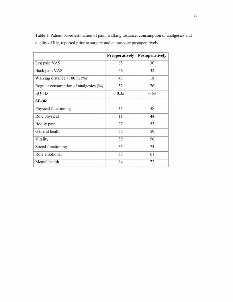
The patients operated on for spinal stenosis had a high level of pain, low quality of life and
low function as measured by walking distance, factors which were all reversed after surgery
(Table 1).
The overall incidence of a peroperative dural lesion was 7.5 %. Patients undergoing
decompressive surgery only had an incidence of 8.5 % as compared to 5.5 % for patients
undergoing decompressive surgery + fusion (p < 0.001). The logistic regression analysis
(Table 2) demonstrated high age, previous surgery and smoking to be risk factors for
sustaining a dural lesion at surgery. At incremental age, a risk increase according to odds ratio
calculation increased by 2.7 % per year of life. For patients aged ≤70 years, the risk for
sustaining a lesion was significantly lower than for the age group >80 years (Table 3). The
risk for the younger patients was 40 (51-60 years) to 56 (61-70 years) percent of that of the
oldest patient group (≥80 years).
The incidence of dural lesions increased with number of levels decompressed from 5.1 % in
one-level decompression to 11.5 % when 4 or more levels were decompressed (Table 4).
At one year after surgery, a significant improvement of back and leg pain, EQ-5D and SF-36
scores was seen (Table 1). In no aspects of the patient based outcome parameters was there
any significant difference in outcome between patients with and without a dural lesion.
Lost to follow-up: For the 22 % of patients who failed to complete the one-year follow-up
questionnaire, neither baseline data nor incidence of peroperative dural lesion differed from
the studied group of 2 875 patients reported.
Discussion

5
It goes without saying that operations within the spinal canal may entail a risk to injure nerve
structures and the dural sac. The improved information preoperatively on the contents of the
spinal canal using MRI gives a good possibility to be prepared for where troubles may arise
during surgery. For spinal stenosis, open, microscopic and also endoscopic techniques have
been developed but only to a limited extent compared [17].
Operations for lumbar spine stenosis are the most common spine operations in the elderly but
the trend of increasing surgery rates noted in Sweden [15] does not seem to be prevalent in
the US [5]. Decompression is the golden standard when surgical treatment is indicated, at
times supplemented with fusion, especially to be considered in spondylolisthesis. Fusion rates
in conjunction with decompression seem to vary a lot over the world and also seem to be
afflicted with higher complication rates [4].
Complication rates in general and dural lesions in particular have been to some extent
sparsely documented in the literature and the complication rates reported probably are
minimum figures. A minor dural lesion noted during surgery, adequately sutured and treated
with a day of bed rest postoperatively is no major issue but in the other end of the spectrum
complication problems such as dural fistulas and cysts, meningitis, arachoiditis and epidural
abscesses can occur. In addition to direct closure by sutures, also fascial, muscular and
artificial grafts exist, further fibrin glue, and, another possibility, sub arachnoid drainage also
may be utilized [10]. Some conflict regarding the long-term outcome after dural lesion exists
[11, 12].
Incidence figures for dural lesions in disc surgeries seem to be in the region of 2 – 6 % [16,
13, 20] with previous surgery being a strong predictor of the complication. In spinal stenosis
surgery higher figures should be expected to be encountered due to the wider exposure of the
dural sac and to the difficulties created by ligamentus hypertrophy and osteophytes on the
facet joints in decompression especially afflicting the nerve roots but also the central cauda.
Most previous studies on dural lesions refer to spinal surgery in general [6, 19, 8, 12] but a
large series from the Scoliosis Research Society [7] demonstrated an incidence of dural
lesions of 3 % but in this study patients previously operated on in the lumbar spine were
excluded. Incidence figures reported must be regarded as minimum figures but most likely are
more correct in prospective than in retrospective studies.

6
The fact that high age and smoking are risk factors for dural injury may indicate that the
strength and elasticity of the dural sac becomes reduced with increasing age and by smoking.
In stenosis surgery previous surgery seems to be a risk factor for dural lesion and with an
increasing number of procedures performed yearly, an increasing number of dural lesions has
to be anticipated in the future. Our study strongly suggests that the long-term results of
decompression in patients sustaining dural lesion are not inferior to those without a
peroperative dural lesion which means that basically a dural lesion is a problem that has to be
solved at time of surgery and if this is adequately carried out the patient will do as well as a
patient without this complication.
The finding in our study of dural lesions being less frequent in patients treated with
concomitant fusion seems to relate to the fact that this patient group is somewhat younger and
is operated on fewer levels. High age, smoking and previous surgery all were identified as risk
factors for sustaining a peroperative lesion. It seems mandatory that decompressive surgery
for spinal stenosis is performed under the best circumstances with good light sources, loupe
magnification or microscopic visualization of the operating field. Further, a surgeon prepared
with maximum study of the preoperative MRI or CT images is probably less prone to run into
trouble during surgery.
The after-treatment when the dural lesion has occurred is usually arbitrarily defined as one
day of bed rest before mobilization. The rather slow healing of dural repair in a canine model
presented by Cain et al 1991 [2] may be interpreted as requiring longer bed rest than 24 hours
but from a clinical perspective normally this time period seems enough.
In conclusion, spinal stenosis surgery when studied in a large patient material from Swespine,
the Swedish Spine Register, demonstrated an incidence of dural lesions during surgery of 7.4
%, somewhat less frequent when decompression was combined with fusion. Risk factors for
sustaining a lesion using a logistic regression model were high age, previous surgery and
smoking. The incidence also increased with number of levels decompressed from 5.1 % in
one-level decompression to 11.5 % when four or more levels were decompressed. The one-
year outcome was not affected negatively in the patient group who sustained a peroperative
lesion.

7
Acknowledgements
The authors highly appreciate statistical help and advice given by Caddie Zhou at the National
Centre for Quality Registers, Lund Sweden. The economical funding by the Swedish
Association of Local Authorities and Regions (SALAR) is also acknowledged.

8
References
1. Atlas SJ, Keller RB, Wu YA, Deyo RA, Singer DE (2005) Long-term outcomes of
surgical and nonsurgical management of lumbar spinal stenosis: 8 to 10 year results
from the maine lumbar spine study. Spine 30(8): 936-943.
2. Cain JE Jr, Lauermann WC, Rosenthal HG, Broom MJ, Jacobs RR (1991) The
histomorphologic sequence of dural repair. Observations in the canine model. Spine
16(Suppl): S319-323.
3. Cammisa FP, Girardi FP, Sangani PK, Parvataneni HK, Cadag S, Sandhu HS (2000)
Incidental durotomy in spine surgery. Spine 25(20): 2663-2667.
4. Deyo RA, Cherkin DC, Loeser JD, Bigos SJ, Ciol MA (1992) Morbidity and mortality
in association with operations on the lumbar spine: the influence of age, diagnosis, and
procedure. J Bone Joint Surg Am 74-A: 536-543.
5. Deyo RA, Mirza SK, Martin BI, Kreuter W, Goodman DC, Jarvik JG (2011) Trends,
major medical complications, and charges associated with surgery for lumbar spinal
stenosis in older adults. JAMA 303(13):1259-1265.
6. Eismont FJ, Wiesel SW, Rothman RH (1981) Treatment of dural tears associated with
spinal surgery. J Bone Joint Surg Am 63-A: 1132-1136.
7. Fu KMG, Smith JS, Polly DW Jr, Perra JH, Sansur CA, Berven SH, Broadstone PA,
Choma TJ, Goytan MJ, Noordeen HH, Knapp DR Jr, Hart RA, Zeller RD, Donaldson
III WF, Boachie-Adjei O, Shaffrey CI (2010) Morbidity and mortality in the surgical
treatment of 10,329 adults with degenerative lumbar stenosis. J Neurosurg Spine 12:
443-446.
8. Jones AA, Stambough JL, Balderston RA, Rothman RH, Booth RE (1989) Long-term
results of lumbar spine surgery complicated by unintended incidental durotomy. Spine
14(4): 443-446.

9
9. Jönsson B, Sjöberg C, Annertz M, Strömqvist B (1997) A prospective and consecutive
study of surgically treated lumbar spinal stenosis. Part II: Five-year follow-up by an
independent observer. Spine 22(24): 2938-2944.
10. Kitchel SH, Eismont FJ, Green BA (1989) Closed subarachnoid drainage for
management of cerebrospinal fluid leakage after an operation on the spine. J Bone
Joint Surg Am 71-A: 984-987.
11. Sin AH, Caldito G, Smith D, Rashidi M, Willis B, Nanda A (2006) Predictive factors
for dural tear and cerebrospinal fluid leakage in patients undergoing lumbar surgery. J
Neurosurg Spine 5: 224-227.
12. Stewart G, Sachs BL (1996) Patient outcomes after reoperation on the lumbar spine. J
Bone Joint Surg Am 78-A: 706-711.
13. Stolke D, Sollman WP, Seifert V (1989) Intra- and postoperative complications in
lumbar disc surgery. Spine 14: 56-59.
14. Strömqvist B, Fritzell P, Hägg O, Jönsson B (2005) One-year report from the Swedish
National Spine Register. Swedish Society of Spinal Surgeons. Acta Orthop 76(Suppl
319): 1-24.
15. Strömqvist B, Fritzell P, Hägg O, Jönsson B. Swedish Society of Spinal Surgeons
(2009) The Swedish Spine Register: development, design and utility. Eur Spine J:
18(Suppl 3): S294-S304.
16. Strömqvist F, Jönsson B, Strömqvist B (2010) Dural lesions in lumbar disc herniation
surgery: incidence, risk factors, and outcome. Eur Spine J 19: 439-442.
17. Thomé C, Zevgaridis D, Leheta O, Bäzner H, Pöckler-Schöniger C, Wöhrle J,
Schmiedek P (2005) Outcome after less-invasive decompression of lumbar spinal
stenosis: a randomized comparison of unilateral laminotomy, bilateral laminotomy,
and laminectomy. J Neurosurg: Spine 3: 129-141.

10
18. Turner JA, Ersek M, Herron L, Deyo R (1992) Surgery for lumbar spinal stenosis.
Attempted meta-analysis of the literature. Spine 17: 1-8
19. Wang JC, Bohlman HH, Riew KD (1998) Dural tears secondary to operations on the
lumbar spine. Management and results after a two-year-minimum follow-up of eighty-
eight patients. J Bone Joint Surg Am 80-A: 1728-32.
20. Weinstein JN, Tosteson TD, Lurie JD, Tosteson AN, Hanscom B, Skinner JS, Abdu
WA, Hilibrand AS, Boden SD, Deyo RA (2006) Surgical vs nonoperative treatment
for lumbar disk herniation: the Spine Patient Outcomes Research Trial (SPORT): a
randomized trial. JAMA 296:2441-2450.
21. Weinstein JN, Tosteson TD, Lurie JD, Tosteson AN, Blood E, Hanscom B, Herkowitz
H, Cammisa F, Albert T, Boden SD, Hilibrand A, Goldberg H, Berven S, An H
SPORT investigators (2008) Surgical versus nonsurgical therapy for lumbar spinal
stenosis. New Eng J Med 358(8): 794-810.
11
Table 1. Patient based estimation of pain, walking distance, consumption of analgesics and
quality of life, reported prior to surgery and at one-year postoperatively.
Preoperatively Postoperatively
Leg pain VAS 63 30
Back pain VAS 56 32
Walking distance <100 m (%) 43 18
Regular consumption of analgesics (%) 52 26
EQ-5D 0.35 0.63
SF-36:
Physical functioning 35 58
Role physical 11 44
Bodily pain 27 53
General health 57 59
Vitality 39 56
Social functioning 55 74
Role emotional 37 61
Mental health 64 72

12
Table 2. Logistic regression analysis of patient related risk factors for dural lesion. Odds ratio
with 95 % confidence interval is given as well as the p-value.
Logistic regression
P-value Odds ratio 95% CI
Age 0.0004 1.027 1.012-1.043
Previous op 0.036 0.699 0.499-0.978
Smoking 0.049 0.696 0.485-0.999
Gender 0.33 1.013 0.652-1.157
Leg p duration 0.87 1.013 0.862-1.191
LBP duration 0.40 1.072 0.909-1.265
Walking ability 0.72 1.124 0.845-1.124
VAS Leg 0.95 1.000 0.993-1.006
VAS Back 0.25 0.996 0.990-1.003

13
Table 3. Age related risk for dural lesion. The figures given in the table relate the Odds ratio
and the p-value for each age group when compared with patients >80 years of age. The Odds
ratio .40 means that patients aged 51-60 years have a 40 % risk of that of patients aged over
80 years.
Odds ratio P-value
≤ 50 0.48 0.053
51-60 0.40 0.002
61-70 0.56 0.027
71-80 0.83 0.455
Table 4. Incidence of dural lesion related to number of levels operated on (%).
Percent
One level 5.1
Two levels 7.7
Three levels 7.6
≥ 4 levels 11.5