environmental/lifestyle factors in the ... - bmc medicine
TRANSCRIPT
REVIEW Open Access
Environmental/lifestyle factors in thepathogenesis and prevention of type 2diabetesHubert Kolb1,2* and Stephan Martin1,2
Abstract
Background: Environmental and lifestyle changes, in addition to the ageing of populations, are generally believedto account for the rapid global increase in type 2 diabetes prevalence and incidence in recent decades.
Discussion: In this review, we present a comprehensive overview of factors contributing to diabetes risk, includingaspects of diet quality and quantity, little physical activity, increased monitor viewing time or sitting in general,exposure to noise or fine dust, short or disturbed sleep, smoking, stress and depression, and a low socioeconomicstatus. In general, these factors promote an increase in body mass index. Since loss of β-cell function is the ultimatecause of developing overt type 2 diabetes, environmental and lifestyle changes must have resulted in a higher riskof β-cell damage in those at genetic risk. Multiple mechanistic pathways may come into play.
Conclusions: Strategies of diabetes prevention should aim at promoting a ‘diabetes-protective lifestyle’ whilstsimultaneously enhancing the resistance of the human organism to pro-diabetic environmental and lifestyle factors.More research on diabetes-protective mechanisms seems warranted.
Keywords: Type 2 diabetes, Environment, Lifestyle, Diabetes risk factors, Diabetes prevention, Diet, Physical activity, β-cells
BackgroundOver the past decades, there has been a major increasein type 2 diabetes (T2D) prevalence in most regions ofthe world [1]. After adjusting for the impact of ageingpopulations, diabetes prevalence in adults (85–95%T2D) almost doubled between 1980 and 2014 world-wide. Increases were more pronounced in low- andmiddle-income countries and in men compared towomen [1].Recognition of the environmental and lifestyle fac-
tors responsible for these changes in theory may leadto the development of strategies to decrease the num-ber of new cases to reach those of 20–40 years earl-ier. This review presents the current state ofknowledge and discusses the possible mechanisms in-volved and the consequences for strategies of diabetesprevention.
Overview of environmental and lifestyle factorsincreasing T2D riskIt is generally believed that an energy-dense Western stylediet in conjunction with a sedentary lifestyle are theprimary cause of T2D [2]. These two factors are also heldresponsible for the current global epidemic of obesity,which is closely associated with the rising rate of T2D [3].At closer analysis, a high body mass index (BMI) appearsto contribute less to an increased risk of T2D than thepresence of increased visceral obesity [4] and/or ectopicfat (liver fat) [5–7]. This fits with the observation thatobese people without metabolic dysregulation have littlevisceral obesity or liver fat [8–10]. Conversely, people whodevelop T2D despite being merely overweight or within anormal weight range, such as in Asia, exhibit visceralobesity and ectopic fat deposition and reduced musclemass, together resulting in a normal or near normal BMI[11–13]. Interestingly, a substantial age-corrected rise ofT2D cases in recent decades was also seen in countrieswith no major change in the availability of food such as inWestern Europe [1]. This suggests that additional environ-mental and lifestyle factors contributed to the increased
* Correspondence: [email protected] of Medicine, University of Duesseldorf, Duesseldorf, Germany2West-German Centre of Diabetes and Health, Duesseldorf Catholic HospitalGroup, Hohensandweg 37, 40591 Duesseldorf, Germany
Diabetes: prevention, management and treatment
© The Author(s). 2017 Open Access This article is distributed under the terms of the Creative Commons Attribution 4.0International License (http://creativecommons.org/licenses/by/4.0/), which permits unrestricted use, distribution, andreproduction in any medium, provided you give appropriate credit to the original author(s) and the source, provide a link tothe Creative Commons license, and indicate if changes were made. The Creative Commons Public Domain Dedication waiver(http://creativecommons.org/publicdomain/zero/1.0/) applies to the data made available in this article, unless otherwise stated.
Kolb and Martin BMC Medicine (2017) 15:131 DOI 10.1186/s12916-017-0901-x
risk of T2D. A list of factors associated with risk of T2D isshown in Box 1.
DietWhen considering the wide range of diet types consumedin different regions of the world, it may not be surprisingthat prospective epidemiological studies vary somewhat inthe association of food groups with incident T2D. In gen-eral, plant food is associated with lower T2D risk thanmeat, low energy density food is considered more protect-ive than high density energy food, associations of fish con-sumption with diabetes risk are variable, and fermenteddairy products may be more beneficial than non-fermentedones. Further, refined grains or sugar-sweetened beveragesconsistently appear to promote obesity and diabetes risk[14–24]. Daily consumption of a handful of nuts may affordsome protection from T2D, despite nuts representing ahigh energy density food [25]. Unfortunately, epidemio-logical studies cannot exclude the impact of confounding
factors such as those of physical activity, which is difficultto assess in queries or interviews.The most recent recommendation by the Government
of the USA no longer focusses on setting limits for theamount of carbohydrates, fat and protein in foods butrather proposes food types or patterns such as a healthyUS-style eating pattern, a Mediterranean diet or avegetarian diet [26].In order to test for cause-effect relationships, the effect of
a given diet on metabolic control has been studiedextensively in controlled trials with the assumption thatshort-term effects may indicate long-term outcomes. In theresults published, most of the diets assessed were reportedto improve metabolic control and lipid status, regardless ofwhether they were high or low in carbohydrates, fats orprotein [27–31]. In many of these studies, given the un-avoidable study effect (Hawthorne effect) on eating behav-iour, participants consumed less calories than before thetrial, at least during the first weeks, and therefore lostweight. In other trials, a hypocaloric diet was given to allstudy groups [31]. As a consequence, weight reduction(and concomitantly less visceral obesity) probably led to im-proved metabolic control, largely independent of the diettried. Hence, the outcome of many dietary trials may giverise to misleading conclusions on the suitability of a diet fordiabetes prevention in the long term.Unfortunately, long-term trials of selected food groups
for diabetes prevention in persons at risk are difficult toperform due to poor compliance. In one successful attempt,a Mediterranean diet supplemented with either 1 L ofvirgin olive oil per week or 30 g nuts per day wascompared, in a randomised trial, with a conventional lowfat diet [32]. After 4 years, both Mediterranean diet groupsexhibited a rate of progression to T2D of approximately50% of the control low fat diet group (PREDIMED trial,group size approximately 140 persons, mean age 67 years,high cardiovascular disease risk). In this trial, calorierestriction was not intended and, on average, there wasless than 1 kg of body weight loss per person, despite themajor reduction of diabetes risk over 4 years. Thissuggests that there are components in the Mediterraneandiet or the overall pattern that may decrease diabetes riskwithout the need of weight reduction. Confirmation ofthese data in a larger trial including also younger personswithout high cardiovascular disease risk would offer arobust basis for a diabetes prevention guideline. Therehave been many trials of lifestyle improvement for theprevention of T2D in persons at increased risk, but thesedo not offer advice on a ‘diabetes-protective’ diet becausedietary guidelines have always been combined with astrategy to reduce body weight [33, 34].Can we learn from the diet of centenarians? Data for
Okinawa, Sicily, Sardinia, Linda Loma (California, USA)and Bama county (China) suggest some similarity to the
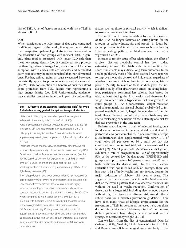
Box 1. Lifestyle characteristics conferring riska for type2 diabetes as suggested by epidemiological studies
Diets poor in fibre, phytochemicals or plant food in general
(relative risk increase by 44% to three-fold [14, 15])
Regular consumption of sugar-sweetened beverages (relative risk
increase by 20–30% compared to non-consumption [22–24])
Little physical activity (leisure time/occupational) (relative risk
approximately 40% higher compared to high total physical activity
[41, 42])
Prolonged TV and monitor viewing/sedentary time (relative risk
increased by approximately 3% per hour television watching [49])
Exposure to road traffic (noise, fine particulate matter) (relative
risk increased by 20–40% for exposure to 10 dB higher noise
level or 10 μg/m3 more of fine dust particles [55–59])
Smoking (relative risk increased by approximately 30%/60% for
light/heavy smokers [85])
Short sleep duration and poor quality (relative risk increased by
approximately 9% for every hour of shorter sleep duration [63])
Low mood/stress/depression (relative risk increase highly
variable, depending on definition of stress and depression)
Low socioeconomic position (relative risk increased by 40–100%
when compared to high socioeconomic level [99–101])
Infection with hepatitis C virus or Chlamydia pneumoniae (no
epidemiological data on relative risk increase available)aAll factors remain significantly associated after statistical
adjustment for body mass index (BMI) and other confounders,
as described in the text. Virtually all non-infectious pro-diabetic
lifestyle characteristics promote an increase in BMI and waist
circumference.
Kolb and Martin BMC Medicine (2017) 15:131 Page 2 of 11

above conclusions from epidemiological studies of a‘healthy’ diet, i.e. a dominance of vegetables and fruit overanimal food, and of whole grains over refined carbohy-drates [35–38]. However, it should not be overlooked thatthere is a substantial contribution from other factors tohealthy ageing, such as genetic background, epigeneticDNA methylation status, physical activity or daily workuntil high age, an active social network, and low smokingrates and alcohol consumption [39, 40].
Occupational and leisure time physical activityEpidemiological studies suggest that high versus low totalphysical activity is associated with a reduction in relativediabetes risk by approximately 30%. All types of leisure timephysical activities as well as occupational physical activitywere found to be inversely associated with diabetes risk [41,42]. The beneficial effect of exercise on insulin sensitivityand glycaemic control (by continuous glucose measure-ment) has also been demonstrated in controlled trials innon-diabetic individuals [43, 44]. Reallocation of 30 min ofsedentary time into moderate to vigorous physical activitywas associated with a 15% difference in HOMA-defined in-sulin sensitivity [45]. The beneficial effects of muscle workdo not simply reflect the burning of calories, since en-hanced physical activity leads to minimal weight loss [46].
Watching TV or sedentary timeThere is a strong association between sedentary time (self-reported or objectively measured) with obesity or incidentdiabetes, independent of the extent of physical activity[47–51]. Increased duration of sedentary behaviour maydouble diabetes risk [47]. In one study, each hour oftelevision watching increased the risk of developingdiabetes over 3.2 years by 3.4% [49]. Not surprisingly, theinteraction appears to be bidirectional – a sedentary life-style promotes obesity and vice versa [52].Recommendations of limiting sedentary time in favour
of being in upright posture and moving are based onshort-term trials (reviewed in [53]) that report beneficialmetabolic effects from moving (without purposeful phys-ical exercise) compared to sitting, including less body fatgain. While sitting at a desk, energy expenditure is just5% above the basal level, whereas the value at least dou-bles within minutes of standing and walking [54].
Housing environment and sleep duration or qualityEpidemiological studies concur in an association betweenincreased exposure to residential traffic, noise, and fine air-borne particulate matter and a higher risk of T2D diagnosisduring the following 5–12 years. The risk was higher byroughly 20–40% for persons exposed to an, at least, 10 dBhigher noise level or to 10 μg/m3 more of fine particulatematter over 10 years, or living on a busy road. It cannot beexcluded that this association is not causal, but extensive
adjustments have been made for age, sex and lifestyle(including BMI and physical activity), as well as forsocioeconomic status, without loss of the observedassociations [55–59].Contributing factors are the duration and quality of
sleep [60]. Night-time exposure to noise or light maycause sleep disturbances [61]. Similar effects have beenreported for shift-workers or for persons with decreasedsleep duration due to extended working hours or leisuretime activities [62]. A recent meta-analysis of prospect-ive studies reported the lowest risk of diabetes for 7–8hours per day of sleep and an increased risk by 9% foreach 1-h shorter sleep duration [63]. Longer sleep dur-ation or day time napping may also be a risk factor forlater diabetes or metabolic syndrome, but findings arenot consistent [63–66]. In controlled trials performed insleep laboratories, sleep restriction for 5 days caused a29% decrease of whole body insulin sensitivity [67], anda decreased glucose disposal rate was observed alreadyafter one night of 4 hours of sleep [68].It is conceivable that other aspects of the housing en-
vironment may also modulate diabetes risk, such as theclimate, UV or ionising radiation, or exposure to toxinsor allergens, but this area is not well researched.
Coffee, tea, alcohol and smokingA recent meta-analysis of coffee consumption confirmedan inverse dose-response relationship between caffeinatedor decaffeinated coffee intake and risk of T2D, with a 25–30% lower risk for drinking three or more cups per day[69]. It still is not clear whether this reflects a causal rela-tionship since controlled short-term trials only reportedsmall changes in insulin and glucose responses to aglucose load after coffee consumption, varying from someimprovement to modest impairment [70–73]. However,other inflammatory risk markers of T2D may be modified[74, 75]. In controlled trials, tea, in particular flavanol-richgreen tea, has been reported to exert a modest improve-ment in glycaemic control if more than three cups or theequivalent amount of green tea catechins were consumed.A meta-analysis of 22 trials reported a mean decrease offasting blood glucose by 1.4 mg/dL [76]. Epidemiologicalstudies suggest a modest decrease of T2D risk by 10–15%in those drinking more than three cups per day [77, 78].The health risks of alcohol intake seem to be dose
dependent. There is now consistent epidemiological datathat moderate alcohol consumption (1–2 drinks per day)may reduce the risk of developing T2D by a mean max-imum of approximately 20%, but possibly only in womenand not in Asian populations [79, 80]. A well-controlledstudy of the consumption of 150 mL of wine for dinnerin patients with T2D observed a modest improvement ofcardiometabolic parameters after 2 years [81].
Kolb and Martin BMC Medicine (2017) 15:131 Page 3 of 11
Conversely, exposure to cigarette smoke both passivelyand actively has been found to be associated with increasedrisk of T2D when compared to non-smokers [82]. Meta-analyses of prospective cohort studies reported a consider-ably higher relative risk of diabetes for heavy smokers (risk~1.6) than for lighter smokers (risk ~1.3) or for formersmokers (risk ~1.2) [83, 84]. Interestingly, a recent studyreported no association between smoking and incidentT2D in a large multi-ethnic cohort, which suggests a morecomplex role of smoking in causing diabetes [85].
Depression and stress as risk factorsStress at work, in social relationships or in other aspects oflife is difficult to define given that it is the impact on theindividual and the coping mechanisms that are probablyrelevant, i.e. perceived stress. Thus, the results of cross-sectional or prospective studies on the association of stresswith T2D have been variable [86–90]. However, a 35-yearstudy of perceived stress in Swedish men reported a signifi-cant association with later diabetes, and a similar result wasobserved in persons with burn-out syndrome [91, 92].More consistent is the observation of an increased diabetesrisk in persons with symptoms of depression or anxiety,and there appears to be a bidirectional relationship betweendepressive mood and diabetes [89, 93–97]. Interestingly,living alone is associated with an increased risk of T2D inmen (hazard ratio 1.89), but not in women [98].
Impact of socioeconomic statusAn inverse association of T2D and socioeconomic positionhas been reported worldwide, also after separate analysis ofhigh-, middle- and low-income countries, independent ofwhether measured by educational level, occupation orincome [99, 100]. Low levels of socioeconomic determi-nants were associated with a 40–60% higher relative riskcompared to the subgroup with high levels. In the EnglishLongitudinal Study of Ageing, the lowest life course socio-economic status group experienced a more than doubledrisk of diabetes [101]. An analysis in Europe found most ofthe difference to be mediated by BMI [102]. A study ofhealth behaviours in Australia found smoking and lack ofphysical activity as a major mediator of the increaseddiabetes incidence in persons with low socioeconomicstatus [103]. A tentative conclusion is that an increasedincome may lower the risk of T2D if accompanied by anappropriate change in diet and lifestyle.
Infections as a cause of T2D?Is there any indication for an infectious origin of T2D? Anon-diabetic partner of a diabetic spouse has a 26% in-creased diabetes risk [104], likely due to a ‘contagious’ life-style. Additionally, adenovirus subtype 36 infections havebeen closely associated with obesity in several regions ofthe world, and a causal relationship was established in
animal experiments [105, 106]. However, adenovirus-36antibodies are uncommon in T2D and are associated withincreased rather than decreased insulin sensitivity [107].Nevertheless, T2D has been clearly associated to certaininfections, such as hepatitis C virus, which may lead tohepatic steatosis, insulin resistance, T2D and cardiovascu-lar disease [108, 109], or Chlamydia pneumoniae, whichmay cause β-cell dysfunction in the context of systemic in-flammation [110]. The diabetes promoting effects of anti-retroviral therapy should also be mentioned here [111].These findings argue against the presence of a specific
infectious agent in the aetiology of T2D but leave roomfor a role of chronic infections and associated systemicinflammation in promoting insulin resistance.
How may unfavourable lifestyle andenvironmental changes cause the current T2Depidemic?Under favourable lifestyle and environmental conditions,people who exhibit a high genetic or epigenetic risk areat increased risk of T2D. Diabetes risk genes seem todirectly or indirectly (via insulin resistance) affect β-cellfunction [112]. Pima Indians are people with a stronggenetic predisposition for T2D and they progress toT2D even when living a ‘normal’ lifestyle in a ‘normal’environment [113]. With an unfavourable change in life-style and environment between 1995 and 2010, non-Pima neighbours also began to exhibit an increased dia-betes rate [113].It seems unlikely that the same type of changes in lifestyle
and environment can be held responsible in countries withwidely different socioeconomic, cultural, environmentaland lifestyle conditions. However, it appears evident thatthe diverse changes in lifestyle and environment have led toan increased prevalence of the primary diabetes risk factor(aside from age), namely a rise in the mean BMI in popula-tions worldwide [3]. Even if overall obesity seems to belevelling off, such as in a region of China [114], increases inabdominal obesity are still rising.In a 13-year observational study in the USA [115], an
initial modestly elevated BMI of 27, when compared to aninitial BMI of 22, resulted in an approximately three-foldincrease of diabetes risk. Therefore, the global obesity epi-demic probably translates some of the changes in lifestyleand environment into a higher T2D risk. However, in theUK, a prospective study from 1984 to 2007 found thatBMI could explain only 26% of the T2D increase [116],leaving room for lifestyle factors with little impact on bodyweight such as lack of physical activity.Epidemiological analyses suggest that many of the dia-
betes risk factors described above contribute at least in partor independently to disease development, such as amountand type of food, sedentary time, physical activity, watchingTV, noise, fine dust, sleep duration, shift working,
Kolb and Martin BMC Medicine (2017) 15:131 Page 4 of 11
emotional stress, socioeconomic status and some infec-tions (for references see above). The various risk factorsare not expected to directly interact with the same targetin the human organism. However, since the loss of insulinproduction is the ultimate cause of developing overt T2D,environmental and lifestyle factors must directly or indir-ectly cause β-cell damage. Concomitant morphologicalchanges in pancreatic islets, either because of β-cell deathor because of dedifferentiation, have indeed been observedin the pathogenesis of T2D [117].Only few environmental or lifestyle factors are ex-
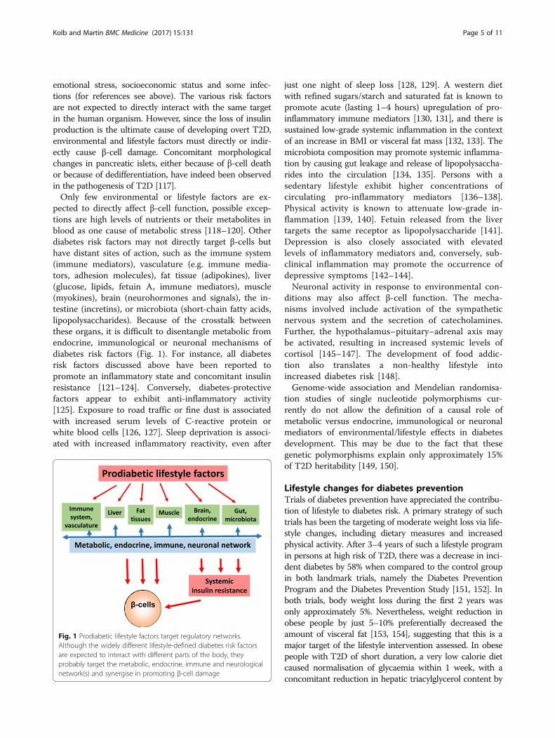
pected to directly affect β-cell function, possible excep-tions are high levels of nutrients or their metabolites inblood as one cause of metabolic stress [118–120]. Otherdiabetes risk factors may not directly target β-cells buthave distant sites of action, such as the immune system(immune mediators), vasculature (e.g. immune media-tors, adhesion molecules), fat tissue (adipokines), liver(glucose, lipids, fetuin A, immune mediators), muscle(myokines), brain (neurohormones and signals), the in-testine (incretins), or microbiota (short-chain fatty acids,lipopolysaccharides). Because of the crosstalk betweenthese organs, it is difficult to disentangle metabolic fromendocrine, immunological or neuronal mechanisms ofdiabetes risk factors (Fig. 1). For instance, all diabetesrisk factors discussed above have been reported topromote an inflammatory state and concomitant insulinresistance [121–124]. Conversely, diabetes-protectivefactors appear to exhibit anti-inflammatory activity[125]. Exposure to road traffic or fine dust is associatedwith increased serum levels of C-reactive protein orwhite blood cells [126, 127]. Sleep deprivation is associ-ated with increased inflammatory reactivity, even after
just one night of sleep loss [128, 129]. A western dietwith refined sugars/starch and saturated fat is known topromote acute (lasting 1–4 hours) upregulation of pro-inflammatory immune mediators [130, 131], and there issustained low-grade systemic inflammation in the contextof an increase in BMI or visceral fat mass [132, 133]. Themicrobiota composition may promote systemic inflamma-tion by causing gut leakage and release of lipopolysaccha-rides into the circulation [134, 135]. Persons with asedentary lifestyle exhibit higher concentrations ofcirculating pro-inflammatory mediators [136–138].Physical activity is known to attenuate low-grade in-flammation [139, 140]. Fetuin released from the livertargets the same receptor as lipopolysaccharide [141].Depression is also closely associated with elevatedlevels of inflammatory mediators and, conversely, sub-clinical inflammation may promote the occurrence ofdepressive symptoms [142–144].Neuronal activity in response to environmental con-
ditions may also affect β-cell function. The mecha-nisms involved include activation of the sympatheticnervous system and the secretion of catecholamines.Further, the hypothalamus–pituitary–adrenal axis maybe activated, resulting in increased systemic levels ofcortisol [145–147]. The development of food addic-tion also translates a non-healthy lifestyle intoincreased diabetes risk [148].Genome-wide association and Mendelian randomisa-
tion studies of single nucleotide polymorphisms cur-rently do not allow the definition of a causal role ofmetabolic versus endocrine, immunological or neuronalmediators of environmental/lifestyle effects in diabetesdevelopment. This may be due to the fact that thesegenetic polymorphisms explain only approximately 15%of T2D heritability [149, 150].
Lifestyle changes for diabetes preventionTrials of diabetes prevention have appreciated the contribu-tion of lifestyle to diabetes risk. A primary strategy of suchtrials has been the targeting of moderate weight loss via life-style changes, including dietary measures and increasedphysical activity. After 3–4 years of such a lifestyle programin persons at high risk of T2D, there was a decrease in inci-dent diabetes by 58% when compared to the control groupin both landmark trials, namely the Diabetes PreventionProgram and the Diabetes Prevention Study [151, 152]. Inboth trials, body weight loss during the first 2 years wasonly approximately 5%. Nevertheless, weight reduction inobese people by just 5–10% preferentially decreased theamount of visceral fat [153, 154], suggesting that this is amajor target of the lifestyle intervention assessed. In obesepeople with T2D of short duration, a very low calorie dietcaused normalisation of glycaemia within 1 week, with aconcomitant reduction in hepatic triacylglycerol content by
Fig. 1 Prodiabetic lifestyle factors target regulatory networks.Although the widely different lifestyle-defined diabetes risk factorsare expected to interact with different parts of the body, theyprobably target the metabolic, endocrine, immune and neurologicalnetwork(s) and synergise in promoting β-cell damage
Kolb and Martin BMC Medicine (2017) 15:131 Page 5 of 11
30%, whereas body weight decreased by only 3% [155].Further, there was substantial recovery of β-cell functionafter 8 weeks of a low calorie diet. A protein-rich, lowcalorie formula diet for meal replacement was found tosubstantially improve metabolic control, decrease theamount of antidiabetic medication and lower body weightin persons with insulin-treated T2D [156].Long-term data are available from the two early diabetes
prevention trials. After a mean follow-up of 15 or 9 years,respectively, the incidence of diabetes was 60% in thecontrol group versus 52% in the intervention group in theDiabetes Prevention Program, and 56% versus 41% in theDiabetes Prevention Study [157, 158]. Thus, the lifestylechanges in the two studies halted the progression towardsovert T2D in a large fraction of study participants for a fewyears, but was much less effective during follow-up. In bothstudies, there was a substantial regain of body weight overtime. It is probable that long-term maintenance of bodyweight reduction would have prevented progression toT2D also in the long term, since this correlation is observedin people at risk of T2D after bariatric surgery [159].
ConclusionsA host of environmental or lifestyle-dependent T2D riskfactors have been described in prospective epidemiologicalstudies, ranging from energy-dense food consumption tolong-term exposure to high levels of fine dust. In thesestudies, the amount of personal, environmental or lifestyleaspects data that can or has been documented is limited,leaving room for confounding. For instance, the number ofmeals prepared at home or the frequency of tooth-brushingare rarely documented although both impact diabetes risk[160, 161] and may therefore confound associations withother lifestyle factors. Further, which environmental orlifestyle facotrs are associated with diabetes risk but do notbear a cause-effect relationship remains unknown.Combining epidemiological data with the experience
from intervention trials may allow to tentatively definediabetes protective factors. A consistent finding from pro-spective studies is the association of plant food-based dietswith a lower T2D risk [14–16, 18, 20]. This fits with theoutcome of the PREDIMED trial of Mediterranean diet,suggesting that the diabetes-protective effect of bodyweight reduction can be replaced, at least in part, by anappropriate quality diet [32]. Prebiotic plant fibres pro-mote growth of a diverse and apparently healthy micro-biota with less endotoxin leakage [162–164]. This mayavoid inflammatory activation of endothelial and Kupffercells in the liver with concomitant hepatocyte dysfunction,as usually observed in response to a western-type high-fatdiet or during non-alcoholic fatty liver disease [165, 166].A fibre-rich diet gives rise to the enhanced production ofshort-chain fatty acids by gut bacteria [167]. There isdirect binding of these products to the free fatty acid
receptor 2 on β-cells and the promotion of cell growthand function [168].A second major diabetes-protective component of plant
food are phytochemicals. A property common to mostphytochemicals is the activation of cell defence and anti-inflammatory genes, for instance, via the Nrf2 signallingpathway [169]. These effects occur body-wide and are alsodemonstrable in β-cells [170].A third major diabetes-protective factor is exercise.
Interestingly, muscle work also activates cell defence andanti-inflammatory pathways via Nrf2 signalling. There is adirect beneficial effect of exercise on β-cell function [171],and also on liver function in non-alcoholic fatty liver dis-ease, although there is no body weight loss [172].The strongest diabetes-protective factor appears to be
the avoidance or correction of a high body fat mass, not-ably visceral and ectopic fat. There is rapid improvement,and often normalisation, of glucose homeostasis withinthe first week after bariatric surgery or initiation of a verylow calorie diet [155, 173]. After Roux-en-Y gastric bypassseveral rapid changes have been noted aside from thelower insulin and plasma glucose levels, including lowerblood levels of leptin and branched amino acids, higherconcentrations of adiponectin and bile acids, and strongerpost prandial increases of GLP-1 and of the satiety peptideYY [173]. However, many of these changes are not seenafter gastric banding or a very low calorie diet, such as noincreased production of GLP-1 [155, 174, 175], bile acids[176, 177], peptide YY [178], or no decreased levels ofbranched amino acids [179]. Therefore, it seems probablethat the major determinant of rapid metabolic improve-ment seen with Roux-en-Y gastric bypass, gastric bandingor a very low calorie diet is the acute negative energy bal-ance, which is similar for the three procedures [180, 181].Obviously, the immediate response to severely restricted
calorie uptake in the days after surgery or during a verylow calorie diet is substantially less post-prandial insulinproduction, thus alleviating stress from β-cells. The lowerlevel of protein synthesis decreases the demand of ATPfrom mitochondria, thereby reducing the amount of con-comitantly released radical oxygen species and allowingrecovery of mitochondrial proteins from oxidative damagesuch as nitrosylation or carbonylation [182, 183]. Indeed,within 1 week there is partial recovery of insulin produc-tion after the introduction of a very low calorie diet, withfurther normalisation in the following months [155, 184].Recovery of β-cell function to varying extents is also seenearly after gastric bypass [185–187].A second immediate response to a very low calorie
intake is the adaptation of metabolic control to the lowamount of digestible carbohydrate. Such adaptation is pri-marily important in the liver, where most of gluconeogen-esis takes place, including the synthesis of glucose fromglycerol of triglycerides and metabolic breakdown of
Kolb and Martin BMC Medicine (2017) 15:131 Page 6 of 11
released fatty acids to the acetate level. Excess acetyl-CoAgives rise to acetoacetyl-CoA and, subsequently, to acet-one and ß-hydroxybutyrate. The latter (reduced) ketonecan be used as a substrate for oxidative energy productionin place of glucose in peripheral tissues and can pass theblood–brain barrier [188].It has been proposed that calorie restriction mimetics
may elicit similar cellular responses as seen during anacute negative balance, including an improved cellularresistance to oxidative stress and an anti-inflammatorymilieu [189, 190]. Many phytochemicals have beenshown to exhibit activities related to calorie restriction,including the grape polyphenol resveratrol [191], anacar-dic acid from cashews, curcumin from the spiceturmeric, garcinol from the fruit of the Kokum tree,epigallocatechin-3-gallate from green tea, and spermi-dine from fermented soy beans or wheat germs [192].Therefore, it is not surprising that plant-based diets havebeen recognised to partly mimic the effects of calorie re-striction [193, 194].Taken together, intervention trials of diabetes preven-
tion indicate that long-term preservation of normogly-caemia in people at high risk of T2D is achievable.Further research on the protective mechanisms associ-ated with physical activity, healthy eating patterns andspecific food components, anti-inflammatory strategies,or with weight reduction via low calorie diet or bariatricsurgery seems warranted (Box 2).
AcknowledgementsSM has received non-restricted support for the conduct of trials of lifestylechange in persons at risk or with type 2 diabetes by Novartis Pharma GmbH,Boehringer Ingelheim Pharma GmbH & Co. KG, Almased Wellness GmbH,Nintendo of Europe GmbH, HMM Holding AG.
FundingThis work was supported by the Gesellschaft von Freunden und Förderernder Heinrich-Heine-Universität Düsseldorf e.V.
Availability of data and materialsNot applicable.
Authors’ contributionsHK and SM conceived and wrote the manuscript. Both authors read andapproved the final manuscript.
Ethics approval and consent to participateNot applicable.
Consent for publicationNot applicable.
Competing interestsThe authors declare that they have no competing interests.
Publisher’s NoteSpringer Nature remains neutral with regard to jurisdictional claims inpublished maps and institutional affiliations.
Received: 14 March 2017 Accepted: 23 June 2017
References1. NDC Risk Factor Collaboration. Worldwide trends in diabetes since 1980: a
pooled analysis of 751 population-based studies with 4.4 millionparticipants. Lancet. 2016;387:1513–30.
2. Chatterjee S, Khunti K, Davies MJ. Type 2 diabetes. Lancet. 2017;389(10085):2239–51. doi:10.1016/S0140-6736(17)30058-2.
3. Ng M, Fleming T, Robinson M, Thomson B, Graetz N, Margono C, et al.Global, regional, and national prevalence of overweight and obesity inchildren and adults during 1980-2013: a systematic analysis for the GlobalBurden of Disease Study 2013. Lancet. 2014;384:766–81.
4. Neeland IJ, Turer AT, Ayers CR, Powell-Wiley TM, Vega GL, Farzaneh-Far R, etal. Dysfunctional adiposity and the risk of prediabetes and type 2 diabetesin obese adults. JAMA. 2012;308:1150–9.
5. Ballestri S, Zona S, Targher G, Romagnoli D, Baldelli E, Nascimbeni F, et al.Nonalcoholic fatty liver disease is associated with an almost twofoldincreased risk of incident type 2 diabetes and metabolic syndrome.Evidence from a systematic review and meta-analysis. J GastroenterolHepatol. 2016;31:936–44.
6. Valenti L, Bugianesi E, Pajvani U, Targher G. Nonalcoholic fatty liver disease:cause or consequence of type 2 diabetes? Liver Int. 2016;36:1563–79.
7. Sattar N, Gill JM. Type 2 diabetes as a disease of ectopic fat? BMC Med.2014;12:123.
8. Phillips CM. Metabolically healthy obesity: definitions, determinants andclinical implications. Rev Endocr Metab Disord. 2013;14:219–27.
9. Roberson LL, Aneni EC, Maziak W, Agatston A, Feldman T, Rouseff M, et al.Beyond BMI: The "Metabolically healthy obese" phenotype & its associationwith clinical/subclinical cardiovascular disease and all-cause mortality – asystematic review. BMC Public Health. 2014;14:14.
10. Bluher M. Are metabolically healthy obese individuals really healthy? Eur JEndocrinol. 2014;171:R209–19.
11. Ma RC, Chan JC. Type 2 diabetes in East Asians: similarities and differenceswith populations in Europe and the United States. Ann N Y Acad Sci. 2013;1281:64–91.
12. Unnikrishnan R, Anjana RM, Mohan V. Diabetes in South Asians: is thephenotype different? Diabetes. 2014;63:53–5.
13. Ding C, Chan Z, Magkos F. Lean, but not healthy: the 'metabolically obese,normal-weight' phenotype. Curr Opin Clin Nutr Metab Care. 2016;19:408–17.
Box 2 Key points
� The global type 2 diabetes (T2D) epidemic is generally
believed to result from environmental and lifestyle changes
� Diabetes risk factors include energy-dense western style
diets, decreased physical activity, increased sitting and
monitor viewing time, exposure to noise or fine dust, short
or disturbed sleep, smoking, stress, depression, and a low
socioeconomic status
� It is suggested that the various diabetes risk factors probably
target different organs of the body, but these are connected
by endocrine, metabolic, immune and neurological networks
� Since the loss of insulin production is the ultimate cause of
developing overt T2D, environmental and lifestyle factors
must directly or indirectly cause β-cell damage
� Epidemiological studies indicate that major
diabetes-protective factors comprise plant food-based diets
and moderate to high intensity muscle work
� It may be possible to reproduce, at least in part, the benefits
of body weight reduction by calorie restriction mimetics
Kolb and Martin BMC Medicine (2017) 15:131 Page 7 of 11
14. Jannasch F, Kroger J, Schulze MB. Dietary patterns and type 2 diabetes: asystematic literature review and meta-analysis of prospective studies. J Nutr.2017;147(6):1174–82.
15. Schwingshackl L, Hoffmann G, Lampousi AM, Knuppel S, Iqbal K,Schwedhelm C, et al. Food groups and risk of type 2 diabetes mellitus: asystematic review and meta-analysis of prospective studies. Eur J Epidemiol.2017. doi:10.1007/s10654-017-0246-y.
16. Fardet A, Boirie Y. Associations between food and beverage groups andmajor diet-related chronic diseases: an exhaustive review of pooled/meta-analyses and systematic reviews. Nutr Rev. 2014;72:741–62.
17. Forouhi NG, Wareham NJ. The EPIC-InterAct Study: a study of the interplaybetween genetic and lifestyle behavioral factors on the risk of type 2diabetes in European populations. Curr Nutr Rep. 2014;3:355–63.
18. Maki KC, Phillips AK. Dietary substitutions for refined carbohydrate thatshow promise for reducing risk of type 2 diabetes in men and women. JNutr. 2015;145:159S–63S.
19. Cespedes EM, Hu FB, Tinker L, Rosner B, Redline S, Garcia L, et al. Multiplehealthful dietary patterns and type 2 diabetes in the Women's HealthInitiative. Am J Epidemiol. 2016;183:622–33.
20. Satija A, Bhupathiraju SN, Rimm EB, Spiegelman D, Chiuve SE, Borgi L, et al.Plant-based dietary patterns and incidence of type 2 diabetes in US menand women: results from three prospective cohort studies. PLoS Med. 2016;13, e1002039.
21. Ma Y, He FJ, Yin Y, Hashem KM, MacGregor GA. Gradual reduction of sugar insoft drinks without substitution as a strategy to reduce overweight, obesity, andtype 2 diabetes: a modelling study. Lancet Diabetes Endocrinol. 2016;4:105–14.
22. Lofvenborg JE, Andersson T, Carlsson PO, Dorkhan M, Groop L, Martinell M,et al. Sweetened beverage intake and risk of latent autoimmune diabetes inadults (LADA) and type 2 diabetes. Eur J Endocrinol. 2016;175:605–14.
23. Wang M, Yu M, Fang L, Hu RY. Association between sugar-sweetened beveragesand type 2 diabetes: a meta-analysis. J Diabetes Investig. 2015;6:360–6.
24. Greenwood DC, Threapleton DE, Evans CE, Cleghorn CL, Nykjaer C, WoodheadC, Burley VJ. Association between sugar-sweetened and artificially sweetenedsoft drinks and type 2 diabetes: systematic review and dose-response meta-analysis of prospective studies. Br J Nutr. 2014;112:725–34.
25. Bao Y, Han J, Hu FB, Giovannucci EL, Stampfer MJ, Willett WC, Fuchs CS.Association of nut consumption with total and cause-specific mortality. NEngl J Med. 2013;369:2001–11.
26. U.S. Department of Health and Human Services. 2015–2020 DietaryGuidelines for Americans. https://health.gov/dietaryguidelines/2015/.Accessed 1 June 2017.
27. Atallah R, Filion KB, Wakil SM, Genest J, Joseph L, Poirier P, et al. Long-termeffects of 4 popular diets on weight loss and cardiovascular risk factors: asystematic review of randomized controlled trials. Circ Cardiovasc QualOutcomes. 2014;7:815–27.
28. Tay J, Luscombe-Marsh ND, Thompson CH, Noakes M, Buckley JD, WittertGA, et al. A very low-carbohydrate, low-saturated fat diet for type 2 diabetesmanagement: a randomized trial. Diabetes Care. 2014;37:2909–18.
29. Yancy Jr WS, Mayer SB, Coffman CJ, Smith VA, Kolotkin RL, Geiselman PJ, etal. Effect of allowing choice of diet on weight loss: a randomized trial. AnnIntern Med. 2015;162:805–14.
30. Hill AM, Harris Jackson KA, Roussell MA, West SG, Kris-Etherton PM. Typeand amount of dietary protein in the treatment of metabolic syndrome: arandomized controlled trial. Am J Clin Nutr. 2015;102:757–70.
31. Tobias DK, Chen M, Manson JE, Ludwig DS, Willett W, Hu FB. Effect of low-fat diet interventions versus other diet interventions on long-term weightchange in adults: a systematic review and meta-analysis. Lancet DiabetesEndocrinol. 2015;3:968–79.
32. Salas-Salvado J, Bullo M, Babio N, Martinez-Gonzalez MA, Ibarrola-Jurado N,Basora J, et al. Reduction in the incidence of type 2 diabetes with theMediterranean diet: results of the PREDIMED-Reus nutrition interventionrandomized trial. Diabetes Care. 2011;34:14–9.
33. Howells L, Musaddaq B, McKay AJ, Majeed A. Clinical impact of lifestyleinterventions for the prevention of diabetes: an overview of systematicreviews. BMJ Open. 2016;6, e013806.
34. Gong QH, Kang JF, Ying YY, Li H, Zhang XH, Wu YH, Xu GZ. Lifestyleinterventions for adults with impaired glucose tolerance: a systematicreview and meta-analysis of the effects on glycemic control. Intern Med.2015;54:303–10.
35. Vasto S, Buscemi S, Barera A, Di Carlo M, Accardi G, Caruso C. Mediterraneandiet and healthy ageing: a Sicilian perspective. Gerontology. 2014;60:508–18.
36. Willcox DC, Scapagnini G, Willcox BJ. Healthy aging diets other than theMediterranean: a focus on the Okinawan diet. Mech Ageing Dev.2014;136–137:148–62.
37. Buettner D. The secrets of long life. National Geographic 2005; November.http://ngm.nationalgeographic.com/2005/11/longevity-secrets/buettner-text.Accessed 21 Mar 2017.
38. Cai D, Zhao S, Li D, Chang F, Tian X, Huang G, et al. Nutrient intake isassociated with longevity characterization by metabolites and elementprofiles of healthy centenarians. Nutrients. 2016;8:564.
39. Shadyab AH, LaCroix AZ. Genetic factors associated with longevity: a reviewof recent findings. Ageing Res Rev. 2015;19:1–7.
40. Loef M, Walach H. The combined effects of healthy lifestyle behaviors on allcause mortality: a systematic review and meta-analysis. Prev Med. 2012;55:163–70.
41. Aune D, Norat T, Leitzmann M, Tonstad S, Vatten LJ. Physical activity andthe risk of type 2 diabetes: a systematic review and dose-response meta-analysis. Eur J Epidemiol. 2015;30:529–42.
42. Smith AD, Crippa A, Woodcock J, Brage S. Physical activity and incidenttype 2 diabetes mellitus: a systematic review and dose-response meta-analysis of prospective cohort studies. Diabetologia. 2016;59:2527–45.
43. DiPietro L, Gribok A, Stevens MS, Hamm LF, Rumpler W. Three 15-min boutsof moderate postmeal walking significantly improves 24-h glycemic controlin older people at risk for impaired glucose tolerance. Diabetes Care. 2013;36:3262–8.
44. Parker L, Shaw CS, Banting L, Levinger I, Hill KM, McAinch AJ, Stepto NK.Acute low-volume high-intensity interval exercise and continuousmoderate-intensity exercise elicit a similar improvement in 24-h glycemiccontrol in overweight and obese adults. Front Physiol. 2016;7:661.
45. Yates T, Henson J, Edwardson C, Dunstan D, Bodicoat DH, Khunti K, DaviesMJ. Objectively measured sedentary time and associations with insulinsensitivity: Importance of reallocating sedentary time to physical activity.Prev Med. 2015;76:79–83.
46. Johns DJ, Hartmann-Boyce J, Jebb SA, Aveyard P. Diet or exerciseinterventions vs combined behavioral weight management programs: asystematic review and meta-analysis of direct comparisons. J Acad NutrDiet. 2014;114:1557–68.
47. Hamilton MT, Hamilton DG, Zderic TW. Sedentary behavior as a mediator oftype 2 diabetes. Med Sport Sci. 2014;60:11–26.
48. de Rezende LF, Rey-Lopez JP, Matsudo VK, do Carmo LO. Sedentarybehavior and health outcomes among older adults: a systematic review.BMC Public Health. 2014;14:333.
49. Rockette-Wagner B, Edelstein S, Venditti EM, Reddy D, Bray GA, Carrion-Petersen ML, et al. The impact of lifestyle intervention on sedentary time inindividuals at high risk of diabetes. Diabetologia. 2015;58:1198–202.
50. Biswas A, Oh PI, Faulkner GE, Bajaj RR, Silver MA, Mitchell MS, Alter DA.Sedentary time and its association with risk for disease incidence, mortality,and hospitalization in adults: a systematic review and meta-analysis. AnnIntern Med. 2015;162:123–32.
51. Dempsey PC, Owen N, Yates TE, Kingwell BA, Dunstan DW. Sitting less andmoving more: improved glycaemic control for type 2 diabetes preventionand management. Curr Diab Rep. 2016;16:114.
52. Golubic R, Wijndaele K, Sharp SJ, Simmons RK, Griffin SJ, Wareham NJ, et al.Physical activity, sedentary time and gain in overall and central body fat: 7-yearfollow-up of the ProActive trial cohort. Int J Obes (Lond). 2015;39:142–8.
53. Levine JA. Sick of sitting. Diabetologia. 2015;58:1751–8.54. Levine JA, Schleusner SJ, Jensen MD. Energy expenditure of nonexercise
activity. Am J Clin Nutr. 2000;72:1451–4.55. Sorensen M, Andersen ZJ, Nordsborg RB, Becker T, Tjonneland A, Overvad K,
Raaschou-Nielsen O. Long-term exposure to road traffic noise and incidentdiabetes: a cohort study. Environ Health Perspect. 2013;121:217–22.
56. Heidemann C, Niemann H, Paprott R, Du Y, Rathmann W, Scheidt-Nave C.Residential traffic and incidence of type 2 diabetes: the German HealthInterview and Examination Surveys. Diabet Med. 2014;31:1269–76.
57. Eze IC, Schaffner E, Foraster M, Imboden M, von Eckardstein A, Gerbase MW,et al. Long-term exposure to ambient air pollution and metabolic syndromein adults. PLoS One. 2015;10, e0130337.
58. Weinmayr G, Hennig F, Fuks K, Nonnemacher M, Jakobs H, Mohlenkamp S,et al. Long-term exposure to fine particulate matter and incidence of type 2diabetes mellitus in a cohort study: effects of total and traffic-specific airpollution. Environ Health. 2015;14:53.
59. Dzhambov AM. Long-term noise exposure and the risk for type 2 diabetes:a meta-analysis. Noise Health. 2015;17:23–33.
Kolb and Martin BMC Medicine (2017) 15:131 Page 8 of 11
60. Li Y, Gao X, Winkelman JW, Cespedes EM, Jackson CL, Walters AS, et al.Association between sleeping difficulty and type 2 diabetes in women.Diabetologia. 2016;59:719–27.
61. Miedema HM, Vos H. Associations between self-reported sleep disturbanceand environmental noise based on reanalyses of pooled data from 24studies. Behav Sleep Med. 2007;5:1–20.
62. Reynolds AC, Banks S. Total sleep deprivation, chronic sleep restriction andsleep disruption. Prog Brain Res. 2010;185:91–103.
63. Shan Z, Ma H, Xie M, Yan P, Guo Y, Bao W, et al. Sleep duration and risk oftype 2 diabetes: a meta-analysis of prospective studies. Diabetes Care. 2015;38:529–37.
64. Xi B, He D, Zhang M, Xue J, Zhou D. Short sleep duration predicts risk ofmetabolic syndrome: a systematic review and meta-analysis. Sleep Med Rev.2014;18:293–7.
65. Kowall B, Lehnich AT, Strucksberg KH, Fuhrer D, Erbel R, Jankovic N, et al.Associations among sleep disturbances, nocturnal sleep duration, daytimenapping, and incident prediabetes and type 2 diabetes: the Heinz NixdorfRecall Study. Sleep Med. 2016;21:35–41.
66. Leng Y, Cappuccio FP, Surtees PG, Luben R, Brayne C, Khaw KT. Daytimenapping, sleep duration and increased 8-year risk of type 2 diabetes in aBritish population. Nutr Metab Cardiovasc Dis. 2016;26:996–1003.
67. Rao MN, Neylan TC, Grunfeld C, Mulligan K, Schambelan M, Schwarz JM.Subchronic sleep restriction causes tissue-specific insulin resistance. J ClinEndocrinol Metab. 2015;100:1664–71.
68. Donga E, van Dijk M, van Dijk JG, Biermasz NR, Lammers GJ, van KralingenKW, et al. A single night of partial sleep deprivation induces insulinresistance in multiple metabolic pathways in healthy subjects. J ClinEndocrinol Metab. 2010;95:2963–8.
69. Ding M, Bhupathiraju SN, Chen M, van Dam RM, Hu FB. Caffeinated anddecaffeinated coffee consumption and risk of type 2 diabetes: a systematicreview and a dose-response meta-analysis. Diabetes Care. 2014;37:569–86.
70. Moisey LL, Kacker S, Bickerton AC, Robinson LE, Graham TE. Caffeinated coffeeconsumption impairs blood glucose homeostasis in response to high and lowglycemic index meals in healthy men. Am J Clin Nutr. 2008;87:1254–61.
71. Ohnaka K, Ikeda M, Maki T, Okada T, Shimazoe T, Adachi M, et al. Effects of16-week consumption of caffeinated and decaffeinated instant coffee onglucose metabolism in a randomized controlled trial. J Nutr Metab. 2012;2012:207426.
72. Robertson TM, Clifford MN, Penson S, Chope G, Robertson MD. A singleserving of caffeinated coffee impairs postprandial glucose metabolism inoverweight men. Br J Nutr. 2015;114:1218–25.
73. Rakvaag E, Dragsted LO. Acute effects of light and dark roasted coffee onglucose tolerance: a randomized, controlled crossover trial in healthyvolunteers. Eur J Nutr. 2016;55:2221–30.
74. Kempf K, Kolb H, Gartner B, Bytof G, Stiebitz H, Lantz I, et al.Cardiometabolic effects of two coffee blends differing in content for majorconstituents in overweight adults: a randomized controlled trial. Eur J Nutr.2015;54:845–54.
75. Wedick NM, Brennan AM, Sun Q, Hu FB, Mantzoros CS, van Dam RM. Effectsof caffeinated and decaffeinated coffee on biological risk factors for type 2diabetes: a randomized controlled trial. Nutr J. 2011;10:93.
76. Zheng XX, Xu YL, Li SH, Hui R, Wu YJ, Huang XH. Effects of green teacatechins with or without caffeine on glycemic control in adults: a meta-analysis of randomized controlled trials. Am J Clin Nutr. 2013;97:750–62.
77. Yang WS, Wang WY, Fan WY, Deng Q, Wang X. Tea consumption and riskof type 2 diabetes: a dose-response meta-analysis of cohort studies. Br JNutr. 2014;111:1329–39.
78. Yang J, Mao QX, Xu HX, Ma X, Zeng CY. Tea consumption and risk of type 2diabetes mellitus: a systematic review and meta-analysis update. BMJ Open.2014;4, e005632.
79. Knott C, Bell S, Britton A. Alcohol consumption and the risk of type 2diabetes: a systematic review and dose-response meta-analysis of morethan 1.9 million individuals from 38 observational studies. Diabetes Care.2015;38:1804–12.
80. Li XH, Yu FF, Zhou YH, He J. Association between alcohol consumption andthe risk of incident type 2 diabetes: a systematic review and dose-responsemeta-analysis. Am J Clin Nutr. 2016;103:818–29.
81. Gepner Y, Golan R, Harman-Boehm I, Henkin Y, Schwarzfuchs D, Shelef I, etal. Effects of initiating moderate alcohol intake on cardiometabolic risk inadults with type 2 diabetes: a 2-year randomized, controlled trial. Ann InternMed. 2015;163:569–79.
82. Zhang L, Curhan GC, Hu FB, Rimm EB, Forman JP. Association betweenpassive and active smoking and incident type 2 diabetes in women.Diabetes Care. 2011;34:892–7.
83. Willi C, Bodenmann P, Ghali WA, Faris PD, Cornuz J. Active smoking and therisk of type 2 diabetes: a systematic review and meta-analysis. JAMA. 2007;298:2654–64.
84. Pan A, Wang Y, Talaei M, Hu FB, Wu T. Relation of active, passive, andquitting smoking with incident type 2 diabetes: a systematic review andmeta-analysis. Lancet Diabetes Endocrinol. 2015;3:958–67.
85. Keith RJ, Al Rifai M, Carruba C, De Jarnett N, McEvoy JW, Bhatnagar A, et al.Tobacco use, insulin resistance, and risk of type 2 diabetes: results from themulti-ethnic study of atherosclerosis. PLoS One. 2016;11, e0157592.
86. Virtanen M, Ferrie JE, Tabak AG, Akbaraly TN, Vahtera J, Singh-Manoux A,Kivimaki M. Psychological distress and incidence of type 2 diabetes in high-risk and low-risk populations: the Whitehall II Cohort Study. Diabetes Care.2014;37:2091–7.
87. Bergmann N, Gyntelberg F, Faber J. The appraisal of chronic stress and thedevelopment of the metabolic syndrome: a systematic review ofprospective cohort studies. Endocr Connect. 2014;3:R55–80.
88. Cosgrove MP, Sargeant LA, Caleyachetty R, Griffin SJ. Work-related stressand type 2 diabetes: systematic review and meta-analysis. Occup Med(Lond). 2012;62:167–73.
89. Pouwer F, Kupper N, Adriaanse MC. Does emotional stress cause type 2diabetes mellitus? A review from the European Depression in Diabetes(EDID) Research Consortium. Discov Med. 2010;9:112–18.
90. Rutters F, Pilz S, Koopman AD, Rauh SP, Pouwer F, Stehouwer CD, et al.Stressful life events and incident metabolic syndrome: the Hoorn study.Stress. 2015;18:507–13.
91. Novak M, Bjorck L, Giang KW, Heden-Stahl C, Wilhelmsen L, Rosengren A.Perceived stress and incidence of type 2 diabetes: a 35-year follow-up studyof middle-aged Swedish men. Diabet Med. 2013;30:e8–16.
92. Melamed S, Shirom A, Toker S, Shapira I. Burnout and risk of type 2diabetes: a prospective study of apparently healthy employed persons.Psychosom Med. 2006;68:863–9.
93. Ford AH, Flicker L, Hankey GJ, Yeap BB, Chubb SA, Golledge J, Almeida OP.Insulin resistance and depressive symptoms in older men: the health inmen study. Am J Geriatr Psychiatry. 2015;23:872–80.
94. Au B, Smith KJ, Gariepy G, Schmitz N. C-reactive protein, depressivesymptoms, and risk of diabetes: results from the English Longitudinal Studyof Ageing (ELSA). J Psychosom Res. 2014;77:180–6.
95. Hasan SS, Clavarino AM, Mamun AA, Kairuz T. Incidence and risk of diabetesmellitus associated with depressive symptoms in adults: evidence fromlongitudinal studies. Diabetes Metab Syndr. 2014;8:82–7.
96. Demakakos P, Zaninotto P, Nouwen A. Is the association betweendepressive symptoms and glucose metabolism bidirectional? Evidence fromthe English Longitudinal Study of Ageing. Psychosom Med. 2014;76:555–61.
97. Khambaty T, Callahan CM, Perkins AJ, Stewart JC. Depression and anxietyscreens as simultaneous predictors of 10-year incidence of diabetes mellitusin older adults in primary care. J Am Geriatr Soc. 2016;65:294–300.
98. Meisinger C, Kandler U, Ladwig KH. Living alone is associated with anincreased risk of type 2 diabetes mellitus in men but not women from thegeneral population: the MONICA/KORA Augsburg Cohort Study. PsychosomMed. 2009;71:784–8.
99. Agardh E, Allebeck P, Hallqvist J, Moradi T, Sidorchuk A. Type 2 diabetesincidence and socio-economic position: a systematic review and meta-analysis. Int J Epidemiol. 2011;40:804–18.
100. Sommer I, Griebler U, Mahlknecht P, Thaler K, Bouskill K, Gartlehner G, MendisS. Socioeconomic inequalities in non-communicable diseases and their riskfactors: an overview of systematic reviews. BMC Public Health. 2015;15:914.
101. Stringhini S, Zaninotto P, Kumari M, Kivimaki M, Batty GD. Lifecoursesocioeconomic status and type 2 diabetes: the role of chronic inflammationin the English Longitudinal Study of Ageing. Sci Rep. 2016;6:24780.
102. Sacerdote C, Ricceri F, Rolandsson O, Baldi I, Chirlaque MD, Feskens E, et al.Lower educational level is a predictor of incident type 2 diabetes in Europeancountries: the EPIC-InterAct study. Int J Epidemiol. 2012;41:1162–73.
103. Williams ED, Tapp RJ, Magliano DJ, Shaw JE, Zimmet PZ, Oldenburg BF.Health behaviours, socioeconomic status and diabetes incidence: theAustralian Diabetes Obesity and Lifestyle Study (AusDiab). Diabetologia.2010;53:2538–45.
104. Leong A, Rahme E, Dasgupta K. Spousal diabetes as a diabetes risk factor: asystematic review and meta-analysis. BMC Med. 2014;12:12.
Kolb and Martin BMC Medicine (2017) 15:131 Page 9 of 11

105. Almgren M, Atkinson R, He J, Hilding A, Hagman E, Wolk A, et al.Adenovirus-36 is associated with obesity in children and adults in Swedenas determined by rapid ELISA. PLoS One. 2012;7, e41652.
106. Pasarica M, Shin AC, Yu M, Ou Yang HM, Rathod M, et al. Humanadenovirus 36 induces adiposity, increases insulin sensitivity, and altershypothalamic monoamines in rats. Obesity (Silver Spring). 2006;14:1905–13.
107. Almgren M, Atkinson RL, Hilding A, He J, Brismar K, Schalling M, et al. Humanadenovirus-36 is uncommon in type 2 diabetes and is associated withincreased insulin sensitivity in adults in Sweden. Ann Med. 2014;46:539–46.
108. Negro F. Facts and fictions of HCV and comorbidities: steatosis, diabetesmellitus, and cardiovascular diseases. J Hepatol. 2014;61:S69–78.
109. Lin YJ, Shaw TG, Yang HI, Lu SN, Jen CL, Wang LY, et al. Chronic hepatitis Cvirus infection and the risk for diabetes: a community-based prospectivestudy. Liver Int. 2016;92:279–82.
110. Rodriguez AR, Plascencia-Villa G, Witt CM, Yu JJ, Jose-Yacaman M, ChambersJP, et al. Chlamydia pneumoniae promotes dysfunction of pancreatic betacells. Cell Immunol. 2015;295:83–91.
111. Betene AD, De Wit S, Neuhaus J, Palfreeman A, Pepe R, Pankow JS, NeatonJD. Interleukin-6, high sensitivity C-reactive protein, and the development oftype 2 diabetes among HIV-positive patients taking antiretroviral therapy.J Acquir Immune Defic Syndr. 2014;67:538–46.
112. Bonnefond A, Froguel P. Rare and common genetic events in type 2diabetes: what should biologists know? Cell Metab. 2015;21:357–68.
113. Esparza-Romero J, Valencia ME, Urquidez-Romero R, Chaudhari LS, HansonRL, Knowler WC, et al. Environmentally driven increases in type 2 diabetesand obesity in Pima Indians and non-Pimas in Mexico over a 15-year period:the Maycoba Project. Diabetes Care. 2015;38:2075–82.
114. Lao XQ, Ma WJ, Sobko T, Zhang YH, Xu YJ, Xu XJ, et al. Overall obesity isleveling-off while abdominal obesity continues to rise in a Chinese populationexperiencing rapid economic development: analysis of serial cross-sectionalhealth survey data 2002-2010. Int J Obes (Lond). 2015;39:288–94.
115. Wang Y, Rimm EB, Stampfer MJ, Willett WC, Hu FB. Comparison ofabdominal adiposity and overall obesity in predicting risk of type 2 diabetesamong men. Am J Clin Nutr. 2005;81:555–63.
116. Hardoon SL, Morris RW, Thomas MC, Wannamethee SG, Lennon LT,Whincup PH. Is the recent rise in type 2 diabetes incidence from 1984 to2007 explained by the trend in increasing BMI?: evidence from aprospective study of British men. Diabetes Care. 2010;33:1494–6.
117. Butler AE, Dhawan S, Hoang J, Cory M, Zeng K, Fritsch H, et al. β-cell deficitin obese type 2 diabetes, a minor role of β-cell dedifferentiation anddegranulation. J Clin Endocrinol Metab. 2016;101:523–32.
118. Kaneto H. Pancreatic beta-cell glucose toxicity in type 2 diabetes mellitus.Curr Diabetes Rev. 2015;11:2–6.
119. Brun T, Maechler P. Beta-cell mitochondrial carriers and the diabetogenicstress response. Biochim Biophys Acta. 1863;2016:2540–9.
120. Ertunc ME, Hotamisligil GS. Lipid signaling and lipotoxicity inmetaflammation: indications for metabolic disease pathogenesis andtreatment. J Lipid Res. 2016;57:2099–114.
121. Kolb H, Mandrup-Poulsen T. The global diabetes epidemic as aconsequence of lifestyle-induced low-grade inflammation. Diabetologia.2010;53:10–20.
122. McNelis JC, Olefsky JM. Macrophages, immunity, and metabolic disease.Immunity. 2014;41:36–48.
123. Herder C, Dalmas E, Boni-Schnetzler M, Donath MY. The IL-1 Pathway intype 2 diabetes and cardiovascular complications. Trends Endocrinol Metab.2015;26:551–63.
124. Berchtold LA, Prause M, Storling J, Mandrup-Poulsen T. Cytokines andpancreatic beta-cell apoptosis. Adv Clin Chem. 2016;75:99–158.
125. Kolb H, Eizirik DL. Resistance to type 2 diabetes mellitus: a matter ofhormesis? Nat Rev Endocrinol. 2012;8:183–92.
126. Lanki T, Hampel R, Tiittanen P, Andrich S, Beelen R, Brunekreef B, et al. Airpollution from road traffic and systemic inflammation in adults: a cross-sectional analysis in the European ESCAPE Project. Environ Health Perspect.2015;123:785–91.
127. Chen JC, Schwartz J. Metabolic syndrome and inflammatory responses tolong-term particulate air pollutants. Environ Health Perspect. 2008;116:612–7.
128. da Costa SA, Ribeiro S. Sleep deprivation and gene expression. Curr TopBehav Neurosci. 2015;25:65–90.
129. Chennaoui M, Sauvet F, Drogou C, Van Beers P, Langrume C, Guillard M, etal. Effect of one night of sleep loss on changes in tumor necrosis factoralpha (TNF-alpha) levels in healthy men. Cytokine. 2011;56:318–24.
130. de Vries MA, Klop B, Janssen HW, Njo TL, Westerman EM, Castro CM.Postprandial inflammation: targeting glucose and lipids. Adv Exp Med Biol.2014;824:161–70.
131. Herieka M, Erridge C. High-fat meal induced postprandial inflammation. MolNutr Food Res. 2014;58:136–46.
132. Thorand B, Baumert J, Doring A, Herder C, Kolb H, Rathmann W, et al. Sexdifferences in the relation of body composition to markers of inflammation.Atherosclerosis. 2006;184:216–24.
133. Choi J, Joseph L, Pilote L. Obesity and C-reactive protein in variouspopulations: a systematic review and meta-analysis. Obes Rev. 2013;14:232–44.
134. Gnauck A, Lentle RG, Kruger MC. The characteristics and function ofbacterial lipopolysaccharides and their endotoxic potential in humans. IntRev Immunol. 2016;35:189–218.
135. Amyot J, Semache M, Ferdaoussi M, Fontes G, Poitout V.Lipopolysaccharides impair insulin gene expression in isolated islets ofLangerhans via Toll-like receptor-4 and NF-kappaB signalling. PLoS One.2012;7, e36200.
136. Martinez-Gomez D, Eisenmann JC, Healy GN, Gomez-Martinez S, Diaz LE,Dunstan DW, et al. Sedentary behaviors and emerging cardiometabolicbiomarkers in adolescents. J Pediatr. 2012;160:104–10.
137. Henson J, Yates T, Edwardson CL, Khunti K, Talbot D, Gray LJ, et al.Sedentary time and markers of chronic low-grade inflammation in a highrisk population. PLoS One. 2013;8, e78350.
138. Gabel L, Ridgers ND, Della Gatta PA, Arundell L, Cerin E, Robinson S, et al.Associations of sedentary time patterns and TV viewing time withinflammatory and endothelial function biomarkers in children. Pediatr Obes.2015;11:194–201.
139. You T, Arsenis NC, Disanzo BL, Lamonte MJ. Effects of exercise training onchronic inflammation in obesity : current evidence and potentialmechanisms. Sports Med. 2013;43:243–56.
140. Gjevestad GO, Holven KB, Ulven SM. Effects of exercise on gene expressionof inflammatory markers in human peripheral blood cells: a systematicreview. Curr Cardiovasc Risk Rep. 2015;9:34.
141. Shen X, Yang L, Yan S, Zheng H, Liang L, Cai X, Liao M. Fetuin A promoteslipotoxicity in beta cells through the TLR4 signaling pathway and the roleof pioglitazone in anti-lipotoxicity. Mol Cell Endocrinol. 2015;412:1–11.
142. Howren MB, Lamkin DM, Suls J. Associations of depression with C-reactiveprotein, IL-1, and IL-6: a meta-analysis. Psychosom Med. 2009;71:171–86.
143. Berk M, Williams LJ, Jacka FN, O'Neil A, Pasco JA, Moylan S, et al. Sodepression is an inflammatory disease, but where does the inflammationcome from? BMC Med. 2013;11:200.
144. Kiecolt-Glaser JK, Derry HM, Fagundes CP. Inflammation: depression fans theflames and feasts on the heat. Am J Psychiatry. 2015;172:1075–91.
145. Kruyt ND, van Westerloo DJ, DeVries JH. Stress-induced hyperglycemia inhealthy bungee jumpers without diabetes due to decreased pancreaticbeta-cell function and increased insulin resistance. Diabetes Technol Ther.2012;14:311–4.
146. Maffei A, Segal AM, Alvarez-Perez JC, Garcia-Ocana A, Harris PE. Anti-incretin,anti-proliferative action of dopamine on beta-cells. Mol Endocrinol. 2015;29:542–57.
147. Kodavanti UP. Stretching the stress boundary: linking air pollution healtheffects to a neurohormonal stress response. Biochim Biophys Acta. 1860;2016:2880–90.
148. Carter A, Hendrikse J, Lee N, Yucel M, Verdejo-Garcia A, Andrews Z, Hall W.The neurobiology of "food addiction" and its implications for obesitytreatment and policy. Annu Rev Nutr. 2016;36:105–28.
149. Fuchsberger C, Flannick J, Teslovich TM, Mahajan A, Agarwala V, Gaulton KJ,et al. The genetic architecture of type 2 diabetes. Nature. 2016;536:41–7.
150. Swerdlow DI. Mendelian randomization and type 2 diabetes. CardiovascDrugs Ther. 2016;30:51–7.
151. Delahanty LM, Pan Q, Jablonski KA, Aroda VR, Watson KE, Bray GA, et al.Effects of weight loss, weight cycling, and weight loss maintenance ondiabetes incidence and change in cardiometabolic traits in the DiabetesPrevention Program. Diabetes Care. 2014;37:2738–45.
152. Tuomilehto J, Lindstrom J, Eriksson JG, Valle TT, Hamalainen H, Ilanne-Parikka P, et al. Prevention of type 2 diabetes mellitus by changes in lifestyleamong subjects with impaired glucose tolerance. N Engl J Med. 2001;344:1343–50.
153. Chaston TB, Dixon JB. Factors associated with percent change in visceralversus subcutaneous abdominal fat during weight loss: findings from asystematic review. Int J Obes (Lond). 2008;32:619–28.
Kolb and Martin BMC Medicine (2017) 15:131 Page 10 of 11
154. Gallagher D, Heshka S, Kelley DE, Thornton J, Boxt L, Pi-Sunyer FX, et al.Changes in adipose tissue depots and metabolic markers following a 1-yeardiet and exercise intervention in overweight and obese patients with type 2diabetes. Diabetes Care. 2014;37:3325–32.
155. Lim EL, Hollingsworth KG, Aribisala BS, Chen MJ, Mathers JC, Taylor R.Reversal of type 2 diabetes: normalisation of beta cell function inassociation with decreased pancreas and liver triacylglycerol. Diabetologia.2011;54:2506–14.
156. Kempf K, Schloot NC, Gartner B, Keil R, Schadewaldt P, Martin S. Mealreplacement reduces insulin requirement, HbA1c and weight long-term intype 2 diabetes patients with >100 U insulin per day. J Hum Nutr Diet.2014;27 Suppl 2:21–7.
157. Diabetes Prevention Program Research Group. Long-term effects of lifestyleintervention or metformin on diabetes development and microvascularcomplications over 15-year follow-up: the Diabetes Prevention ProgramOutcomes Study. Lancet Diabetes Endocrinol. 2015;3:866–75.
158. Lindstrom J, Peltonen M, Eriksson JG, Ilanne-Parikka P, Aunola S, Keinanen-Kiukaanniemi S, et al. Improved lifestyle and decreased diabetes risk over13 years: long-term follow-up of the randomised Finnish DiabetesPrevention Study (DPS). Diabetologia. 2013;56:284–93.
159. Sjoholm K, Sjostrom E, Carlsson LM, Peltonen M. Weight change-adjustedeffects of gastric bypass surgery on glucose metabolism: 2- and 10-year resultsfrom the Swedish Obese Subjects (SOS) study. Diabetes Care. 2016;39:625–31.
160. Zong G, Eisenberg DM, Hu FB, Sun Q. Consumption of meals prepared athome and risk of type 2 diabetes: an analysis of two prospective cohortstudies. PLoS Med. 2016;13, e1002052.
161. Kuwabara M, Motoki Y, Sato H, Fujii M, Ichiura K, Kuwabara K, Nakamura Y.Low frequency of toothbrushing practices is an independent risk factor fordiabetes mellitus in male and dyslipidemia in female: A large-scale, 5-yearcohort study in Japan. J Cardiol. 2016. doi:10.1016/j.jjcc.2016.10.008.
162. Brandsma E, Houben T, Fu J, Shiri-Sverdlov R, Hofker MH. The immunity-diet-microbiota axis in the development of metabolic syndrome. Curr OpinLipidol. 2015;26:73–81.
163. Delzenne NM, Cani PD, Everard A, Neyrinck AM, Bindels LB. Gutmicroorganisms as promising targets for the management of type 2diabetes. Diabetologia. 2015;58:2206–17.
164. Xiao S, Fei N, Pang X, Shen J, Wang L, Zhang B, et al. A gut microbiota-targeted dietary intervention for amelioration of chronic inflammationunderlying metabolic syndrome. FEMS Microbiol Ecol. 2014;87:357–67.
165. Miele L, Valenza V, La Torre G, Montalto M, Cammarota G, Ricci R, et al.Increased intestinal permeability and tight junction alterations innonalcoholic fatty liver disease. Hepatology. 2009;49:1877–87.
166. Festi D, Schiumerini R, Eusebi LH, Marasco G, Taddia M, Colecchia A. Gutmicrobiota and metabolic syndrome. World J Gastroenterol. 2014;20:16079–94.
167. Boets E, Gomand SV, Deroover L, Preston T, Vermeulen K, De Preter V, et al.Systemic availability and metabolism of colonic-derived short-chain fattyacids in healthy subjects: a stable isotope study. J Physiol. 2017;595:541–55.
168. Priyadarshini M, Wicksteed B, Schiltz GE, Gilchrist A, Layden BT. SCFAreceptors in pancreatic beta cells: novel diabetes targets? Trends EndocrinolMetab. 2016;27:653–64.
169. Stefanson AL, Bakovic M. Dietary regulation of Keap1/Nrf2/ARE pathway:focus on plant-derived compounds and trace minerals. Nutrients. 2014;6:3777–801.
170. Qin S, Hou DX. Multiple regulations of Keap1/Nrf2 system by dietaryphytochemicals. Mol Nutr Food Res. 2016;60:1731–55.
171. Li T, He S, Liu S, Kong Z, Wang J, Zhang Y. Effects of different exercisedurations on Keap1-Nrf2-ARE pathway activation in mouse skeletal muscle.Free Radic Res. 2015;49:1269–74.
172. Keating SE, Hackett DA, George J, Johnson NA. Exercise and non-alcoholic fattyliver disease: a systematic review and meta-analysis. J Hepatol. 2012;57:157–66.
173. Nguyen KT, Korner J. The sum of many parts: potential mechanisms forimprovement in glucose homeostasis after bariatric surgery. Curr Diab Rep.2014;14:481.
174. Korner J, Bessler M, Inabnet W, Taveras C, Holst JJ. Exaggerated glucagon-like peptide-1 and blunted glucose-dependent insulinotropic peptidesecretion are associated with Roux-en-Y gastric bypass but not adjustablegastric banding. Surg Obes Relat Dis. 2007;3:597–601.
175. Svendsen PF, Jensen FK, Holst JJ, Haugaard SB, Nilas L, Madsbad S. The effectof a very low calorie diet on insulin sensitivity, beta cell function, insulinclearance, incretin hormone secretion, androgen levels and body compositionin obese young women. Scand J Clin Lab Invest. 2012;72:410–9.
176. Lips MA, de Groot GH, Berends FJ, Wiezer R, Van Wagensveld BA, Swank DJ,et al. Calorie restriction and Roux-en-Y gastric bypass have opposing effectson circulating FGF21 in morbidly obese subjects. Clin Endocrinol (Oxf). 2014;81:862–70.
177. Jorgensen NB, Dirksen C, Bojsen-Moller KN, Kristiansen VB, Wulff BS,Rainteau D, Clausen TR, et al. Improvements in glucose metabolism earlyafter gastric bypass surgery are not explained by increases in total bile acidsand fibroblast growth factor 19 concentrations. J Clin Endocrinol Metab.2015;100:E396–406.
178. Rodieux F, Giusti V, D'Alessio DA, Suter M, Tappy L. Effects of gastric bypassand gastric banding on glucose kinetics and gut hormone release. Obesity(Silver Spring). 2008;16:298–305.
179. Lips MA, Van Klinken JB, van Harmelen V, Dharuri HK, 't Hoen PA, Laros JF,van Ommen GJ, et al. Roux-en-Y gastric bypass surgery, but not calorierestriction, reduces plasma branched-chain amino acids in obese womenindependent of weight loss or the presence of type 2 diabetes. DiabetesCare. 2014;37:3150–6.
180. Isbell JM, Tamboli RA, Hansen EN, Saliba J, Dunn JP, Phillips SE, et al. Theimportance of caloric restriction in the early improvements in insulinsensitivity after Roux-en-Y gastric bypass surgery. Diabetes Care. 2010;33:1438–42.
181. Lips MA, de Groot GH, Van Klinken JB, Aarts E, Berends FJ, Janssen IM, et al.Calorie restriction is a major determinant of the short-term metaboliceffects of gastric bypass surgery in obese type 2 diabetic patients. ClinEndocrinol (Oxf). 2014;80:834–42.
182. Supale S, Li N, Brun T, Maechler P. Mitochondrial dysfunction in pancreaticbeta cells. Trends Endocrinol Metab. 2012;23:477–87.
183. Peinado JR, Diaz-Ruiz A, Fruhbeck G, Malagon MM. Mitochondria inmetabolic disease: getting clues from proteomic studies. Proteomics. 2014;14:452–66.
184. Malandrucco I, Pasqualetti P, Giordani I, Manfellotto D, De Marco F, AlegianiF, et al. Very-low-calorie diet: a quick therapeutic tool to improve beta cellfunction in morbidly obese patients with type 2 diabetes. Am J Clin Nutr.2012;95:609–13.
185. Polyzogopoulou EV, Kalfarentzos F, Vagenakis AG, Alexandrides TK.Restoration of euglycemia and normal acute insulin response to glucose inobese subjects with type 2 diabetes following bariatric surgery. Diabetes.2003;52:1098–103.
186. Martinussen C, Bojsen-Moller KN, Dirksen C, Jacobsen SH, Jorgensen NB,Kristiansen VB, et al. Immediate enhancement of first-phase insulin secretionand unchanged glucose effectiveness in patients with type 2 diabetes afterRoux-en-Y gastric bypass. Am J Physiol Endocrinol Metab. 2015;308:E535–44.
187. Nannipieri M, Mari A, Anselmino M, Baldi S, Barsotti E, Guarino D, et al. Therole of beta-cell function and insulin sensitivity in the remission of type 2diabetes after gastric bypass surgery. J Clin Endocrinol Metab. 2011;96:E1372–9.
188. Longo VD, Mattson MP. Fasting: molecular mechanisms and clinicalapplications. Cell Metab. 2014;19:181–92.
189. Brewer RA, Gibbs VK, Smith Jr DL. Targeting glucose metabolism for healthyaging. Nutr Healthy Aging. 2016;4:31–46.
190. Lopez-Lluch G, Navas P. Calorie restriction as an intervention in ageing. JPhysiol. 2016;594:2043–60.
191. Cao Y, Jiang X, Ma H, Wang Y, Xue P, Liu Y. SIRT1 and insulin resistance. JDiabetes Complications. 2016;30:178–83.
192. Marino G, Pietrocola F, Madeo F, Kroemer G. Caloric restriction mimetics:natural/physiological pharmacological autophagy inducers. Autophagy.2014;10:1879–82.
193. Pall ML, Levine S. Nrf2, a master regulator of detoxification and alsoantioxidant, anti-inflammatory and other cytoprotective mechanisms, israised by health promoting factors. Sheng Li Xue Bao. 2015;67:1–18.
194. Rodriguez-Ramiro I, Vauzour D, Minihane AM. Polyphenols and non-alcoholicfatty liver disease: impact and mechanisms. Proc Nutr Soc. 2016;75:47–60.
Kolb and Martin BMC Medicine (2017) 15:131 Page 11 of 11