foodborne agents causing illness
DESCRIPTION
ToxicologyTRANSCRIPT
Foodborne Agents Causing Illness
6
Throughout our lifetimes we are subjected to risks and hazards of all kinds. The food supply in the United States is one of the most abundant, nutritious, and safest on earth. However, there is no absolute degree of safety, not even for the food we consume.
Foods should be safer today than in the "good old days," due to the knowledge we have gained of bacteria and sanitation, as well as to in· creased regulations. However, due to large· scale, high·speed food pro· cessing, alteration of traditional processing methods resulting in less con· trol of microorganisms, proliferation of heat·and·eat convenience foods, and nationwide distribution with increased potential for mishandling, it is possible for outbreaks of foodborne illness to occur that involve many people. This is evident by the five-state outbreak of salmonellosis that occurred in 1985. The number of reported outbreaks and cases fluctuates from year to year, but since 1967 the number of cases per 100,000 people has tended to increase. Part of this increase may be due to more complete reporting of foodborne illness, and part may be due to an actual increase in the number of cases of such illness.
TYPES OF FOOD HAZARDS
The agents that cause human illness and can be transmitted by foods are bacteria, viruses, fungi, parasites, chemicals, and toxins naturally present in plants and animals. Bryan (1973) listed approximately 200 etiologic agents of foodborne illness.
In 1961, the Communicable Disease Center (since renamed the Centers for Disease Control, or CDC), became responsible for maintaining records and reporting foodborne illnesses in the United States. It is well recognized that all outbreaks or cases of foodborne illness are not reported to the CDC. However, the data collected by the CDC are the best that we have. Annual summaries of foodborne 'illnesses have been published since 1966. The confirmed etiologies of fooJborne outbreaks for 1981 (CDC 1983b) are listed in Table 6.1. In 1981, ::lS well as in other years, bacteria were involved in most of the outbreaks and cases.
195 G. J. Banwart, Basic Food Microbiology© Van Nostrand Reinhold 1989
196 BASIC FOOD MICROBIOLOGY
TABLE 6.1. ETIOLOGY OF CONFIRMED FOODBORNE DISEASE OUTBREAKS,
CASES, AND DEATHS IN THE UNITED STATES IN 1981
Outbreaks Cases Deaths
Etiology No. % No. % No. %
Bacterial Bacillus cereus 8 (3.2) 74 (0.9) 0 (0.0) Campylobacter jejuni 10 (4.0) 487 (5.6) 0 (0.0) Clostridium botulinum 11 (4.4) 22 (0.3) 1 (3.1) Clostridium perJringens 28 (11.2) 1,162 (13.4) 2 (6.3) Salmonella 66 (26.4) 2,456 (26.8) 21 (65.6) Shigella 9 (3.6) 351 (4.1) 0 (0.0) Staphylococcus aureus 44 (17.6) 2,934 (33.9) 1 (3.1) Streptococcus Group A 2 (0.8) 307 (3.5) 0 (0.0) Streptococcus Group D 1 (0.4) 24 (0.3) 0 (0.0) Vibrio cholerae non·OI 1 (0.4) 4 «0.1) 0 (0.0) Vibrio parahaemolyticus 2 (0.8) 13 (0.2) 0 (0.0) Yersinia enterocolitica 2 (0.8) 326 (3.8) 0 (0.0) Other 1 (0.4) 48 (0.6) 0 (0.0) TOTAL 185 (74.0) 8,208 (93.2) 25 (78.1)
Chemical Ciguatoxin 15 (6.0) 152 (1.8) 3 (9.4) Heavy metals 2 (0.8) 4 «0.1) 0 (0.0) Monosodium glutamate 2 (0.8) 4 «0.1) 0 (0.0) Mushroom poisoning 11 (4.4) 25 (0.3) 3 (9.4) Scombrotoxin 7 (2.8) 67 (0.8) 0 (0.0) Other 14 (5.6) 75 (0.9) 0 (0.0) TOTAL 51 (20.4) 327 (3.8) 6 (18.8)
Parasitic Giardia lamblia 1 (0.4) 61 (0.7) 0 (0.0) Trichinella spiralis 7 (2.8) 70 (0.8) 1 (3.1)
-TOTAL 8 (3.2) 131 (1.5) 1 (3.1)
Viral Hepatitis A 6 (2.4) 128 (1.5) 0 (0.0) TOTAL 6 (2.4) 128 (1.5) 0 (0.0)
Confirmed Total 250 (100.0) 8,794 (100.0) 32 (100.0)
SOURCE: CDC (1983).
Not all of the diseases that may be transmitted by foods can be de· scribed thoroughly in this text. Therefore, those organisms and illnesses that have been reported most frequently are stressed.
Definition of an Outbreak
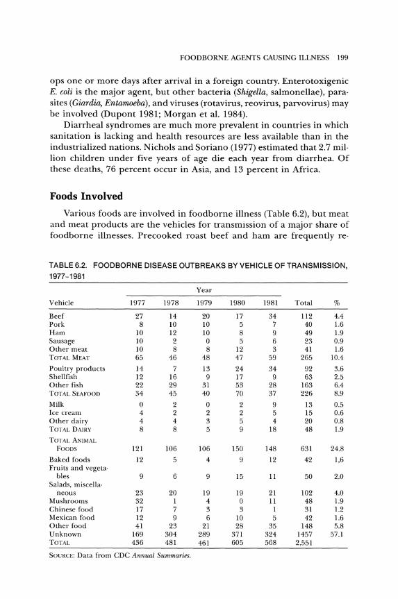
A foodborne disease outbreak is defined by the CDC as an incident in which two or more persons experience a similar illness, usually gastro· intestinal, after ingesting a common food, and epidemiological analysis implicates the food as the source of the illness. For botulism or chemical poisoning, one case constitutes an outbreak (see Figure 6.1).
/ ,-----,
INITIAL REPORT OF ILLNESS
Two or more persons ill ~
j Not compatible with food borne
outbreak
Acute gastroenteritis
Botulism, trichinosis,
chemical, etc.
Compatible with food borne outbreak;
same time & symptoms; common food consumed /\
List on Register;
No outbreak investigation
r-------,
Interstate consumer product; notify
regulatory agent
/\ ...--------, Beef or poultry report to USDA
Other interstate products report
to FDA
J OUTBREAK INVESTIGATION
(Including notification of appropriate local, state, or
federal agencies)
Epidemiology Laboratory Environmental ·Describe disease ·Food samples Hygiene -Define persons -Human specimens -Describe food -Incriminate food -Environmental events
~ L'W"" I -~;~~;:;;'"e po'"'
~I CONTROL I~ Figure 6.1. A scheme for handling food borne disease complaints, to be implemented by state and local health departments. Courtesy of CDC.
197
198 BASIC FOOD MICROBIOLOGY
A microbial foodborne illness may result from ingesting a food con· taining either pathogenic microorganisms or a toxin or poison. When a pathogenic microorganism is the etiologic agent, the illness is called an infection. If a toxin or poison is the causative agent, the illness is called a food intoxication or food poisoning.
Epidemiology
Epidemiology attempts to identify the cause and the mode of trans· mission of infections and to suggest and evaluate methods for controL
The diagnosis of the specific disease is important for treatment and controL With a known etiology, acceptable therapy can be prescribed, dangers from handling patients with infections can be avoided, and the patient can be informed of the possible course of the illness.
Confirmed etiologies are those in which laboratory evidence is ob· tained and fulfill specific criteria of the CDC. The present reporting sys· tem involves many people and agencies. If the affected people do not seek medical help, if the doctor does not report the illness, or if there is no further investigation to confirm the cause of the illness, it is not recorded.
The data recorded by the CDC showed that bacterial agents accounted for 92 outbreaks and 3,270 cases in 1976. This increased to 185 outbreaks and 8,208 cases in 1981 (Table 6.1). It has been estimated that less than 10 percent of the actual outbreaks and cases are reported.
For the United States, estimates as high as 10 or 20 million cases a year have been made. Of the reported outbreaks, only about 50 percent have a confirmed etiology.
The data in Table 6.1 show that two bacterial agents, staphylococci and salmonellae, account for 44 percent of the outbreaks and more than 60 percent of the cases. The salmonellae accounted for more than 65 percent of the deaths due to foodborne agents. The three most promi· nent agents are salmonellae, S. aureus, and C. perfringens, not only in the United States but also in Canada (Todd 1983a), the Netherlands (Beckers 1982), England and Wales (Roberts 1982), and Europe in generaL The most commonly reported cause of foodborne illness in India is staphylo· coccal ent~rotoxin (Hobbs 1982).
Other important bacteria have been C. botulinum and Shigella, with B. cereus showing an increase. Recent additions are Vibrio parahaemolyticus, Yersinia enterocolitica, and Campylobacter jejuni. Pasteurized milk was the ve· hicle for Listeria monocytogenes (Fleming et aL 1985).
Cholera is an important illness worldwide, spread by poor sanitation and contaminated food. There are illnesses, such as traveler's diarrhea, in which symptoms resemble those of several foodborne diseases. The CDC defines traveler's diarrhea as an acute intestinal illness that devel·
FOODBORNE AGENTS CAUSING ILLNESS 199
ops one or more days after arrival in a foreign country. Enterotoxigenic E. coli is the major agent, but other bacteria (Shigella, salmonellae), para· sites (Giardia, Entamoeba), and viruses (rotavirus, reovirus, parvovirus) may be involved (Dupont 1981; Morgan et al. 1984).
Diarrheal syndromes are much more prevalent in countries in which sanitation is lacking and health resources are less available than in the industrialized nations. Nichols and Soriano (1977) estimated that 2.7 mil· lion children under five years of age die each year from diarrhea. Of these deaths, 76 percent occur in Asia, and 13 percent in Africa.
Foods Involved
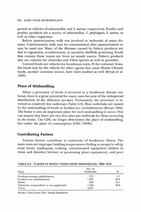
Various foods are involved in foodborne illness (Table 6.2), but meat and meat products are the vehicles for transmission of a major share of foodborne illnesses. Precooked roast beef and ham are frequently reo
TABLE 6.2. FOODBORNE DISEASE OUTBREAKS BY VEHICLE OF TRANSMISSION,
1977-1981
Year
Vehicle 1977 1978 1979 1980 1981 Total %
Beef 27 14 20 17 34 112 4.4 Pork 8 10 10 5 7 40 1.6 Ham 10 12 10 8 9 49 1.9 Sausage 10 2 0 5 6 23 0.9 Other meat 10 8 8 12 3 41 1.6 TOTAL MEAT 65 46 48 47 59 265 10.4
Poultry products 14 7 13 24 34 92 3.6 Shellfish 12 16 9 17 9 63 2.5 Other fish 22 29 31 53 28 163 6.4 TOTAL SEAFOOD 34 45 40 70 37 226 8.9
Milk 0 2 0 2 9 13 0.5 Ice cream 4 2 2 2 5 15 0.6 Other dairy 4 4 3 5 4 20 0.8 TOTAL DAIRY 8 8 5 9 18 48 l.9
TOTAL ANIMAL FOODS 121 106 106 150 148 631 24.8
Baked foods 12 5 4 9 12 42 1,6 Fruits and vegeta·
bles 9 6 9 15 11 50 2.0 Salads, miscella·
neous 23 20 19 19 21 102 4.0 Mushrooms 32 1 4 0 11 48 l.9 Chinese food 17 7 3 3 1 31 l.2 Mexican food 12 9 6 10 5 42 l.6 Other food 41 23 21 28 35 148 .5.8 Unknown 169 304 289 371 324 1457 57.1 TOTAL 436 481 461 605 568 2,551
SOlJRCE: Data from CDC Annual Summaries.
200 BASIC FOOD MICROBIOLOGY
ported as vehicles of salmonellae and S. aureus, respectively. Poultry and poultry products are a source of salmonellae, C. perfringens, S. aureus, as well as other organisms.
Before pasteurization, milk was involved in outbreaks of many dis· eases. Unfortunately, milk may be contaminated after pasteurization or may be used raw. Many of the illnesses caused by fishery products are due to ciguatoxin, scombrotoxin, or paralytic shellfish poisoning. Foods that contain these toxins are from an unsafe source. Fishery products also are vehicles for clostridia and Vibrio species as well as parasites.
Canned foods are vehicles for botulinum toxin. If the container leaks, the foods may be the vehicle for other agents that cause illness. Chinese foods, another common source, have been studied as well (Bryan et al 1982).
Place of Mishandling
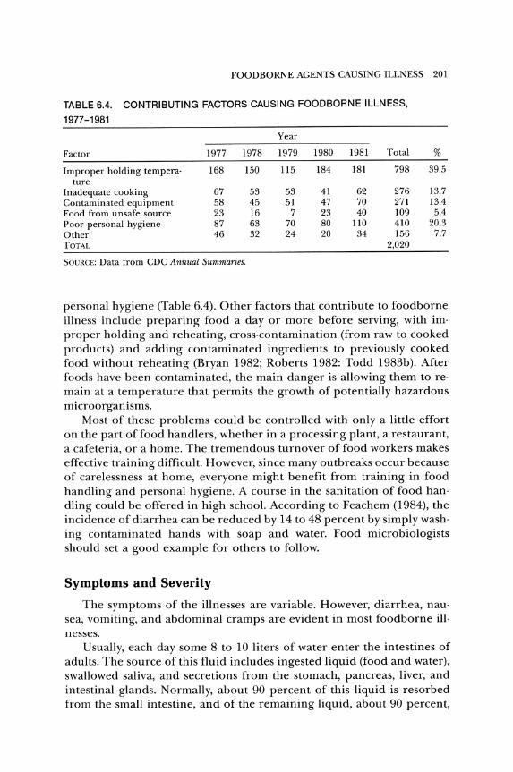
When a processor of foods is involved in a foodborne disease out· break, there is a great potential for many cases because of the widespread distribution of the defective product. Fortunately, the processor is involved in relatively few outbreaks (Table 6.3). Most outbreaks are caused by the mishandling of foods in foodservice establishments (Bryan 1982). The home is also an important place for such mishandling to occur. One can assume that there are very few cases per outbreak for those occurring in the home. The CDC no longer determines the place of mishandling, but rather the place of consumption (CDC 1983b).
Contributing Factors
Various factors contribute to outbreaks of foodborne illness. The main ones are improper holding temperatures (failing to properly refrigerate food), inadequate cooking, contaminated equipment (failure to clean and disinfect kitchen or processing plant equipment), and poor
TABLE 6.3. PLACES IN WHICH FOODS WERE MISHANDLED, 1968-1978
Place
Food-processing establishment Food-service establishment Home Unknown, unspecified, or not applicable TOTAL
SOURCE: Data from CDC Annual Summaries.
No. of Outbreaks
171 1,707
639 1,704 4,221
%
4.1 40.4 15.1 40.4
FOODBORNE AGENTS CAUSING ILLNESS 201
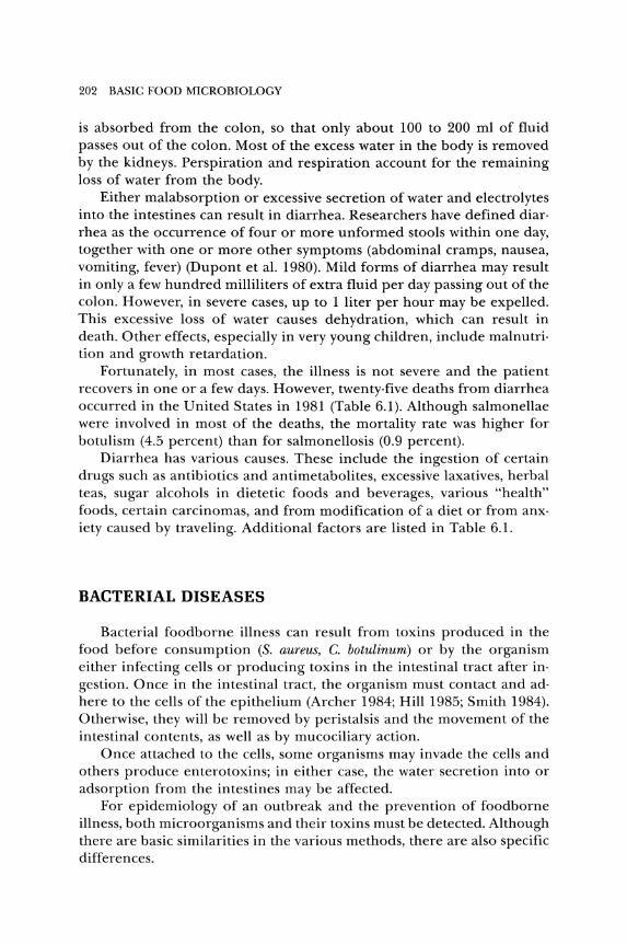
TABLE 6.4. CONTRIBUTING FACTORS CAUSING FOODBORNE ILLNESS,
1977-1981
Year
Factor 1977 1978 1979 1980 1981 Total %
Improper holding tempera· 168 150 115 184 181 798 39.5 ture
Inadequate cooking 67 53 53 41 62 276 13.7 Contaminated equipment 58 45 51 47 70 271 13.4 Food from unsafe source 23 16 7 23 40 109 5.4 Poor personal hygiene 87 63 70 80 110 410 20.3 Other 46 32 24 20 34 156 7.7 TOTAL 2,020
SOVRCE: Data from CDC Annual Summaries.
personal hygiene (Table 6.4). Other factors that contribute to foodborne illness include preparing food a day or more before serving, with im· proper holding and reheating, cross·contamination (from raw to cooked products) and adding contaminated ingredients to previously cooked food without reheating (Bryan 1982; Roberts 1982: Todd 1983b). After foods have been contaminated, the main danger is allowing them to reo main at a temperature that permits the growth of potentially hazardous microorganisms.
Most of these problems could be controlled with only a little effort on the part of food handlers, whether in a processing plant, a restaurant, a cafeteria, or a home. The tremendous turnover of food workers makes effective training difficult. However, since many outbreaks occur because of carelessness at home, everyone might benefit from training in food handling and personal hygiene. A course in the sanitation of food han· dling could be offered in high school. According to Feachem (1984), the incidence of diarrhea can be reduced by 14 to 48 percent by simply wash· ing contaminated hands with soap and water. Food microbiologists should set a good example for others to follow.
Symptoms and Severity
The symptoms of the illnesses are variable. However, diarrhea, nau· sea, vomiting, and abdominal cramps are evident in most foodborne ill· nesses.
Usually, each day some 8 to 10 liters of water enter the intestines of adults. The source of this fluid includes ingested liquid (food and water), swallowed saliva, and secretions from the stomach, pancreas, liver, and intestinal glands. Normally, about 90 percent of this liquid is resorbed from the small intestine, and of the remaining liquid, about 90 percent,
202 BASIC FOOD MICROBIOLOGY
is absorbed from the colon, so that only about 100 to 200 ml of fluid passes out of the colon. Most of the excess water in the body is removed by the kidneys. Perspiration and respiration account for the remaining loss of water from the body.
Either malabsorption or excessive secretion of water and electrolytes into the intestines can result in diarrhea. Researchers have defined diar· rhea as the occurrence of four or more unformed stools within one day, together with one or more other symptoms (abdominal cramps, nausea, vomiting, fever) (Dupont et al. 1980). Mild forms of diarrhea may result in only a few hundred milliliters of extra fluid per day passing out of the colon. However, in severe cases, up to 1 liter per hour may be expelled. This excessive loss of water causes dehydration, which can result in death. Other effects, especially in very young children, include malnutri· tion and growth retardation.
Fortunately, in most cases, the illness is not severe and the patient recovers in one or a few days. However, twenty· five deaths from diarrhea occurred in the United States in 1981 (Table 6.1). Although salmonellae were involved in most of the deaths, the mortality rate was higher for botulism (4.5 percent) than for salmonellosis (0.9 percent).
Diarrhea has various causes. These include the ingestion of certain drugs such as antibiotics and antimetabolites, excessive laxatives, herbal teas, sugar alcohols in dietetic foods and beverages, various "health" foods, certain carcinomas, and from modification of a diet or from anx· iety caused by traveling. Additional factors are listed in Table 6.1.
BACTERIAL DISEASES
Bacterial foodborne illness can result from toxins produced in the food before consumption (S. aureus, C. botulinum) or by the organism either infecting cells or producing toxins in the intestinal tract after in· gestion. Once in the intestinal tract, the organism must contact and ad· here to the cells of the epithelium (Archer 1984; Hill 1985; Smith 1984). Otherwise, they will be removed by peristalsis and the movement of the intestinal contents, as well as by mucociliary action.
Once attached to the cells, some organisms may invade the cells and others produce enterotoxins; in either case, the water secretion into or adsorption from the intestines may be affected.
For epidemiology of an outbreak and the prevention of foodborne illness, both microorganisms and their toxins must be detected. Although there are basic similarities in the various methods, there are also specific differences.
FOODBORNE AGENTS CAUSING ILLNESS 203
Staphylococcal Intoxication
This illness accounted for over 17 percent of the outbreaks and al· most 34 percent of the cases of reported foodborne illnesses in the United States in 1981 (Table 6.1). However, the actual extent of this ill· ness is not known.
CHARACTERISTICS OF THE INTOXICATION. The characteristics of an illness can aid in determining the causative agent in an outbreak of foodborne illness.
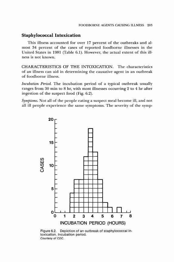
Incubation Period. The incubation period of a typical outbreak usually ranges from 30 min to 8 hr, with most illnesses occurring 2 to 4 hr after ingestion of the suspect food (Fig. 6.2).
Symptoms. Not all of the people eating a suspect meal become ill, and not all ill people experience the same symptoms. The severity of the symp·
CI) W
20
15
CI) 10 ~ ()
5
r-
~
I- -I- -I- -I- -I- -f-- - l-I-
l-
f--
l-
f--
f--
f--
l-
f--
l-
I- 1 r-II n II I o
012345678
INCUBATION PERIOD (HOURS)
Figure 6.2. Depiction of an outbreak of staphylococcal in· toxication. Incubation period. Courtesy of CDC.
204 BASIC FOOD MICROBIOLOGY
TABLE 6.5. PERCENTAGE OF ILL PERSONS EXPERIENCING SPECIFIC SYMPTOMS DUE TO STAPHYLOCOCCAL INTOXICATION
Outbreaks
Symptoms 2 3 4
Nausea 76 100 50 Vomiting 70 44 100 77 Diarrhea 19 67 100 82 Abdominal cramps 71 71 58 15 Chills 25 46 Headache 42 6 41 Prostration 63 Weakness 68 Leg cramps Muscle soreness 5 Collapse 9 Hypotension 1 Fever 25 7 21
- = Not reported. SOURCE: Data from CDC Morbidity and Mortality Weekly Reports.
5
41 76 73 16
35
73 11
24
toms varies with the concentration of enterotoxin in the food, the amount of food consumed, and the susceptibility of the individual. The principal symptoms listed in Table 6.5 are nausea, vomiting, abdominal cramps, and diarrhea.
Duration and Therapy. Symptoms of the illness usually subside after one or two days. The illness is rarely fatal. Due to the sudden onset and short duration of the illness, treatment usually is not needed. However, hospi· talization is required in cases in which shock, dehydration, and extensive vomiting have occurred. In these cases, therapy includes replacing lost fluids and electrolytes. According to Holmberg and Blake (1984), 10 per· cent of the victims seek hospital care.
ETIOLOGIC AGENT. This illness is called an intoxication because the etiologic agent is an enterotoxin. Payne and Wood (1974) listed six known staphylococcal enterotoxins (A, B, C, D, E, F), (SEA, SEB, SEC, SED, SEE, SEF) based on serological reactions. Although SEC) and SEC2
react with the same antibody and are often lumped together as SEC, they also react with distinct minor antibodies. The presence of a third SEC, enterotoxin C3, has also been reported (Reiser et al. 1984).
Enterotoxin A is the most frequently encountered enterotoxin in food poisoning outbreaks in the United States. In New Zealand, Jarvis and Harding (1972) found enterotoxins C and D to be more prevalent than SEA. SEB rarely is involved in food poisoning outbreaks in the United States.
FOOD BORNE AGENTS CAUSING ILLNESS 205
Properties of the Enterotoxins. Staphylococcal enterotoxins are simple pro· teins with a molecular weight between 25,000 and 35,000. They are read· ily soluble in water and salt solutions.
The heat stability is an important characteristic of staphylococcal enterotoxins. Enterotoxin B is more heat resistant than A or D. The heat resistance is influenced by the medium in which it is heated (composi· tion, pH). In foods, the enterotoxins are not completely inactivated by normal cooking, pasteurization, or other usual heat treatments. Tatini (1976) stated that thermal processing cannot be relied upon to inactivate these toxins. Further, he found that heated toxins had greater biological activity than unheated toxin when tested at the same dose level.
Action of Enterotoxins. Researchers used monkeys to study enterotoxin· induced emesis (vomiting) (Elwell et al. 1975). Their results indicate that enterotoxin is not absorbed from the intestine. Orally ingested enter· otoxin is thought to mediate emesis by acting on sites in the intestine. This stimulus apparently is transferred by the vagus and sympathetic nerves to the vomiting center, which is part of the central nervous system. The vomiting center somehow induces retroperistalsis of the stomach and small intestine, resulting in emesis. The exact systems involved in the vomiting syndrome have not been determined. The known facts and hypotheses have been discussed and described elsewhere (Robins· Browne 1980; Thompson and Malagelada 1982; Van Miert et al. 1983). Since the nervous system is involved, it has been suggested that the enter· otoxins should be called neurotoxins. However, neuronal binding of SEA in the intestinal tract was not demonstrable (Beery et al. 1984).
The action of staphylococcal enterotoxins in the diarrheal syndrome is not known. Staphylococcal enterotoxin shows an affinity to the walls of the stomach and the small and large intestines. If sufficient enterotoxin is present in consumed food, it causes inflammation and irritation of the lining in the stomach and intestinal tract. Working with flounder intes· tine in vitro, the data of Huang, Chen, and Rout (1974) suggested that enterotoxin stimulates active sodium and chloride secretion. Others found that enterotoxin B did not interfere with water absorption in the guinea pig ileum, but that staphylococcal delta toxin did inhibit absorp' tion in the jejunum and ileum (Kapral et al. 1974).
Amount of Enterotoxin Needed for Illness. There are no definite data concern· ing the minimum amount of enterotoxin needed to cause symptoms in a human. Gilbert (1974) listed estimates that ranged from 0.015 to 0.357 p,g of enterotoxin per kilogram of body weight. Besides body weight, indio viduals vary in their sensitivity to enterotoxins.
FOODS INVOLVED. Various foods have been involved in staphylococ· cal intoxication since 1974 (Table 6.6). More than 35 percent of the out·
206 BASIC FOOD MICROBIOLOGY
TABLE 6.6. FOODS INVOLVED IN STAPHYLOCOCCAL INTOXICATIONS 1976-1981
Years
Food 1976-1978 1979-1981 Total %
Beef 3 5 8 4.5 Ham 22 19 41 23.3 Pork 1 1 2 1.1 Other meat 5 7 12 6.8 Poultry 7 16 23 13.1 Shellfish 1 0 1 0.6 Other fish 0 1 1 0.6 Milk 0 1 1 0.6 Other dairy 1 2 3 1.7 Bakery products 3 10 13 7.4 Fruits and vegetables 1 1 2 1.1 Salads 13 13 26 14.8 Other foods 6 6 12 6.8 Unknown 11 20 31 17.6 TOTAL 74 102 176
SOURCE: Data from CDC Annual Summaries.
breaks involved red meats, with 23 percent being caused by contami· nated ham. Fresh meats are involved less often than cured meats since, being perishable, they are refrigerated. Cured meats are not as perishable and may be mishandled by being allowed to remain at room temperature. Also, it is difficult for S. aureus to compete with the microbial flora on fresh meat as compared to that on cured meat.
The poultry products involved are usually either barbecued or in salads. Other salads (potato, macaroni, and tuna fish) have been implicated in more than 14 percent of the outbreaks. Bakery products containing custard or cream are important offenders.
In many outbreaks, the food is cooked, then contaminated with S. aureus during handling and held at a temperature for growth and production of enterotoxin.
THE ORGANISM (S. AUREUS). S. aureus is described briefly in Chapter 3. These Gram-positive cocci produce colonies that may be white or have pigment ranging from yellow to orange. The color is influenced by growth conditions as well as strain variation.
In both aerobic and anaerobic conditions, S. aureus produces acid from mannitol, glucose, lactose, and maltose_ Aerobically, many carbohydrates are metabolized with the production of acid, but acid is not produced from arabinose, cellobiose, dextrin, inositol, raffinose, rhamnose, or xylose.
Typical S. aureus strains produce a-toxin, coagulase, and a heat-stable nuclease_
FOODBORNE AGENTS CAUSING ILLNESS 207
SOURCES. Some reports call S. aureus ubiquitous because it is so widespread (air, dust, clothing, floors, water, sewage, and insects). The principal source of S. aureus is the human nose, although it is found on the skin, especially on the hands, in infected wounds, burns, boils, pimples, acne, in nose and throat discharges, and in feces_ The primary site on the hands is the fingertips, which relates to the habit of handling one's nose with the fingers. The extent of nasal carriers is difficult to determine, but surveys have shown the carrier rate to vary from 6 percent to over 60 percent of the population_ People associated with hospitals tend to have a higher carrier rate than the normal population_
Animals are a source of S. aureus. Most of the strains of S. aureus isolated from animals tend to have characteristics different from those asso· ciated with people (Devriese 1980; Kibenge, Wilcox, and Perret 1982). The organism is found in food-processing operations (Harvey, Patterson, and Gibbs 1982; Notermans, Dufrenne, and Van Schothorst 1982), and at a high level on and in healthy food handlers (Holmberg and Blake 1984).
GROWTH. Researchers believe that 105 to 106 cells of S. aureus per gram of food must generally be present before the production of enterotoxin reaches a level that can cause intoxication. Due to the normally low numbers in food, multiplication must occur. By knowing and understanding the factors affecting the growth of S. aureus, we can control the growth, enterotoxin production, and outbreaks of staphylococcal intoxication. The general factors affecting the growth of S. aureus are described in other sections of this text. S. aureus is a relatively poor competitor, and various bacteria can inhibit or outgrow it. This inhibitory interrelationship of other bacteria with S. aureus is important in preventing toxin pro· duction in foods. It may be a primary reason for certain foods to be less involved than others in outbreaks of staphylococcal intoxication. In foods with aw of 0.90 to 0.95 or with 5 to 10 percent salt, S. aureus can dominate because most other bacteria cannot grow.
Strains oflactic streptococci (S. lac tis and S. cremoris) inhibited S. aureus inoculated into milk prior to cheese making (Ibrahim 1978). Since cheese has been a vehicle of staphylococcal enterotoxin, it is evident that microbial competition cannot be relied upon to always inhibit growth and toxin production of S. aureus. Also, it has been suggested that some competitive organisms may degrade the enterotoxins.
Obviously, S. aureus is able to grow on or in foods that have been involved in staphylococcal intoxication. Some outbreaks have occurred by holding food at room temperature for less than 4 hr. Longer incubation times increase the risk. The behavior (growth or inhibition) of S. aureus has been reported for various cheeses (Koenig and Marth 1982;
208 BASIC FOOD MICROBIOLOGY
Magrini, Chirife, and Parada 1983), pumpkin pie (Wyatt and Guy 1981), potatoes (Tamminga et al. 1978), and meat (Lui ten , Marchello, and Dry· den 1982). The maximum temperature for growth of S. aureus was in· creased by the addition of salt, monosodium glutamate, or soy sauce (Hurst and Hughes 1983).
Sublethal treatments and other factors (pH, aw , Eh, temperature) discussed in Chapter 4 act to control growth and enterotoxin production of S. aureus. Growth was not observed at aw 0.85 or below, pH 4.3 or less, or at 8°C or less (Notermans and Heuvelman 1983). Enterotoxin produc· tion required higher aw , pH, or temperature than that needed for growth but was mainly inhibited by the effect of aw •
When warm or hot foods are refrigerated, a long time may be needed to cool the food sufficiently to prevent growth of S. aureus. Foods with gravies or sauces cool slower than do those without, and the substrate is more readily available for growth of organisms.
TOXIN PRODUCTION. S. aureus produces several toxins, including those listed in Table 6.7. The main difference between exotoxins and endotoxins is not whether they are outside or inside the cell, but rather their structure. Exotoxins are proteins with little or no nonprotein resi· dues. Endotoxins are primarily polysaccharide and lipid complexes (lipo· polysaccharides). Food microbiologists are concerned with the enterotox· ins, which, by definition, are exotoxins, since they are protein as well as being found free from the cell.
Not all strains of S. aureus are enterotoxigenic. Schroeder (1967) esti· mated that only 4 percent of the staphylococcal strains in milk were capa· ble of producing enterotoxin. In a survey of S. aureus in meat, dairy prod· ucts, and other foods, Payne and Wood (1974) found that 125 of 200 strains (62.5 percent) produced enterotoxins. Wieneke (1974) found a somewhat higher ratio of enterotoxigenic strains in cooked food than in
TABLE 6.7. SOME TOXINS OF STAPHYLOCOCCUS AUREUS
Toxin
a-toxin J3-toxin (phospholipase C) 'Y-toxin I)-toxin Hyaluronidase Staphylococcal coagulase Staphylokinase Leukocidin Epidermolytic Enterotoxins (A, B, C, D, E, F) Toxic shock toxin
Action
Hemolytic, dermonecrotic, lethal Hemolytic Hemolytic Hemolytic, enteric Spreading factor Coagulates plasma Fibrinolytic Kills leucocytes Exfoliation Emetic Lymphoid hyperplasia
FOODBORNE AGENTS CAUSING ILLNESS 209
raw food. The higher ratio may be due to people contaminating cooked foods with human strains. Such strains are more frequently enterotoxi· genic than are strains from animal or other sources.
The production of SEA results from a chromosomal gene (Mallonee, Glatz, and Pattee 1982; Pattee and Glatz 1980; Shafer and Iandolo 1978b). Shafer and Iandolo (l978a) reported that SEB production of strains S6 and 277 is determined by a chromosomal gene or genes and suggested that this might be the case for many enterotoxigenic strains of S. aureus. Dyer and Iandolo (1981) proposed that SEB production is dependent on at least two unlinked genes. Altboum, Hertman, and Sarid (1985) reo ported that elimination of plasmid pZA10 from strain 6344 caused the loss of SEB and SEC] production. According to these researchers, the plasmid can be transferred and confer toxigenicity to other S. aureus strains, and chromosomal DNA can integrate the plasmid.
The conditions necessary for growth and enterotoxin production have been reviewed (Smith, Buchanan, and Palumbo 1983). In general, toxin production occurs in a more narrow range of environmental char· acteristics than those discussed for growth in Chapter 4.
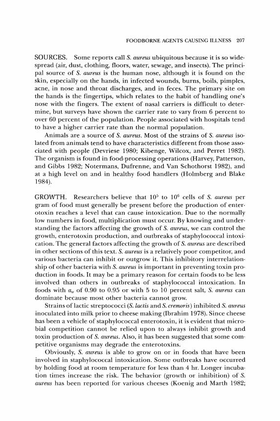
The relationship of growth of S. aureus and SEB production was reo ported by Markus and Silverman (1969) and is shown in Figure 6.3. The rate of synthesis of SEB is greater than that for SEA, so higher concentra· tions of SEB are obtained. Tweten and Iandolo (1983) suggested that a precursor of SEB is bound to the cell membrane. At some stage, mature SEB is formed and released by the membrane into a specialized compart· ment in the cell wall. From there, it is released to the exterior environ· ment.
A constant dissolved oxygen (DO) level of 100 percent stimulated growth, but enterotoxin production was not observed (Carpenter and Silverman 1974). A DO of 10 percent yielded a higher level of enter· otoxin than did a DO of either 100 percent or 50 percent.
Aerobically, certain strains of S. aureus produce enterotoxin at a pH of 4.8, but anaerobically, no enterotoxin is found at pH 5.4 (Barber and Deibel 1972). Their results indicate that biological acid production can· not be relied upon to inhibit S. aureus in fermented sausage, and they recommended chemical acidulation. Since the toxin is produced at a higher level aerobically than anaerobically, they suggested sampling aero· bic portions of foods for the toxin. Mixing and cross·sectional sampling merely dilute the toxin with anaerobic portions of the food, which con· tain little or no toxin.
S. aureus (A100) grew and produced enterotoxin in precooked bacon stored aerobically either at 37°C and a minimum aw of 0.84, or at 20°C and an aw of 0.89 (Lee, Silverman, and Munsey 1981). Anaerobically, the organism required a minimum aw of 0.90 at 37°C, and 0.94 at 20°C.
210 BASIC FOOD MICROBIOLOGY
200
160
E ...... 120 01 ~
a:l
Z X 80 o b It: LLI
!z LLI
40
TIME
BACTERIA
ENTEROTOXIN B
Figure 6.3. Growth and Enterotoxin B production of S. aureus. courtesy of Markus and Silverman (1969).
en ::i en Z ct (l) It: o
10'~ It: LLI a:l ::i :;) z (l)
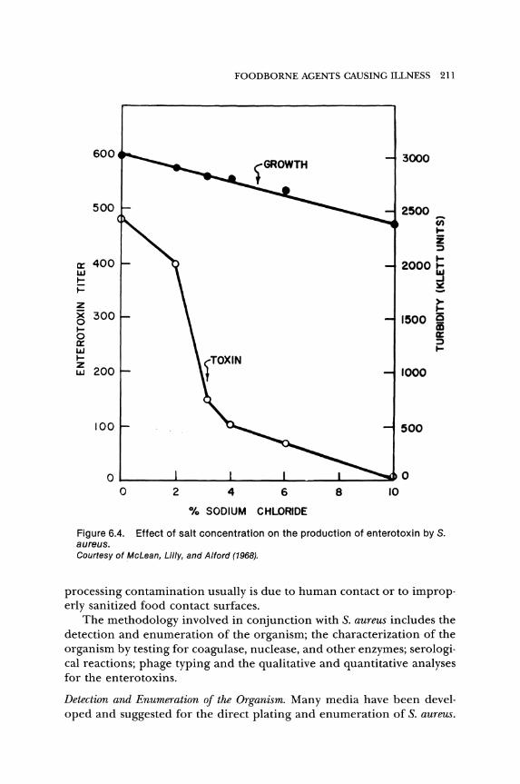
o ..J
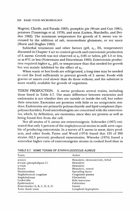
The effect of sodium chloride on growth and enterotoxin production was reported by McLean, Lilly, and Alford (1968) and is shown in Figure 6.4.
METHODOLOGY. Foods or other samples are examined for S. aureus and the enterotoxins to demonstrate postprocessing contamination, to determine a potentially hazardous product, or to confirm a causative agent in a foodborne illness. Since S. aureus is sensitive to heat and sani· tizing agents, the presence of the organism or its toxins in processed food or on equipment generally indicates poor sanitation or handling. Post-
FOODBORNE AGENTS CAUSING ILLNESS 211
600 3000
500 2500 u; ~ z :;)
a:: 400 2000 § w ~
~ lII: ..... Z >
~ g 300 1500 is ~ as 0 a:: a:: :;)
w ~ ~ z
200 w 1000
100 500
o 1--__ ....... ___ .......... __ ---'1.....-__ --'-__ --'"' ... 0
o 2 4 6 8 10
% SODIUM CHLORIDE
Figure 6.4. Effect of salt concentration on the production of enterotoxin by S. aureus. courtesy of McLean, Lilly, and Alford (1968).
processing contamination usually is due to human contact or to improp· erly sanitized food contact surfaces.
The methodology involved in conjunction with S. aureus includes the detection and enumeration of the organism; the characterization of the organism by testing for coagulase, nuclease, and other enzymes; serologi· cal reactions; phage typing and the qualitative and quantitative analyses for the enterotoxins.
Detection and Enumeration of the Organism. Many media have been devel· oped and suggested for the direct plating and enumeration of S. aureus.
212 BASIC FOOD MICROBIOLOGY
The FDA (1978) method uses Baird-Parker agar_ This medium contains potassium tellurite as a selective agent and egg yolk and tellurite as differential agents_ On this medium, S_ aureus colonies appear circular, smooth, convex, moist, gray to jet-black, frequently with an off-white margin, due to reduction of the tellurite to elemental tellurium_ They are surrounded by an opaque zone and an outer clear zone due to the reaction on the emulsified egg yolk_ When touched with an inoculating needle, the colonies have a buttery to gummy consistency_ There may be variations to this description_ Typical colonies are selected for further study_
A collaborative study revealed that Baird-Parker (BP) agar is satisfactory for recovery of cells stressed or injured by heat or other processing conditions (Baer et al. 1975)_
, Baird-Parker agar, modified by substituting pig plasma for egg yolk, uses the coagulase reaction for differentiation and is as effective as the original agar for growing stressed S_ aureus cells (Becker, Terplan, an9 Zaadhof 1983; Idziak and Mossel 1980)_ The addition of bovine fibrinogen to plasma-modified BP agar was found to be effective in detecting S. aureus and also determines the nuclease reaction (Beckers et al. 1984; Chopin et al. 1985).
Characterization_ Colonies typical of S. aureus on an agar surface are selected for further testing and characterization. These tests may include various fermentations of carbohydrates, the presence of coagulase, heatstable nuclease or lysozyme, and determining the resistance to chemical inhibitors or antibiotics (Baker 1984). None of these tests, or a combination of them is an absolutely reliable indicator of enterotoxin formation by the organism.
The coagulase test is considered the most reliable single test for differentiating potentially pathogenic S. aureus. However, not all coagulasepositive strains produce enterotoxins. On the other hand, there are reports that coagulase-negative strains produce enterotoxin or have been involved in cases of staphylococcal intoxication (Lotter and Genigeorgis 1975). Some enterotoxigenic strains of staphylococci found to be coagulase negative on first isolation, become coagulase positive after subculturing several times, and some strains may lose the ability to produce coagulase but remain enterotoxigenic.
The coagulase reaction in clotting blood serum was reviewed by Tager (1974). According to this review, there are three main reactions, ending with the conversion of fibrinogen to fibrin and formation of a clot. According to Baird-Parker (1972), over 90 percent of the strains of S. aureus produce a coagulase. Several tests have been suggested and evaluated to detect the presence of coagulase. A tube test is used to determine free coagulase, and a slide test detects a clumping factor. These are distinct
FOODBORNE AGENTS CAUSING ILLNESS 213
entities. A latex slide agglutination test to determine clumping factor and protein A simultaneously was described by Essers and Radebold (1980) and reported to be as accurate as the tube test and more accurate than the slide test by Doern (1982).
Although other organisms may produce nucleases, the heat stability of S. aureus nuclease is unique. It is generally agreed that the enzyme loses but little activity by boiling for 30 min. It retained 10 percent of its activity after heating at 100°C for 180 min, 120°C for 34 min, or l30°C for 16 min (Erickson and Deibel 1973).
The basic test for staphylococcal nuclease was described by Lachica, Genigeorgis, and Hoeprich (1971). Since then, several suggestions for im· provement have been made (Ibrahim 1981; Pham and Davis 1979; Stad· houders, Hassing, and Galesloot 1980). However, other staphylococci produce thermostable nuclease (Gudding 1983). These nucleases can be distinguished from each other by seroinhibition, the inhibition of nuclease activity by specific antibodies (Gudding 1983; Lachica, Genigeorgis, and Hoeprich 1979).
The thermostable nuclease test might be used as a screening method for foods to detect the possible presence of high populations of S. aureus or enterotoxin (Tatini et al. 1975; Tatini, Cords, and Gramoli 1976). This system seemed to be applicable to certain foods, but was of questionable value for others. Ibrahim and Baldock (1981) always detected nuclease in cheese that contained enterotoxin.
Medwid and Grant (1980) questioned the value of the thermonuclease test for foods containing high numbers of Streptococcus faecalis subsp. liquejaciens, since this organism inactivated the enzyme.
Phage Typing. Some bacteriophages are very specific for the cells they will lyse, while other phages have a wider spectrum of susceptible cells. Obviously, for phage typing of S. aureus, those phages that have a limited spectrum of susceptible cells are desirable.
The International Committee on Nomenclature of Bacteria established a Subcommittee on Phage Typing of Staphylococci in 1953. The Staphylococcus Reference Laboratory of the British Public Health Laboratory Service in London became the International Reference Center for the subcommittee. This subcommittee established a set of phages for routine testing of S. aureus. In 1961, it became the World Health Organization Centre for Staphylococcus Phage Typing. A description of the system used for phage typing was reported by CDC (1976b) and Parker (1972).
Phage typing is of particular help in the epidemiological study of staphylococcal intoxications or infections. Various strains isolated during an outbreak from patients, foods, and food handlers can be tested for
214 BASIC FOOD MICROBIOLOGY
their phage patterns. This information makes it possible to differentiate between a strain responsible for the outbreak and unrelated strains.
Smith (1972) stated that there are only two good reasons for typing S. aureus, either by phages or antisera. The main reason is for epidemio· logical investigations, and the second reason is to give a label to the or· ganism for research work.
Enterotoxin Detection. The detection of enterotoxin is important. If an out· break occurs and isolates of S. aureus are obtained from a food, the enter· otoxins produced can be compared to the enterotoxin causing the intoxi· cation. In one outbreak involving dried milk, no S. aureus isolates were detected. In this case, the organisms multiplied, produced the toxin, and then were killed by heat during the processing. In cases such as this, the food can be analyzed for the enterotoxin.
The principal methods for detecting enterotoxins can be separated into biological and serological systems.
A biological system can be some living entity or part thereof. The best living entity to determine enterotoxin activity is a human. However, humans are not always readily available. Also, the amount of enterotoxin cannot be determined, because people vary in their sensitivity to enter· otoxin.
Animals such as monkeys, chimpanzees, dogs, pigs, cats, and kittens have been tested as biological systems. Monkeys and chimpanzees can be given the enterotoxin orally, whereas the other animals are injected either intraperitoneally or intravenously. Because vomiting is the first symptom to occur and is the most readily observable reaction to enter· otoxin, animals such as rodents that do not have a vomiting mechanism are not used. Pigeons, frogs, tropical fish, nematodes, or various protozoa or bacteria show no obvious reaction with enterotoxins. Tissues includ· ing human intestinal cells, rabbit gut segments, chicken embryos, tissue cultures (HEp-2 and HeLa cells) and isolated enzyme systems have been tested for assay of enterotoxin. Although crude enterotoxins have shown some effect on some of these systems, the purified toxins have revealed little or no effect.
Since monkeys or chimpanzees can be fed the enterotoxin orally, they are the preferred test animal. The monkey is not as sensitive to enterotoxin as humans are and the sensitivity of chimpanzees is somewhere between the sensitivities of humans and of monkeys. Another disadvantage is that these animals are expensive to maintain and, as they are used to assay enterotoxin, they tend to develop a resistance (a limited immunity) to the toxins. Thus, their usefulness for enterotoxin detection is limited. A direct skin test on sensitized guinea pigs reportedly has a sensitivity of 10 to 100 pg of SEB per milliliter of prepared food sample (Scheuber et al. 1983).
FOODBORNE AGENTS CAUSING ILLNESS 215
Tests using animals were necessary before the enterotoxins were purified and serological tests could be developed_ It is evident that the animal tests are still important for the detection of any new types of enterotoxins, or testing the toxicity of isolated compounds, or a particular chemical portion of a toxin_
Serological systems that have been suggested for assaying enterotoxins include Ouchterlony double-diffusion plate (Ouchterlony 1968), Oudin single-gel diffusion tube, Oakley double-gel diffusion tube, microslide (Casman and Bennett 1965), polyvalent single radial immunodiffusion (Meyer and Palmieri 1980), hemagglutination inhibition, reversed passive hemagglutination (Yamada, Igarishi, and Terayama 1977), immunofluorescence, radioimmunoassay and enzyme-linked immunosorbent assay (ELISA) tests_
The Ouchterlony system was adapted for use on a glass slide by Wadsworth_ A modification of this microslide method was used by Casman and Bennett (1965) to detect enterotoxin in foods. Bennett (1971) stated that 0.005 J-tg of enterotoxin A per gram of cheese was detectable. In order to determine low levels of enterotoxin, procedures of extraction, separation, and concentration are needed (Bennett and McClure 1980).
According to a collaborative study reported by Bennett and McClure (1976), the microslide gel double-diffusion test has a high degree of specificity, is simple, and has good reproducibility in the identification of enterotoxins. This system has been adopted as official by the AOAC.
The various diffusion tests have been used with varying results. The radioimmunoassay (RIA) technique reportedly can detect as little as 0.1 ng/ml of SEA and 0.5 ng/ml of SEB (Areson, Charm, and Wong 1980). However, the radioactive label deteriorates and should be replaced after about two months. Radioactive waste is created. The system also requires expensive equipment and scarce and expensive purified toxins.
To overcome some of the problems of the RIA, Saunders and Bartlett (1977) suggested an enzyme immunoassay (EIA). Using an EIA, Kauffman (1980) stated that it was sensitive to 2 ng SEA/ml. He considered this method to be sensitive and precise enough to serve as an alternative to the RIAThe FDA guidelines for acceptable enterotoxin detection is 1 to 2 ng/g of food_
Procedures using ELISA now have the greatest potential for the detection of recognized enterotoxins. First, a measured amount (100 J-t1) of purified enterotoxin is added to wells in a polystyrene plate. After sufficient time at 4°C to allow the toxin to bind to the plastic, the liquid is removed and the wells filled with serum albumin to block any residual binding sites on the plastic. Then the wells are emptied, washed with buffer, and known antitoxins, to which an enzyme such as peroxidase or alkaline phosphatase is conjugated, are added_ After about 2 hr, the wells are aspirated to remove the liquid and washed to remove excess conju-
216 BASIC FOOD MICROBIOLOGY
gated antitoxin. Then a substrate is added which the conjugated enzyme can change to a colored compound. If there is no enterotoxin, there is nothing to which the antitoxin can react. It, along with the enzyme, are removed by washing, and there will be no color reaction when the sub· strate is added. It is evident that the amount of enterotoxin is related to the amount of enzyme·conjugated antitoxin that remains, and that is reo lated to the intensity of color that is formed by the enzyme acting on the substrate for a specified time. The testing of known amounts of enter· otoxin is used to establish the color intensities produced in a specified time, so that the ELISA is a quantitative procedure. Extensive purifica· tion of the toxin in the sample is not needed for the ELISA system. There are several variations to this basic system.
Using a competitive ELISA, SEA, SEB, or SEC concentrations of 0.1 ng or less were measured (Stiffler·Rosenberg and Fey 1978). Four ver· sions of the ELISA were compared by Fey, Pfister, and Ruegg (1984). They stated that the sandwich with labeled antibody was the best, with a sensi· tivity of 0.1 ng of enterotoxin per milliliter. To detect enterotoxin in food, the ELISA is as sensitive as the RIA (Freed et al. 1982; Kuo and Silverman 1980) and more sensitive than the microslide technique (Lenz et al. 1983).
Injection of an antigen into an animal results in the production of antibodies. A single B cell produces a single antibody specific for a single antigenic determinant. However, the B-cell population is polyclonal in nature and produces heterogeneous, polyclonal antibodies even when a highly purified antigen is injected. If the B cells were a monoclonal population, a large amount of single antibody would be produced. It is difficult to culture individual B cells. However, when B cells are fused with myeloma (tumor-producing) cells, the resultant hybridoma combines the antibody production of the B cell and the reproduction of the myeloma cell. The hybridoma can be cultured and antibody harvested from the culture supernatant, or it can be obtained from the ascites fluid of a host animal bearing a hybridoma tumor.
The monoclonal antibodies produced by the hybridoma are very specific for a particular antigenic determinant. However, monoclonal antibodies do not readily cross-link when binding to antigen. Cross-linking is important in diagnostic tests, which depend on precipitation reactions, such as the gel-diffusion tests. Also, if two or more antigens share a common determinant recognized by the monoclonal antibody, crossreactions can occur.
The above is a rather simplistic description of monoclonal antibody production. More complete descriptions of production or use can be found in many publications, including articles by L0vborg (1984), Sherman (1984), and van Hell and Helmich (1984). Monoclonal antibodies
FOODBORNE AGENTS CAUSING ILLNESS 217
have been produced for staphylococcal enterotoxins (Edwin et al. 1984; Meyer et al. 1984; Thompson, Ketterhagen, and Bergdoll 1984).
Although the biological activity of the enterotoxins is not measured by these tests, Bergdoll (1970) stated that correlation between the two is adequate to justify the use of serological tests to analyze for enterotoxins. Chang and Dickie (1971) interpreted their observations with enterotoxin B to mean that antigenic sites and toxin sites are probably not the same. They did not speculate on whether this would invalidate the serological tests as methods for the analysis of the enterotoxins. Other researchers reported that heating in gelatin caused both immunological inactivation and biological inactivation of SEB, indicating a possible relationship (Notermans et al. 1983).
CONTROL OF STAPHYLOCOCCAL INTOXICATION. It should be relatively simple and easy to prevent staphylococcal intoxication. The obvious control measure would be to keep the organisms out of foods. For those S. aureus that do invade food, methods to destroy them or to prevent their growth and enterotoxin production can be employed. If toxin is produced, then destruction of the toxin is needed.
Prevent Contamination. It is impossible to keep all foods free from S. aureus due to the ubiquitous character of the organisms. However, it would seem possible to use common sense measures to keep the contamination low.
Humans are the main reservoir of the organism. The health, hygiene, and work habits of food handlers can influence the level of contamina· tion of foods. People who are sick should not be allowed to handle food. They contaminate the food not only with S. aureus, but also with other disease organisms. People with colds, sinusitis, or other respiratory dis· turbances are good sources of S. aureus, as are boils, pimples, acne, and infected cuts. It is not possible to segregate all carriers of S. aureus, since a sizable portion of the population may be involved. However, those people who are obviously contaminators of food should be removed from food·handling areas.
S. aureus has been isolated from knives and slicers during investiga· tions of outbreaks of food poisoning. Food·handling equipment should be designed so that it can be properly and effectively cleaned and sani· tized. The cleaning and sanitizing of equipment are discussed in Chap· ter 9.
Prevent Growth and Toxin Production. The main control of S. aureus is to hold the food at a temperature unsatisfactory for growth. There is little or no growth below 4°C or above 46°C.
Toxin production occurs in a narrower temperature range than cellu· lar growth. Troller (1976) suggested a range of 10° to 45°C. Greater
218 BASIC FOOD MICROBIOLOGY
amounts of enterotoxin are produced in the range of 33° to 38°C than at either higher or lower temperatures.
Obviously, to warm the product for serving, or to cool leftover heated food, the temperature of the food must go through the growth range. This range must be traversed as quickly as possible. Three hours has been suggested as the maximum time allowed. To facilitate cooling of leftover food, the food should be placed in shallow layers in shallow pans. The deeper the food, the longer it will take to cool. The warm food should not be held at room temperature to cool, but should be placed in the refrigerator. This means that the refrigeration system must be adequate to cool these potential extra loads. The refrigerator cannot be jammed full of hot food, since no refrigerator can handle such a condition.
In fermented sausage products, S. aureus can be controlled by lower· ing the pH with starter cultures or chemical acidulation (Daly et al. 1973; Raccach 1981). A combination of a culture and acidulation or chemical inhibitor is more effective than either treatment alone. Adding solutes or drying of foods to lower the aw below 0.85 should prevent growth and enterotoxin production of S. aureus.
Dipping poultry carcasses in a 5 percent potassium sorbate solution reduced the growth of inoculated S. aureus (To and Robach 1980). In model systems, concentrations above 3 percent were needed to inhibit 50 percent growth of S. aureus (Lahellec, Fung, and Cunningham 1981). Butylated hydroxyanisole (BHA) was reported to be lethal to S. aureus at 100 {tg/ml (Degre and Sylvestre 1983). Although Parada, Chirife, and Magrini (1982) found some growth inhibition of S. aureus in a model sys· tem by 100 {tg/ml of either BHA or butylated hydroxy toluene, neither was effective in delaying growth in a cheese spread (aw of 0.976).
Destroy S. aureus, Inactivate Toxin. Heating is the principal method used to kill S. aureus cells as well as other organisms. Although heat may kill the cells, if enterotoxin has been produced, the toxin may persist, because, in a food, the enterotoxin is more heat stable than the organism.
Not only is the temperature important, the time of exposure is also important. The temperature and time needed to kill an organism depend on factors such as the heat resistance of the organism, the number of cells present, the age of the cells, the type offood or suspending medium, the ingredients added to the food, the pH, a"" and previous treatment of the food or organism (stress conditions). Thermal processing is described elsewhere in the text, so only a brief preview is given here.
To help describe the heat resistance of an organism, D and z values are used. The D value is used to describe the time· temperature relation· ship to the killing of an organism. A l·D is the time needed at a specified temperature to reduce the number of cells by 90 percent, or, conversely,
FOODBORNE AGENTS CAUSING ILLNESS 219
10 percent of the cells survive this treatment. The z value is a measure of the effect of a change in temperature on the resistance of an organism. It is the of or °c required for the thermal resistance to change by a factor of lO. Most of the thermal evaluations are reported in of; however, when possible, the of have been converted to °C.
Angelotti, Foter, and Lewis (1961) determined the D values of various organisms in foods. In custard heated at 60°C, the D value for S. aureus 196E was 7.82 min, while for S. aureus Ms 149 it was 7.68 min. This means that if 100 cells of S. aureus Ms 149 were present per gram of custard, heating for 7.68 min at 60°C would reduce this number to 10. In another 7.68 min, there would be only 1 cell per 10 gram and after another 7.68 min, only 0.1 of a cell per gram, or 1 cell per 10 grams of custard would be viable. The importance of aseptic methods to keep the original popu· lation as low as possible should be quite evident. In chicken a la king, the respective D values for S. aureus 196E and Ms 149 were 5.37 and 5.17 min. This indicates that the organisms are more susceptible to heat in chicken a la king than in custard, since the D values are lower for the organisms in chicken a la king. Other D values for S. aureus are listed in Chapter 12.
It is relatively easy to kill S. aureus by normal pasteurization or cook· ing procedures. The problem lies in recontamination of the heated food with S. aureus. Deboning poultry or slicing of ham after cooking furnishes opportunities for recontamination from the hands of food handlers. With the destruction or reduction of the indigenous flora during heating, the recontaminating cells of S. aureus are not competitively inhibited. If this recontaminated food is allowed to remain at temperatures for growth of S. aureus, enterotoxin production may occur, resulting in a potential outbreak of staphylococcal intoxication. To reduce the occurrence of staphylococcal intoxications, it is important that strict sanitation and hygiene be followed anywhere that food is handled.
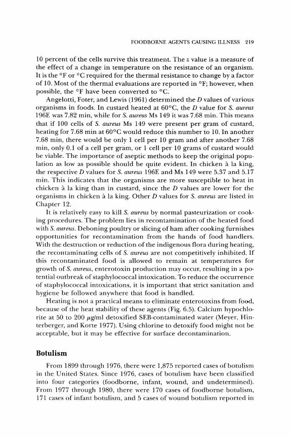
Heating is not a practical means to eliminate enterotoxins from food, because of the heat stability of these agents (Fig. 6.5). Calcium hypochlorite at 50 to 200 {tg/ml detoxified SEB-contaminated water (Meyer, Hinterberger, and Korte 1977). Using chlorine to detoxify food might not be acceptable, but it may be effective for surface decontamination.
Botulism
From 1899 through 1976, there were 1,875 reported cases of botulism in the United States. Since 1976, cases of botulism have been classified into four categories (foodborne, infant, wound, and undetermined). From 1977 through 1980, there were 170 cases of foodborne botulism, 171 cases of infant botulism, and 5 cases of wound botulism reported in
220 BASIC FOOD MICROBIOLOGY
200
100
!~ 50
_ 40 U)
I.IJ 30 ~ ::::)
~ 20 2
10
! 5 4
3 - NEGATIVE
2 + POSITIVE (AT LEAST 1.0 J.l0 ENTEROTOXIN A)
100 104.4 110.5 115.5 121.1 TEMPERATURE °c
126.6
Figure 6.5. Thermal inactivation of staphylococcal enterotoxin. Courtesy of Hilker et al. (1968).
the United States (CDC 1979, 1981; Gunn and Terranova 1979; Morris and Hatheway 1980).
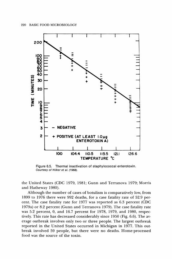
Although the number of cases of botulism is comparatively low, from 1899 to 1976 there were 992 deaths, for a case fatality rate of 52.9 per· cent. The case fatality rate for 1977 was reported as 6.3 percent (CDC 1979a) or 8.2 percent (Gunn and Terranova 1979). The case fatality rate was 5.2 percent, 0, and 16.7 percent for 1978, 1979, and 1980, respec· tively. This rate has decreased considerably since 1950 (Fig. 6.6). The avo erage outbreak involves only two or three people. The largest outbreak reported in the United States occurred in Michigan in 1977. This out· break involved 59 people, but there were no deaths. Home-processed food was the source of the toxin.
100
90
80
70
60
a: W m 50 ::E ::::l Z
40
30
20
10
0 1880'61 '62
CASES DEATHS
FOOD BORNE AGENTS CAUSING ILLNESS 221
,..,.---".,..,-----------.... ,----
~ ..." ...... --~ *
'83 '84 '86 '88 '87 '88 'S8 '70 '71 '72 '73 '74 '76 '78 '77 '78 '78 '80
* NOT AVAILABLE FOR 1979 AND 1980
Figure 6.6 Foodborne botulism-death-to-case ratios, by 10-year periods, 1899-1973. Courtesy of CDC.
ETIOLOGIC AGENT. Botulism is a neuroparalytic intoxication caused by neurotoxins produced by toxigenic strains of Clostridium botulinum. Eight antigenically distinct toxin types (A, B, C], C2, D, E, F, and G) are recognized. Sometimes types C) and C2 are grouped as C, but they are serologically distinct. Types A, B, E, and F cause botulism in humans. Although types C and D have been reported as causing illness in humans, the involvement is rare and uneventful. Types C and D affect animals. Type C is especially involved with birds and type D with cattle. Swine are moderately to highly resistant to most of the toxins.
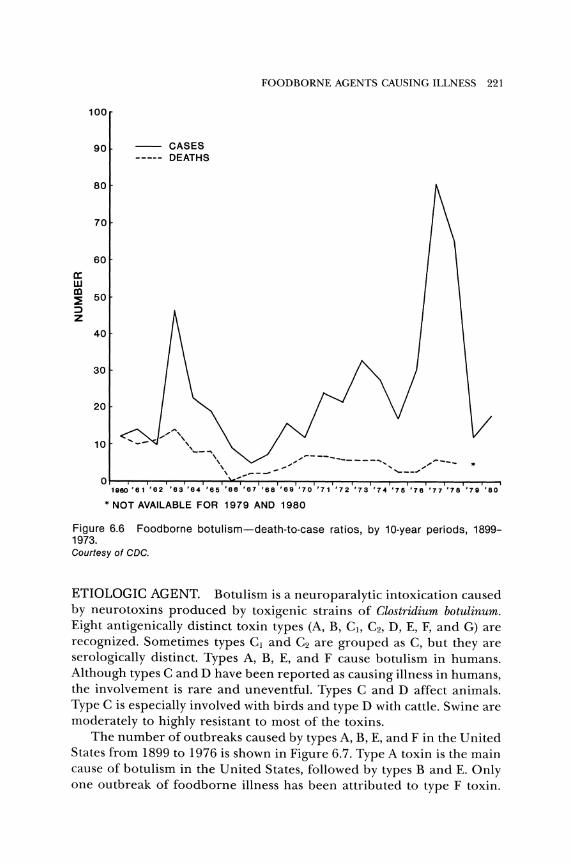
The number of outbreaks caused by types A, B, E, and F in the United States from 1899 to 1976 is shown in Figure 6.7. Type A toxin is the main cause of botulism in the United States, followed by types Band E. Only one outbreak of foodborne illness has been attributed to type F toxin.
222 BASIC FOOD MICROBIOLOGY
26
a: 24 4( W >- 22
a: 20 W Q.
18 t/) W 16 t/) 4( U 14
u. 12 0 a: 10 W m 8 ::E :::J
6 Z
Z 4 4( w ::E 2
0
UNKNOWN TYPE A TYPE B· TYPE E TYPE F
1899' 1900-09 1910-19 1920-29 1930-391940-491950-59 1980-691970-77
*1-YEAR PERIOD .* 8-YEAR PERIOD
10-YEAR PERIODS
tlNCLUDES ONE OUTBREAK OF 58 CASES, 1977
Figure 6.7. Number of cases of botulism, by type of toxin. Courtesy of CDC.
No human cases of type G have been reported in the United States, but the organism has been isolated from necropsy samples of four adults and one infant in Switzerland (Sonnabend et al. 1981). Type G toxin was demonstrated in the serum of three of these cases.
Most of the type A outbreaks occur in the western states, while type B is predominant in the east and central United States. This correlates with the finding that type A spores predominate in the soil in the west, while type B spores predominate in the east and central areas. In Europe, type B botulism is the predominant type, which correlates with the occurrence of type B spores in the soil. Although not prominent in the contiguous forty·eight states of the United States, type E botulism has accounted for more than 40 percent of the outbreaks in Japan, Canada, and the Scandanavian countries, and for almost all of the cases in Alaska.
It is generally agreed that for foodborne botulism, the C. botulinum grows in the food and produces the neurotoxin, which is then ingested orally. However, for wound botulism, the organism produces the toxin
FOOD BORNE AGENTS CAUSING ILLNESS 223
in the infected wound, in vivo. Infant botulism apparently results from the ingestion of the organism with subsequent intraintestinal production of the toxin. Hence, there may be an unusual circumstance in which the toxin might be formed in the intestinal tract of adults after ingestion of the organism or its spores.
During the logarithmic growth phase, the toxin accumulates intrace1-lularly, with small amounts found extracellularly. Beyond the logarithmic phase of growth, by cell lysis, the toxin is released to the extracellular medium.
Properties of the Toxins. The toxins are simple proteins that are water soluble, heat sensitive, and acid stable. On a molar basis, the toxins produced by C. botulinum are the most lethal natural products known. Since the toxins are protein, they are antigenic. Various molecular weights ranging up to 900,000 have been reported. According to Sugiyama (1980), these large compounds are complexes of toxins and nontoxic material. The molecular weights range from 128,000 to 170,000 depending upon the type of toxin and culture. The toxins are dichains composed of two single-chain polypeptides of different length combined by a sulfide linkage. Proteolytic organisms form these dichains naturally. For nonproteolytic strains, trypsinization of the toxin components produces similarly linked dichains of the pep tides. This has been called "activation." The presence of two dissimilar components was shown for type C2 toxin (Iwasaki, Ohishi, and Sakaguchi 1980; Ohishi 1983b). Neither component manifested the original toxicity, but when mixed together and trypsinized, the original toxicity was restored.
Action of the Toxins. Except for wound or infant botulism, the preformed toxin is considered to be ingested orally. To react with and affect the nerves, the toxin must traverse the barrier of the gastrointestinal tract and be transported to the susceptible nerves. The action on the nerves causes neuromuscular blockage, paralyzing the muscles.
The type of food in the GI tract can affect the toxin and its action. The food might protect the toxins from the enzymes or other destructive actions, such as by stomach acids. Food increases or decreases the secretion of digestive juices. The food might combine with the toxin, forming larger particles less able to penetrate the intestinal wall. Foods may affect the rate of peristalsis, which increases or decreases the time the toxin is in an area of the intestine affording the greatest opportunity for penetration of the wall.
The major site for absorption of the toxin is the small intestine (Bonventre 1979; Ohishi 1983a). Perhaps only a small amount of the ingested toxin is absorbed, but Bonventre (1979) estimated that about lO 11 molecules of toxin reaching the peripheral nerve endings is sufficient to cause
224 BASIC FOOD MICROBIOLOGY
symptoms of botulism in adults. Also, the large intestine may be an abo sorption site, since, according to Lamanna (1968), intrarectal instillation of toxin into monkeys and rabbits caused death, although the animals lived longer than when the toxin was given orally. The different toxins are absorbed at different rates (Sugii, Ohishi, and Sakaguchi 1977).
That the toxin is "wasted" in the alimentary tract is evident from work with animals. When type C toxin is administered to mink intraperitoneally, only 50 MLD (mouse lethal dose) causes death. Oral administration of 4,000 MLD causes death, but with 2,500 MLD, the mink survive. With pigs, the oral to intravenous ratio of amount of toxin needed to cause death is 16,700:1 (Smith, Davis, and Libke 1971). Data from various outbreaks indicate that not all of the toxin penetrates the intestinal wall, and that it can be present in the intestines for a prolonged period.
The toxin passes with the lymph through the thoracic duct and is dumped into the bloodstream. It is carried by the vascular system to the nerves. Lamanna (1968) reported that when the lymph system was prevented from emptying into the bloodstream, animals fed botulinum toxin did not develop symptoms of botulism.
At the nerves, the toxin attaches to the presynaptic terminals of cholinergic nerves, and interferes with the release of acetylcholine at the myoneural junctions (Sellin 1981; Sugiyama 1980). The failure of impulses to be transmitted across the nerve fiber junctions results in paralysis of the muscles, which are controlled by the nerves.
CHARACTERISTICS OF THE INTOXICATION. Since the toxins affect the nerves, this illness differs from most other foodborne illnesses.
Incubation Period. After food contaminated with botulinum toxin is ingested, the usual time for symptoms or signs of botulism to appear is 12 to 48 hr (CDC 1983b). However, this time may vary from 2 hr to 8 days, depending upon the amount of toxin ingested, the type of toxin, the resistance of the individual, and perhaps even the type of food. Generally, if the incubation period is less than 24 hr, the person will be more severely affected, and death is more likely than if the incubation period is longer.
Attack Rates. Although botulinum toxin is the most potent natural poison known, not everyone who consumes contaminated food will acquire symptoms or succumb to the toxin. This has been related to the amount of contaminated food ingested. However, there have been reports that merely tasting a food and spitting it out resulted in intoxication and death. This led to the supposition that toxin could be absorbed directly from buccal exposure. The contention of Lamanna, Hillowalla, and AI-
FOODBORNE AGENTS CAUSING ILLNESS 225
ling (1967) is that the normal swallowing reflex transfers the toxin from the mouth to the GI tract.
Symptoms and Signs. The cardinal features of botulism are as follows:
1. Fever is absent but may develop if a complicating infection occurs. 2. Mental status is normal. Patients may be anxious or agitated and
some are unusually drowsy; however, in the absence of secondary complications, patients are responsive.
3. The pulse rate is normal or slow, but tachycardia may occur if hy· potension develops.
4. Although vision may be blurred, numbness and paresthesias are absent, and a sensory deficit does not occur.
5. Neurological manifestations are symmetrical.
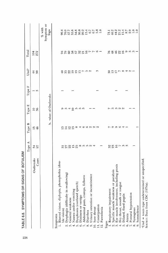
The symptoms and signs from cases reported to the Centers for Disease Control are listed in Table 6.8. The first symptoms of illness are gastrointestinal. Nausea or vomiting, substernal burning or pain, abdominal distension, and decreased bowel sounds may occur. Some cases have initial transitory diarrhea but subsequently become constipated. These symptoms and signs may mislead physicians to diagnose the illness as appendicitis, bowel obstruction, or even diaphragmatic myocardial infarction. The mucous membranes of the mouth, tongue, and pharynx may be red, dry, and painful, which might be diagnosed as pharyngitis.
Sometimes other illnesses are diagnosed as botulism. Investigation of 438 suspect botulism outbreaks by CDC (1974a) revealed that only 75 were actually botulism, and the rest were due to other disorders.
The neural symptoms usually begin with the eyes and face, and paralysis progresses downward to the throat, chest, and extremities. When the diaphragm and chest muscles become fully involved, respiration is not possible. Death is due to asphyxiation. This usually occurs in three to six days. In nonfatal cases, complete recovery may take several months. Mental processes are usually clear during the illness.
Therapy and Duration. Prompt clinical, epidemiological, and laboratory efforts are required because each hour is critical to the survival of a patient with botulism. The patient should be hospitalized for treatment.
The treatment of botulism can be separated into three parts: removing unabsorbed toxin from the alimentary tract, neutralizing toxin with antitoxin, and treating the symptoms, such as respiratory distress.
The sooner the antitoxin is administered, the better the chances for recovery. The reduction of the mortality rate in recent years has been attributed to the prompt treatment of the patients and administration of antitoxin. In some cases, guanidine hydrochloride is used as an adjunct.
"" "" T
AB
LE
6.8
. S
YM
PT
OM
S O
R S
IGN
S O
F B
OT
UL
ISM
G"
>
Ty
pe
A
Ty
pe
B
Ty
pe
E
Ty
pe
F U
nd"
To
tal
Ou
tbre
aks
34
15
10
I 44
10
4 C
ases
97
46
36
3
90
272
% w
ith
Sy
mp
tom
or
1\.
'lI
ber
of
Ou
tbre
aks
Sig
n
Sy
mp
tom
s I.
B
lurr
ed v
isio
n. d
iplo
pia
. p
ho
top
ho
bia
(do
u·
ble
visi
on)
31
13
9 40
94
90
.4
2.
Dys
phag
ia (
diff
icul
ty i
n s
wal
low
ing)
27
14
3
35
79
76.0
3.
G
ener
al w
eakn
ess
22
12
4 22
60
57
.7
4.
Nau
sea
and
/or
vo
mit
ing
15
13
10
19
58
55
.8
5.
Dy
sph
on
ia (
con
fuse
d s
peec
h)
25
8 5
19
57
54.8
6.
D
izzi
ness
or
vert
igo
8 4
5 15
32
30
.8
7.
Ab
do
min
al p
ain
. cr
amp
s, f
ulln
ess
5 6
3 7
21
20.2
8.
D
iarr
hea
5
6 5
16
15.4
9.
U
rin
ary
ret
enti
on
or
inco
nti
nen
ce
2 2
2 7
6.7
10.
So
re t
hro
at
4 2
7 6.
7 II
. C
on
stip
atio
n
2 3
6 5.
8 12
. P
ares
thes
ia
1.0
Sig
ns
I.
Res
pir
ato
ry i
mp
airm
ent
32
7 7
30
76
73.1
2.
S
peci
fic
mus
cle
wea
knes
s o
r pa
raly
sis
23
9 3
13
48
46.2
3.
E
ye m
uscl
e in
volv
emen
t, i
ncl
ud
ing
pto
sis
16
9 3
17
46
44.2
4.
D
ry t
hro
at,
mo
uth
or
ton
gu
e 7
6 2
7 22
21
.2
5.
Dil
ated
, fi
xed
pu
pil
s 3
4 2
8 16
15
.4
6.
Ata
xia
3 1
4 9
8.7
7.
Po
stu
ral
hy
po
ten
sio
n
2 3
2.9
8.
Nys
tagm
us
1 3
1.0
9.
So
mn
ole
nce
1.
0
"Un
d =
tox
in t
ype
un
det
erm
ined
or
unsp
ecif
ied.
SO
CRCE
: D
ata
from
CD
C (
197
4a).
FOODBORNE AGENTS CAUSING ILLNESS 227
This chemical seems to compensate for neural effects of the toxin. The use of germine in combination with guanidine is beneficial in the treat· ment (Cherington, Soyer, and Greenberg 1972).
The treatment of symptoms is especially important with respiratory difficulty or failure. Tracheotomy of the patient is used to assist in breathing. In a severe case, a mechanical respirator may be needed to maintain breathing.
With very little exposure to the toxin, the patient may remain asymptomatic or develop symptoms with little distress that pass in a few days. With increased amounts of toxin and development of respiratory failure, so that a respirator is needed, it may take several months for the patient to recover. Partial paralysis may persist for six to eight months (Bryan 1973).
One reason given for the lower mortality rate in Europe compared to the United States is that type B is prevalent in Europe and type A in the United States. Type A toxin is known to bind rapidly to tissue. In one case, 24 hr between ingestion and treatment was sufficient for the type A toxin to adhere irreversibly to the patient's myoneural tissue (Dillon et al. 1969). The antitoxin reacts with free toxin. When the toxin is irreversibly bound, administration of antitoxin has little effect on the recov· ery of the patient. Muscles injected with type A toxin were paralyzed up to seven days, while type B caused paralysis for less than five days (Sellin, Thesleff, and Dasgupta 1983).
Although immunization of people could be accomplished, the rare occurrence of botulism makes widespread immunization impractical. Immunization is recommended for laboratory or other personnel who are exposed to the toxin.
FOODS AS VEHICLES OF THE TOXIN. Usually botulism is associated with foods that have been given a preservation treatment, stored for some time, and consumed without appropriate heating. The preservation treatment in these foods is inadequate to destroy the spores that were present in the food.
The foods involved in botulism outbreaks in which the toxin type was determined are listed in Table 6.9. In more than 54 percent of the outbreaks, vegetables were the vehicle of the toxin. Fish, fruit, and condiments were other important vehicles, whereas meat, poultry, and dairy products have been involved rarely in botulism. Home-processed foods accounted for the majority of the outbreaks (72 percent), while commercially processed foods were involved in less than 10 percent of the outbreaks. Unknown vehicles accounted for slightly less than 20 percent of the outbreaks (Table 6.10). Although commercially processed foods have been involved in fewer outbreaks than home-processed foods, the inci·
228 BASIC FOOD MICROBIOLOGY
TABLE 6.9. FOODS INVOLVED IN BOTULISM OUTBREAKS IN WHICH THE TYPE OF TOXIN WAS DETERMINED, 1899-1977
Botulinum Toxin Type
Food Group A B E F A+B Total %
Vegetables 115 31 1 2 149 54.4 Fishery products 11 4 25 40 14.6 Fruit 22 7 29 10.6 Condiments 17 5 22 8.0 Beef 6 1 8 2.9 Dairy products 3 2 5 1.8 Pork 2 1 3 1.1 Poultry 2 2 4 1.5 Other 8 3 3 14 5.1 TOTAL 186 56 29 1 2 % 67.9 20.4 10.6 0.3 0.7
SOURCE: CDC (1979b).
dents which involve commercial foods are publicized, while those caused by home-packed foods rarely are mentioned. The reason is the widespread distribution of the commercial packs. If 500 cans of home-packed corn are contaminated, chances are only one of these will be involved in botulism in one family. If 500 cans of commercially canned corn are contaminated, they could cause botulism in 500 families. So far, this has not happened in the United States.
The outbreaks of botulism due to commercially packed foods since 1960 caused some health "experts" to suggest that families should process food at home. Data certainly do not support the theory that home canning reduces the number of botulism outbreaks.
THE ORGANISM. The genus Clostridium is divided into four groups according to spore formation and gelatin liquefaction. Clostridium botulinum is in Group II. In this group, the spores are terminal and gelatin is hydrolyzed.
The species includes a heterogeneous group of strains that are divided into types A through G based on the antigenic neurotoxins that are produced. These seven types are divided into four groups according to their deoxyribonucleic acid homologies and biochemical, physiological, and serological characteristics. The members of these groups are as follows: group 1, type A and proteolytic strains of types B, C, D, and F; group II, type E and non proteolytic strains of types Band F; group III, non proteolytic strains of types C and D; and group IV, type G (Buchanan and Gibbons 1974).
The optimum temperatures for growth are 30° to 40°C for group 1, 25° to 37°C for group II and 30° to 37°C for groups III and IV
Nl
Nl
<J:)
TA
BL
E 6
.10.
O
UT
BR
EA
KS
OF
FO
OD
BO
RN
E B
OT
UL
ISM
AT
TR
IBU
TE
D T
O C
OM
ME
RC
IAL
LY
PR
OC
ES
SE
D O
R H
OM
E·P
RO
CE
SS
ED
FO
OD
S,
1899
-197
7
1900
-19
10-
1920
-19
30-
1940
-19
50-
1960
-19
70-
Sou
rce
of
Foo
d 18
99
1909
19
19
1929
19
39
1949
19
59
1969
19
77
Tot
al
%
Hom
e pr
oces
sed
48
77
135
120
50
42
74
548
71.6
C
omm
erci
ally
pr
oces
sed
0 1
14
26
6 1
2 10
6
66
8.6
Unk
now
n 0
0 8
13
13
13
51
26
28
152
19.8
TO
TAL
1 2
70
116
154
134
103
78
108
766
SOUR
CE: C
DC
(l9
79b)
.
230 BASIC FOOD MICROBIOLOGY
organisms. Group I organisms digest milk, group II coagulate milk with a soft curd but do not digest it, group III organisms do not change milk, and those in group IV digest milk slowly. Other differences and simi· larities were described by Lynt, Kautter, and Solomon (1982).
Toxigenic Strains. Apparently most strains of C. botulinum are capable of producing a toxin; however, this ability may be lost due to laboratory cultivation, and non toxigenic clostridia have been isolated from both mao rine and terrestrial environments.
Inoue and Iida (1970) suggested that production of toxin by C. botuli· num is influenced by prophages in the cells. By curing the cells of the phage, some toxigenic strains lose the ability to produce toxin. With rein· fection by phage, the cells regain toxigenicity. It has been established that the phage type can determine the toxin type that is produced and not all lysogenic phages will confer toxigenicity on the infected cells (Hari· haran and Mitchell 1976; Oguma, !ida, and Inoue 1975). Most of the non· toxigenic strains that were converted to the toxigenic state reportedly were not stable to serial transfer through cooked meat medium (Oguma 1976).
Some strains of C. botulinum cured of their prophages by researchers remained toxigenic (Eklund et aL 1970). The researchers suggested that the organisms might carry more than one prophage, or alternatively that not all C. botulinum toxins are induced by bacteriophage.
The studies of phage inducement of toxin formation primarily have used type C and D strains. The relationship of phage to toxigenicity of strains causing human botulism has not been evaluated sufficiently.
Sources. The organism is widely distributed in nature. It occurs in soil apparently throughout the world. The type of C. botulinum depends upon the locality. The factors that affect the distribution of the different toxin types are not known. Healthy animals may be carriers of types of organisms to which they are immune, thus serving as reservoirs for reo plenishing the organisms in the soil and spreading the organisms to var· ious places.
Bottom sediments of marshes, lakes, and coastal ocean waters contain C. botulinum. The primary type in marine environments in northern areas appears to be type E; however, other toxin types of the organisms also are found. Type E is common in the intestine of fish taken from certain waters. The organism has been isolated from the environment of trout farms and from trout grown in these establishments (Cann and Taylor 1982) and in a salmon hatchery (Eklund et aL 1982). The organism does not multiply in living fish, but it does multiply in aquatic vegetation and bottom deposits. In marshes, the growth of algae reduces the oxygen con· tent of the water to a level that allows C. botulinum to multiply.
FOODBORNE AGENTS CAUSING ILLNESS 231
Since the organism is widely distributed in nature, it is evident that food, especially vegetables and fish, can be contaminated during produc· tion, harvesting, or processing. C. botulinum has been reported in com· mercial honey (Huhtanen et al. 1981; Kautter et al. 1982; Midura et al. 1979), corn syrup (Kautter et al. 1982) and liver sausage (Hauschild and Hilsheimer 1983). Fortunately, if present, the spores are usually in low numbers, and they can germinate only if the conditions of microenviron· ments of the food are favorable. After germination, sufficient time is needed for growth and toxin production.
GROWTH AND TOXIN PRODUCTION. The foods involved in out· breaks of botulism obviously present an environment or microenviron· ments that allow the growth of C. botulinum.
Various minimum and maximum temperatures have been reported for growth and toxin production. Eklund, Wieler, and Poysky (1967) reo ported growth and toxin production of a strain of type B C. botulinum at 3.3°C. The maximum temperature of growth for types A and B is about 48°C. Type E can grow in a range from 3.3° to 45°C. Growth and toxin production of type E is less pronounced at 37°C than at either 30° or 22°C (Ajmal 1968). A higher number of cells developed at 30° than at 22°C, but no difference was found for toxin production, indicating that the amount of toxin produced per cell was greater at 22° than at 30°C. When incubated at 4°C, growth and toxin produ~ir~\,occurred in four to five weeks. '\,,'
Type A (14 strains) and proteolytic type B (13 strains) did not multi· ply, but decreased in number during holding in cooked meat medium at 100C for eighteen weeks (Smelt and Haas 1978). At 12°C some strains formed toxin in three to four weeks. At 4°C, non proteolytic type Band type F cultures grew in tryptone glucose yeast extract broth, but not in crabmeat (Solomon, Kautter, and Lynt 1982). For growth in crabmeat, a temperature of 12°C was needed.
At pH 4.8, the growth of C. botulinum was detected in only one food (pineapple rice pudding) of the many tested (Townsend, Vee, and Mercer 1954). No growth was found at any pH below 4.8. Several other reports have confirmed that C. botulinum fails to grow below pH 4.8 (Baird·Parker and Freame 1967; Huhtanen et al. 1976; Ito et al. 1976; Segner, Schmidt, and Boltz 1971). These and similar reports have been the basis for the FDA setting an upper limit of pH 4.6 for acid foods. These require less heat treatment in the canning process than do those foods over pH 4.6. However, growth and toxin production have been detected at pH 4.3 to 4.6 when media with high amounts of protein in suspension were used (Smelt et al. 1982; Tanaka 1982).
In some outbreaks of botulism, the pH values of the foods involved
232 BASIC FOOD MICROBIOLOGY
were below that regarded as necessary for growth and toxin production. One explanation is that there are pH gradients in some foods so that some microenvironments are acceptable for growth and toxin produc· tion. Another possibility is that the growth of other organisms can result in acceptable microenvironments (Anderson 1984; Montville 1982; Od· laug and Pflug 1979).
The effect of salt on the growth and toxin production varies with the types and strains of C. botulinum. An aqueous phase level of 5.5 percent salt in homogenized cod was sporostatic to two strains of C. botulinum type E (Boyd and Southcott 1971). In lumpfish caviar, no toxin was formed at water phase salt concentrations of 5.56 percent or greater (Hauschild and Hilsheimer 1979).
C. botulinum is an anaerobic organism. The clostridia lack cyto· chromes, cytochrome oxidase, catalase, and peridoxase, so the oxidation· reduction potential (Eh) must be low for growth to occur (Lund and Wy· att 1984). C. botulinum type E inoculated into meat and fish produced toxin under both aerobic and anaerobic conditions on incubation (Ajmal 1968). There are microenvironments in these foods which have low oxidation-reduction potentials favorable for the growth of this organism.
Sugiyama and Yang (1975) inoculated spores of C. botulinum into fresh mushrooms, which were then packaged and stored at 20°C. The respiration of the mushrooms removed the free oxygen and allowed the spores to germinate and the cells to produce toxin in three to four days. The mushrooms appeared to be edible. No toxin was detected in mushrooms held at 4°C.
Since vacuum packaging in gas-impermeable plastic produces anaerobic conditions, it was reasoned that this practice might encourage the growth of C. botulinum and result in toxic foods. The rate of toxin production is higher in vacuum-packaged fish than in fish packaged without vacuum (Pace and Krumbiegel 1983). The slowest rate of toxin production is in unpackaged products. Research on vacuum packaging of raw fish has revealed that when the fish are stored at lOoC or less, spoilage is evident before toxin is detected (Eyles and Warth 1981). This safety margin is reduced if storage is at 20°C. Flushing the package with CO2
may increase the risk of botulism, since CO2 tends to inhibit spoilage organisms but enhance the germination of C. botulinum spores and cellular growth. However, 100 percent CO2 delays toxin production (Doyle 1983). Vacuum-packaged and modified atmosphere-packaged foods must be stored below 4°C to assure safety (Notermans, Dufrenne, and Keijbets 1981; Stier et al. 1981).
The interaction of microorganisms is both stimulatory and inhibitory to growth of C. botulinum. The growth of other organisms tends to reduce the Eh of the growth medium, which may then allow growth of C. botuli-
FOODBORNE AGENTS CAUSING ILLNESS 233
num. Some organisms, such as yeasts, may produce growth factors favor· able for C. botulinum.
The growth of C. botulinum in foods can cause a foul, putrid odor which should serve as a warning to the consumer. In many of the out· breaks of botulism, either a patient or an asymptomatic participant stated that the food had an off.odor or flavor, but these warning signs were ignored. Some people have a high tolerance to off.flavors or off· odors. In some foods (smoked, spiced, fermented), slight off.flavors or off.odors are difficult to detect.
There are reports that toxin is present in spores (spore·bound toxin) of C. botulinum (Booth et al. 1972; Yamakawa, Nishida, and Nakamura 1983). Phagocytosis releases the spore toxin, which has the same lethal effect as other free toxins.
METHODOLOGY. The methodology of C. botulinum involves the detec· tion and enumeration of the organism, the characterization of the or· ganism, or qualitative or quantitative determinations of the neurotoxins.
Detection and Enumeration. Due to the hazards involved, before working with C. botulinum, a person should be protected by suitable toxoids.
The demonstration of botulinum toxin in a food implicated in a botu· lism outbreak is often all that is done. However, the detection and isola· tion of C. botulinum from the food furnishes confirmatory evidence. Also, methods of detection, isolation, identification, and enumeration are ba· sic in determining the distribution of the orgamism in nature.
A system for detecting the organism is described in the Bacteriological Analytical Manual (FDA 1978). The medium for detection of C. botulinum is anaerobic egg yolk agar or liver veal·egg yolk agar.
Characterization. There is a widely held view that no organism isolated from a suspected case of botulism should be identified as C. botulinum unless it is toxigenic. Therefore, the outstanding characteristic of a strain of C. botulinum is the toxin it produces.
Once an organism has been isolated and found to be a Gram·positive rod that forms spores and is anaerobic, other tests can be made. These include digestion of gelatin, milk, and meat; fermentation of various car· bohydrates; indole production; hemolysis; and lecithinase and lipase activity. Various biochemical reactions of C. botulinum types are listed in Table 6.11. However, there may be disagreement on some of these reactions. Others are listed by Dezfulian and Dowell (1980).
It is evident from this information that biochemical tests can be considered reliable only to a limited extent in determining or distinguishing C. botulinum. This is because different toxin types of C. botulinum, or even strains within toxin types, represent quite distinct metabolic groups.

234 BASIC FOOD MICROBIOLOGY
TABLE 6.11. REACTIONS OF CLOSTRIDIUM BOTULINUM
Proteolytic N on·Proteolytic Types Types
A,B,C,D,F B,C,D,E,F G
Digestion of Gelatin +" + + Casein + + Meat + N
Acids produced from Glucose + + Fructose + + Mannose + Lactose Salicin V
Other tests Indole Urease Nitrates reduced Lipase + + Hemolysis + + +
.,+ = positive reaction; - = no reaction: V = variable reactions; N = insufficient data.
Serological Reactions. Besides the toxins, the organism has three sources of antigenic substances: the flagellar antigens (H), the somatic antigens (0), and the spore antigens. The flagellar antigens show a narrow range of specificity, and the somatic antigens are often common to all members of a species or type. The spore antigens are type specific and are dissimi· lar to either the flagellar or somatic antigens of the vegetative cell (Solo· mon et al. 1969).
Toxin Detection. The Bacteriological Analytical Manual (FDA 1978) describes a procedure for detection of preformed toxin in food and for detection of toxins produced by organisms in the food with an enrichment proce· dure.
An extract is analyzed for toxin by intraperitoneal injection of mice. The injected mice are observed over a period of 92 hr. Death indicates the presence of toxin. The mouse neutralization test for toxin has been accepted as official by the AOAC.
Serological techniques, as described for detection of staphylococcal enterotoxin, have been tested for the analysis of botulinum toxins. The ELISA system has been adapted for type A (Notermans, Dufrenne, and Van Schothorst 1978), type B (Kozaki et al. 1979), type E (Notermans, Dufrenne, and Kozaki 1979) and type G toxin (Lewis et al. 1981). Cross· reactivity and nonspecific reactivity are problems not only for the ELISA, but also for other serological systems for analyzing for botulinum toxins.
FOODBORNE AGENTS CAUSING ILLNESS 235
Dezfulian and Bartlett (1984) produced specific type A antitoxin by selective suppression of the immunological response of inoculated rabbits to unwanted antigens and subsequent immunization with a toxoid_ Even then, there was some cross-reaction of the antitoxin with culture filtrates of type B organisms_ Monoclonal antibodies to type C 1 and D toxins were produced and used in the ELISA to detect these toxins (Oguma et aL 1984)_
CONTROL OF BOTULISM. For botulism to occur, the spores or cells of toxigenic C. botulinum must be present in the environment and gain access to the food; viable cells or spores must remain in the food after processing; and the food must have an environment favorable for germination and outgrowth of the spores and growth of the vegetative cells to produce the toxin; then the food must be eaten cold or with insufficient heating to destroy the toxin_
Therefore, the principal methods of controlling staphylococcal intoxication also are important in the control of botulism. Quite simply, we can prevent contamination of the food, prevent growth and toxin production, destroy the organisms or toxin, and not eat suspect foods.
Perhaps the best method of controlling botulism is to heat the food during processing to a temperature that will destroy the spores of toxigenic C. botulinum. However, there are many foods, such as cured meat products, that would suffer organoleptically if these heat treatments were used. For these foods, additives are needed that will inhibit toxin production by C. botulinum contaminants.
Prevent Contamination. Generally, C. botulinum gains access to food as a soilborne or dustborne contaminant. Vegetables are harvested from the soil or in close proximity to the soiL Washing of vegetables will remove some soil and associated organisms, but it is difficult to remove all organisms by this method.
Fish and marine animals feeding in waters that contain C. botulinum can be contaminated. Most of the contamination is in the intestines of fish_ Proper cleaning and eviscerating procedures result in minimal contamination of the fish flesh_
High levels of contamination are more difficult to destroy than are low levels or, ideally, no spores. Therefore, sanitary practices are very important.
Prevent Growth and Toxin Production_ Foods that do not have an environment conducive to the growth and toxin production of C. botulinum will not become toxic.
One of the simplest methods to inhibit the growth of microorganisms
236 BASIC FOOD MICROBIOLOGY
is to hold the food at temperatures below which growth will occur. Since certain strains of C. botulinum can grow and produce toxins at SoC or less, normal refrigerator temperatures cannot be relied upon to control toxin formation in foods. Freezing does not destroy the toxin or the spores of C. botulinum, but as long as the food is frozen, germination and growth of the vegetative cells with toxin production does not occur. Frozen foods should not be allowed to thaw and remain in that condition for extended periods. C. botulinum does not grow at an Ow of 0.93 or less. Foods dried below this level should not become toxic (there is the possibility that toxin could be formed prior to drying).
A chemical added to cured meats is sodium nitrite. This chemical is important in color fixation of cured meat, as well as in inhibiting growth and toxin formation by C. botulinum. Various other chemicals have been tested for inhibition of the organism. These chemicals and nitrites are discussed in Chapter 11.
During processing, the destruction of microbial competitors, the reo moval of air by heating (lowering the redox potential), and the destruc· tion of cellular tissues with the release of cellular fluids, favors the growth of C. botulinum.
Destruction ofC. botulinum. Since this organism produces spores, it is more difficult to destroy than is S. aureus. The method used to destroy the spores is thermal processing.
Most of the outbreaks of botulism have been caused by consumption of toxic heat·processed food. Thus, it may seem odd that heating is the only presently acceptable method of destroying the spores of C. botulinum. The spores of C. botulinum vary in their heat resistance. Type E spores are the least resistant and thus are most easily destroyed by heat.
Due to outbreaks of botulism from eating contaminated fish, it was recommended that during smoking, the fish be held at 82.2°C for 30 min. Pace and Krumbiegel (1973) examined commercially smoked fish and found that 0.9 percent to 2.0 percent contained C. botulinum type E spores, although the fish had been subjected to 82.2°C for 30 min.
Various results of heat processing are obtained undoubtedly because of the difference in heat resistance of various strains of type E spores. One group of researchers determined the thermal-death-time of five strains of type E spores in fish paste (Crisley et al. 1968). The D values in this medium at 80°C varied from an average of 1.6 min to 4.3 min for the five strains.
Boiling (l00°C) is not recommended to destroy spores of type A or B, since several hours would be required. Processing of low-acid foods is accomplished at a temperature of 121.1 °C. A 12D thermal process (see Chapter 12) is considered essential for low-acid canned foods. With this
FOODBORNE AGENTS CAUSING ILLNESS 237
treatment, the probability of survival of C. botulinum spores is very remote_
Of the clostridial spores, those of C. botulinum are included in the most radiation-resistant group_ The spores vary in their resistance, with type F generally considered to be the most resistant and type E spores the least resistant, with types A and B intermediate_
A combination of radiation and heat has a synergistic effect on destruction of spores of C. botulinum. Spores that are exposed to low-level radiation are more susceptible to heat inactivation than untreated spores. The heat is also of value in halting enzyme action and inactivating any toxin that might be present. Thus, pasteurization levels of radiation followed by heating at values less than those presently required may be beneficial in controlling C. botulinum. Heating before radiation does not lower the radiation resistance, but radiation lowers the heat resistance of the spores.
Inactivation of Toxin. Radiation pasteurization does not inactivate preformed botulinum toxin, whereas heat pasteurization does. The heat stability of botulinum toxin is considerably less than that of staphylococcal enterotoxin. The types of botulinum toxin vary in their heat resistance. The time necessary to inactivate the toxins depends upon the amount of toxin present, the temperature, and the substrate in which they are heated. The toxins are more stable at acid pH levels and are most stable about pH 5.0. Various times and temperatures have been recommended for inactivating botulinum toxins (Bradshaw, Peeler, and Twedt 1979, 1981; Woodburn et al 1979).
Although the toxins are relatively heat labile, being inactivated in 2 min or less at 90°C, this heat treatment usually is not suggested for destroying toxins formed in canned foods. Bryan (1973) recommended that before serving, canned food should be heated to boiling (about 100°C) and held for 5 min to 15 min.
The resistance to chemicals depends upon the type of toxin, temperature, pH, and other substances in the medium. In water, the purified toxin is sensitive to chlorine, bromine, or iodine. Free available chlorine with a residual of 1 mg/L (1 ppm) will destroy at least 99.9 percent of all the types of botulinum toxin in 5 min or less. Type E toxin is the most resistant to destruction by chlorine.
Canned food that is not normal should not be consumed. In most outbreaks of botulism involving canned food, one or more people noted swelling of the can, bubbles in the food, off-odors, or turbid liquid. If the food is suspicious, it should be returned to the store, if purchased, or disposed of in a manner so that it is not consumed by animals or by other people.
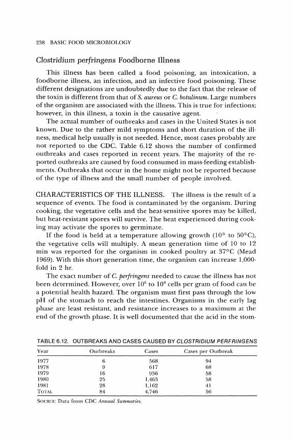
238 BASIC FOOD MICROBIOLOGY
Clostridium perfringens Foodborne Illness
This illness has been called a food poisoning, an intoxication, a foodborne illness, an infection, and an infective food poisoning. These different designations are undoubtedly due to the fact that the release of the toxin is different from that of S. aureus or C. botulinum. Large numbers of the organism are associated with the illness. This is true for infections; however, in this illness, a toxin is the causative agent.
The actual number of outbreaks and cases in the United States is not known. Due to the rather mild symptoms and short duration of the ill· ness, medical help usually is not needed. Hence, most cases probably are not reported to the CDC. Table 6.12 shows the number of confirmed outbreaks and cases reported in recent years. The majority of the reported outbreaks are caused by food consumed in mass-feeding establishments. Outbreaks that occur in the home might not be reported because of the type of illness and the small number of people involved.
CHARACTERISTICS OF THE ILLNESS. The illness is the result of a sequence of events. The food is contaminated by the organism. During cooking, the vegetative cells and the heat-sensitive spores may be killed, but heat-resistant spores will survive. The heat experienced during cooking may activate the spores to germinate.
If the food is held at a temperature allowing growth (100 to 50°C), the vegetative cells will multiply. A mean generation time of 10 to 12 min was reported for the organism in cooked poultry at 37°C (Mead 1969). With this short generation time, the organism can increase 1,000-fold in 2 hr.
The exact number of C. perfringens needed to cause the illness has not been determined. However, over 101i to 108 cells per gram of food can be a potential health hazard. The organism must first pass through the low pH of the stomach to reach the intestines. Organisms in the early lag phase are least resistant, and resistance increases to a maximum at the end of the growth phase. It is well documented that the acid in the stom-
TABLE 6.12. OUTBREAKS AND CASES CAUSED BY CLOSTRIDIUM PERFRINGENS
Year Outbreaks Cases Cases per Outbreak
1977 6 568 94 1978 9 617 68 1979 16 936 58 1980 25 1,463 58 1981 28 1,162 41 TOTAl. 84 4,746 56
SOVRCE: Data from CDC Annual Summaries.
FOODBORNE AGENTS CAUSING ILLNESS 239
ach combines with consumed proteins, causing an increase in the overall pH. This effect of protein on the stomach acidity, and resultant protec· tion of the bacterial cells, may be the factor in the involvement of protein foods in outbreaks of the illness.
When the remaining organisms reach the small intestine, they find an environment acceptable for multiplication and sporulation. The sporulating cells produce the enterotoxin, which is released during cell lysis. The organisms may sporulate in some foods, but usually, by the time the food is toxic, it is not palatable (Craven, Blankenship, and McDonel 1981 ).
Incubation Period. The interval of time between the meal and onset of illness for an outbreak of C. perfringens food poisoning is shown in Figure 6.8. In this example, the range is 2 to 29 hr, with a median of 13 hr. CDC (1983b) listed the incubation period as 9 to 15 hr.
Attack Rate. Not everyone who eats the contaminated food becomes ill. The reasons for the escape of some people are similar to those described for staphylococcal intoxication and botulism. People with hyperacid stomachs may be protected, due to the death of C. perfringens cells. Previous contact or any immunity apparently has little effect, if any, on acquiring the illness when contaminated food is ingested.
The disease is not prevented in parenterally immunized animals that possess neutralizing serum antibody against the toxin (N iilo 1971). In a survey of human sera, a majority contained antibody against C. perfringens enterotoxin (Uemura et al. 1974). They speculated that antibody could be induced by the acute C. perfringens food poisoning or be due to the continuous presence of the organism and small amounts of toxin in asymptomatic people.
Symptoms. The symptoms reported in four outbreaks of C. perfringens illness are listed in Table 6.13. The two prominent symptoms of the illness are diarrhea and abdominal cramps. Nausea and headache were fairly important in two outbreaks but were of minor occurrence in the others. Fever, vomiting, dizziness, and bloody stools may occur, but they are rare.
In young people, the symptoms are usually mild, but in elderly, ill, or debilitated people, the illness can be severe. Although rare, death has occurred in cases complicated by other illness.
Duration and Therapy. The illness is usually of short duration, from less than 12 hr up to 24 hr. A few cases may persist for 48 hr. The illness is followed by a complete and uneventful recovery. Although weakness may be evident the day after symptoms begin, a person usually can return to work or normal activity. Due to the rather mild symptoms and short duration, usually no therapy is needed.
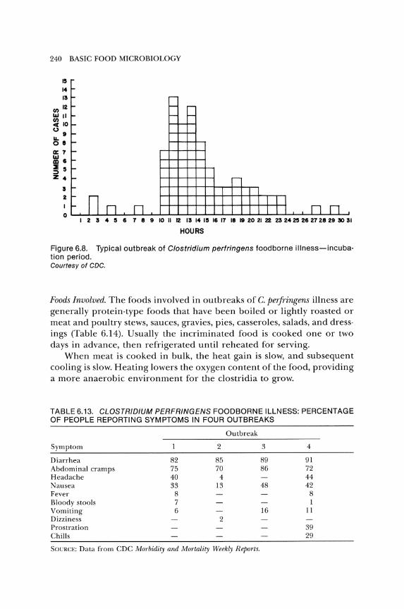
240 BASIC FOOD MICROBIOLOGY
15
14 13
II) 12 10111 ~ 10
~9 08
0:7
~ 6
~ !5 Z 4
3 2
o
---
- - -- - --r-I-r-I-
r-i- nn l- n
----
r-
I I I I I I nn
I 2 3 4 !5 6 1 8 9 10 II 12 13 14 15 16 11 18 19 20 21 22 23 2425 26 2128 29 ~ 31
HOURS
Figure 6.8. Typical outbreak of Clostridium perfringens food borne illness-incubation period. Courtesy of CDC.
Foods Involved. The foods involved in outbreaks of C. perfringens illness are generally protein-type foods that have been boiled or lightly roasted or meat and poultry stews, sauces, gravies, pies, casseroles, salads, and dressings (Table 6.14). Usually the incriminated food is cooked one or two days in advance, then refrigerated until reheated for serving.
When meat is cooked in bulk, the heat gain is slow, and subsequent cooling is slow. Heating lowers the oxygen content of the food, providing a more anaerobic environment for the clostridia to grow.
TABLE 6.13. CLOSTRIDIUM PERFRINGENS FOODBORNE ILLNESS: PERCENTAGE OF PEOPLE REPORTING SYMPTOMS IN FOUR OUTBREAKS
Outbreak
Symptom 2 3 4
Diarrhea 82 85 89 91 Abdominal cramps 75 70 86 72 Headache 40 4 44 Nausea 33 13 48 42 Fever 8 8 Bloody stools 7 1 Vomiting 6 16 11 Dizziness 2 Prostration 39 Chills 29
SOCRCE: Data from CDC Morbidity and Mortality Weekly Reports.
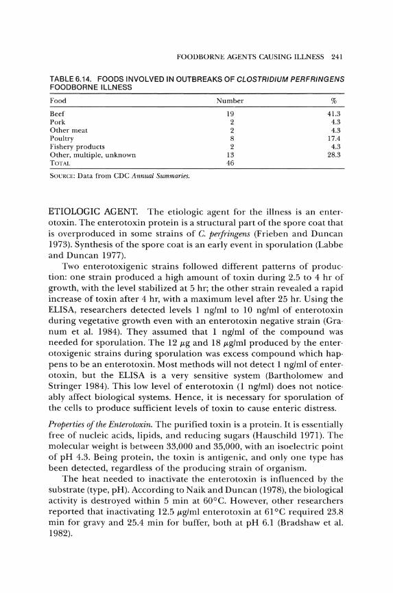
FOODBORNE AGENTS CAUSING ILLNESS 241
TABLE 6.14. FOODS INVOLVED IN OUTBREAKS OF CLOSTRIDIUM PERFRINGENS FOODBORNEILLNESS
Food
Beef Pork Other meat Poultry Fishery products Other, multiple, unknown TOTAL
SOCRCE: Data from CDC Annual Summaries.
Number
19 2 2 8 2
13 46
%
41.3 4.3 4.3
17.4 4.3
28.3
ETIOLOGIC AGENT. The etiologic agent for the illness is an enter· otoxin. The enterotoxin protein is a structural part of the spore coat that is overproduced in some strains of C. perfringens (Frieben and Duncan 1973). Synthesis of the spore coat is an early event in sporulation (Labbe and Duncan 1977).
Two enterotoxigenic strains followed different patterns of produc· tion: one strain produced a high amount of toxin during 2.5 to 4 hr of growth, with the level stabilized at 5 hr; the other strain revealed a rapid increase of toxin after 4 hr, with a maximum level after 25 hr. Using the ELISA, researchers detected levels 1 ng/ml to 10 ng/ml of enterotoxin during vegetative growth even with an enterotoxin negative strain (Gra· num et al. 1984). They assumed that 1 ng/ml of the compound was needed for sporulation. The 12 J-tg and 18 J-tg/ml produced by the enter· otoxigenic strains during sporulation was excess compound which hap· pens to be an enterotoxin. Most methods will not detect 1 ng/ml of enter· otoxin, but the ELISA is a very sensitive system (Bartholomew and Stringer 1984). This low level of enterotoxin (l ng/ml) does not notice· ably affect biological systems. Hence, it is necessary for sporulation of the cells to produce sufficient levels of toxin to cause enteric distress.
Properties of the Enterotoxin. The purified toxin is a protein. It is essentially free of nucleic acids, lipids, and reducing sugars (Hauschild 1971). The molecular weight is between 33,000 and 35,000, with an isoelectric point of pH 4.3. Being protein, the toxin is antigenic, and only one type has been detected, regardless of the producing strain of organism.
The heat needed to inactivate the enterotoxin is influenced by the substrate (type, pH). According to N aik and Duncan (1978), the biological activity is destroyed within 5 min at 60°C. However, other researchers reported that inactivating 12.5 J-tg/ml enterotoxin at 61°C required 23.8 min for gravy and 25.4 min for buffer, both at pH 6.1 (Bradshaw et al. 1982).
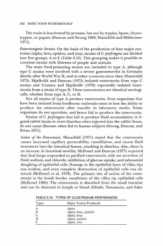
242 BASIC FOOD MICROBIOLOGY
The toxin is inactivated by pronase, but not by trypsin, lipase, chymotrypsin, or papain (Duncan and Strong 1969; Hauschild and Hilsheimer 1971).
Enterotoxigenic Strains. On the basis of the production of four major exotoxins (alpha, beta, epsilon, and iota), strains of C. perfringens are divided into five groups, A to E (Table 6.15). This grouping makes it possible to correlate strains with illnesses of people and animals.
The main food-poisoning strains are included in type A, although type C strains were involved with a severe gastroenteritis in Germany shortly after World War II, and in other countries since then (Hauschild 1973). Skjelkvale and Duncan (1975) isolated enterotoxin from type C strains and Uemura and Skjelkvale (1976) reportedly isolated enterotoxin from a strain of type D. These enterotoxins are identical serologically, whether from type A, C, or D.
Not all strains of type A produce enterotoxin. Even organisms that have been isolated from foodborne outbreaks seem to lose the ability to produce the enterotoxin after transfer in laboratory media. Some organisms do not sporulate, and hence fail to produce the enterotoxin.
Strains of C. perfringens that fail to produce fluid accumulation in ligated rabbit ileum or overt diarrhea when injected into the rabbit ileum, do not cause illnesses when fed to human subjects (Strong, Duncan, and Perna 1971).
Action of the Enterotoxin. Hauschild (1971) stated that the enterotoxin causes increased capillary permeability, vasodilation, and excess fluid movement into the intestinal lumen, resulting in diarrhea. Also, there is an increase in intestinal motility. McDonel and Duncan (1977) reported that ileal loops responded to purified enterotoxin, with net secretion of fluid, sodium, and chloride, inhibition of glucose uptake, and substantial sloughing of epithelial cells. Damage to the epithelial layer of villus tips was evident, and even complete destruction of epithelial cells was observed (McDonel et al. 1978). The primary site of action of the enterotoxin is the brush border membrane of the villus tip epithelial cells (McDonel 1980). The enterotoxin is absorbed from the small intestine and can be detected in lymph or blood (Ohishi, Yamamoto, and Saka-
TABLE 6.15. TYPES OF CLOSTRIDIUM PERFRINGENS
Types
A B C o E
Major Toxins Produced
alpha alpha, beta, epsilon alpha, beta alpha, epsilon alpha, iota
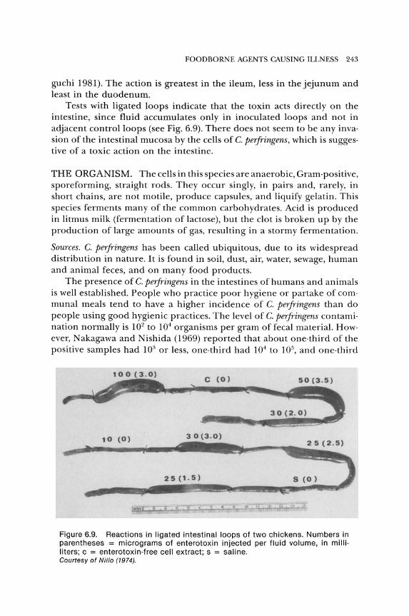
FOODBORNE AGENTS CAUSING ILLNESS 243
guchi 1981). The action is greatest in the ileum, less in the jejunum and least in the duodenum.
Tests with ligated loops indicate that the toxin acts directly on the intestine, since fluid accumulates only in inoculated loops and not in adjacent control loops (see Fig. 6.9). There does not seem to be any inva· sion of the intestinal mucosa by the cells of C. perfringens, which is sugges· tive of a toxic action on the intestine.
THE ORGANISM. The cells in this species are anaerobic, Gram·positive, sporeforming, straight rods. They occur singly, in pairs and, rarely, in short chains, are not motile, produce capsules, and liquify gelatin. This species ferments many of the common carbohydrates. Acid is produced in litmus milk (fermentation of lactose), but the clot is broken up by the production of large amounts of gas, resulting in a stormy fermentation.
Sources. C. perfringens has been called ubiquitous, due to its widespread distribution in nature. It is found in soil, dust, air, water, sewage, human and animal feces, and on many food products.
The presence of C. perfringens in the intestines of humans and animals is well established. People who practice poor hygiene or partake of com· munal meals tend to have a higher incidence of C. perfringens than do people using good hygienic practices. The level of C. perfringens contami· nation normally is 102 to 104 organisms per gram of fecal material. How· ever, Nakagawa and Nishida (1969) reported that about one·third of the positive samples had 103 or less, one· third had 104 to 105, and one·third
c (0) 50 (3.5)
30(2.0)
,0 (0) 30(3.0)
25 (1.5)
Figure 6.9. Reactions in ligated intestinal loops of two chickens. Numbers in parentheses = micrograms of enterotoxin injected per fluid volume, in milli· liters; c = enterotoxin·free cell extract; s = saline. Courtesy of Niilo (1974).
244 BASIC FOOD MICROBIOLOGY
had 106 or more C. perfringens per gram from normal human intestines. Others found that some healthy aged adults persistently excreted from 107 to 109 C. perfringens per gram of feces (Yamagishi et al. 1976). However, these may not be enterotoxigenic types.
With the widespread distribution of C. perfringens in the environment, and the fact that the organism can produce spores that are resistant to adverse conditions, the organism is a logical common contaminant of food.
Growth. Although C. perfringens is an anaerobe, it is aerotolerant. There· fore, strict anaerobic conditions are not needed for growth. The redox potentials of meat products are favorable for growth of the micro· orgamsm.
Good growth occurs between pH 5.5 and 8.0, and no growth occurs below pH 5.0 or above pH 9.0. At pH 5.5, vegetative growth occurred, but not sporulation and enterotoxin production (Labbe and Duncan 1974). They reported the optimum range for enterotoxin production as pH 6.5 to 7.3.
C. perfringens grows readily at temperatures between 20° and 50°C, with maximum growth between 37° and 47°C. Mead (1969) found no growth at either 15° or 52°C. Reportedly, some strains show limited growth at 15°C. A long lag phase is a characteristic of growth at low temperatures. Sporulation and toxin production were optimum at 37°C (Labbe and Duncan 1974). No sporulation or toxin production was noted at 46°C.
At aU! values less than 0.995, the rate of growth is reduced. The mini· mum aw for growth depends upon the solute, temperature, pH, and other factors. The absolute minimum aw for growth appears to be 0.93 (Kang et al. 1969). In this case, glycerol was used to lower the aw •
C. perfringens can grow in concentrations of curing salts (sodium ni· trite, and nitrate) considerably higher than those used in normal curing operations (Gough and Alford 1965). Ando (1975) reported that with 2 percent NaCl, a level of 0.05 percent (500 ppm) NaN02 was needed to completely prevent outgrowth of germinated spores of C. perfringens. Lower concentrations permitted some outgrowth.
METHODOLOGY. The clinical and epidemiological pattern of C. per· fringens foodborne illness is so characteristic that it is almost diagnostic. However, it is necessary to analyze appropriate samples in order to con· firm the presence of the organism. Because the enterotoxin is released primarily during sporulation, and most of the sporulation occurs in vivo, it is assumed that the analysis of food for the enterotoxin may be of little value. In one study, however, researchers inoculated ground and auto·
FOODBORNE AGENTS CAUSING ILLNESS 245
claved chicken meat with C. perfringens and analyzed it for enterotoxin (AI-Obaidy et al. 1985). They reported a level of 0.76 p,g/g of enterotoxin after incubation for 14 hr, indicating that the toxin might be detected in foods involved in gastroenteritis due to C. perfringens.
To help establish C. perfringens as the cause of a foodborne outbreak, three laboratory tests are suggested: (1) organisms of same serotype in epidemiologically incriminated food and stool of ill individuals; (2) isolation of organisms with same serotype in stool of most ill individuals and not in stool of controls; or (3) demonstration of greater than 105 C. perfringens cells per gram of incriminated food, provided the food sample was properly handled.
Detection and Enumeration ofC. perfringens. Food should always be analyzed promptly. This is especially true for C. perfringens detection because unprotected cells are sensitive to low temperatures. A buffered glycerolsodium chloride solution mixed in equal portions with a food and stored in dry ice at - 56°C protected cells of C. perfringens (Harmon and Placencia 1978). The number of C. perfringens was only slightly lowered by this treatment.
For the enumeration of C. perfringens, tryptose sulfite cycloserine (TSC) agar without egg yolk was effective (Harmon, Kautter, and Peeler 1971). This medium with or without added egg yolk emulsion is recommended for enumeration of C. perfringens (AOAC 1985; APHA 1984; FDA 1978). The medium is differential because of the presence of sodium bisulfite and ferric ammonium citrate. Bacteria that reduce sulfite to sulfide produce black colonies due to the formation of iron sulfide. Since many organisms can reduce sulfite to sulfide, D-cycloserine is added to select for clostridia.
For enumeration of this organism in foods, the MPN technique using thioglycollate-cycloserine (Debevere 1979), iron milk (Abeyta 1983; Abeyta, Michalovskis, and Wekell 1985; St. John, Matches, and Wekell 1982), or rapid perfringens medium (DeBoer and Boot 1983; Erickson and Deibel 1978) reportedly is simpler and more effective than direct plating systems.
Confirmational Tests. C. perfringens is the only clostridial species that reduces sulfite, is nonmotile, reduces nitrate, and produces a stormy fermentation in milk (Buchanan and Gibbons 1974).
Nitrate reduction and motility are determined by stab inoculation of nitrate motility medium. It is recognized that all strains do not reduce nitrate in this medium. However, when the medium is modified by adding 0.5 percent galactose and glycerol, then good nitrate reduction is obtained (Hauschild and Hilsheimer 1974).
246 BASIC FOOD MICROBIOLOGY
Serology. Not all of the strains are typable with the presently available antisera. The many problems associated with serotyping C. perfringens were reviewed by Stringer, Turnbull, and Gilbert (1980).
Bacteriocins. The specificity of bacteriocins can be used to type microor· ganisms. Watson (1985) used fifty bacteriocins to type 802 isolates of the organism. A greater proportion of those strains involved in foodborne illness were bacteriocinogenic than were other isolates.
Toxin Detection-Enterotoxin. Although the determination of enterotoxin may not be important epidemiologically, it is important when studying the role of toxin in pathogenesis and the mechanisms of enterotoxic ac· tion. Various systems have been used for the assay of the enterotoxin.
Ligated intestinal loops have been used to assay for the enterotoxin (Hauschild, Hilsheimer, and Rogers 1971; Niilo 1974). When toxin is injected into a ligated loop, it causes fluid accumulation (Fig. 6.9). Measurement of this fluid as compared to control loops is an indication of the amount of toxin that is injected. Since it is inconvenient to assay toxin in this manner, other methods have been devised.
Intradermal injection of the enterotoxin into rabbits and guinea pigs causes erythema around the injection site in 1 to 2 hr, and reaches a maximum in 18 to 24 hr. The diameter of this reddened area is related to the concentration of enterotoxin (Niilo 1975). The reaction is distinctive for the enterotoxins. According to Hauschild (1970), this system is 1,000 times as sensitive as the ligated intestinal loop, as well as being more rapid and accurate.
Various mammalian cell lines have been tested. African green monkey kidney (Vero) and dog kidney (MDCK) cells are sufficiently sensitive to assay for the enterotoxin (Giugliano, Stringer, and Drasar 1983; McDonel and McClane 1981).
The immunodiffusion methods listed for assaying S. aureus enterotoxin can be used to assay for C. perfringens enterotoxin. According to Skjelkvale and Uemura (1977), both the reversed passive hemagglutination and counterimmunoelectrophoresis assays are rapid and reliable systems. The radioimmunoassay detects enterotoxin at a level of 1 ng/ml (Stelma et al. 1983).
Various ELISA systems are specific and sensitive for C. perfringens enterotoxin. In one study, as little as 25 ng/ml of enterotoxin was detected with an indirect ELISA, while 1 ng/ml was detected with a fourlayer sandwich ELISA (McClane and Strouse 1984). Other researchers have detected 5 ng/g of enterotoxin in feces with a sandwich ELISA (Bartholomew et al. 1985).
FOODBORNE AGENTS CAUSING ILLNESS 247
CONTROL OF C. PERFRINGENS FOOD POISONING. The three general methods for control of this foodborne illness are (1) limit or prevent contamination of the food; (2) prevent or inhibit growth of the organism; or (3) destroy the organism. Any combination of these methods also may be used. Since most, if not all, of the enterotoxin is produced in the intestinal tract, systems to prevent production or to destroy the toxin are not applicable_
Limit OT PTevent Contamination. Due to the ubiquitous character of the organism, preventing contamination of food cannot be relied upon as a means of control. However, this does not mean that we should ignore good sanitary practices_
PTevent Gmwth. The presence of a few C. peTfTingens cells in a food product is not considered to be a health hazard. The hazardous level is some 106
to 108 cells per gram of food. The prevention of germination of spores with outgrowth and multiplication of the vegetative cells is the only practical method to control the illness caused by C. peTfTingens_
One simple method of control is to cook and serve the food without an extended holding period. If this is not possible, refrigeration of the food in small quantities for quick cooling will inhibit growth. The minimum temperature of growth is about 15°C. Any acceptable refrigerator should be able to maintain this temperature unless it is overloaded. The lower the temperature of the refrigerator, the sooner the food will be cooled. As the temperature approaches DoC, the population tends to decline, since the vegetative cells are not stable at such low temperatures. Even the spores appear to be damaged at low refrigerator or freezer temperatures.
The maximum temperature for growth is 50° to 52°C. Above 52°C, no growth should occur, and the vegetative cells may die. There was greater than a 99 percent reduction in the number of C. peTjTingens on cooked beef cubes when held at 53.3°C (Brown and Twedt 1972).
Destmy the OTganism. Vegetative cells of C. peTfTingens are destroyed by thorough cooking, but heat-resistant spores can survive_ Subjecting the spores to sublethal temperatures stimulates germination_ The heat resistance of the spores varies from strain to strain; hence, heating to a certain temperature is not required, since the killing effect is dependent upon the strain of organism that is present.
If heat-resistant spores are present, it is not possible to heat foods sufficiently to inactivate all of them without damaging the organoleptic properties of the food. It must be assumed that there are surviving spores in the cooked food, and the food must be kept hot until it is served_
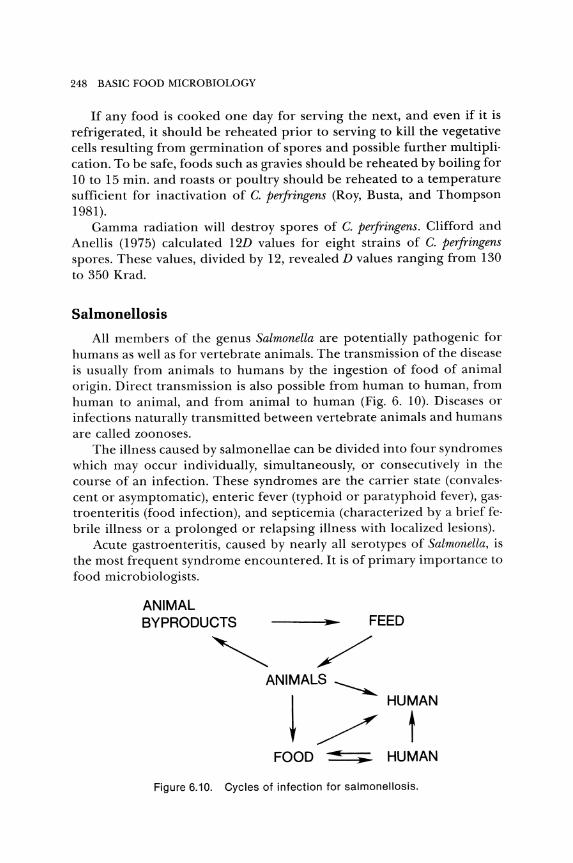
248 BASIC FOOD MICROBIOLOGY
If any food is cooked one day for serving the next, and even if it is refrigerated, it should be reheated prior to serving to kill the vegetative cells resulting from germination of spores and possible further multiplication_ To be safe, foods such as gravies should be reheated by boiling for 10 to 15 min_ and roasts or poultry should be reheated to a temperature sufficient for inactivation of C. perfringens (Roy, Busta, and Thompson 1981).
Gamma radiation will destroy spores of C. perfringens. Clifford and Anellis (1975) calculated 12D values for eight strains of C. perfringens spores. These values, divided by 12, revealed D values ranging from 130 to 350 Krad.
Salmonellosis
All members of the genus Salmonella are potentially pathogenic for humans as well as for vertebrate animals. The transmission of the disease is usually from animals to humans by the ingestion of food of animal origin. Direct transmission is also possible from human to human, from human to animal, and from animal to human (Fig. 6. 10). Diseases or infections naturally transmitted between vertebrate animals and humans are called zoonoses.
The illness caused by salmonellae can be divided into four syndromes which may occur individually, simultaneously, or consecutively in the course of an infection. These syndromes are the carrier state (convalescent or asymptomatic), enteric fever (typhoid or paratyphoid fever), gastroenteritis (food infection), and septicemia (characterized by a brief febrile illness or a prolonged or relapsing illness with localized lesions).
Acute gastroenteritis, caused by nearly all serotypes of Salmonella, is the most frequent syndrome encountered. It is of primary importance to food microbiologists.
ANIMAL BYPRODUCTS FEED
ANIMALS ~
~ /HUrN FOOD .... HUMAN
Figure 6.10. Cycles of infection for salmonellosis.

FOODBORNE AGENTS CAUSING ILLNESS 249
According to CDC (l983b), salmonellosis accounted for 26.4 percent of the reported outbreaks, 26.8 percent of the cases, and 65.6 percent of the deaths due to foodborne illness in 1981. Only the number of cases of S. aureus intoxication surpassed salmonellosis in these categories. The reported isolations of Salmonella from humans (1968-1980) is shown in Figure 6.11. Most isolations occur during the warm months. The number of reported isolations each year appears to increase.
The increase in the number of cases has been attributed to better diagnosis, laboratory procedures, and reporting. However, in those areas in which diagnosis and reporting have always been adequate, increases have occurred in the number of outbreaks and cases of salmonellosis. The prevalence of salmonellosis is increasing, although much energy has been expended to control this illness. This increase of Salmonella in both human and animal sources is associated with national and international commerce in animals, feeds, and foods; with large·scale, intensive animal raising; and with increased use of ready-to-serve or heat-and-eat foods.
It has been estimated that only 1 to lO percent of the actual number of cases are reported. There may be over 2,000,000 cases of salmonellosis each year in the United States. Many cases are mild and no professional care is solicited_ Salmonellae may be the major cause of bacterial gastroenteritis and foodborne illness in developed countries.
The economic loss due to Salmonella was estimated to be at least $300 million annually in the United States (Foster et al. 1970). The cost includes medical care, hospitalization, lost time and income through absence from work, death of animals, decreased production of animals, loss or reduced value of contaminated products, testing and control proce-
g
" .. '" "' ~ '" "' "' ~ I ;!, f/l
"' I-
~ 0 !!)
4000
3500
3000
2500
2000
1500
1000
500
1968 1969 1970 1971 1972 1973 197-4 1915 1976 1977 1878 1871 11180
DATE or REPORT TO CDC
Figure 6.11. Reported number of isolations of salmonellae from humans by month, 1968-1980.
250 BASIC FOOD MICROBIOLOGY
dures, and recall of products from market channels. A more recent estimate (CDC 1978b) is that the annual cost for salmonellosis is $1.5 billion for medical expenses alone. Depending on the circumstances, the average cost per case ranged from $277 to $4,548 for 12 outbreaks (Todd 1985). The median cost per case was $769. At this rate, 2 million cases cost more than $1.5 billion per year in the United States.
More deaths are caused by salmonellae and salmonellosis than by botulism. The death-to-case ratio is rather low, with a case fatality ratio of 0.26 percent from 1962 to 1968 (CDC 1971) and 0.15 percent for 1974 (CDC 1975). In 1981, the number of deaths jumped to 21 for a case-tofatality ratio of 0.86 percent (CDC 1983b). Most deaths occur in very young or old people.
CHARACTERISTICS OF THE ILLNESS. Salmonellosis has been considered to be an infection caused by the action of the organism in the intestine. However, there is evidence that a toxin or toxins may be involved.
Incubation Period. In thirty-four random outbreaks reported by the CDC, the range for the incubation period varied from 1 hr to eight days. The usual incubation period is reported as 6 to 48 hr (CDC 1983b).
Symptoms. The symptoms of nine random outbreaks reported by CDC are listed in Table 6.16. The symptoms and their severity depend on the number of organisms and the serotype of Salmonella, as well as the resistance of the host. The most reported symptom is diarrhea, followed by abdominal cramps, fever, nausea, vomiting, chills, and headache.
TABLE 6.16. SALMONELLOSIS: PERCENTAGE OF PEOPLE EXPERIENCING SYMPTOMS IN RANDOM OUTBREAKS
Outbreak
Symptom 2 3 4 5 6 7 8 9
Diarrhea 87 100 100 96 93 75 93 96 95 Diarrhea (bloody) 4 5 5 Abdominal cramps 70 47 70 81 86 82 76 66 57 Fever 68 82 85 62 80 48 97 43 Nausea 53 69 80 62 69 70 52 38 Vomiting 53 82 80 40 40 62 26 54 19 Chills 38 54 70 79 52 86 Headache 36 66 60 65 63 29 Dizziness 42 Muscle aches 70 95
"- = Not reported; however, the symptom might have existed. SOURCE: Data from CDC Morbidit,r and Mortality Weekly Reports.
FOODBORNE AGENTS CAUSING ILLNESS 251
Duration. In normal, healthy adults, the gastroenteritis usually lasts for only two to three days, but if further infection occurs, the illness may persist for months or years, and it may result in death. Thomas and Mog· ford (1970) stated that the duration of infection was longer than two months in nearly 25 percent of the cases, and intermittent excretion of the organisms was observed in 20 percent of the cases. Children are more susceptible than adults to prolonged excretion of the organism.
Therapy. The majority of the cases need no therapy. When hospitalization is required, an attempt is made to stabilize loss of fluids and prevent dehydration, as well as to maintain the electrolyte balance.
The administration of broad· spectrum antibiotics usually gives no ap· preciable benefit; the salmonellae become resistant to antibiotics. Treat· ment with antibiotics can cause the asymptomatic carrier state to become an active case (Saroglou and Bisno 1978).
Eating yogurt was suggested as part of the therapy for the salmo· nellosis outbreak involving 2 percent milk in 1985. Although yogurt did not prevent salmonellosis in rats, it did significantly reduce the mortality as well as other effects (Hitchins et al. 1985a, 1985b).
FOODS INVOLVED. Various foods have been the vehicle for transmis· sion of salmonellae. Most of these foods are of animal origin or contami· nated by foods of animal origin, as shown in Table 6.17.
The involvement of meat and meat products continues to increase and accounted for more than 50 percent of the known foods from 1977 to 1981. During 1977, there were several outbreaks and more than 180
TABLE 6.17. FOODS INVOLVED IN SALMONELLOSIS, 1977-1981, NUMBER
OF OUTBREAKS
Year
Food 1977 1978 1979 1980 1981 Total %
Beef 11 3 4 4 13 35 14.9 Pork 2 7 2 2 1 14 6.0 Other meat 1 3 1 0 0 5 2.1 Poultry 6 4 3 7 9 29 12.3 Eggs 0 0 2 1 0 3 1.3 Fishery prod· 0 0 0 0 2 2 0.9
ucts Dairy products 4 4 2 3 5 18 7.7 Bakery products 1 0 1 1 1 4 1.7 Salads 3 2 2 3 2 12 5.1 Other, unknown 13 22 27 18 33 113 48.1 TOTAL 41 45 44 39 66 235
SOURCE: CDC Annual Reports.
252 BASIC FOOD MICROBIOLOGY
cases of salmonellosis related to the consumption of precooked roast beef. These, as well as outbreaks in 1975 and 1976, stimulated action by the USDA to require specific times and temperatures for heating beef roasts (USDA 1983).
Poultry and poultry products have been implicated in many out· breaks of salmonellosis. Since the USDA required the pasteurization of egg products, these have been eliminated as a significant vehicle for sal· monella organisms. The failure to cook chicken and turkey sufficiently to eliminate the natural contamination is the usual contributing factor causing salmonellosis.
Raw milk has been the vehicle for salmonellae causing salmonellosis throughout the world (Bryan 1983; CDC 1984a; Reilly et al. 1983; Tacket et al. 1985a). All milk should be pasteurized before human consump· tion, but even pasteurized milk has been implicated in outbreaks of salmonellosis (Bryan 1983; CDC 1984b). A widespread outbreak in 1985, affecting over 16,000 people in several states, involved pasteurized milk. This incident should make us aware that pasteurization systems and proper procedures for handling pasteurized milk are needed to prevent salmonellosis and perhaps other illnesses.
Apple cider involved in an outbreak was produced from apples that had fallen onto the ground on which animal waste had been used as fertilizer. Although the apples were washed, sufficient salmonellae remained to cause illness. S. eastbourne was isolated from the environment in which cocoa beans were processed (CDC 1974b), apparently causing the contamination of chocolate candy which was involved in a widespread outbreak. No source was determined for S. napoli, which was isolated from chocolate bars involved in several cases of illness (Greenwood and Hooper 1983).
ETIOLOGIC AGENT. The mechanism by which the salmonellae cause gastroenteritis has not been fully explained. Perhaps more than one system is involved. The heating of food to destroy salmonellae results in a safe product. Hence, it was believed that salmonellosis was an infection that required high numbers of the organism to cause the illness. However, the heating needed to kill the organism also might inactivate any heat-labile toxins produced by the organisms.
The number of ingested Salmonella needed to cause an infection var· ies with the strain of the organism and the characteristics of the individual ingesting the organisms. Healthy adult males can ingest 105 to 107
cells (depending upon the strain of the organism) before symptoms of the illness occur. However, person-to-person transmission in hospitals indicates that only a few organisms are needed to cause illness. Blaser and Newman (1982) reviewed eleven outbreaks and calculated that, in six of
FOODBORNE AGENTS CAUSING ILLNESS 253
these, less than 1,000 ingested organisms causerl the illness. In a series of cases involving chocolate bars, it was estimated that an infective dose was roughly fifty organisms (Greenwood and Hooper 1983). The very young, aged, debilitated, and undernourished, and individuals with other illness are more susceptible than normal healthy individuals to infection by Salmonella.
The pH of the gastric juice can have a marked effect on salmonellae (Giannella, Broitman, and Zamcheck 1971). Healthy gastric juice with low pH (1-2) kills small numbers of salmonellae, but when it is deficient (pH 3.0 or higher), an otherwise harmless number of cells may survive.
When salmonellae reach the small intestine, they can live and multi· ply. The extent of multiplication depends on factors such as the peristal· tic rate, attachment to the mucosa, the composition of the intestinal flora, the ingestion of various therapeutic agents (such as antibiotics), exposure to radiation, and presence of iron·deficiency diseases.
Attachment of the salmonellae to the intestinal mucosa is considered to be essential for the organisms to remain in the intestine and cause illness. Although invasion of the intestinal epithelium is needed for ex· cess secretions (Giannella 1979), invasion, per se, is not sufficient to cause salmonellosis (Formal, Hale, and Sansonetti 1983). The results of rectal biopsies of twenty·two patients varied from normal mucosa to edema, hemorrhages, and sloughing (Day, MandaI, and Morson 1978). Some strains of salmonellae are known to cause mucosal damage and increased fluid output, some cause minimal mucosal damage and in· creased fluid, while others cause damage and no increase in fluid output. Salmonella infection causes lesions in the ilea of pigs (Wilcock 1979) and calves (Wray and Sojka 1978).
Researchers in one study suggested that S. typhimurium causes fluid secretion by altering the sodium and chloride transport systems (Giannella et al. 1975). When infected, the intestinal mucosa showed an increase in adenyl cyclase activity, which results in ileal secretion of fluids. In are· view, Smith (1977) suggested that an extracellular product is involved, because the microvilli degenerate before coming into contact with the bacteria.
Only four of thirteen strains of Salmonella tested produced a positive reaction in the rabbit ligated gut loop when living organisms were used (Sakazaki et al. 1974). When culture filtrates were tested in the ligated loops, eleven of thirteen strains of Salmonella gave a positive reaction. The researchers contended that the culture filtrates contained enter· otoxic activities.
The rabbit ileal loop model was used to detect enterotoxin produced by salmonellae (Sedlock and Deibel 1978). The serotypes that were tested varied in their ability to produce the toxin, but even old stock cultures
254 BASIC FOOD MICROBIOLOGY
maintained this trait. This indicates that the action is not due to a plasmid.
A factor isolated from S. typhimurium caused cells of Chinese hamster ovary (CHO) to elongate (Sandefur and Peterson 1977). This action is indicative of an enterotoxin. They found that this effect was blocked by antisera for cholera toxin and the B fragment of cholera toxin.
It is quite evident that most, if not all, strains of Salmonella produce enterotoxins that elongate CHO cells and cause secretion in gut loop tests (Baloda et al. 1983; Sharma, Singh, and Singh 1983). Jiwa and Mans· son (1983) and Alouf (1982) reported that both heat-labile and heat-stable enterotoxins are produced by some strains of salmonellae_ Although the enterotoxin usually reacts with antitoxin to cholera enterotoxin, one study reported a salmonella enterotoxin that did not cross-react with antisera to either cholera or heat-labile E. coli enterotoxins (Baloda et al. 1983).
Other researchers reported that, in salmonellosis, the cyclic adenosine monophosphate (cAMP) is increased in epithelial cells in a manner similar to that of cholera (Peterson et al. 1983). They stated that it was not clear whether the increase in cAMP was due to enterotoxins, to prostaglandins formed during the inflammatory response to the bacteria, or to both systems.
THE ORGANISMS_ The organisms in this genus are separated on the basis of their somatic (0) and flagellar (H) antigens. The antigenic classification of Salmonella is known as the Kauffmann-White scheme. New species or serotypes are continually being found and added to the many already classified. In 1964, there were about 900 known serotypes. Now there are more than 1,700 serotypes (CDC 1982a). The Arizona group has been incorporated into the Kauffmann-White scheme for Salmonella by Rohde (1979).
Although there are many serotypes, only about fifty cause nearly all of the outbreaks and cases of salmonellosis. Each year, some ten serotypes account for more than 60 percent of the isolations of Salmonella. The ten most prominent serotypes isolated from humans for the years 1977 through 1980 are listed in Table 6.18. S. typhimurium is the most frequently reported serotype isolated from both human and nonhuman sources (Table 6.19). Also, S. typhimurium is the most frequently isolated serotype in many other countries.
Growth Factors. Most strains of salmonellae can grow in a simple medium consisting of ammonium nitrogen, mineral salts, and glucose. A few strains need essential growth factors, primarily vitamins. The minimum water activity for growth is between 0.93 and 0.96. The salt concentration
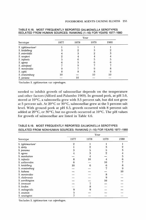
FOOD BORNE AGENTS CAUSING ILLNESS 255
TABLE 6.18. MOST FREQUENTLY F.!EPORTED SALMONELLA SEROTYPES ISOLATED FROM HUMAN SOURCES: RANKING (1-10) FOR YEARS 1977-1980
Year
Serotype 1977 1978 1979 1980
S. typhimurium" 1 1 1 1 S. heidelberg 3 2 3 2 S. enteritidis 4 3 2 3 S. newport 2 4 4 4 S. infantis 5 6 5 5 S. agona 6 5 6 6 S. saintpaul 7 9 7 7 S. montevideo 9 7 9 8 S. typhi 8 8 8 9 S. oranienburg 10 10 10 S.javiana 10
"Includes S. typhimurium var copenhagen.
needed to inhibit growth of salmonellae depends on the temperature and other factors (Alford and Palumbo 1969). In ground pork, at pH 5.0, stored at lOoe, a salmonella grew with 3.5 percent salt, but did not grow at 5 percent salt. At 20°C or 30°C, salmonellae grew at the 5 percent salt level. With ground pork at pH 6.5, growth occurred with 8 percent salt added at 20°C, or 30°C, but no growth occurred at lOoe. The pH values for growth of salmonellae are listed in Table 4.6.
TABLE 6.19. MOST FREQUENTLY REPORTED SALMONELLA SEROTYPES
ISOLATED FROM NONHUMAN SOURCES: RANKING (1-10) FOR YEARS 1977-1980
Serotype
S. typhimurium" S. derby S. panama S. agona S. manhattan S. infantis S. weltevreden S. heidelberg S. oranienburg S. habana S. montevideo S. cholerasuis S. worthington S. tennessee S. london S. meleagridis S. anatum S. newington
"Includes S. typhimurium var copenhagen.
1977
2 1 3 4
8 6
10
9 5 7
1978
1 2 5 3 4
10
6
7 8 9
Year
1979 1980
1 1 3 2 5 3 2 4
5 4 6
10 7 6 8 7 9
10 8 9
10
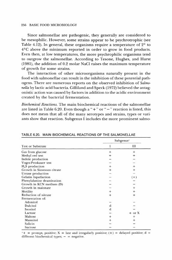
256 BASIC FOOD MICROBIOLOGY
Since salmonellae are pathogenic, they generally are considered to be mesophilic. However, some strains appear to be psychrotrophic (see Table 4.12). In general, these organisms require a temperature of 2° to 4°C above the minimum reported in order to grow in food products. Even then, at low temperatures, the more psychrophilic organisms tend to outgrow the salmonellae. According to Tesone, Hughes, and Hurst (1981), the addition of 0.2 molar NaCI raises the maximum temperature of growth for some strains.
The interaction of other microorganisms naturally present in the food with salmonellae can result in the inhibition of these potential path· ogens. There are numerous reports on the observed inhibition of Salmo· nella by lactic acid bacteria. Gilliland and Speck (1972) believed the antag· onistic action was caused by factors in addition to the acidic environment created by the bacterial fermentation.
Biochemical Reactions. The main biochemical reactions of the salmonellae are listed in Table 6.20. Even though a "+" or" -" reaction is listed, this does not mean that all of the many serotypes and strains, types or vari· ants show that reaction. Subgenus I includes the more prominent salmo·
TABLE 6.20. MAIN BIOCHEMICAL REACTIONS OF THE SALMONELLAE
Test or Substrate
Gas from glucose Methyl red test Indole production Voges·Proskauer test H"S production Growth in Simmons citrate Urease production Gelatin liquefaction Phenylalanine deamination Growth in KCN medium (D) Growth in malonate Motility Reduction of nitrate Fermentation of:
Adonitol Dulcitol Inositol Lactose Maltose Mannitol Salicin Sucrose
+ +
+ +
+ +
d d
+ +
"+ = prompt, positive; X late and irregularly positive; (+) different biochemical types; - = negative.
Subgenus·'
III
+ +
+ +
(+)
+ + +
+ or X + +
delayed positive; d =

FOODBORNE AGENTS CAUSING ILLNESS 257
nellae serotypes, and subgenus III consists of the organisms commonly referred to as the Arizona group, now designated as S. arizona.
Motility. Although the organisms are said to be motile, there are mutants that lose this ability, and two serotypes, S. pullorum and S. gallinarum are not motile and do not possess flagella.
Sources. As a genus, the salmonellae are said to be ubiquitous. They are worldwide and found in or on soil, air, water, sewage, animals, humans, food, feed, processing equipment, and some plant products. Some serotypes tend to be localized in a region or a country, but with national and international travel and trade, the organisms are readily disseminated_
The natural habitat of the organisms is the intestinal tract of humans and animals. Thus, it is logical that humans, animals, and their environments are the primary sources of Salmonella.
Few animals, if any, are born with intestinal contamination by Salmonella.Just as human infants are highly susceptible to salmonellosis, so are very young animals_ Some young animals may survive salmonellosis and become temporary or long-term shedders of the organisms. However, it is more likely that the animal is continually infected by some source of Salmonella.
The sources of salmonellae for infection of domestic animals include the parent stock, feed, water, rodents, wildlife, pets, humans, insects, arachnids, soil, and vegetation. The salmonellae can spread from animal to animal during production and processing. They spread from animals to people by direct interactions of humans and livestock, wild animals and pets, as well as through poor food-handling practices and the consumption of raw or undercooked foods of animal origin.
The relationship of Salmonella in feed to human salmonellosis is apparent with S. agona_ Prior to 1971, the serotype was reported from human hosts in the United States only six times. It was isolated from imported fish-meal on several occasions from 1970 to 1972_ Since these isolations, it has been found in domestic animals, and since 1973 it has ranked as one of the ten most prevalent salmonellae from humans (see Table 6.18). This suggests the involvement of feed in a chain or cycle of infection (see Fig. 6.10).
The role of free-living (wild) animals as a source of Salmonella was thought to be quite important. Now it is thought that rodents, birds, and other wild animals may be victims of their environment rather than the source of the organisms. Even so, these free-living animals can transfer the organism from infected areas to clean areas.
Since the waste materials from infected farms contain salmonellae, the use of this material as fertilizer for crops or pastures can spread the potentially infectious material to contaminate future animals. Rain fall-
258 BASIC FOOD MICROBIOLOGY
ing on the farms, feed lots, or fields containing contaminated wastes can wash the organism into streams and lakes. Besides the runoff from ani· mal quarters, sewage sludge and effluents from abattoirs and poultry· processing plants contain salmonellae. These polluted waters can cause infection of farm animals and wild animals that drink from them.
Various pets are potential sources of salmonellae. They may contami· nate food or directly transmit these organisms to humans. Pet turtles are an important source of Salmonella for transmission primarily to children. An estimated 280,000 cases of turtle·associated salmonellosis occurred in the United States in 1970 and 1971 (Lamm et al 1972). The interstate shipment of turtles less than 4 inches long was banned by the FDA in 1975, because studies showed that 14 percent of the reported salmonellosis in the United States was linked to pet turtles. However, some turtles are still being sold in the United States, and some 3 or 4 million are raised each year for export.
The direct transmission of Salmonella from animals to humans poses a problem in zoos that have children's petting areas or other open areas such as the aviary (Komorowski and Hensley 1973).
Although the cooking of raw meat and poultry will destroy salmonellae, the raw foods are a source of contamination to the food preparer and the kitchen equipment and can cross-contaminate cooked foods and other foods that are eaten raw.
Fish and shellfish are contaminated by water that contains waste products from humans and animals. Salmonellae were recovered from oysters, clams, and crabs harvested from both the east and west coasts of Florida (Fraiser and Koburger 1984). The east coast site was approved for shellfish harvest, but 25 percent of the trapped crabs and more than 13 percent of the clams contained salmonellae. The west coast site was an unclassified harvesting area, and the incidence of salmonellae in clams was higher than 43 percent. The highest level of contamination reported was 2.2 salmonellae per 100 grams of shellfish.
Fruits and vegetables are rarely involved in outbreaks of salmonellosis. However, vegetables can be contaminated by organisms present in animal wastes, or polluted irrigation water used on crops. Velaudapillai, Niles, and Nagaratnam (1969) found that l.3 percent of the vegetables sampled were infected with Salmonella, Shigella, or enteropathogenic E. coli. Salmonella serotypes were found on celery, leeks, spinach, green beans, and carrots. Wild olives contained S. sandiego. Samples of lettuce (68.3 percent) and fennel (7l.9 percent) yielded one or more serotypes of salmonellae (Ercolani 1976). Salmonellae were isolated from over 22 percent of the vegetable samples analyzed by Tamminga, Beumer, and Kampelmacher (1978a). Other researchers found that four samples of vegetables (radish, carrot, celery and spinach) offifty examined contained

FOODBORNE AGENTS CAUSING ILLNESS 259
salmonellae (Rude et al. 1984). They suggested that vegetables eaten raw should be considered a source of salmonellae in outbreaks of salmo· nellosis. Further, raw vegetables may be a source of human parasites. Co· conut products are believed to be contaminated by salmonellae in the soil. This contamination is transferred to the inner, edible portion duro ing processing.
Humans may be one of the main reservoirs of Salmonella. If not a source, humans can act as the vehicle for transfer of the organisms from one area to another.
Food handlers or workers in animal by· product plants often have a high rate of carriage of Salmonella. Although they might not have an active infection, the children of these workers have been observed to have higher rates of gastroenteritis than children of other workers. This indio cates that a worker who handles products that contain Salmonella can bring the organisms home to the other members of the family.
Survival in Nature. The natural habitat of Salmonella is the intestinal tract of animals. Excreted organisms are found in fecal material, food, and various parts of the environment. To cause salmonellosis, they must be able to survive in these unnatural surroundings.
Salmonellae in slurry (liquid manure) sprayed onto grass may survive from eighteen days (Taylor and Burrows 1971) to thirty· three weeks (Find· lay 1972). Jones, Hagler, and Hamilton (1977) found that the survival of salmonellae in cattle slurry was influenced by the normal microbial flora. In sterilized slurry, S. dublin multiplied and survived for more than 370 days, while in natural slurry, there was no multiplication and shorter sur· vival times. Survival time in a pasture is influenced by grass cover, sun· light, temperature, and rainfall.
Effluent from apparently healthy pigs was applied to land (Chandler and Craven 1981). The salmonellae were detected for eight months from soil and nearly two months from pasture. S. typhimurium survived in soil for seven to forty·two days, depending upon soil moisture and temper· ature (Zibilske and Weaver 1978).
Liu, Snoeyenbos, and Carlson (1969) found that the survival of salmo· nellae in meat and bone meal was related to water activity and holding temperature. As water activity and temperature increased from low lev· els, the survival decreased. At an aw of 0.82 and temperature of 50°C, a 5·log reduction occurred in 72 hr. However, at 10 percent moisture (aw
about 0.55) at 4°C, there was essentially no reduction in viable cells of S. senjtenberg 775W for 20 days. Elevated temperatures, oxygen, and unsatu· rated fatty acids accelerate the death of S. oranienburg in fish meal (Lam· precht and Elliot 1974).
When fingertips were contaminated with 500 to 2,000 cells of S. an·
260 BASIC FOOD MICROBIOLOGY
atum, the cells could be recovered 3 hr later. Contamination, followed by washing, eliminated levels of less than 1,000, but when 6,400 were inoculated, they could be recovered 10 min after washing. Even fewer than 100 cells of S. anatum inoculated onto fingertips infected samples of corned beef and ham handled 10 min after exposure (Pether and Gil· bert 1971). This shows how Salmonella can be spread by workers handling first contaminated products and then clean products.
Survival in Food. There is less destruction of inoculated salmonellae duro ing pan drying than during spray drying of egg white. Pan drying is more similar to the natural drying of materials. Once dried, the organisms can survive for long periods at room temperature or below. Salmonellae were isolated from dried eggs after forty·one weeks at 20°C, and at sixty·five weeks when held at 2°C.
Salmonellae can survive up to eleven weeks on vegetables held at 2° to 4°C, and seven weeks at room temperature. In acid foods, survival is relatively short, but if salmonellae survive freezing, such as in certain fruits, they may remain viable for considerable periods. Salmonella can survive at least thirteen months on frozen poultry held at - 21°C. In ice cream, the organisms can survive for several years.
Salmonellae inoculated into cold· pack cheese food survived in excess of twenty·seven weeks at 4.4°C, when no preservative (potassium sorb ate) or acids were added (Park, Marth, and Olson 1970). Six Salmonella strains were inoculated into sheep milk at 104 cells/ml and processed into Man· chego cheese (Medina, Gaya, and Nunez 1982). The organisms were abo sent from all lots of eight·week Manchego cheese.
The cells of S. typhimurium tended to die when present in yogurt (Rubin 1985). The presence of casein and an increased pH of yogurt pro· tected the cells from the inhibitory agent, lactic acid.
When inoculated into a beef.pork mixture at a level of 104 cells per gram, S. dublin survived pepperoni processes and persisted after forty· two to forty·three days of drying (Smith et al. 1975). S. typhimurium was more sensitive than S. dublin to the acid condition of Lebanon bologna during processing and aging (Smith et al. 1975b).
Salmonellae inoculated into chocolate bars at a level of about 106
cells/g were viable after storage for nine months (Tamminga et al. 1976). The reduction was greater in bitter chocolate than in milk chocolate. S. typhimurium inoculated at a level of about 1,000/g of chocolate was not detected in 55 g after fifteen months (Tamminga et al. 1977). S. eastbourne survived better than S. typhimurium.
In a review, Bryan (1968) summarized the survival of salmonellae. These organisms can survive in the low range of temperature and relative humidity. According to references cited by him, they can survive in con·
FOODBORNE AGENTS CAUSING ILLNESS 261
taminated earth and pasture for more than 200 days, cloth for 228 days, plastic cover slips for 93 days, sweeper dust for 10 months, rodent feces for 148 days, roach pellets for 199 days, poultry feces for more than 9 days, dried cattle feces for more than 1,000 days, on egg shells from 21 to 350 days, in dried whole eggs for more than 4 years and in meat salad for 77 days.
BASIC METHODOLOGY. It is not the intent of this text to precisely describe the procedures needed to obtain samples, or to detect or enu· merate salmonellae in the samples. For the procedures, the reader is reo ferred to FDA (1978), AOAC (1985), APHA (1984), and USDA (1971). Sam· pIing and testing plans for salmonellae were reviewed by Olson (1975).
We are concerned primarily with the analysis of food samples, swabs from food surfaces and water, and environmental swabs, although the analysis of other samples may be required, even in a food microbiology laboratory.
In many foods, the organisms have been subjected to debilitating or sublethal processes such as freezing, desiccation, extremes of pH, heat, osmotic pressures, or curing of ingredients during the manufacture or storage of the product. When present, salmonellae usually comprise a very small component of the total population. For most foods, there is no tolerance for Salmonella. Even one Salmonella detected in a 25·g sample is considered to be adulteration of the food (exceptions are raw chicken and meat). Hence, it is necessary to detect very low levels of these organ· isms. This small number of salmonellae, especially in comparison to the larger number of other organisms, means that the presence of salmonellae is unlikely to be demonstrated by direct plating on selective media, and that an enrichment procedure in broth is needed.
The basic procedure for the examination of foods for Salmonella consists of preenrichment and enrichment in broths, streaking and detection on selective-differential agars, and then characterizing typical colonies to confirm that they are salmonellae. Confirmation is done by means of biochemical tests, serological typing and, for a few serotypes, phage typing. It is evident that a long time period is needed before results of any kind are obtained. Hence, many attempts have been made to develop rapid procedures. Besides the basic procedure, some of these rapid methods are discussed.
Preenrichment. The preenrichment phase is accomplished by incubating the sample in a non inhibitory broth (usually in the ratio of one gram of sample to nine of broth) for 18 to 24 hr at 37°C. Shorter preenrichment times (4 to 6 hr) have been suggested, but longer preenrichment times (18 to 24, or 48 hr) have been more effective (D'Aoust 1981; Kafel 1981).
262 BASIC FOOD MICROBIOLOGY
The recovery of salmonellae from dried food products may be improved by reduced hydration systems (Andrews, Wilson, and Poelma 1983; Poelma, Andrews, and Wilson 1984; Wilson et al. 1985).
Media that have been used for pre enrichment include lactose broth, trypticase soy broth, reconstituted nonfat dry milk, nutrient broth, and buffered peptone water.
Generally, processed foods that may contain sublethally injured sal· monellae are preenriched. Raw or heavily contaminated products are put directly into a selective enrichment broth. The review by Andrews (1985) pointed out that preenrichment increased the recovery of salmonellae from these foods. However, for the examination of oysters, Miller and Koburger (1984) reported greater recovery by direct enrichment in sele· nite cystine broth than by using a preenrichment broth.
Enrichment. The enrichment process is the creation of a special environ· ment which permits the growth or selection of the desired species or group of microorganisms from a mixture of microorganisms with which they are likely to be found. The procedure usually functions by furnish· ing conditions for the desired organisms to outgrow all others. Ideally, an enrichment system for salmonellae should allow these organisms to grow and inhibit the growth of all other organisms.
It has been difficult to develop the ideal enrichment medium. Since there are some 1,700 serotypes of Salmonella, any selective agent is likely to be inhibitory to one or more serotypes or to be ineffective in inhibit· ing the nonsalmonellae that are present.
Foods vary in their nutrient content, pH, inhibitory substances, and other factors that may influence the growth of Salmonella. Therefore, when a food is added to an enrichment medium, usually in a ratio of 1:9, the characteristics of the medium are changed. Each food will alter the medium in its own peculiar way. Therefore, to be effective, a different enrichment medium should be designed for each food.
The enrichment broths commonly used in the United States are sele· nite cystine (SC), tetrathionate (T), tetrathionate brilliant green (TBG), and TT. Rappaport's medium (with modifications) reportedly is an effec· tive broth for salmonellae enrichment (Fricker and Girwood 1985; Tong· pim et al. 1984; Vassiliadis et al. 1984). However, it has never received much attention in the United States.
The enrichment broths are used as secondary enrichment of samples that have been preenriched (usually 1 ml of sample from the preenriched broth to 10 ml of enrichment broth), and as direct enrichment for raw or heavily contaminated food. Most analytical procedures suggest adding 25 g of food sample to 225 ml of broth. For feed samples, 30 g is added to 100 ml of broth. Due to multiple sampling of a food lot to assure the
FOODBORNE AGENTS CAUSING ILLNESS 263
absence of Salmonella, the analysis of larger quantities of pooled material from these samples has been suggested.
Some foods have certain peculiarities that call for special procedures for better detection of Salmonella. When gelatin is added in enrichment media, the viscous suspension that results can interfere with Salmonella analysis. Rose (1972) suggested adding gelatinase to the enrichment medium. The enzyme was not inhibitory to salmonellae, and better recovery was made from gelatin artificially inoculated with S. typhimurium.
When milk is analyzed, the acid produced causes formation of curds. Salmonella trapped inside these clots of milk are not detected. The addition of trypsin aids detection by digesting the protein of the milk, but apparently it does not injure the salmonellae.
The inoculated enrichment broth is incubated at either 37° or 43°C for 18 to 24 hr, and then a loopful is streaked onto an agar surface that is selective and differential.
Isolation. Many more Salmonella-positive samples are found when two or three agars are used. Due to the results of collaborative studies, researchers recommended retaining the use of bismuth sulfite (BS) agar, but recommended that salmonella-shigella agar and brilliant green (BG) agar be replaced by Hektoen enteric (HE) agar and xylose lysine desoxycholate (XLD) agar for detection and isolation of salmonellae (Sanders 1981). Bailey, Cox, and Thomson (1983) reported that BG and HE were superior to BS agar for recovery of Salmonella. Hawa, Morrison, and Fleet (1984) used a dulcitol bile novobiocin agar for isolating salmonellae from chicken carcasses.
It is recommended that BS plates be observed after 24 hr and, if no typical salmonellae colonies are observed, the plates should be incubated for another 24 hr before they are called negative.
With preenrichment for 24 hr, enrichment for 24 hr and BS incubation for 48 hr, a total of 96 hr (four days) is needed even for samples with no Salmonella, before any decision can be made.
The procedure to be used (type of preenrichment medium or no preenrichment, types of enrichment and plating media, as well as incubation temperatures) is dictated somewhat by the type of sample being analyzed, as well as the types of organisms and serotypes of Salmonella that might be present.
It is difficult to compare media for the preenrichment, enrichment, and isolation for Salmonella. If salmonellae are present in the sample at a high level, almost any of the typical media can be used to detect them. If the organisms are at a very low level, some of the samples for preenrichment or direct enrichment may not contain any Salmonella. To overcome these circumstances, many samples need to be tested and the results analyzed by an acceptable statistical system.
264 BASIC FOOD MICROBIOLOGY
In all of the research that has been done, it is evident that the more different types of broths and agars that are used, the greater is the chance for recovery of Salmonella, especially if sublethally injured and at a very low level, with various other organisms at high levels.
Biochemical Characterization. If no typical colonies are found on the isola· tion medium, this is satisfactory evidence that the sample is Salmonella· negative. If typical colonies are detected, this is presumptive evidence that the sample may be Salmonella· positive. For further characterization of the organisms in these typical colonies, both biochemical and serological tests are needed. Neither the biochemical nor the serological tests are specific for Salmonella, and neither system will detect all the serotypes of the genus.
Rapid and easily run methods have long been the desire of microbiologists. One method employed to get a quick reaction is to use a large inoculum in a small amount of medium.
With the number of biochemical tests that need to be run, considerable time and material are required to prepare the media, inoculate, incubate, determine the reactions, and classify the cultures. To cut down on the time and cost, rapid methods, composite media, and simplified test kits have been developed. Commercial preparations have the potential benefit of providing many laboratories with standard materials for use in assessing bacterial reactions. These commercial systems include the API 20E, Enteric-Tek, Enterotube II, Micro-ID, Minitek, Repliscan II, and Sensititre systems.
These systems have certain things in common. They are all miniaturized and rapid tests based primarily on reports in the literature by various workers. None of the systems agrees 100 percent with the conventional tube method for determining reactions. The common opinion is that these tests are acceptable for general use.
Serology. Serological considerations are used in the immunization of humans and animals for certain diseases caused by salmonellae, for testing the serum of humans or animals for potential carriers of the organisms, and for determining the serotype of an isolated test culture. The serological reactions of the test culture are needed to supplement the informa· tion obtained from biochemical tests.
The salmonellae possess 0, H, and K antigens (Kauffman 1972). The o (somatic) antigens are considered to be a constitutive part of the cell wall, but they may extend beyond this barrier. They are a complex of lipopolysaccharide (LPS) and protein. The detailed structure of the 0 antigen varies from strain to strain. This variation forms the basis for the serological differences of the organisms. The 0 antigens are considered to be heat stable, resisting boiling for 21;2 hr.
Salmonella can change in colony morphology form smooth to rough

FOOD BORNE AGENTS CAUSING ILLNESS 265
(S--> R variation). These rough forms are caused by mutations that block the synthesis of polysaccharides. Thus, they have no 0 side chains, lose o agglutinating ability, and have a reduced virulence.
The H antigens, or flagellar antigens, are found only in motile cuI· tures. They are destroyed at 100°C (heat labile), and by dilute alcohol or acid. Flagella are composed of the protein flagellin, with a molecular weight of about 40,000. Many species of salmonellae have two types of flagellar antigens (phase 1 and phase 2). A single bacterium manifests only one of these.
Phase 1 antigens are specific and are shared by only a few serotypes of salmonellae, while phase 2 antigens are nonspecific and are common to many serotypes.
Usually, only the 0 and H antigens are determined when typing a culture, although the Vi antigen is sometimes of interest. The cultures are typed according to the Kauffmann·White scheme. This system is out· lined by Kauffmann (1972) and Krieg and Holt (1984).
The specific H or 0 antigens are determined by cell genetics. Due to mutations or phage conversions, the antigens can be altered. This may account for the multiplicity of serotypes and the overlapping patterns of antigenicity.
If there is an agglutination with the 0 antisera, it is not proof that the culture is a salmonella. There are cross·reactions with the Salmonella o antisera and nonsalmonellae. Some strains of closely related orga· nisms, especially strains of E. coli, Citrobacter, Shigella, and Enterobacter, either share certain somatic antigens or have antigens similar to those of salmonellae (Refai and Rohde 1975). Other unrelated microorganisms reportedly react with Salmonella 0 antisera (Aksoycan and Saganak 1977; Corbel 1975).
The H antigens are determined by a tube test. A motile organism is necessary for reaction with H antigens. Since S. pullorum and S. gallinarum are not motile, they will not react. For the exact procedure for detecting H antigens, the reader is referred to AOAC (1985) or FDA (1978).
The medium in which the organism is grown can influence the reac· tion with H antiserum (Banwart and Kreitzer 1972; Stamper and Banwart 1974).
Besides biochemical and serological tests, for epidemiological pur· poses phage typing, antimicrobial resistance patterns (resistotyping), and plasmid typing may be used for characterizing salmonellae (Brunner et al. 1983; Farrar 1983; Riley et al. 1983; Somerville, Nhlapo, and Alberts 1983).
OTHER METHODS. The conventional procedure requires consider· able material and time to detect Salmonella. Other tests have been devised to obtain results more quickly and easily. Usually rapid tests result in a
266 BASIC FOOD MICROBIOLOGY
loss of accuracy or precision. However, even with the conventional test, not all of the Salmonella serotypes are detected in all cases. The results obtained with a rapid test should at least compare favorably with those obtained with the conventional test (FDA 1978).
To detect the low number of salmonellae in most foods, the sample must be incubated in a growth medium to enrich the salmonellae prior to analysis, even by the "rapid" methods.
Hydrophobic·Crid-Membrane Filtration (HCMF). After preenrichment and enrichment, a portion of enrichment broth is passed through an HGM filter (En tis et al. 1982). The filter is transferred to a selective agar surface and, after incubation, observed for salmonellae. Presumptive colonies must be evaluated biochemically and serologically as in the conventional test. The HGMF system has been adopted by the AOAC.
Phage Systems. The use of bacteriophage has been suggested for detection, characterization, and the typing of certain serotypes of salmonellae.
To detect S. typhosa (S. typhi) in soil samples, Kande1aki (1964) added typhoid indicator phage to the sample. An increase in the phage titer during incubation was evidence that S. typhi was present in the sample. The phage titer increase detected more positive samples than did the bacteriological plating system Kandelaki used.
For determining the character of colonies on selective agars, one team of researchers (Cherry et al. 1954) suggested using bacteriophages. According to them, the method is rapid, simple, and quite specific. They used the 0-1 phage of Felix and Callow. This system was reevaluated by We1kos, Schreiber, and Baer (1974). They tested 652 strains of Salmonella and found that the phage (0-1) reacted with 640 (98.2 percent). Of 1,463 nonsalmonellae strains, only E. coli strains were lysed. Of 239 strains of E. coli tested with 1012 plaque-forming units/ml (PFU/ml), 14 (5.9 percent) were susceptible to lysis. Gunnarsson, Hurvell, and ThaI (1977) found that 98 percent of 5,287 strains of salmonellae were lysed by 0-1 phage. They warned that monophasic strains of subgenus III and strains of subgenus IV usually are not sensitive to the 0-1 phage.
The increase in 0-1 phage was determined with high performance liquid chromatography to detect salmonellae in milk and feces (Hirsch and Martin 1983; Crane, Martin, and Hirsch 1984). A plaque system using 0-1 phage reportedly can detect salmonellae in milk in 24 hr (Hirsch and Martin 1984). Since not all strains of salmonellae are affected by the 0·1 phage, Glidel and Fey (1981) suggested adding a G47 phage to the 0-1 phage to increase the number of strains that are lysed.
Immunoassays. There are many immunoassay systems, some of which are discussed for the detection of S. aureus enterotoxin. The immunoassays
FOODBORNE AGENTS CAUSING ILLNESS 267
of interest in salmonellae detection are those using a labeled reactant (usually the antibody).
For the radioimmunoassay (RIA), a radioactive substance is conju· gated onto one of the reactants. Although the RIA is a sensitive system, there are problems (see S. aureus enterotoxin detection, page 215).
When a fluorescent compound is conjugated onto a reactant in salmonellae determinations, it is called the fluorescent antibody (FA) technique. This test has been adopted by the AOAC (Sanders 1975). After an enrichment procedure, a direct or indirect FA test can be used.
For the direct test, the specific antibody is conjugated with a flit orescent dye. When this conjugate is added to a solution containing homologous antigen, they will react, and the product will be fluorescent in ultraviolet light.
For the indirect method, if the antibody is produced in a rabbit, rabbit serum is injected into another animal, such as a goat, to produce antirabbit serum. This goat antirabbit serum is conjugated with the fluorescent dye. For the test, the antigen is reacted with the specific antiserum produced in the rabbit. Then the conjugated goat antirabbit serum is added, which will react with the antiserum produced in the rabbit.
The advantage of the indirect method is that, as long as the antibodies are produced in a rabbit, only one conjugated antiserum is needed (goat antirabbit). This conjugate will work in FA tests for clostridia, staphylococci, salmonellae, and any organisms, as long as the specific antiserum to the antigens of the organism are produced in a rabbit.
Besides the requirement for a good fluorescent (ultraviolet) microscope and well-trained personnel, the diagnostic sera used to detect salmonellae by the FA is expensive. The serum should react with all of the serotypes of Salmonellae and not cross-react with the nonsalmonellae. This type of serum is not yet available.
In many surveys, the FA procedure reveals more Salmonella-positive samples than the conventional culture test. Various reasons for this have been suggested. One proposal is that the FA is more sensitive than culturing. A more logical explanation is that cross-reactions with nonsalmonellae by the FA antisera result in more apparently Salmonella-positive samples. Thus, the samples that appear positive with the FA technique must be run through the conventional test for confirmation.
When an enzyme is conjugated onto a reactant, it is called an enzyme immunoassay (EIA) (see S. aureus enterotoxin detection, page 215). For salmonellae testing, this and the other immunoassays might be used to detect the organisms as well as their enterotoxins.
For their EIA, Mattingly and Gehle (1984) stated that 106 Salmonellal ml were needed for detection. This means that the salmonellae in the samples must be enriched prior to analysis. However, the enrichment
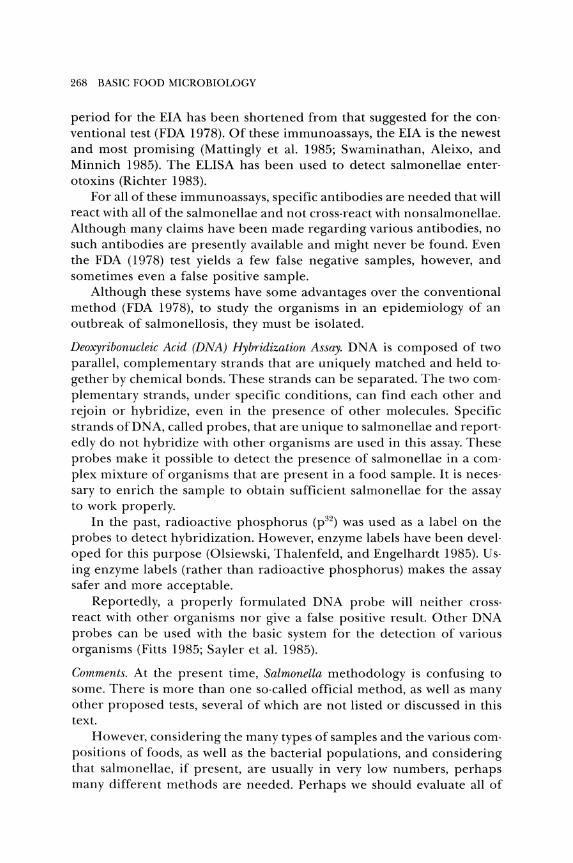
268 BASIC FOOD MICROBIOLOGY
period for the EIA has been shortened from that suggested for the conventional test (FDA 1978)_ Of these immunoassays, the EIA is the newest and most promising (Mattingly et aL 1985; Swaminathan, Aleixo, and Minnich 1985)_ The ELISA has been used to detect salmonellae enterotoxins (Richter 1983)_
For all of these immunoassays, specific antibodies are needed that will react with all of the salmonellae and not cross-react with nonsalmonellae_ Although many claims have been made regarding various antibodies, no such antibodies are presently available and might never be found_ Even the FDA (1978) test yields a few false negative samples, however, and sometimes even a false positive sample_
Although these systems have some advantages over the conventional method (FDA 1978), to study the organisms in an epidemiology of an outbreak of salmonellosis, they must be isolated_
Deoxyribonucleic Acid (DNA) Hybridization Assay_ DNA is composed of two parallel, complementary strands that are uniquely matched and held together by chemical bonds_ These strands can be separated_ The two complementary strands, under specific conditions, can find each other and rejoin or hybridize, even in the presence of other molecules_ Specific strands of DNA, called probes, that are unique to salmonellae and reportedly do not hybridize with other organisms are used in this assay_ These probes make it possible to detect the presence of salmonellae in a complex mixture of organisms that are present in a food sample_ It is necessary to enrich the sample to obtain sufficient salmonellae for the assay to work properly_
In the past, radioactive phosphorus (p32) was used as a label on the probes to detect hybridization_ However, enzyme labels have been developed for this purpose (Olsiewski, Thalenfeld, and Engelhardt 1985)_ Using enzyme labels (rather than radioactive phosphorus) makes the assay safer and more acceptable_
Reportedly, a properly formulated DNA probe will neither crossreact with other organisms nor give a false positive result_ Other DNA probes can be used with the basic system for the detection of various organisms (Fitts 1985; Sayler et aL 1985)_
Comments_ At the present time, Salmonella methodology is confusing to some_ There is more than one so-called official method, as well as many other proposed tests, several of which are not listed or discussed in this text
However, considering the many types of samples and the various compositions of foods, as well as the bacterial populations, and considering that salmonellae, if present, are usually in very low numbers, perhaps many different methods are needed_ Perhaps we should evaluate all of
FOODBORNE AGENTS CAUSING ILLNESS 269
the potential media and systems with each food to develop the best system for each food_
The results of research have shown that, with the FDA (1978) method, the more types of media that are used, the greater the likelihood that all Salmonella-positive samples will be detected_ With nearly all of the socalled rapid methods, only one medium is used_ Are these systems that superior to using a selective agar isolation system? It seems doubtfuL Obtaining a false positive sample may increase the cost of food due to reexamination, reprocessing or discarding the suspect food_ However, missing a Salmonella-positive sample with a resultant salmonellosis outbreak can result in an even higher cost to the processor.
CONTROL OF SALMONELLA. The measures needed to control Salmonella and salmonellosis are essentially the same as those needed for the control of S. aureus and the clostridia (prevent contamination, prevent growth, destroy the organisms). The number of cases of typhoid fever due to S. typhi has been drastically reduced_ Proper sewage disposal, protection and chlorination of communal water supplies, restricting the harvesting of aquatic food from contaminated water, pasteurization of certain products, sanitary control of food processing and sales establishments, exclusion of typhoid carriers from food-handling occupations, and immunization programs, have reduced the incidence of typhoid fever in the United States.
Since the prevalence of salmonellosis is increasing, it is evident that these procedures are not effective in controlling this illness; other measures need to be incorporated_
There is an incentive for the food industry to market products with no salmonellae. Section 402 of the Federal Food, Drug and Cosmetic Act defines a food to be adulterated if it bears or contains any poisonous or deleterious substance that may render it injurious to health, and if it has been prepared, packed, or held under unsanitary conditions whereby it may become contaminated with filth or whereby it may be rendered injurious to health_ Foods containing Salmonella or other pathogens fall within this definition.
The control of salmonellae in foods includes the acquisition of Salmonella-free raw materials, with processing, storage, and distribution under conditions that prevent the increase of Salmonella and, ideally, a terminal treatment of the food to destroy any salmonellae that may be present.
Prevent Contamination. Because salmonellae are ubiquitous, keeping them out of food might seem to be a hopeless task_ However, it is generally believed that, with a sincere effort by all factions of feed and food pro-
270 BASIC FOOD MICROBIOLOGY
duction, handling, processing, and preparation, the contamination of food by salmonellae can be prevented or reduced.
A major cycle of infection is animal by·product to feed, feed to ani· mals, and animals to food to humans (Fig. 6. 10). There are other cycles, such as human to food to human or even human to human. If the cycle of feed to animals to humans can be broken, the incidence of salmo· nellosis might be reduced significantly.
There should be Salmonella·free breeding stock, feed, and water, strict sanitary practices, segregation of sick animals, and sale of only healthy animals. Sanitation includes many facets, such as pest control, cleaning and disinfecting housing, removal and disposal of waste, storing feed so that it is not contaminated, preventing workers from carrying diseases with them from one farm area to another, and keeping out visitors. There are sanitary requirements for dairy farms, but other farming practices are usually left to the farmer. However, Oosterom, Van Erne, and Van Schothorst (1982) reported that using practical farming situations, even with sanitary precautions, it was impossible to produce Salmonella-free pigs.
To reduce or eliminate Salmonella from chickens, a system called competitive exclusion shows promise (Blanchfield et al. 1984; Goren et al. 1984; Pivnick et al. 1982; Schneitz, Seuna, and Rizzo 1981). It is based on colonization of the gastrointestinal epithelial surface by indigenous organisms. Feeding or otherwise inoculating fecal, cecal, or anaerobic cultures from adult fowl into the intestines of day-old chicks will allow these bacteria to occupy the attachment sites. This deters or prevents salmonellae from colonizing the intestines of the chicks. Competitive exclusion also protected turkeys from salmonellae (Reid and Barnum 1983). The feeding of antibiotics to animals can alter the indigenous flora, allowing salmonellae to colonize the intestinal tract (Barrow, Smith, and Tucker 1984; Que and Hentges 1985).
For seafoods, the habitat in which such animals grow must be free of Salmonella. The first step is to prevent untreated sewage or water from entering streams, lakes, or oceans. Shellfish, such as oysters, are believed to cleanse themselves of salmonellae and E. coli in 48 to 72 hr if placed in clean water. At an initial level of 400 salmonellae per gram, oysters contained no detectable cells after 2.5 days, whereas 900 cells per gram required 3.5 days (Son and Fleet 1980). However, clean beds are needed for the production and harvesting of shellfish, since the depuration process cannot be relied upon for the elimination of all pathogens.
In the preparation of food, one of the important factors for spreading Salmonella is the cross-contamination from raw to cooked food. Until such time as raw poultry or meat products can be produced with no Salmonella, we must assume that the organisms will be present. Handling of these
FOODBORNE AGENTS CAUSING ILLNESS 271
products and then handling ready-to-eat products transfers the bacteria from the raw to the ready-to-eat products.
Thorough daily cleaning and sanitizing of the processing plant and equipment can reduce the dissemination of salmonellae and other organisms into the environment and onto foods. Testing for salmonellae should be part of the control effort. If the organisms are in the plant environment, there is a good chance they will be in the finished product. Cleaning and sanitizing are discussed further in Chapter 10.
Foster (1969) pointed out that elimination of salmonellae from foods would not necessarily eliminate human salmonellosis. We also need to consider the human carrier as well as household pets. The human carrier is important in the processing plant, in the food-preparation and serving industry, and in the home.
Foods have been placed into five categories in terms of hazard to human health (Foster 1971). No safe level of salmonellae has been established, but it is evident that illness is more likely when the number ingested is increased. Three hazard characteristics were established for a particular food. These are (1) the food or an ingredient is a significant source of Salmonella; (2) there is no control step (heating) in the process to destroy the organism; and (3) if the product is mishandled, growth can occur, resulting in an increased hazard. The higher the number of hazard characteristics of a food, the more potentially dangerous it is. A food with no hazard characteristics is not considered dangerous, foods with one or two are intermediate, and foods with all three are hazardous. The susceptibility of people varies, with infants, the aged, and the infirm the most susceptible to Salmonella infection. With this aspect included, the five categories are established:
Category I foods possess any of the hazard characteristics and are intended for use by infants, the aged, or the infirm.
Categories II, III, IV, and V foods are for general uses. Category II foods possess all three hazard characteristics. Category III foods have two of the hazard characteristics. Category IV foods have one hazard characteristic. Category V foods have no hazard characteristics_
With these hazard categories, it is evident that fewer samples of foods in category V need to be tested (if negative results are obtained), than those in category I or II. Considering 25-g samples, it was proposed that the food should be accepted if there are no positive samples in fifteen samples (Category III, IV, or V), thirty samples (Category II), and sixty samples (Category I) or, if no more than one positive sample is detected in twenty-four samples (III, IV, V), forty-eight samples (Category II) or
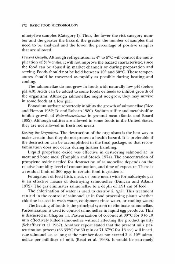
272 BASIC FOOD MICROBIOLOGY
ninety· five samples (Category I). Thus, the lower the risk category num· ber and the greater the hazard, the greater the number of samples that need to be analyzed and the lower the percentage of positive samples that are allowed.
Prevent Growth. Although refrigeration at 4° to 5°C will control the multi· plication of Salmonella, it will not improve the hazard characteristic, since the food can be abused in market channels or during preparation and serving. Foods should not be held between 10° and 50°C. These temper· atures should be traversed as rapidly as possible during heating and cooling.
The salmonellae do not grow in foods with naturally low pH (below pH 4.0). Acids can be added to some foods or feeds to inhibit growth of the organisms. Although salmonellae might not grow, they may survive in some foods at a low pH.
Potassium sorb ate reportedly inhibits the growth of salmonellae (Rice and Pierson 1982; To and Robach 1980). Sodium sulfite and metabisulfite inhibit growth of Enterobacteriaceae in ground meat (Banks and Board 1982). Although sulfites are allowed in some foods in the United States, they are not allowed in fresh red meats.
Destroy the Organisms. The destruction of the organisms is the best way to make certain that they do not present a health hazard. It is preferable if the destruction can be accomplished in the final package, so that recon· tamination does not occur during further handling.
Liquid propylene oxide was effective in destroying salmonellae in meat and bone meal (Tompkin and Stozek 1974). The concentration of propylene oxide needed for destruction of salmonellae depends on the relative humidity, level of contamination, and time of exposure. There is a residual limit of 300 j.tg/g in certain food ingredients.
Fumigation of feed (fish, meat, or bone meal) with formaldehyde gas is an effective means of destroying salmonellae (Duncan and Adams 1972). The gas eliminates salmonellae to a depth of 1.91 cm of feed.
The chlorination of water is used to destroy S. typhi. This treatment can aid in the control of salmonellae in food· processing plants whether chlorine is used in wash water, equipment rinse water, or cooling water.
The heating of foods is the principal system to eliminate salmonellae. Pasteurization is used to control salmonellae in liquid egg products. This is discussed in Chapter 11. Pasteurization of coconut at 80°C for 8 to 10 min effectively killed salmonellae without affecting the product quality (Schaffner et aL 1967). Another report stated that the present milk pas· teurization process (63.33°C for 30 min or 71.67°C for 16 sec) will inacti· vate salmonellae, as long as the number does not exceed 3 X 10 12 salmo· nellae per milliliter of milk (Read et aL 1968). It would be extremely
FOOD BORNE AGENTS CAUSING ILLNESS 273
unlikely that this level of salmonellae would be attained without spoilage, due to other bacteria.
Although bacteria have an increased heat resistance in dried prod· ucts, it is not impossible to eliminate salmonellae from dry animal feed. Carrol and Ward (1967) found a combination of 87.8°C for 10 min con· sistently reduced salmonellae to a nondetectable level in fish meal. Van Cauwenberge, Bothaste, and Kwolek (1981) stored salmonellae· inoculated corn flour (10 and 15 percent moisture) at 49°C. After 24 hr, 99.9 percent of the salmonellae cells were destroyed. However, heating salmonellae· inoculated poultry feed in a microwave oven (reaching a temperature of 186°C), in a hot air oven at 110°C for 1 hr, or in flowing steam for 10 min failed to consistently eliminate the salmonellae (Bur· dick et al. 1983).
The cooking of foods usually destroys any salmonellae that may be present. S. senftenberg 775W is killed during the normal processing of frankfurters in the smokehouse when the internal temperature of the product reaches 7l.1°C (Palumbo, Huhtanen, and Smith 1974). Heating pepperoni to 60°C or Lebanon bologna to 5l.7°C eliminated 104 inocu· lated cells of salmonellae (Smith et al. 1975a, 1975b). Goodfellow and Brown (1978) determined D values (time needed to destroy 90 percent of the population) for salmonellae in a ground beef system. The D values they reported were 5l.6°C (61-62 min), 57.2°C (3.8-4.2 min), and 62.7°C (0.6-0.7 min). These D values were used to establish the times and tem· peratures needed to cook roast beef to assure the death of any salmo· nellae.
Salmonellae are only moderately resistant to radiation treatment. AI· though the needed radiation dose varies with the type of product, conditions ofradiation, and level of contamination, Thornley (1963) suggested that 0.5 Mrad would reduce the number of salmonellae by a factor of 107•
Ostovar, Pereira, and Gallop (1971) found that 0.5 Mrad was sufficient to destroy salmonellae in smoked whitefish. Another team of researchers reported that, for frozen meat, 0.6 Mrad would reduce Salmonella numbers by a factor of at least 105 (Ley et al. 1970). For decontamination of herring meal, radiation doses of 0.8 to l.3 Mrad were recommended (Underdal and Rossebo 1972). Mulder, Notermans, and Kampelmacher (1977) suggested treating poultry carcasses with 0.25 Mrad to eliminate salmonellae. More recently it was reported that, after treatment with 0.25 Mrad at 5°C, the poultry might still contain salmonellae, but that, after storage of this treated poultry at -18°C for three months, no salmonellae were detected (Mulder 1982). Pearce (1979) found that salmonellae were quite stable in stored casein, but treatment with 0.5 Mrad eliminated these organisms from the protein.
The low doses of radiation needed to eliminate salmonellae have no

274 BASIC FOOD MICROBIOLOGY
apparent adverse effect on the flavor or nutritional quality of the food or feed. It would seem to be a very useful process for animal feed, since gamma irradiation can be accomplished after the feed has been packed into impervious bags.
Shigellosis (Bacillary Dysentery)
This illness accounts for less than 10 percent of the reported out· breaks of foodborne illness in the United States. Since only a few states report this illness to CDC, it is probably more widespread than the data indicate. The predominant mode of transmission is human to human, but contaminated water and food also are important.
Sometimes the illness is called the filth disease, because it is associ· ated with poor personal hygiene and sanitation. The illness is often prominent in areas where there are large groups of people. Summer camps, mental institutions, Indian reservations, and low socioeconomic urban communities are the high·risk population areas. The increase in infant day care centers has resulted in another high risk situation.
The mortality rate appears to be greatest in infants and in adults over fifty years of age. Most of the cases are children under ten years, and the highest attack rate is children in the one-to-four age group. This is probably due to their lack of training in personal hygiene at this young age.
In the twenty-to-twenty·nine age group, there is a higher rate of illness in females than in males. This is believed to be due to women's having closer contact with children, especially sick children, than do men of this age group.
Although considered a disease of the poor, the affluent have acquired the illness through travel to foreign areas, such as Mexico, Central America, and the Far East.
Shigellosis is primarily an illness of humans, but other primates, including monkeys and chimpanzees, can acquire the disease (Rout et al. 1975). By using procedures such as starvation, folic acid deficiency, control of normal intestinal flora, and administration of opium to slow peristaltic action, experimental shigellosis has been established in mice and guinea pigs (Nelson and Haltalin 1972; Maier and Hentges 1972). The ligated small intestine of rabbits serves as an animal model for studying shigellosis.
PATHOGENESIS. The pathogenesis of Shigella is rather complex. After reaching the small intestine, the first requirement is the attachment to and penetration of cells of the intestinal mucosa. If the cells cannot invade, they are avirulent. After invasion, shigellae multiply and cause the destruction of cells (Levine et al. 1973). This results in mucosal ulcera-
FOODBORNE AGENTS CAUSING ILLNESS 275
tion. Speelman, Kabir, and Islam (19S4) studied colonic lesions in thirty· three patients with shigellosis. Their findings indicated that the rectosig· moid area is the most frequently and most severely affected, and that during the course of illness, the colonic lesions extend in a proximal direction. In some cases, lesions extended to the splenic flexure, the dis· tal transverse colon, and the proximal transverse colon. In 15 percent of the patients, pancolitis was evident.
Shiga toxin, an enterotoxin produced by S. dysenteriae 1, the most viru· lent Shigella, was purified by one group of researchers (Brown et al. 19S2). The toxin was cytotoxic to HeLa cells, caused secretion in rabbit ileal loops, and was lethal to mice. S. sonnei and S. jlexneri also produce toxins that are antigenically and biologically related to Shiga toxin of S. dysenter· iae (Keusch and Jacewicz 1977; O'Brien et al. 1977). These three species, as well as S. boydii, produce a toxin that causes morphological changes in Chinese hamster ovary cells (Takeda, Okamoto, and Miwatani 1979).
The exact role of the enterotoxins in shigellosis has not been estab· lished, but there are various speculations as to their action (Fernandez et al. 19S4; Formal, Hale, and Sansonetti 19S3; Mathias et al. 19S0). The mechanism of the pathogenic action of shigellae and their toxins is not known exactly. The illness may be an infection caused by the organism, some form of intoxication, or a combination of these actions.
CHARACTERISTICS OF THE ILLNESS. The number of organisms needed to cause the infection may be rather low, since person·to·person transfer is the main mode of transmission. One study reported that as few as ten organisms of virulent strains produced illness in human vol· unteers (Levine et al. 1973).
In most outbreaks of shigellosis there is an index case. One person develops symptoms of the disease, and then one or two days later, others became ill. If the index case is a food handler, a foodborne outbreak can occur. An example of an outbreak is shown in Figure 6.12.
Incubation Period. The time from ingestion of the organisms until symp' toms appear varies with the individual (health, age), number of organ· isms ingested, and virulence of the organism. CDC (l 9S3b) listed the incubation period for shigellosis as 12 to 50 hr. Due to person· to· person spread, it sometimes is difficult to determine the exact times for the in· fections.
Symptoms. The symptoms of shigellosis as listed in several outbreaks are shown in Table 6.21. The main symptom is diarrhea. Infections associ· ated with mucosal ulceration usually cause more fever, abdominal pain, and rectal bleeding than do toxin· induced diarrheas.
In the United States, the disease is usually self.limiting and rather
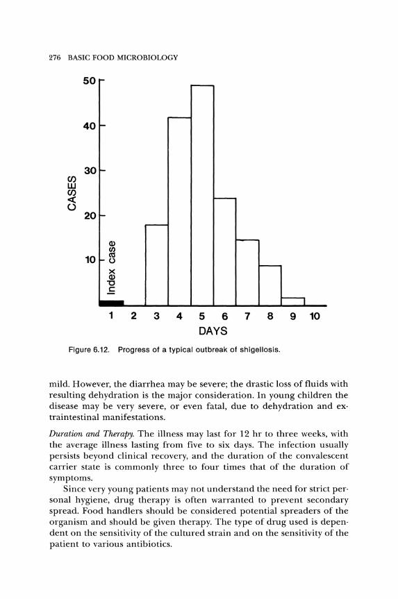
276 BASIC FOOD MICROBIOLOGY
C/) w C/) « o
50
40
20
10
Q) en t\'l ()
X Q) "0 C
1 2 3 4 5 6 7 8
DAYS
Figure 6.12. Progress of a typical outbreak of shigellosis.
9 10
mild. However, the diarrhea may be severe; the drastic loss of fluids with resulting dehydration is the major consideration. In young children the disease may be very severe, or even fatal, due to dehydration and ex· traintestinal manifestations.
Duration and Therapy. The illness may last for 12 hr to three weeks, with the average illness lasting from five to six days. The infection usually persists beyond clinical recovery, and the duration of the convalescent carrier state is commonly three to four times that of the duration of symptoms.
Since very young patients may not understand the need for strict personal hygiene, drug therapy is often warranted to prevent secondary spread. Food handlers should be considered potential spreaders of the organism and should be given therapy. The type of drug used is dependent on the sensitivity of the cultured strain and on the sensitivity of the patient to various antibiotics.

FOODBORNE AGENTS CAUSING ILLNESS 277
TABLE 6.21. SHIGELLOSIS: PERCENTAGE OF PEOPLE EXPERIENCING
SYMPTOMS IN EIGHT OUTBREAKS
Outbreak
Symptom 2 3 4 5 6 7 8
Diarrhea 100 98 91 92 100 98 100 91 Bloody 6 6 II 9 23 6 5 24 Mucus 19 19 19 Tenesmus 31 54 17 31 54
Abdominal pain or cramps 94 85 64 76 79 85 16 69
Chills 56 54 57 27 51 54 Fever 53 47 57 78 95 47 27 76 Nausea 41 59 47 56 59 35 57 Vomiting 33 27 21 53 49 27 13 48 Headache 66 43 29 51 66 57 Muscle ache 55 55 Weakness 71
SOCRCE: CDC Repoyts.
After recovery from shigellosis, an apparent state of immunity often develops, but repeated infections may occur. Immunization of a popula· tion would not be practical, due to the low incidence of shigellosis. In institutions or other areas of high incidence, immunization would be beneficial.
Foods Involved. As might be expected, the foods that have been involved in most outbreaks are those that are handled the most. These are salads (potato, tuna, shrimp, macaroni, and chicken). The ingredients may be clean, but during the preparation, the salad is contaminated by hand manipulation or mixing. The organisms can multiply readily in moist foods held at room temperature, so even a small inoculum can cause a large·scale outbreak of shigellosis.
THE ORGANISMS. The genus Shigella contains four species: S. dysenter· iae, S. jlexneri, S. boydii, and S. sonnei. They are separated mainly by sero· logical tests and DNA relatedness. Each of the four species contains sev· eral biotypes or serotypes. In the United States more than 60 percent of the isolates are S. sonnei, and more than 25 percent are S. jlexneri. On a relative basis, S. sonnei is decreasing and S. jlexneri is increasing in the frequency of isolation. Typically, the shigellae are nonmotile, fail to fer· ment lactose, and are anaerogenic (do not produce visible gas from fer· mentation of carbohydrate).
Sources. The normal habitat is the intestinal tract of human beings and other primates. Isolation from other animals is rare.
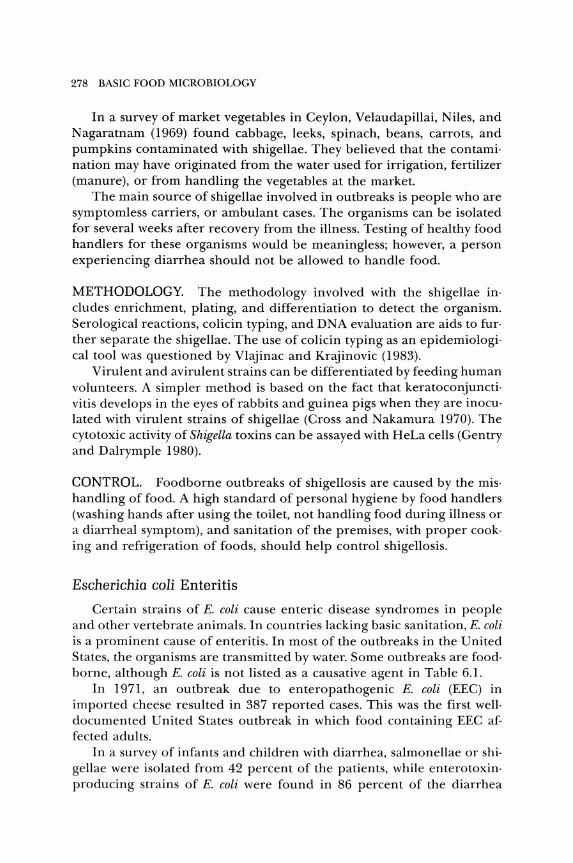
278 BASIC FOOD MICROBIOLOGY
In a survey of market vegetables in Ceylon, Velaudapillai, Niles, and N agaratnam (1969) found cabbage, leeks, spinach, beans, carrots, and pumpkins contaminated with shigellae. They believed that the contamination may have originated from the water used for irrigation, fertilizer (manure), or from handling the vegetables at the market.
The main source of shigellae involved in outbreaks is people who are symptomless carriers, or ambulant cases. The organisms can be isolated for several weeks after recovery from the illness. Testing of healthy food handlers for these organisms would be meaningless; however, a person experiencing diarrhea should not be allowed to handle food.
METHODOLOGY. The methodology involved with the shigellae includes enrichment, plating, and differentiation to detect the organism. Serological reactions, colicin typing, and DNA evaluation are aids to further separate the shigellae. The use of colicin typing as an epidemiological tool was questioned by Vlajinac and Krajinovic (1983).
Virulent and avirulent strains can be differentiated by feeding human volunteers. A simpler method is based on the fact that keratoconjunctivitis develops in the eyes of rabbits and guinea pigs when they are inoculated with virulent strains of shigellae (Cross and Nakamura 1970). The cytotoxic activity of Shigella toxins can be assayed with HeLa cells (Gentry and Dalrymple 1980).
CONTROL. Foodborne outbreaks of shigellosis are caused by the mishandling of food_ A high standard of personal hygiene by food handlers (washing hands after using the toilet, not handling food during illness or a diarrheal symptom), and sanitation of the premises, with proper cooking and refrigeration of foods, should help control shigellosis.
Escherichia coli Enteritis
Certain strains of E. coli cause enteric disease syndromes in people and other vertebrate animals. In countries lacking basic sanitation, E. coli is a prominent cause of enteritis. In most of the outbreaks in the United States, the organisms are transmitted by water. Some outbreaks are foodborne, although E. coli is not listed as a causative agent in Table 6.l.
In 1971, an outbreak due to enteropathogenic E. coli (EEC) in imported cheese resulted in 387 reported cases. This was the first welldocumented United States outbreak in which food containing EEC affected adults.
In a survey of infants and children with diarrhea, salmonellae or shigellae were isolated from 42 percent of the patients, while enterotoxinproducing strains of E. coli were found in 86 percent of the diarrhea

FOOD BORNE AGENTS CAUSING ILLNESS 279
group and 41 percent of the control group (Rudoy and Nelson 1975). This indicates that in enteric diseases of young people, E. coli may be as important as salmonellae or shigellae.
ETIOLOGIES. The role of E. coli in enteric disease, especially the diar· rheal syndrome, is quite complex. Although all virulent strains can be called enteropathogenic, there are classical enteropathogenic strains, in· vasive strains that cause illness similar to shigellae, strains that produce enterotoxins, and a more recently recognized enterohemorrhagic E. coli (Levine and Edelman 1984).
Classical enteropathogenic strains of E. coli that are neither invasive nor produce enterotoxin (ST or LT) have been reported (Levine et al. 1978; Robins-Browne et al. 1982; Toledo et al. 1983). The colonization of the intestines by enteropathogenic strains can result in moderate to severe injury to the mucosa (Rothbaum et al. 1982; Ulshen and Rollo 1980). It has not been established that mucosal injury causes the gastroenteritis. The possibility that a toxin different from the conventional enterotoxins is involved in the diarrheal syndrome has been proposed (Klipstein et al. 1978). Other researchers have suggested that a shigalike cytotoxin may playa role in enteropathogenic E. coli gastroenteritis (Cleary et al. 1985). In a study of an enteropathogenic strain, Ryder, Kaslow, and Wells (1979) found that it produced a heat-stable enterotoxin.
The invasive strains of E. coli cause a dysentery (fever, cramps, and diarrhea containing blood and mucus) similar to that caused by shigellae (DuPont 1978; Sakazaki et al. 1974). The invasive strains can be distin· guished by the development of keratoconjunctivitis when placed on the guinea pig eye, or by the penetration of HeLa or HEp·2 cells (Mehlman et al. 1982).
Enterotoxigenic E. coli (ETEC) may form a heat-labile (LT) , a heatstable (ST), or both LT and ST toxins. The heat-labile toxin of E. coli is readily destroyed at 60°C. It is similar to cholera enterotoxin (choleragen) immunologically, structurally, and functionally. Cholera antitoxin reacts not only with choleragen and Salmonella enterotoxins, but also with the LT of E. coli. Both choleragen and LT are composed of two subunits, designated as A and B.
Acting alone, neither A nor B is toxic. For toxicity, the B subunit binds with host cell membrane receptors and acts as the messenger for the A subunit, which activates adenyl cyclase (Gill and Richardson 1980; Moss et al. 1984). This alters the cyclic adenosine monophosphate metabolism in intestinal cells, which induces an enhanced secretion of fluid and electrolytes into the intestine, resulting in the diarrheal syndrome in the same manner as cholera toxin.
In one study, researchers separated the A and B subunits of LT and
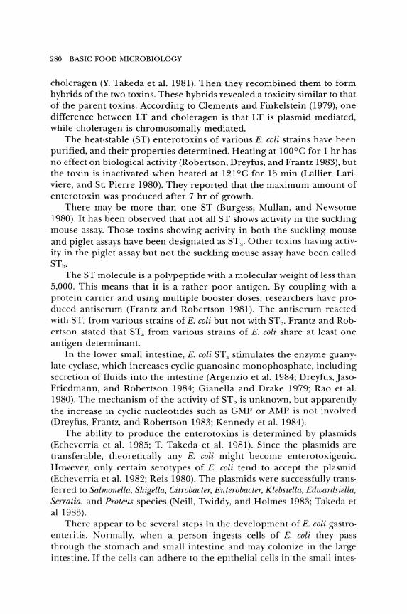
280 BASIC FOOD MICROBIOLOGY
choleragen (Y. Takeda et al. 1981). Then they recombined them to form hybrids of the two toxins. These hybrids revealed a toxicity similar to that of the parent toxins. According to Clements and Finkelstein (1979), one difference between LT and choleragen is that LT is plasmid mediated, while choleragen is chromosomally mediated.
The heat·stable (ST) enterotoxins of various E. coli strains have been purified, and their properties determined. Heating at 100°C for 1 hr has no effect on biological activity (Robertson, Dreyfus, and Frantz 1983), but the toxin is inactivated when heated at 121°C for 15 min (Lallier, Lari· viere, and St. Pierre 1980). They reported that the maximum amount of enterotoxin was produced after 7 hr of growth.
There may be more than one ST (Burgess, Mullan, and Newsome 1980). It has been observed that not all ST shows activity in the suckling mouse assay. Those toxins showing activity in both the suckling mouse and piglet assays have been designated as ST a. Other toxins having activo ity in the piglet assay but not the suckling mouse assay have been called STb •
The ST molecule is a polypeptide with a molecular weight of less than 5,000. This means that it is a rather poor antigen. By coupling with a protein carrier and using multiple booster doses, researchers have pro· duced antiserum (Frantz and Robertson 1981). The antiserum reacted with STa from various strains of E. coli but not with STb • Frantz and Rob· ertson stated that STa from various strains of E. coli share at least one antigen determinant.
In the lower small intestine, E. coli STa stimulates the enzyme guany· late cyclase, which increases cyclic guanosine monophosphate, including secretion of fluids into the intestine (Argenzio et al. 1984; Dreyfus, Jaso· Friedmann, and Robertson 1984; Gianella and Drake 1979; Rao et al. 1980). The mechanism of the activity of ST b is unknown, but apparently the increase in cyclic nucleotides such as GMP or AMP is not involved (Dreyfus, Frantz, and Robertson 1983; Kennedy et al. 1984).
The ability to produce the enterotoxins is determined by plasm ids (Echeverria et al. 1985; T. Takeda et al. 1981). Since the plasmids are transferable, theoretically any E. coli might become enterotoxigenic. However, only certain serotypes of E. coli tend to accept the plasmid (Echeverria et al. 1982; Reis 1980). The plasm ids were successfully trans· ferred to Salmonella, Shigella, Citrobacter, Enterobacter, Klebsiella, Edwardsiella, Serratia, and Proteus species (Neill, Twiddy, and Holmes 1983; Takeda et al 1983).
There appear to be several steps in the development of E. coli gastro· enteritis. Normally, when a person ingests cells of E. coli they pass through the stomach and small intestine and may colonize in the large intestine. If the cells can adhere to the epithelial cells in the small intes·
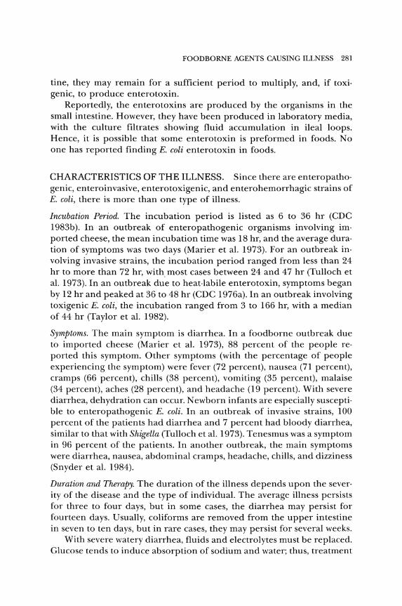
FOODBORNE AGENTS CAUSING ILLNESS 281
tine, they may remain for a sufficient period to multiply, and, if toxi· genic, to produce enterotoxin.
Reportedly, the enterotoxins are produced by the organisms in the small intestine. However, they have been produced in laboratory media, with the culture filtrates showing fluid accumulation in ileal loops. Hence, it is possible that some enterotoxin is preformed in foods. No one has reported finding E. coli enterotoxin in foods.
CHARACTERISTICS OF THE ILLNESS. Since there are enteropatho· genic, enteroinvasive, enterotoxigenic, and enterohemorrhagic strains of E. coli, there is more than one type of illness.
Incubation Period. The incubation period is listed as 6 to 36 hr (CDC 1983b). In an outbreak of enteropathogenic organisms involving im· ported cheese, the mean incubation time was 18 hr, and the average dura· tion of symptoms was two days (Marier et al. 1973). For an outbreak in· volving invasive strains, the incubation period ranged from less than 24 hr to more than 72 hr, with most cases between 24 and 47 hr (Tulloch et al. 1973). In an outbreak due to heat·labile enterotoxin, symptoms began by 12 hr and peaked at 36 to 48 hr (CDC 1976a). In an outbreak involving toxigenic E. coli, the incubation ranged from 3 to 166 hr, with a median of 44 hr (Taylor et al. 1982).
Symptoms. The main symptom is diarrhea. In a foodborne outbreak due to imported cheese (Marier et al. 1973), 88 percent of the people reo ported this symptom. Other symptoms (with the percentage of people experiencing the symptom) were fever (72 percent), nausea (71 percent), cramps (66 percent), chills (38 percent), vomiting (35 percent), malaise (34 percent), aches (28 percent), and headache (19 percent). With severe diarrhea, dehydration can occur. Newborn infants are especially suscepti· ble to enteropathogenic E. coli. In an outbreak of invasive strains, 100 percent of the patients had diarrhea and 7 percent had bloody diarrhea, similar to that with Shigella (Tulloch et al. 1973). Tenesmus was a symptom in 96 percent of the patients. In another outbreak, the main symptoms were diarrhea, nausea, abdominal cramps, headache, chills, and dizziness (Snyder et al. 1984).
Duration and Therapy. The duration of the illness depends upon the sever· ity of the disease and the type of individual. The average illness persists for three to four days, but in some cases, the diarrhea may persist for fourteen days. Usually, coliforms are removed from the upper intestine in seven to ten days, but in rare cases, they may persist for several weeks.
With severe watery diarrhea, fluids and electrolytes must be replaced. Glucose tends to induce absorption of sodium and water; thus, treatment
282 BASIC FOOD MICROBIOLOGY
with oral glucose-electrolyte solutions has been suggested (Klipstein and Engert 1978).
Foods Involved. The only well-documented foodborne outbreaks in the United States involved soft cheese. In other countries, outbreaks have been associated with consumption of dairy products, salads containing raw vegetables, meat and meat products, poultry, fish, and baked products.
Number of Organisms Needed. Although newborn humans and animals are highly susceptible, adults are more resistant. In a survey by Rudoy and Nelson (1975), EEC was isolated from 41 percent of a control group of children without diarrhea. This indicates that, even if a few organisms are present, they do not cause illness if the numbers are kept under control by the body defenses or by interactions with other organisms in the intestine.
One study found that all of their human volunteers developed diarrhea 11 to 48 hr after ingestion of 106 to 1010 colony-forming units of E. coli (Donta et al. 1974). The type and number of cells of E. coli and the age and physical condition of the host determine the occurrence and the severity of the illness.
NATURE OF THE ORGANISM. Escherichia coli is an important organism in the microbiology of foods. Besides being involved in gastroenteritis, the organisms in this species are considered to be indicators of possible fecal contamination and can cause spoilage of some foods. There are more than 160 serotypes of E. coli.
Sources. The main habitat of E. coli is the intestinal tract of humans and animals. However, fecal contamination causes it to spread to other environments, especially soil and water.
Researchers analyzing various foods of animal origin found enterotoxigenic E. coli in each type (Sack et al. 1977). They believed that these foods are potentially important vehicles for these toxigenic organisms, especially if the food is not handled properly.
EEC was detected in about 10 percent of 2,000 samples of cheese (Fantasia et al. 1975). None of the foods examined from a village in the Philippines (Echeverria et al. 1978) or Ethiopia (Jiwa, Krovacek, and Wadstrom 1981) contained enterotoxigenic E. coli.
METHODOLOGY. There is no simple culture medium or system that will differentiate pathogenic from nonpathogenic E. coli. After enrichment and isolation of E. coli, it is necessary to characterize the organisms with biochemical, serological, and other tests.
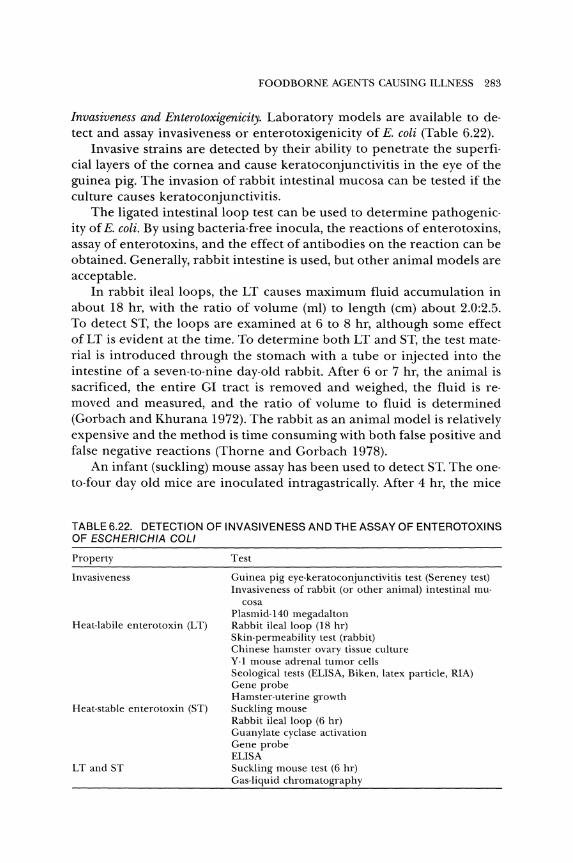
FOODBORNE AGENTS CAUSING ILLNESS 283
Invasiveness and Enterotoxigenicity. Laboratory models are available to de· tect and assay invasiveness or enterotoxigenicity of E. coli (Table 6.22).
Invasive strains are detected by their ability to penetrate the superfi· cial layers of the cornea and cause keratoconjunctivitis in the eye of the guinea pig. The invasion of rabbit intestinal mucosa can be tested if the culture causes keratoconjunctivitis.
The ligated intestinal loop test can be used to determine pathogenic· ity of E. coli. By using bacteria·free inocula, the reactions of enterotoxins, assay of enterotoxins, and the effect of antibodies on the reaction can be obtained. Generally, rabbit intestine is used, but other animal models are acceptable.
In rabbit ileal loops, the LT causes maximum fluid accumulation in about 18 hr, with the ratio of volume (ml) to length (cm) about 2.0:2.5. To detect ST, the loops are examined at 6 to 8 hr, although some effect of LT is evident at the time. To determine both LT and ST, the test mate· rial is introduced through the stomach with a tube or injected into the intestine of a seven·to·nine day·old rabbit. After 6 or 7 hr, the animal is sacrificed, the entire GI tract is removed and weighed, the fluid is reo moved and measured, and the ratio of volume to fluid is determined (Gorbach and Khurana 1972). The rabbit as an animal model is relatively expensive and the method is time consuming with both false positive and false negative reactions (Thorne and Gorbach 1978).
An infant (suckling) mouse assay has been used to detect ST. The one· to·four day old mice are inoculated intragastrically. After 4 hr, the mice
TABLE 6.22. DETECTION OF INVASIVENESS AND THE ASSAY OF ENTEROTOXINS OF ESCHERICHIA COLI
Property
Invasiveness
Heat-labile enterotoxin (LT)
Heat-stable enterotoxin (ST)
LT and ST
Test
Guinea pig eye-keratoconjunctivitis test (Sereney test) Invasiveness of rabbit (or other animal) intestinal mu-
cosa Plasmid-140 megadalton Rabbit ileal loop (18 hr) Skin-permeability test (rabbit) Chinese hamster ovary tissue culture Y-I mouse adrenal tumor cells Seological tests (ELISA, Biken, latex particle, RIA) Gene probe Hamster-uterine growth Suckling mouse Rabbit ileal loop (6 hr) Guanylate cyclase activation Gene probe ELISA Suckling mouse test (6 hr) Gas-liquid chromatography
284 BASIC FOOD MICROBIOLOGY
are sacrificed and the ratio of intestinal tract plus contents weight to the remaining body weight is determined. A ratio of greater than 0.83 indio cates the presence of ST. Researchers reported, however, that the mouse test was not sufficiently sensitive (Merson et al. 1980). Frantz and Robert· son (1981) found the assay to be impractical for screening large numbers of isolates. They stated that it was fairly expensive and did not detect all forms of ST. A dog assay detected more ST-producing strains than did the mouse assay (N alin et al. 1978).
Rabbit skin tests have been used to detect LT_ The preparation is in· jected intracutaneously, followed 24 hr later by an injection of Evans blue dye. Areas of induration and bluing are read 1 to 2 hr later. Young rabbits show a better reaction than do old rabbits.
Certain cell lines can be used to assay for these enterotoxins. The LT causes elongation of Chinese hamster ovary cells.
Originally flat Y-1 mouse adrenal tumor cells become rounded when exposed to the enterotoxins (Gurwith 1977). Vero (African green monkey) cells appear enlarged, thick-walled, and refractile, with several filamentous tendrils in response to E. coli LT (Speirs et al. 1977). The ST has no effect on these three cell lines (Giugliano, Stavric, and Konowalchuck 1982). The Y- 1 test for LT and the suckling mouse test for ST were adopted as official first action by the AOAC (Lovett and Peeler 1984).
Another team of researchers described a gas-liquid chromatographic method that could detect and differentiate LT and ST (Brooks et al. 1984). The activation of guanylate cyclase was used to detect ST (Waldman et al. 1984). They claimed it was more sensitive than the suckling mouse test. The increase in uterine weight after inoculation i.p. into cycling hamsters was used to detect the presence of LT (Alleva and Lamanna 1984). Cholera toxin also gave this response.
Various serological tests similar to those used to detect staphylococcal enterotoxins have been used for E. coli LT (Honda et al. 1981; Tsukamoto et al. 1980; Yano et al. 1982), and ST (Giannella, Drake, and Luttrell 1981). A staphylococcal coagglutination test for LT was developed by Brill, Wasilavskas, and Richardson (1979) and modified by later researchers (Honda et al. 1983a). Monoclonal antibodies have been developed for both LT and ST (Belisle, Twiddy, and Holmes 1984; Brandwein et al. 1985).
Enzyme-linked immunosorbent assays (ELISA) have been developed to detect LT (Honda, Sato, and Miwatani 1984; Merson et al. 1980; Ristaino, Levine, and Young 1983; Sack et al. 1980; Svennerholm and Wiklund 1983) as well as ST (Klipstein et al. 1984; Thompson et al. 1984). At the present time, the ELISA systems seem to be the most promising for the detection of toxins as well as other antigenic materials.
Methods have been developed to detect enterotoxigenic E. coli (ETEC)
FOODBORNE AGENTS CAUSING ILLNESS 285
by means of DNA colony hybridization using DNA or gene probes (Mose· ley et aL 1980; Patamaroj, Seriwatana, and Echeverria 1983). According to these authors, this system can detect ETEC in food or stools without the need to enrich the sample. The use of this system for determining ETEC among isolated organisms was approved as official first action by the AOAC (Hill and Payne 1984).
CONTROL. The control of the illness is similar to that for other enteric diseases. As far as foodborne illness is concerned, the number of cases would not warrant the immunization of all humans in developed coun· tries but might be worthwhile for people at risk in developing countries. Some data suggest that there is a natural development of immunity in an endemic area (Black et aL 1981).
To prevent contamination, Bryan (1973) stated simply that we should practice personal hygiene, prepare foods in a sanitary manner, protect and treat (chlorinate) water, and dispose of sewage in a sanitary manner.
The easiest way to prevent growth of the organism is to provide an unacceptable environment for E. coli.
Since apparently large numbers of cells are needed to cause illness in other than newborn babies, simply cooking the food sufficiently pro· vides a safe product.
Bacillus cereus Gastroenteritis
Under normal circumstances, B. cereus is not considered to be a path· ogen. Foodborne illness due to B. cereus rarely is reported in the United States. The number of reported outbreaks is much higher in European countries and is one of the major causes of foodborne illness in Hungary.
There are two distinct syndromes caused by enterotoxins produced by B. cereus. One enterotoxin causes an illness similar to that produced by the S. aureus enterotoxin. The primary symptoms are nausea, vomit· ing, and abdominal cramps. Diarrhea may occur in the later stages of some cases. The incubation period usually is less than three hr. A second enterotoxin results in an illness similar to that caused by the C. perfringens enterotoxin. In this case, the effects are primarily in the lower intestinal tract, and the primary symptom is diarrhea. The incubation period is about 10 to 12 hr.
In both syndromes, fever is absent, indicating that toxins are in· volved. Usually, the symptoms last about 12 to 24 hr, but some cases, especially of the emetic type, may be severe. Due to the usually mild ill· ness, with short duration, people might not seek medical aid. Therefore, most cases probably are undetected and unreported.
High numbers of B. cereus (106 to 109/g) have been associated with
286 BASIC FOOD MICROBIOLOGY
foods involved in the illness. Due to multiplication of the cells in the food before analysis, these numbers may be higher than when the food was ingested.
THE ORGANISM. B. cereus is an aerobic, Gram·positive, motile, spore· forming rod. It hydrolyzes esculin and starch, reduces litmus milk and nitrate, and is catalase positive.
Since many cells (> 105/g of food) are needed to cause foodborne illness, the food must be abused sometime during preparation. In out· breaks in England due to rice, the rice was boiled and then allowed to sit at room temperature for 12 hr to three days. The rice was either boiled or fried before being served. The original heating was not sufficient to kill the spores of B. cereus but could activate the spores to germinate. Holding the rice at room temperature allowed the vegetative cells to mul· tiply to levels that caused illness.
Source. B. cereus is common in soil and dust, so it is logical that foods that are readily contaminated by soil and dust will contain the organism. Plant products (cereals, flour, starch, bakery products, spices), animal products, and mixtures of ingredients (spaghetti sauce, pudding, soup mixes, gravy mixes) can contain a few or many cells or spores of B. cereus.
There is a similarity between foods involved in illnesses due to B. cereus and to C. perfringens. In either case, the food is prepared ahead of time in large batches and is not properly cooled before reheating (if needed) and serving. The reheating is not sufficient to destroy the cells.
METHODOLOGY. To detect B. cereus a spread plate system using mannitol·egg yolk·polymyxin (MYP) agar is suggested (FDA 1978). On this medium, the organism produces pink colonies surrounded by a dense precipitate (lecithinase·positive reaction). B. cereus is mannitol negative. Harmon, Kautter, and McClure (1984) found MYP slightly superior to two other media (polymyxin pyruvate-egg yolk-mannitol-brom thymol blue and trypticase-soy-polymyxin blood agars), but all three were satisfactory for detection of B. cereus.
Selected colonies are isolated and the organisms tested for the Gram reaction and biochemical tests. A method for differentiating B. cereus was described by Harmon (1982) and was adopted by the AOAC as its interim official first action.
ENTEROTOXIN. Turnbull (1981) discussed the various toxins of B. cereus. Gilbert and Kramer (1984) compared the various properties of the diarrheal and emetic enterotoxins. The diarrheal toxin is a protein with a molecular weight of about 50,000, while the emetic toxin is a peptide (not antigenic) and has a molecular weight of less than 5,000.
FOODBORNE AGENTS CAUSING ILLNESS 287
The diarrheal toxin can be detected by the rabbit ileal loop test or skin·permeability system of either rabbits or guinea pigs. Monkey feeding with observation of vomiting is the system used to detect emetic enter· otoxin.
CONTROL. The methods of control are the same as those for C. per· fringens. Keeping B. cereus out of the food would appear to be a difficult task. However, proper holding temperatures (55°C or higher, or below lO°C) for the food would prevent growth of the organisms. Multiplica· tion of the cells is needed to attain sufficient numbers to cause illness.
Streptococcal Infections
Group A (Streptococcal pyogenes) may be present in food. This organism causes scarlet fever or septic sore throat. Besides a sore throat, symptoms may include vomiting and diarrhea.
A beta·hemolytic group A streptococcus caused an outbreak of pharo yngitis (CDC 1982b). The food involved was not determined. Outbreaks of pharyngitis believed due to contaminated salads were reported by CDC (l984c).
Although group A streptococci usually are involved, group G strepto· cocci occasionally cause pharyngitis. Among the possible symptoms are nausea, vomiting, and diarrhea. An outbreak in 1979 involving chicken salad was reported by Stryker, Fraser, and Facklam (1982).
A group C streptococcus contaminating homemade cheese caused fe· ver, chills, and vague constitutional symptoms (CDC 1983a).
When streptococcal foodborne infection is mentioned, the organisms in group D, often referred to as enterococci, or fecel streptococci, are considered to be the etiologic agent of the illness.
The reported number of outbreaks due to enterococci is not signifi· cant In cases in which enterococci have been involved, the incubation period ranged from 2 to 30 hr. The symptoms included nausea, vomiting, and diarrhea. Usually, the symptoms were milder than those in other foodborne illnesses. The duration was usually only a few hours.
Some people believe that the enterococci do not cause foodborne illness, and some are convinced that these organisms are involved in out· breaks. Other workers are unsure, but they realize that more convincing proof is needed to relate enterococci to foodborne illness.
Illness Due to Vibrios
The genus Vibrio is discussed in Chapter 3. The two important species causing gastroenteritis are V. cholerae and V. parahaemolyticus. Other Vibrio species such as V. fluvialis (Tacket et aL 1982) and V. mimicus (Ciufecu,
288 BASIC FOOD MICROBIOLOGY
N acescu, and Israil 1983) have been involved in causing diarrhea in humans_
V. CHOLERAE. This organism causes cholera in humans_ Generally, the ingestion of 106 to 1010 cells is needed to cause the illness_ The normally low pH of the stomach will kill unprotected cells, but they can pass through the stomach if the pH is altered, such as by the ingestion of antacids_ Those cells that pass into the small intestine must be able to adhere to the mucosa on which they grow and produce an enterotoxin_ It is believed that the flagellum is associated with attachment, since nonmotile cells generally cannot adhere to the intestine_ From 24 to 48 hr after ingestion, symptoms of cholera may occur. The symptoms include profuse watery diarrhea, abdominal cramps, nausea, and vomiting_ In severe cases, a liter or more of water may be lost each hour, resulting in rapid dehydration, shock, and eventually, death_
The principal strains of V. cholerae that cause illness are in serogroup 0:1, although other strains have been involved_ The 0:1 serogroup contains two biovars, the classical and the eltor (Krieg and Holt 1984)_ There are two subtypes, Ogawa and Inaba_ A third subtype, Hikjima, has been proposed_ Although the toxin is produced in response to a gene located on the chromosome of the cell, not all 0:1 strains are enterotoxigenic.
The cholera enterotoxin (CT) and E. coli heat-labile toxin (LT) share some antigenic properties_ The CT has a molecular weight of about 84,000_ It consists of five B subunits arranged in a ring in which there is an A subunit. The B subunits combine to the membrane receptor, GM1 ganglioside, in the intestine, which then allows the A subunit to traverse the cell membrane and activate the membrane-bound adenyl cyclase, thereby causing an increase in cyclic adenosine mono phosphate (cAMP)_ This causes an inhibition of absorption of sodium and chloride at the villus tip, which is accompanied by water being dumped into the intestine_ There is no structural damage to the intestinal mucosa_ The presence of bile acids seems to enhance the excretion of water into the intestine (Eidels, Proia, and Hart 1983; Lange, Hansson, and Lonnroth 1983; Middlebrook and Dorland 1984)_
Cholera toxin is determined by methods similar to those used for E. coli LT, such as the skin reaction (Kuroki 1981), CHO cells (Nozawa, Yokota, and Kuwahara 1978), animal models (Richardson, Giles, and Kruger 1984; Spira, Sack, and Froehlich 1981), and immunological reactions (Beutin et aL 1984; Holmes and Twiddy 1983; Ito, Kuwahara, and Yokota 1983; Shah, Kauffman, and Boutin 1982)_ A gene probe for detecting enterotoxigenic strains was described by Hanchalay et aL (1985)_
Cholera is associated with poor sewage treatment and disposal, inadequate treatment of drinking water, and crowded conditions with poor
FOODBORNE AGENTS CAUSING ILLNESS 289
sanitation. Although it is important in Asia and Africa, it has been reo ported only rarely in countries with proper sanitation. Cholera was not reported in the United States from 1911 to 1973, when a single case oc· curred in Texas. In 1978, there were eight cases of cholera in Louisiana, apparently caused by eating inadequately cooked crabs (CDC 1978). Since 1978, cases involving V. cholerae have occurred in Florida and Texas due to consumption of raw oysters and water, as well as foods contami· nated with the water (CDC 1981d; Johnston et al. 1983; Shandera et al. 1983). Non·0:1 and toxigenic 0:1 V. cholerae were involved in these cases.
The various cases of cholera have increased interest in the presence of V. cholerae in United States coastal waters. It has been reported in the gulf coast from Texas to Florida (De Paola et al. 1984; Hood et al. 1983; Roberts et al. 1982), the Chesapeake Bay (Colwell et al. 1981) and Califor· nia waters (Kenyon et al. 1984). The contamination of coastal waters means that shellfish (oysters, clams) present the greatest risk when consumed. Also, fish from these waters may contain V. cholerae.
V. PARAHAEMOLYTICUS. Although perhaps not as important as cholera on a worldwide basis, the prevalence of this illness seems to have increased. In 1968, V. parahaemolyticus was not listed as an etiological agent of foodborne illnesses by CDC. In 1971, there were three outbreaks with 370 cases and in 1972, six outbreaks with 701 cases. From 1975 to 1981 there was an average of two outbreaks per year.
The organism was recognized as a cause of illness in Japan during the early 1950s. During the summer months, the organism accounts for a majority of the reported foodborne illnesses in Japan.
The illness was thought to be unique to Japan. However, due to a spreading epidemic of cholera in the early 1970s, techniques to isolate vibrios were improved, and diarrheal stools were examined for vibrios. It was discovered that V. parahaemolyticus was an important cause of foodborne illness, not only in Japan, but also in the United States and other countries.
The Illness. The main symptom is diarrhea. Other symptoms are listed in Table 6.23.
The incubation period usually is 12 to 24 hr with a range of 4 to 96 hr. The usual duration is two to three days, but it ranges from a few hours to ten days. If treatment is needed, fluid replacement and antibiotic medication may be used.
Foods that have been incriminated in outbreaks in the United States include steamed crabs, crab salad (made from canned crabmeat), raw crab, processed lobster, boiled shrimp, roasted oysters, and raw oysters.
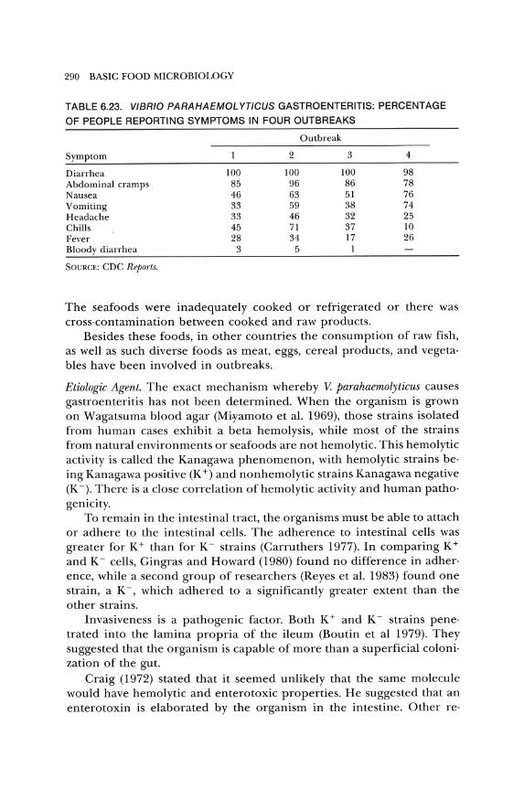
290 BASIC FOOD MICROBIOLOGY
TABLE 6.23. VIBRIO PARAHAEMOL YTiCUS GASTROENTERITIS: PERCENTAGE
OF PEOPLE REPORTING SYMPTOMS IN FOUR OUTBREAKS
Outbreak
Symptom 2 3 4
Diarrhea 100 100 100 98 Abdominal cramps 85 96 86 78 Nausea 46 63 51 76 Vomiting 33 59 38 74 Headache 33 46 32 25 Chills 45 71 37 10 Fever 28 34 17 26 Bloody diarrhea 3 5 1
SOURCE: CDC Reports.
The seafoods were inadequately cooked or refrigerated or there was cross·contamination between cooked and raw products.
Besides these foods, in other countries the consumption of raw fish, as well as such diverse foods as meat, eggs, cereal products, and vegeta· bles have been involved in outbreaks.
Etiologic Agent. The exact mechanism whereby V. parahaemolyticus causes gastroenteritis has not been determined. When the organism is grown on Wagatsuma blood agar (Miramoto et al. 1969), those strains isolated from human cases exhibit a beta hemolysis, while most of the strains from natural environments or seafoods are not hemolytic. This hemolytic activity is called the Kanagawa phenomenon, with hemolytic strains be· ing Kanagawa positive (K +) and nonhemolytic strains Kanagawa negative (K -). There is a close correlation of hemolytic activity and human patho· genicity.
To remain in the intestinal tract, the organisms must be able to attach or adhere to the intestinal cells. The adherence to intestinal cells was greater for K + than for K - strains (Carruthers 1977). In comparing K +
and K - cells, Gingras and Howard (1980) found no difference in adher· ence, while a second group of researchers (Reyes et al. 1983) found one strain, a K-, which adhered to a significantly greater extent than the other strains.
Invasiveness is a pathogenic factor. Both K + and K - strains pene· trated into the lamina propria of the ileum (Boutin et al 1979). They suggested that the organism is capable of more than a superficial coloni· zation of the gut.
Craig (1972) stated that it seemed unlikely that the same molecule would have hemolytic and enterotoxic properties. He suggested that an enterotoxin is elaborated by the organism in the intestine. Other reo
FOODBORNE AGENTS CAUSING ILLNESS 291
searchers found a pathogenic strain that was not hemolytic (Sakurai et al. 1974). They believed that a factor or factors other than a hemolysin was responsible for the illness. Yet another team reported that antisera to the hemolysin did not prevent accumulation of fluid in ileal loops when challenged with living cells of either K + or K - strains (Honda et al. 1983b). This would indicate that a substance other than the hemolysin was responsible for this action.
A study by Bradshaw and others (1984) stated that there is no satisfac· tory evidence for a filterable enterotoxin, but that the invasive and adhe· sive traits substantiate the theory that the Kanagawa hemolysin is the primary virulence factor.
It is not known how many cells need to be ingested to cause the ill· ness. Smith (1971) stated that 106 to 109 cells were needed, depending on the condition of the stomach, type and quantity of food eaten, and buffering capability of the ingested food. Accidental ingestion of 105 cells caused symptoms in a laboratory worker. Using rabbit ileal loops, Twedt, Peeler, and Spaulding (1980) found that concomitant inoculation of other nonvirulent vibrios can increase the number of cells of V. parahae· molyticus needed to cause dilation of the loops.
Nature oj the Organism. V. parahaemolyticus is a Gram·negative, straight or curved, facultatively anaerobic rod. It is halophilic, requiring 1 to 3 per· cent salt for growth. The cells grow in broth with 8 percent, but not 10 percent salt (Kourany and Vasquez 1975). The minimum aw is about 0.94. Usually the pH range for growth is 5 to 11 (Twedt, Spaulding, and Hall 1969). The optimum is pH 7.6 to 8.6.
The organism is sensitive to cold temperatures, so the minimum for growth is usually listed as 10° to 13°C. The upper temperature is about 42° or 43°C, with an optimum between 30° and 37°C. The effect of low temperature on the cells is related to the type of product and the temper· ature of storage. When exposed to 2°C, the membrane is damaged (Van den Broek and Mossel 1977). The presence of salt tends to protect the cells from low· temperature damage.
Smith (1971) stated that the organism has a very short generation time of 9 to 11 min. The average generation time of four strains was 13.6 min (Lee 1973).
The natural habitat is coastal and estuarine ocean water throughout the world. It lives in sediment during the cold winter months, and, as the temperature rises in late spring, the organism colonizes the water and animal life. Direct relationships have been demonstrated between the water temperature, abundance of the organism in the water, and out· breaks of vibrio foodborne illness. The organism has been isolated from a variety of seafoods.

292 BASIC FOOD MICROBIOLOGY
Methodology. V. parahaemolytieus, if present in a food, is usually in low numbers. Hence, an enrichment procedure using a broth such as glucose salt teepol (GST) is used (FDA 1978). For chill-stressed cells, GST supplemented with magnesium or ferric iron salts increases recovery (MaLin and Beuchat 1980). A loopful of the enriched sample is streaked onto thiosulfate citrate bile salts sucrose (TCBS) agar (FDA 1978). On this medium, the organism appears as round colonies 2 to 3 mm in diameter with green or blue centers. The salt concentration (1 percent) and pH (8.4), plus other inhibitors, suppress the growth of most organisms except halophiles, such as V. parahaemolytieus. Sucrose-fermenting organisms produce yellow colonies. These include V. eholerae, some enterics and S. aureus. Organisms besides V. parahaemolytieus that can form green colonies include some vibrios, pseudomonads, and enterics.
A hydrophobic grid membrane filtration system using V. parahaemolytieus sucrose agar was described by Entis and Boleszczuk (1983). They claimed that significantly higher counts were obtained with their system than with the FDA (1978) method.
Kourany (1983) found TCBS to be of dubious. value in certain cases and suggested a specially supplemented trypticase soy agar for detection of V. parahaemolytieus.
Since organisms besides V. parahaemolytieus can grow on the isolation media, biochemical tests are needed to differentiate it from other vibrios as well as other Gram-negative organisms.
The organism has 0, H, and K antigens. Serological typing schemes using the K and H antigens have been devised (Beuchat 1982; Shinoda et al. 1983).
Control. The control of V. parahaemolytieus foodborne illness should be relatively simple. The organism is readily killed by heat. When inoculated at a level of 10o/ml in shrimp homogenate, no survivors were found after heating at 100°C for 1 min (Vanderzant and Nickelson 1972). According to CDC (1978a), for boiled seafoods, Louisiana law requires a minimum of 7 mIll of boiling to ensure the destruction of pathogens. Delmore and Crisley (1979) reported D values at temperatures from 49° to 55°C for nine strains of V. parahaemolytieus. At 55°C, the D ranged from 0.03 to 0.24 min in sterile clam homogenate.
Although cooking seafoods will destroy the organisms, it is difficult to change the habit in some cultures of eating raw seafood. The use of radiation treatment of seafoods might be a solution.
Campylobacter
Prior to 1980, Campylobaeter was not listed as a causative agent of foodborne gastroenteritis by CDC. G.jejuni was involved in five outbreaks in 1980 and ten in 1981 (CDC 1983b).
FOODBORNE AGENTS CAUSING ILLNESS 293
Analysis of 8,097 fecal samples obtained from eight hospitals in var· ious areas of the United States yielded twice as many isolations of C. jejuni as salmonellae (4.6 percent to 2.3 percent) (Blaser et al. 1983). Similar findings were reported by other researchers (Drake et al. 1981; Svedhem and Kaijser 1980). Stool samples of patients admitted to an Australian hospital were examined for various potential pathogens (Cavanagh et al. 1980). The percent of stools containing these organisms was as follows: C. jejuni, 13 percent; salmonellae, 20 percent; shigellae, 4 percent; and parasites, 13 percent.
If C. jejuni is so prominent, why was it not listed as a cause of food· borne illness in the CDC reports prior to 1980? With the development of simplified and selective methods to isolate C. jejuni from stools (Skir· row 1977), the number of confirmed cases has increased, and only con· firmed cases are listed by CDC. Although there are several species of Campylobacter, the one most often associated with human distress is C. JeJuni.
The organism is a Gram·negative, slender, curved to spiral rod. The cells have a single polar flagellum and move with a corkscrewlike motion.
C. jejuni does not ferment or oxidize carbohydrates. Energy is ob· tained from amino acids or from intermediates of the tricarboxylic acid cycle. It does not hydrolyze gelatin, casein, deoxyribonucleic acid, or es· culin. The organism is catalase and oxidase positive and reduces nitrate to nitrite; most strains hydrolyze hippurate.
It is microaerophilic with 5 percent O 2 and 10 percent CO2 optimal for growth. The minimum temperature for growth is 32° to 35°C and the maximum is about 44° to 45°C. At 42°C it grows in 1.5 percent but not in 2 percent NaCl. At 25°C, 1.0 to 2.5 percent salt enhances the death of the organism, and it is sensitive to 1.0 percent salt at 4°C (Doyle and Roman 1982a).
The minimum pH for growth generally is 5.3, but some strains grow at pH 5.1 (Gill and Harris 1983) and one strain showed growth at pH 4.9 (Doyle and Roman 1981).
The illness is a typical gastroenteritis, with the dominant symptoms being diarrhea and abdominal pain, usually with fever. Other symptoms include nausea, vomiting, mucus or blood in the stool, headache, chills, fatigue, backache, and dehydration. The illness may mimic appendicitis (Chan, Stringel, and MacKenzie 1983; Pitkanen et al. 1983), and can cause abortion (Gilbert et al. 1981). The illness may be severe in some patients. Smith and Blaser (1985) reported on two fatalities associated with C. je· juni. The incubation period usually is 2 to 5 days but may range from 1 to 11 days. The duration of the illness usually is 1 to 3 days but may last for three weeks. The organism may be excreted for over a year (Richard· son, Koornhof, and Bokkenheuser 1981). If the illness is severe, treat· ment with erythromycin and fluid replacement may be needed.
294 BASIC FOOD MICROBIOLOGY
C. jejuni can invade the intestinal epithelium and cause damage to the mucosa. In calves, AI-Mashat and Taylor (1980) found a thickening of the wall of the ileum, inflammation of the jejunal and ileal mucosa, an enlargement of mesenteric lymph nodes, as well as dilated capillaries in the mucosa of the small intestine_ Invasion and lesions in the intestinal tract also were noted with experimentally infected hamsters (Humphrey, Montag, and Pittman 1985) and newly-hatched chicks (Welkos 1984).
Mills and Bradbury (1984) stated that little is known of the mechanism of the pathogenicity of the organism. Since fever is a symptom, it would seem to be an infection. However, the researchers have detected toxin production in twenty-four of thirty-two strains of C. jejuni (RuizPalacios et al. 1983). Culture supernatants caused fluid secretion in rat ileal loops, but not in rabbit loops or the infant mouse assay. It caused elongation ofCHO cells. Cholera antitoxin prevented the toxic response, indicating a serological similarity to cholera enterotoxin, E. coli LT, and possibly the enterotoxins of salmonellae. These observations were confirmed by Klipstein and Engert (1984, 1985) and McCardell, Madden, and Lee (1984).
Domestic and wild animals are the main reservoirs of C. jejuni. Hence, foods derived from animal sources tend to be those involved as vehicles for infection of humans by the organism. Drinking raw (untreated) water is the highest risk factor (Hopkins, Olmsted, and Istre 1984). They listed further risks (in descending order) as drinking raw (unpasteurized) milk, having a cat in the home, and eating undercooked chicken. Besides the ingestion of contaminated water or food, the organism can be transferred directly from animals to humans or from humans to humans (Norkrans and Svedhem 1982).
The exact mechanism by which milk becomes contaminated is not known. However, milk from cows with Campylobacter mastitis might be used. Also, according to CDC (1981e), up to 60 percent of healthy cows shed Campylobacter in their feces. Without very careful handling, fecal contamination of milk might occur. The incidence of C. jejuni in farm bulk milk is about 1 to 2 percent (Lovett, Francis, and Hunt 1983).
Milk has been involved in several outbreaks of campylobacteriosis (CDC 1981a, 1984d; Hudson et al. 1984; Potter et al. 1983; Wright et al. 1983). In a survey of raw milk, deBoer, Hartog, and Borst (1984) concluded that there was no association between developing campylobacteriosis and consuming raw milk. They, as well as Doyle and Roman (1982c), reported that C. jejuni survived longer in sterile milk than in raw milk when held at 4°C.
C. jejuni is quite prevalent in poultry-processing plants (LuechtefeId and Wang 1981; Oosterom et al. 1983b; Wempe et al. 1983) and on the carcasses of the processed poultry (Rayes, Genigeorgis, and Farver 1983; Shanker et al. 1982). Fortunately, C. jejuni tends to die during refrigerated
FOODBORNE AGENTS CAUSING ILLNESS 295
holding so there is less contamination at the retail store than immediately after processing (Kinde, Genigeorgis, and Pappaioanou 1983)_ From a survey of chicken livers, Barot, Mosenthal, and Bokkenheuser (1983) concluded that most of the contamination with C jejuni occurred at the processing plant.
The organism has been detected on red meats in processing plants and at retail stores (Stern et aL 1985; Turnbull and Rose 1982)_ Koidis and Doyle (1983) reported that C jejuni survived well in refrigerated ground beef. The extent of survival depended upon the strain of the organism. Conversely, Bolton, Dawkins, and Robertson (1982) and Oosterom et al. (1983a) found that cold storage reduced the level of C jejuni on carcasses below detectable levels.
When food is contaminated, it is usually at a low leveL Due to temperature and oxygen requirements, the organism generally does not multiply in food. Hence, it is evident that low levels of contamination are sufficient to cause illness in humans, although the exact number of cells needed is not known. Only a low-level infection occurs during direct transfer from animals or people to the recipient. A level of 90 C jejuni was the minimum infective dose causing diarrhea in 90 percent of inoculated chickens (Ruiz-Palacios. Escamilla, and Torres 1981).
Various methods have been used to detect the presence of C jejuni in foods. Since a relatively low number of cells are in foods, an enrichment procedure usually is needed. Selective enrichment broths are generally a basal medium to which various supplements and antibiotics are added to stimulate the growth of C jejuni and inhibit other organisms (Barot and Bokkenheuser 1984; Doyle and Roman 1982a; Rogol et aL 1985; Rothenberg, Stern, and Westhoff 1984). After incubation of the inoculated enrichment broth in a microaerobic environment at 42°C, a portion is surface plated onto a selective agar surface_ In some cases, the sample may be plated directly without enrichment. The selective agars tend to be similar to the selective enrichment broths, with 1.5 to 2.0 percent agar added for solidification (Fricker 1985; Moskowitz and Chester 1982; N g, Stiles, and Taylor 1985; Stern 1982). After incubation microaerobically at 42°C, the inoculated surfaces are observed for typical colonies of C jejuni.
These can be screened by microscopic observation (phase-contrast) for motility and morphology, and then for oxidase and catalase-positive characteristics. After these simple tests, other biological and biochemical tests can be run (Lior 1984; Roop, Smibert, and Krieg 1984; Stern 1982). Serological and other tests may be performed to further confirm the presence of C jejuni (Bradbury et aL 1984; Harvey and Greenwood 1983; Kosunen, Bang, Hurme 1984; Penner, Hennessy, and Congi 1983; Tenover et al. 1984, 1985; Wong et al. 1985).
The control of C jejuni enteritis is similar to that for salmonellae.
296 BASIC FOOD MICROBIOLOGY
Foods derived from animal sources should be handled as though they contain the organism. G.jejuni is not very heat resistant. In skim milk, the D values for G.jejuni ranged from 7.2 to 12.8 min at 48°C and 0.74 to 1.0 min at 55°C (Doyle and Roman 1981). In ground beef, these values were 5.9 to 6.3 min at 50°C and 0.2 to 0.35 min at 58°C (Koidis and Doyle 1983). In autoclaved ground chicken, the D values ranged from 20.5 min at 49°C to 0.79 min at 57°C (Blankenship and Craven 1982). Hence, pas· teurization of milk and cooking of beef and poultry should destroy any G. jejuni in these products. Svedhem, Kaijser, and Sjogren (1981) suggested heating food to at least 60°C for 15 min to prevent infection. Gill and Harris (1984) recommended minimal cooking of poultry at 190°C for 20 min to eliminate G. jejuni.
The organism is more sensitive than salmonellae to gamma radiation (Lambert and Maxcy 1984). Competitive exclusion in young chicks worked similarly to that for salmonellae (Soerjadi-Liem, Snoeyenbos, and Weinack 1984).
Brucellosis
The number of cases of brucellosis has generally decreased in the United States. It has become a disease of people who handle cattle and swine. This includes veterinarians, farmers, and packinghouse workers. The workers in the kill department are most subject to contamination, but workers handling the carcasses and meat also have become infected.
In 1975, there were twenty·four cases of brucellosis caused by the ingestion of unpasteurized dairy products, such as raw domestic milk and imported cheese. The consumption of raw dairy products continues to cause about 10 percent of the cases of brucellosis (Bryan 1983).
QFever
Coxiella burnetii causes Q fever, a rickettsial zoonosis, in humans. Cattle, sheep, and goats are the usual reservoirs of the organism. Usually, transmission to humans is by inhaling aerosols derived from contami· nated animal products (including excreta). The organism is a hazard to people working in meat· packing plants and handling raw meat (McKelvie 1980). Infected cows shed the organism in their milk. The pasteurization of milk was adjusted so that the treatment inactivates G. burnetii.
Yersinia
Yersinia enterocolitica is a member of the family Enterobacteriaceae. For most strains, the temperature growth range in nutrient broth is 1 to 44°C (Sutherland and Varum 1977), and, at 25°C, the pH range for growth is about 4.6 to 9.6 (Stern, Pierson, and Kotula 1980).

FOODBORNE AGENTS CAUSING ILLNESS 297
The organism produces several disease syndromes in people (enteritis, terminal ileitis, mesenteric lymphadenitis, erythema nodosum, polyarthritis, septicemia, and metastatic abscesses in various organs). The gastroenteritis is characterized by diarrhea (which may be bloody), abdominal pain, fever, and vomiting. The illness may last from two days to several months. The illness may mimic appendicitis and, in some outbreaks, appendectomies have been performed before yersiniosis was suspected (Black et al. 1978; Shayegani et al. 1983). The organism has been isolated from the appendices of patients (Fukushima et al. 1981).
Y. enterocolitica can be found almost anywhere in nature, but only certain serotypes are involved in human infection. These serotypes are prevalent in swine (Fukushima et al. 1983; Walker and Grimes 1985), and have been isolated from dogs (Fukushima et al. 1984). Hence, although the organism has been isolated from various animals and food products derived from animals, these isolations are important only if they are serotypes that can cause human yersiniosis.
The ability of the organism to grow at refrigeration temperatures, makes refrigerated animal foods, such as milk, a potential hazard. Milk that was either improperly pasteurized or contaminated after pasteurization has been involved in various outbreaks (Black et al. 1978; CDC 1982c; Tacket et al. 1984).
A fermented soybean product, tofu, was a vehicle for outbreaks (Aulisio et al. 1983; Tacket et al. 1985b). Besides foods, the organism is found in water and can be acquired by direct transfer to people from animals or from other people.
Since fever and bloody diarrhea are symptoms of gastroenteritis, an invasive type of infection similar to shigellosis would be suspected. A plasmid is associated with invasiveness and other virulence factors (Kay, Wachsmuth, and Gemski 1982).
Some strains of Y. enterocolitica produce an enterotoxin similar to E. coli ST, which stimulates the activity of guanylate cyclase (Inoue et al. 1983; Rao et al. 1979). The results of Schiemann (1981) indicated that the enterotoxin has no role in pathogenesis. Hence, the exact role of invasiveness and enterotoxin needs further evaluation. Also, the extent of involvement of persiniae in human illness is not known.
The methodology for Yersinia has been discussed by various researchers (Bercovier et al. 1984; Hill, Payne, and Aulisio 1983; and Sack 1984).
Listeria
The first human illness due to Listeria monocytogenes was recognized in 1929. The organism is present in soil and has been detected in many species of animals. The infection of humans usually occurs orally, such as
298 BASIC FOOD MICROBIOLOGY
with contaminated food, or possibly by direct contact with infected animals_ About 1 percent of humans excrete the organism_
The organism is a small Gram-positive rod with a tendency toward a diplobacillary form_ Although it is aerobic, it grows better at reduced O 2
and increased CO2 levels_ It is more motile at 25°C than at 37°C. The cells can multiply at pH 9_0 and at 10 percent NaCl. It can grow at 4°C, which makes it a problem in refrigerated foods. It ferments a variety of sugars, producing acid but no gas.
There are several manifestations of listeriosis, including septicemia, (which in pregnant women can lead to abortion or stillbirth), endocarditis, pneumonia, conjunctivitis, pharyngitis, cutaneous papules, and pustules, urethritis, and meningitis. Septicemia may result in the invasion of organs such as the spleen, liver, or adrenal gland. The generalized infection may mimic other diseases such as typhoid fever or infectious mononucleosis.
A rather large outbreak of listeriosis was described by CDC (1985a). According to this report, the outbreak occurred in California due to consumption of contaminated cheese, with eighty-six reported cases and twenty-nine deaths. Of these twenty-nine deaths, there were eight neonatal deaths and thirteen stillbirths. The other eight were non-neonatal. There are indications that forty-eight or more deaths may have occurred in this outbreak.
There have been other recent outbreaks of listeriosis. In 1983, fourteen persons died in Massachusetts after consuming pasteurized milk. Although considered to be heat sensitive, the organism is believed to be able to survive pasteurization by living parasitically within white blood cells present in milk.
In August, 1985, soft cheese produced in Ohio was found to contain L. monocytogenes and fortunately was recalled before any illness occurred.
Although most of the problems with Listeria have been associated with the dairy industry, fowl are considered to be a natural reservoir of the organism. Fortunately, most poultry products are cooked sufficiently to kill the organisms before consumption.
Others
Although not listed as major causes of gastroenteritis, several organisms produce enterotoxins. These include species of the genera Aeromonas (Buchanan and Palumbo 1985; Goodwin et al. 1983; Jiwa 1983; Ljungh, Eneroth, and Wadstrom 1982), Citrobacter (Wadstrom 1976), Enterobacter (Klipstein, Engert, and Short 1977), Klebsiella (Klipstein, Engert, and Short 1977), and Kluyvera (Farmer et al. 1981). Clostridium difJicile and its enterotoxin (toxin A) have been associated with a severe gastroin-
FOODBORNE AGENTS CAUSING ILLNESS 299
testinal illness in patients who have ingested certain antimicrobial agents (Mulligan 1984; Thelestam and Florin 1984). Some strains of Clostridium sordellii also produce an enterotoxin (Yamakawa et al. 1983). The extent ofthe role of these organisms as agents of foodborne illness has not been fully evaluated.
FUNGAL ILLNESSES
Fungi are a diverse group of organisms that include molds, yeasts, rusts, smuts, and mushrooms. These organisms can cause many diseases in plants, animals, and humans.
In humans, the fungi can cause mycoses, allergies, or toxicoses. Mycoses are diseases resulting from the invasion of living cells by the fungi. Allergies are diseases resulting from the development of hyper sensitivity to fungal antigens. Toxicoses consist of illnesses due to ingesting toxic fungal metabolites formed in the food (mycotoxicoses) and the mycetisms caused by ingesting toxic fungal fruiting bodies_
Besides producing toxins, the fungi can degrade the food so that it is deficient in certain nutrients, or the mycosis can upset the metabolism of the animal so that a nutrient deficiency occurs_ Our concern is primarily with mycotoxicoses.
Mycotoxins
Mycotoxins are secondary metabolites produced by fungi and can cause unnatural or deleterious biological changes in plants, animals, humans or microorganisms. The mycotoxins may be contained within the spore or fungal thallus, or they may be secreted into the growth substrate.
Mushroom poisoning was experienced by people in ancient times. Cases of mushroom poisoning still occur in the United States and elsewhere (Kendrick and Shimizu 1984).
Ergotism, or St. Anthony's Fire, was reported in the Middle Ages. This illness is caused by eating cereals infected with Claviceps purpurea. This fungus can grow and produce toxic alkaloids on wheat, barley, rye, oats, and wild grasses. About forty alkaloids have been isolated from the fungus. These alkaloids can cause constriction of peripheral blood vessels with violent pain, intense burning, and gangrene of the extremities, or they may cause convulsions and hallucinations, as well as abortion in pregnant women_ Some of the ergot alkaloids are important therapeutic agents (Esser and Diivell 1984; Rehacek 1984). The last major outbreak of ergotism occurred in France in 1951. The maximum level of ergot
300 BASIC FOOD MICROBIOLOGY
allowed is 0.1 percent dry weight in barley, oats, and triticale, and 0.3 percent in wheat and rye (USDA 1978).
Alimentary toxic aleukia (ATA), or septic angina, has occurred in the Soviet Union at infrequent intervals. Symptoms of this illness include a burning sensation in the mouth, stiffness of the tongue, and diarrhea, nausea, vomiting, and perspiration. After these symptoms, there is a quiescent period, followed sometime later by leukopenia, weakness, hemorrhages of the skin and mucous membranes, necrotic areas in the mouth, throat and skin, gangrenous pharyngitis, fever, and recovery or death. The fatality rate varies from 2 to 80 percent.
This illness (ATA) was especially severe during World WarIl. The So· viets did not have farm labor to harvest the grain in the fall. When the grain that was harvested during the following spring was consumed, it caused outbreaks of AT A. Investigations revealed several toxin· producing fungi on ovewintered grain, with Fusarium predominating.
Also, during the period of World War II, rice that Japan imported from other Asian countries was responsible for outbreaks of an illness called "yellow rice disease," which caused several deaths. The illness was associated with the invasion of rice by Penicillium islandicum, P. citrinum, and P. citreoviride.
In 1929, Fleming described a substance from a Penicillium mold that he called penicillin. During the search for other antibiotics, several sub· stances were found that are more toxic to animals and humans than to microorganisms. Even with the finding of these toxic substances, there was little activity in mycotoxin research until 1960, when over 100,000 turkeys died in England. An investigation revealed that the deaths were due to peanut meal, in which Aspergillus jlavus had produced mycotoxins called aflatoxins.
With an increased interest in fungal toxins, more than 150 species of fungi nave been reported to be capable of producing substances that are toxic. The number of mycotoxins has been reported as over 100 (Pollock 1983), about 200 (Wyatt 1980), and several hundreds (Stark and Demain 1980). New mycotoxins are found each year. Many genera of molds can produce mycotoxins, but fortunately most molds produce no toxins. Not all species of a mycotoxin· producing genus, and not all strains of a mycotoxin· producing· species, are toxigenic.
TYPES OF TOXINS. Apparently most mycotoxins do not affect hu· mans. However, those that might are listed in Table 6.24. A few, such as aflatoxins, citrinin, luteoskyrin, patulin, penicillic acid, rugulosin, and sterigmatocystin are carcinogens (Stark and Demain 1980). Although a discussion of every individual mycotoxin is not possible in this text, it seems desirable to investigate a few of those that might affect humans.
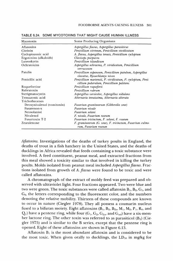
FOODBORNE AGENTS CAUSING ILLNESS 301
TABLE 6.24. SOME MYCOTOXINS THAT MIGHT CAUSE HUMAN ILLNESS
Mycotoxin
Aflatoxins Citrinin Cyclopiazonic acid Ergotoxins (alkaloids) Luteoskyrin Ochratoxins
Patulin
Penicillic acid
Roquefortine Rubratoxin Sterigmatocystin Tenuazonic acid Trichothecenes
Deoxynivalenol (vomitoxin) Fusarenon·x Neosolaniol Nivalenol Fusartoxin T-2
Zearalenone
Some Producing Organisms
Aspergillus jlavus, Aspergillus parasiticus Penicillium citrinum, Penicillium vividicatum A. flavus, Aspergillus tenuis, Penicillium cyclopium Claviceps purpurea Penicillium islandicum Aspergillus ochraceus, P. viridicatum, Penicillium
verrucosum Penicillium expansum, Penicillium patulum, Aspergillus
clavatus, Byssochlamys nivea Penicillium martensii, P. viridicatum, P. cyclopium, Peni·
cillium puberulum, Penicillium palitans Penicillium roque forti Penicillium rubrum Aspergillus versicolor, Aspergillus nidulans Alternaria tenuissima, Alternaria alterata
Fusarium graminearum (Gibberella zeae) Fusarium nivale Fusarium solani F. nivale, Fusarium roseum Fusarium tricinctum, F. solani, F. roseum F. graminearum (G. zeae), F. tricinctum, Fusarium culmo·
rum, Fusarium roseum
Aflatoxins. Investigations of the deaths of turkey poults in England, the deaths of trout in a fish hatchery in the United States, and the deaths of ducklings in Africa revealed that feeds containing a toxic substance were involved. A feed constituent, peanut meal, and extracted fractions from this meal showed a toxicity similar to that involved in killing the turkey poults. Molds isolated from peanut meal included Aspergillus flavus. Frac· tions isolated from growth of A. flavus were found to be toxic and were called aflatoxins.
A chromatograph of the extract of moldy feed was prepared and ob· served with ultraviolet light. Four fractions appeared. Two were blue and two were green. The toxic substances were called aflatoxin B), B2, G), and G2, the letters corresponding to the fluorescent color, and the numbers denoting the relative mobility. Thirteen of these compounds are known to occur in nature (Ciegler 1978). They all possess a coumarin nucleus fused to a bifuran moiety. Eight aflatoxins (B), B2, B2a, MI, M2, PI, Ro, and QI) have a pentene ring, while four (G 1, G2 , G2a, and Gml) have a six·mem· ber lactone ring. The other toxin was referred to as parasiticol (B3) (Cie' gler 1975) and is similar to the B series, except that the pentene ring is opened. Eight of these aflatoxins are shown in Figure 6.13.
Aflatoxin BI is the most abundant aflatoxin and is considered to be the most toxic. When given orally to ducklings, the LDso in mg/kg for
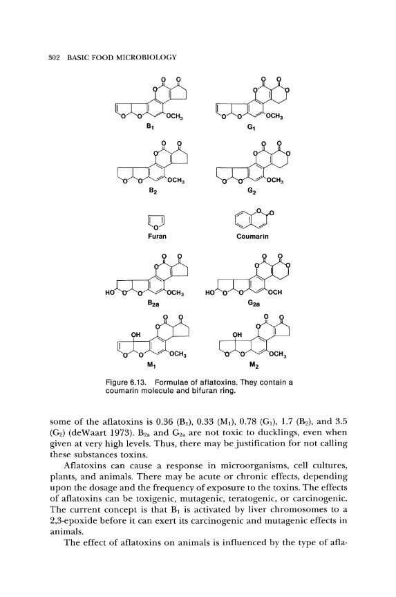
302 BASIC FOOD MICROBIOLOGY
I 1(6 ~CH3 B1
o o Furan
1(6 HO~CH3
B2a
11M ~::: G1
~<>yo VJ Coumarin
1M HO~; G2a
Figure 6.13. Formulae of aflatoxins. They contain a coumarin molecule and bifuran ring.
some of the aflatoxins is 0.36 (BJ), 0.33 (MJ), 0.78 (GJ), 1.7 (B2)' and 3.5 (G2) (deWaart 1973). B2a and G2a are not toxic to ducklings, even when given at very high levels. Thus, there may be justification for not calling these substances toxins.
Aflatoxins can cause a response in microorganisms, cell cultures, plants, and animals. There may be acute or chronic effects, depending upon the dosage and the frequency of exposure to the toxins. The effects of aflatoxins can be toxigenic, mutagenic, teratogenic, or carcinogenic. The current concept is that BJ is activated by liver chromosomes to a 2,3·epoxide before it can exert its carcinogenic and mutagenic effects in animals.
The effect of aflatoxins on animals is influenced by the type of afla·
FOODBORNE AGENTS CAUSING ILLNESS 303
toxin, species of animal, age, weight, health, and diet. Environmental stresses may alter the susceptibility of the animal.
Very young animals are more susceptible to aflatoxicosis than are mao ture animals. Species of animals vary in their resistance. In farm poultry, ducklings are the most susceptible, followed by turkey poults, goslings, and domestic chicks. For farm animals, the order from high susceptibility to resistant is young pigs, pregnant sows, calves, fattening pigs, mature cattle, and sheep.
Acute aflatoxicosis has been characterized by hemorrhage in tissues, anorexia, hepatitis, and death of animals. The liver is the primary tissue that is affected. However, the spleen, pancreas, kidneys, and other tissues may be involved. Aflatoxin also causes fragility of the capillaries, so there is a greater tendency for bruising (Huff et al. 1983). The clinical signs of BJ toxicosis in goats included decreased feed consumption, some loss of body weight, mucopurulent nasal discharge, dyspenea, coughing, leth· argy, icterus, diarrhea, and subnormal body temperature 24 to 48 hr be· fore death (Clark et al. 1984). They found that the clinicopathological changes included increases in red blood cell count, packed cell volume, hemoglobin concentration, serum bilirubin concentration and serum ac· tivities of aspartate aminotransferase, isocitric dehydrogenase, and or· nithine carbamyl transferase. At necropsy, goats fed aflatoxin BJ showed evidence of ascites, pale livers, petechial hemorrhages, nasal discharge, and icterus, as well as bile duct proliferation, hepatocytic karyomegaly, hepatocellular degeneration, pneumonia, rhinitis, and proximal renal tu· bular nephrosis (Miller et al. 1984). Similar effects have been noted after BJ was fed to other animals (Applebaum and Marth 1983; Ketterer et al. 1982; Wyatt et al. 1985). Reduced egg production was noted when BJ was fed to pullets at 0.7 mg/kg body weight, or at higher levels (Exarchos and Gentry 1982). The effect of aflatoxins on rats was reduced when cabbage or cauliflower was included in the diet (Boyd and Stoewsand 1981; Boyd, Babish, and Stoewsand 1982).
Aflatoxin is carcinogenic in ducklings, rainbow trout, rats, and ferrets (Detroy, Lillehoj, and Ciegler 1971). They stated that some reports indio cate that aflatoxin induces carcinomas in pigs, hamsters, guinea pigs, mice, and sheep. A monkey developed a cancer due to aflatoxin inges· tion (Gopalan, Tulpule, and Krishnamurthi 1972). Although the carcino· genic activity is primarily in the liver, aflatoxin might induce tumor for· mation in other organs.
There is ample evidence that aflatoxins produce acute and chronic illness in animals. What is their effect when ingested by humans? The deliberate feeding of aflatoxins to human subjects has not been reported. The only information concerning humans is from instances in which the food, naturally contaminated with aflatoxins, has been consumed. Thus,
304 BASIC FOOD MICROBIOLOGY
evidence for human aflatoxicosis is not based on scientific experiments. Information accumulated after an incident is often incomplete.
Aflatoxin Bl was detected in specimens of stools, urine, liver, brain, and kidney obtained from Thai children who died of encephalopathy and fatty degeneration of the liver (Shank et al. 1971). In another out· break, caused by consuming moldy maize, 106 people died and 291 showed signs of hepatic dysfunction (Krishnamachari et al. 1975).
Attempts have been made to associate the high incidence of liver can· cer in humans in certain areas to the presence of toxigenic molds. New· berne (1974) listed evidence associating aflatoxins with cirrhosis of the liver, as well as hepatoma, hepatitis, and Reye's syndrome. Stora, Dvor· ackova, and Ayraud (1983) further associated aflatoxin Bl with Reye's syndrome and the deaths of five children. At autopsy, aflatoxin Bl was detected in the liver at levels of 120 to 810 {tg/kg. They reported fatty degeneration of the liver, kidneys, myocardium, and fibers of the striated muscles. Wray and Hayes (1980) reported liver cancer in a patient whose serum contained aflatoxin B1• In a study of the diets of ninety people with liver cancer, and ninety controls in the Philippine Islands, research· ers found that the mean aflatoxin load per day was 440 percent greater for people with liver cancer than for the controls (BulataoJayme et al. 1982). Dichter (1984) considered the current levels of aflatoxin exposure from consuming peanuts and peanut products, and, on the basis of epi· demiological data of populations in Africa and Thailand, estimated some fifty·eight cases of liver cancer annually in the United States. We are ex· posed to aflatoxins from other products and activities, so the rate may be higher. There is surely sufficient evidence, if not from scientific exper· iments, that aflatoxins can cause human illness, including liver carci· noma.
Any food that becomes contaminated with spores of toxigenic A. fla· vus or A. parasiticus is capable of supporting growth of the mold and, if held in an environment favorable for growth and toxin production, can become a source of aflatoxin. Foods found to contain aflatoxins are listed in Table 6.25. In the United States the main concern has been with afla· toxin on peanuts, corn, and cottonseed.
Peanuts can become contaminated with mold and aflatoxin during growth, harvesting, or storage. Peanuts grown in soil used consistently for peanuts tend to have a higher accumulation than when crops are rotated. The type of soil and rainfall following dry weather influences pod split· ting of peanuts (Graham 1982). The infection of peanuts also is associ· ated with mechanical damage during or after harvesting. It is essential that the harvested peanuts be dried to 8 percent or less moisture, and stored under conditions so that the moisture content will not increase.
When contaminated peanuts are pressed to remove the oil, most of
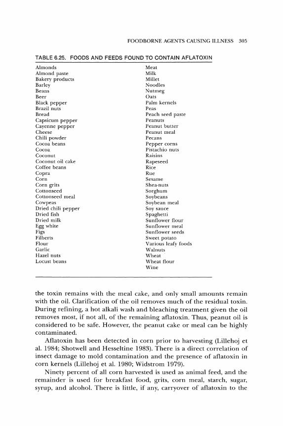
FOODBORNE AGENTS CAUSING ILLNESS 305
TABLE 6.25. FOODS AND FEEDS FOUND TO CONTAIN AFLATOXIN
Almonds Meat Almond paste Milk Bakery products Millet Barley Noodles Beans Nutmeg Beer Oats Black pepper Palm kernels Brazil nuts Peas Bread Peach seed paste Capsicum pepper Peanuts Cayenne pepper Peanut butter Cheese Peanut meal Chili powder Pecans Cocoa beans Pepper corns Cocoa Pistachio nuts Coconut Raisins Coconut oil cake Rapeseed Coffee beans Rice Copra Rue Corn Sesame Corn grits Shea·nuts Cottonseed Sorghum Cottonseed meal Soybeans Cowpeas Soybean meal Dried chili pepper Soy sauce Dried fish Spaghetti Dried milk Sunflower flour Egg white Sunflower meal Figs Sunflower seeds Filberts Sweet potato Flour Various leafy foods Garlic Walnuts Hazel nuts Wheat Locust beans Wheat flour
Wine
the toxin remains with the meal cake, and only small amounts remain with the oil. Clarification of the oil removes much of the residual toxin. During refining, a hot alkali wash and bleaching treatment given the oil removes most, if not all, of the remaining aflatoxin. Thus, peanut oil is considered to be safe. However, the peanut cake or meal can be highly contaminated.
Aflatoxin has been detected in corn prior to harvesting (Lillehoj et al. 1984; Shotwell and Hesseltine 1983). There is a direct correlation of insect damage to mold contamination and the presence of aflatoxin in corn kernels (Lillehoj et al. 1980; Widstrom 1979).
Ninety percent of all corn harvested is used as animal feed, and the remainder is used for breakfast food, grits, corn meal, starch, sugar, syrup, and alcohol. There is little, if any, carryover of aflatoxin to the
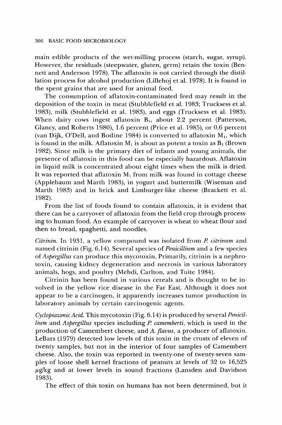
306 BASIC FOOD MICROBIOLOGY
main edible products of the wet· milling process (starch, sugar, syrup). However, the residuals (steepwater, gluten, germ) retain the toxin (Ben· nett and Anderson 1978). The aflatoxin is not carried through the distil· lation process for alcohol production (Lillehoj et al. 1978). It is found in the spent grains that are used for animal feed.
The consumption of aflatoxin·contaminated feed may result in the deposition of the toxin in meat (Stubblefield et al. 1983; Trucksess et al. 1983), milk (Stubblefield et al. 1983), and eggs (Trucksess et al. 1983). When dairy cows ingest aflatoxin -Bb about 2.2 percent (Patterson, Glancy, and Roberts 1980), 1.6 percent (Price et al. 1985), or 0.6 percent (van Dijk, O'Dell, and Bodine 1984) is converted to aflatoxin M I, which is found in the milk. Aflatoxin MI is about as potent a toxin as BI (Brown 1982). Since milk is the primary diet of infants and young animals, the presence of aflatoxin in this food can be especially hazardous. Aflatoxin in liquid milk is concentrated about eight times when the milk is dried. It was reported that aflatoxin MI from milk was found in cottage cheese (Applebaum and Marth 1983), in yogurt and buttermilk (Wiseman and Marth 1983) and in brick and Limburger·like cheese (Brackett et al. 1982).
From the list of foods found to contain aflatoxin, it is evident that there can be a carryover of aflatoxin from the field crop through process· ing to human food. An example of carryover is wheat to wheat flour and then to bread, spaghetti, and noodles.
Citrinin. In 1931, a yellow compound was isolated from P. citrinum and named citrinin (Fig. 6.14). Several species of Penicillium and a few species of Aspergillus can produce this mycotoxin. Primarily, citrinin is a nephro· toxin, causing kidney degeneration and necrosis in various laboratory animals, hogs, and poultry (Mehdi, Carlton, and Tuite 1984).
Citrinin has been found in various cereals and is thought to be in· volved in the yellow rice disease in the Far East. Although it does not appear to be a carcinogen, it apparently increases tumor production in laboratory animals by certain carcinogenic agents.
Cyclopiazonic Acid. This mycotoxin (Fig. 6.14) is produced by several Penicil· lium and Aspergillus species including P. camemberti, which is used in the production of Camembert cheese, and A. flavus, a producer of aflatoxin. LeBars (1979) detected low levels of this toxin in the crusts of eleven of twenty samples, but not in the interior of four samples of Camembert cheese. Also, the toxin was reported in twenty·one of twenty·seven sam· pIes of loose shell kernel fractions of peanuts at levels of 32 to 16,525 J.tg/kg and at lower levels in sound fractions (Lansden and Davidson 1983).
The effect of this toxin on humans has not been determined, but it
OH
HOOC~9 O~CHa
CHs CHa
Citrinin
FOODBORNE AGENTS CAUSING ILLNESS 307
CHI
Cyclopiazonic Acid
COOH OH 0
ObH2-CH-NH-CO~9 ~CHa
CI
J---;f0 COXOH
Ochratoxin A Patulin
Peniciliic Acid Roquefortine
o CHa H H H OH II I I I I
OH «I C-O-~-A-1-~~_ H H H H c-o HO ~ I I I 1/
c=c-c-C-C 1 1 I I H H H H
Sterigmatocystin Zearalenone
Figure 6.14. Formulae of various mycotoxins.
was found to be mutagenic to two strains of Salmonella typhimurium using the Ames test (Sorenson, Tucker, and Simpson 1984).
Luteoskyrin. This mycotoxin is produced by Penicillium islandicum. In reo views by Mislivec (1981) and Stark (1980), it is listed as a carcinogen to rats and mice. When given orally, it causes liver cirrhosis and hepatomas. It binds to DNA and is mutagenic to yeast. In areas in which rice is a
308 BASIC FOOD MICROBIOLOGY
major constituent of the diet, the presence of this toxin is associated with a high incidence of liver cancer in humans.
Ochratoxin A. This is the most common and toxic member of a group of related toxins (Fig. 6.14). It was first discovered as a product of Aspergillus ochraceus. Also, it is produced by other aspergilli and penicillia (see Table 6.24). This toxin has been found in various grains (wheat, barley, oats, corn), beans, peanuts, and as residues in animal carcasses. In hogs, the residues are greatest in the kidney and declining amounts in lean meat, liver, and fat (Madsen, Mortensen, and Hald 1982). In Denmark, hog car· casses are condemned if the ochratoxin residual in kidneys exceeds 25 !kg/kg.
The ingested toxin can cause various syndromes in many types of animals. However, it affects pimarily the kidneys (nephrotoxin). The ef. fect of ochratoxin A on humans has not been determined, but there may be a relationship of renal disease of humans to the consumption of food containing this toxin.
Patulin. This mycotoxin (Fig. 6.14) is produced by some 19 species of fungi, including those listed in Table 6.24. Being produced by P. patulum, it was given the trivial name patulin. Also, it has been named claviformin, expansin, and clavicin. It inhibits a wide variety of bacteria and for a time was used as an antibiotic. However, side effects of intestinal distress, vom· iting, and diarrhea ended its use as a therapeutic agent. The toxin is mutagenic (Mayer and Legator 1979), terotogenic (Ciegler, Beckwith, and Jackson 1976) and carcinogenic (Stark and Demain 1980).
This toxin has been found in fruit products, especially apple juice (Brackett and Marth 1979a; Scott and Bullerman 1975). During the alco· holic fermentation of apple juice, patulin is converted to other chemicals (Stinson, Osman, and Bills 1979).
PR Toxin. This mycotoxin is produced by Penicillium roqueforti, the mold used in the production of Roquefort, blue, and Gorgonzola cheese. Scott (1981) stated that it was one of the most acutely toxic metabolites of P. roqueforti. It is mutagenic to bacteria and cytotoxic to various cell lines. The toxin causes degenerative changes in the liver and kidney of rats. When it was injected into rats, researchers observed breathing difficul· ties, motor incoordination, and flaccid paralysis (Polonelli et al. 1978). Chen, Chen, and Wei (1982) concluded that the effects of PR toxin in animals are increased capillary permeability and direct damage to the lungs, heart, and kidneys. The effect on humans has not been deter· mined.
Although it is produced by P. roque forti, the toxin was not produced in blue-veined cheese (Polonelli et al. 1978). The toxin is not stable in cheese.
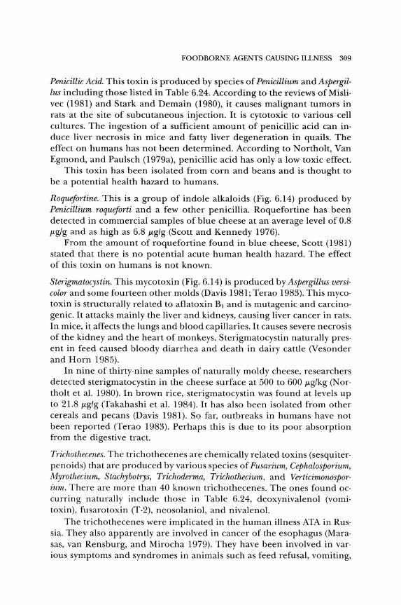
FOODBORNE AGENTS CAUSING ILLNESS 309
Penicillic Acid. This toxin is produced by species of Penicillium and Aspergil· lus including those listed in Table 6.24. According to the reviews of Misli· vec (1981) and Stark and Demain (1980), it causes malignant tumors in rats at the site of subcutaneous injection. It is cytotoxic to various cell cultures. The ingestion of a sufficient amount of penicillic acid can in· duce liver necrosis in mice and fatty liver degeneration in quails. The effect on humans has not been determined. According to Northolt, Van Egmond, and Paulsch (1979a), penicillic acid has only a low toxic effect.
This toxin has been isolated from corn and beans and is thought to be a potential health hazard to humans.
RoqueJortine. This is a group of indole alkaloids (Fig. 6.14) produced by Penicillium roqueforti and a few other penicillia. Roquefortine has been detected in commercial samples of blue cheese at an average level of 0.8 JAg/g and as high as 6.8 JAg/g (Scott and Kennedy 1976).
From the amount of roquefortine found in blue cheese, Scott (1981) stated that there is no potential acute human health hazard. The effect of this toxin on humans is not known.
Sterigmatocystin. This mycotoxin (Fig. 6.14) is produced by Aspergillus versi· color and some fourteen other molds (Davis 1981; Terao 1983). This myco· toxin is structurally related to aflatoxin BJ and is mutagenic and carcino· genic. It attacks mainly the liver and kidneys, causing liver cancer in rats. In mice, it affects the lungs and blood capillaries. It causes severe necrosis of the kidney and the heart of monkeys. Sterigmatocystin naturally pres· ent in feed caused bloody diarrhea and death in dairy cattle (Vesonder and Horn 1985).
In nine of thirty-nine samples of naturally moldy cheese, researchers detected sterigmatocystin in the cheese surface at 500 to 600 JAg/kg (Northolt et al. 1980). In brown rice, sterigmatocystin was found at levels up to 21.8 JAg/g (Takahashi et al. 1984). It has also been isolated from other cereals and pecans (Davis 1981). So far, outbreaks in humans have not been reported (Terao 1983). Perhaps this is due to its poor absorption from the digestive tract.
Trichothecenes. The trichothecenes are chemically related toxins (sesquiterpenoids) that are produced by various species of Fusarium, Cephalosporium, Myrothecium, Stachybotrys, Trichoderma, Trichothecium, and Verticimonosporium. There are more than 40 known trichothecenes. The ones found occurring naturally include those in Table 6.24, deoxynivalenol (vomitoxin), fusarotoxin (T-2), neosolaniol, and nivalenol.
The trichothecenes were implicated in the human illness ATA in Russia. They also apparently are involved in cancer of the esophagus (Marasas, van Rensburg, and Mirocha 1979). They have been involved in various symptoms and syndromes in animals such as feed refusal, vomiting,
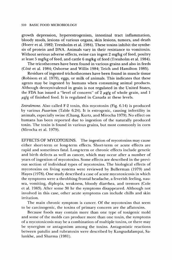
310 BASIC FOOD MICROBIOLOGY
growth depression, hyperestrogenism, intestinal tract inflammation, bloody stools, lesions of various organs, skin lesions, tumors, and death (Hoerr et al. 1982; Trenholm et al. 1984). These toxins inhibit the synthesis of protein and DNA. Animals vary in their resistance to vomitoxin. Without serious adverse effects, swine can ingest 2 mg/kg of feed, poultry at least 5 mg/kg of feed, and cattle 6 mg/kg of feed (Trenholm et al. 1984).
The tricothecenes have been found in various grains and also in feeds (Cote et al. 1984; Osborne and Willis 1984; Teich and Hamilton 1985).
Residues of ingested trichothecenes have been found in muscle tissue (Robison et al. 1979), eggs, or milk of animals. This indicates that these agents may be ingested by humans when consuming animal products. Although deoxynivalenol in grain is not regulated in the United States, the FDA has issued a "level of concern" of 2 jlg/g of whole grain, and 1 jlg/g of finished feed. It is regulated in Canada at these levels.
Zearalenone. Also called F-2 toxin, this mycotoxin (Fig. 6.14) is produced by various Fusarium (Table 6.24). It is estrogenic, causing infertility in animals, especially swine (Chang, Kurtz, and Mirocha 1979). No effect on humans has been reported due to ingestion of the naturally produced toxin. The toxin is found in various grains, but most commonly in corn (Mirocha et al. 1979).
EFFECTS OF MYCOTOXINS. The ingestion of mycotoxins may cause either short-term or long-term effects. Short-term or acute effects are rapid and sometimes fatal. Long-term or chronic effects include genetic and birth defects as well as cancer, which may occur after a number of years of ingestion of mycotoxins_ Some effects are described in the previous section of individual types of mycotoxins. The biological effects of mycotoxins on living systems were reviewed by Bullerman (1979) and Hayes (1978). One study described a case of acute mycotoxicosis in which the symptoms were a throbbing frontal headache, a feverish feeling, nausea, vomiting, diplopia, weakness, bloody diarrhea, and tremors (Cole et al. 1983). After some 30 hr the symptoms disappeared. Although not involved in this case, other acute symptoms can include chills and skin irritation.
The main chronic symptom is cancer. Of the mycotoxins that seem to be carcinogenic, the toxins of primary concern are the aflatoxins.
Because foods may contain more than one type of toxigenic mold and some of the molds can produce more than one toxin, the symptoms of a mycotoxicosis may be a combination of multiple toxins, or there may be synergism or antagonism among the toxins. Antagonistic reactions between patulin and rubratoxin were described by Kangsadalampai, Salunkhe, and Sharma (1981).
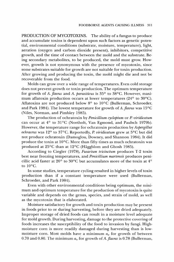
FOODBORNE AGENTS CAUSING ILLNESS 311
PRODUCTION OF MYCOTOXINS. The ability of a fungus to produce and accumulate toxins is dependent upon such factors as genetic potential, environmental conditions (substrate, moisture, temperature), light, aeration (oxygen and carbon dioxide present), inhibitors, competitive growth, and the time of contact between the mold and the substrate. Being secondary metabolites, to be produced, the mold must grow. However, growth is not synonymous with the presence of mycotoxin, since some substrates suitable for growth are not suitable for toxin production. After growing and producing the toxin, the mold might die and not be recoverable from the food.
Molds can grow over a wide range of temperatures. Even cold storage does not prevent growth or toxin production. The optimum temperature for growth of A. flavus and A. parasiticus is 35° to 38°C. However, maximum aflatoxin production occurs at lower temperatures (240 to 30°C). Aflatoxins are not produced below 8° to lOoC (Bullerman, Schroeder, and Park 1984). The lowest temperature for growth of A.flavus was 15°C (Niles, Norman, and Pimbley 1985).
The production of ochratoxin by Penicillium cyclopium or P. viridicatum can occur at 4° to 31°C (Northolt, Van Egmond, and Paulsch 1979b). However, the temperature range for ochratoxin production by Aspergillus ochraceus was 12° to 37°C. Reportedly, P. viridicatum grew at 5°C but did not produce ochratoxin (Damoglou, Downey, and Shannon 1984). It did produce the toxin at lO°e. More than fifty times as much ochratoxin was produced at 25°C than at 12°C (Haggblom and Ghosh 1985).
According to Ciegler (1978), Fusarium tricinctum produces T-2 toxin best near freezing temperatures, and Penicillium martensii produces penicillic acid faster at 20° to 30°C but accumulates more of the toxin at 4° to lO°e.
In some studies, temperature cycling resulted in higher levels of toxin production than if a constant temperature were used (Bullerman, Schroeder, and Park 1984).
Even with other environmental conditions being optimum, the minimum and optimum temperature for the production of mycotoxin is quite variable and depends on the genus, species, and strain of mold, as well as the mycotoxin that is elaborated.
Moisture satisfactory for growth and toxin production may be present in foods prior to or during harvesting, before they are dried adequately. Improper storage of dried foods can result in a moisture level adequate for mold growth. During harvesting, damage to the protective covering of foods increases the susceptibility of the food to invasion by fungi. Highmoisture corn is more readily damaged during harvesting than is lowmoisture corn. Most molds have a minimum aw for growth of between 0.70 and 0.80. The minimum aw for growth of A.flavus is 0.78 (Bullerman,
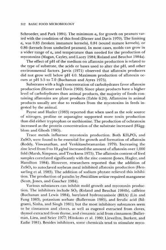
312 BASIC FOOD MICROBIOLOGY
Schroeder, and Park 1984). The minimum aw for growth on peanuts varied with the condition of this food (Diener and Davis 1970). The limiting aw was 0.83 (broken immature kernels), 0.84 (sound mature kernels), or 0.86 (kernels from unshelled peanuts). In most cases, molds can grow in a wider range of aw and temperature than needed for the production of mycotoxins (Magan, Cayley, and Lacey 1984; Roland and Beuchat 1984a).
The effect of pH of the medium on aflatoxin production is related to the type of substrate, the acids or bases used to alter the pH, and other environmental factors. Jarvis (1971) observed that aflatoxin producers did not grow well below pH 4.0. Maximum production of aflatoxin occurs at pH 5.5 to 7.0 (Buchanan and Ayres 1975).
Substrates with a high concentration of carbohydrates favor aflatoxin production (Diener and Davis 1969). Since plant products have a higher level of carbohydrates than animal products, the majority of foods containing aflatoxins are plant products (Table 6.24). Aflatoxins in animal products usually are due to residues from the mycotoxins in feeds ingested by the animal.
Payne and HazIer (1983) reported that when used as the sole source of nitrogen, proline or asparagine supported more toxin production than did either tryptophan or methionine. The production of ochratoxin increased as the protein concentration of the substrate increased (Haggblom and Ghosh 1985).
Trace metals influence mycotoxin production. Both KH2P04 and ZnS04 were found to be essential for growth and formation of aflatoxin (Reddy, Viswanathan, and Venkitasubramanian 1979). Increasing the zinc level from 0 to 10 t-tg/ml increased the amount of aflatoxin over 1,000 fold (Marsh, Simpson, and Trucksess 1975). The aflatoxin content offeed samples correlated significantly with the zinc content (Jones, Hagler, and Hamilton 1984). However, researchers reported that the addition of ZnS04 to autoclaved soybean meal inhibited aflatoxin production (Hensarling et al. 1983). The addition of sodium phytate relieved this inhibition. The production of patulin by Penicillium urticae required manganese (Scott, Jones, and Gaucher 1984).
Various substances can inhibit mold growth and mycotoxin production. The inhibitors include S02 (Roland and Beuchat 1984b), caffeine (Buchanan and Lewis 1984), butylated hydroxyanisole (BHA) (Lin and Fung 1983), potassium sorbate (Bullerman 1985), and ferulic acid (Bilgrami, Sinha, and Singh 1981); but the most inhibitory substances seem to be cinnamon and cloves, as well as eugenol extracted from cloves, thymol extracted from thyme, and cinnamic acid from cinnamon (Bullerman, Lieu, and Seier 1977; Hitokoto et al. 1980; Llewellyn, Burkett, and Eadie 1981). Besides inhibitors, some chemicals tend to stimulate myco-
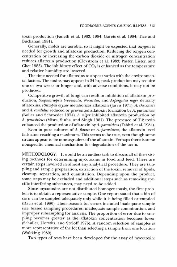
FOODBORNE AGENTS CAUSING ILLNESS 313
toxin production (Fanelli et al. 1983, 1984; Gareis et al. 1984; Tice and Buchanan 1981).
Generally, molds are aerobic, so it might be expected that oxygen is needed for growth and aflatoxin production. Reducing the oxygen con· centration or increasing the carbon dioxide or nitrogen concentration reduces aflatoxin production (Clevstrom et al. 1983; Paster, Lister, and Chet 1983). The inhibitory effect of CO2 is enhanced as the temperature and relative humidity are lowered.
The time needed for aflatoxins to appear varies with the environmen· tal factors. The toxins may appear in 24 hr, peak production may require one or two weeks or longer and, with adverse conditions, it may not be produced.
Competitive growth of fungi can result in inhibition of aflatoxin pro· duction. Scopulariopsis brevicaulis, Nocardia, and Aspergillus niger detoxify aflatoxins. Rhizopus oryzae metabolizes aflatoxin (Jarvis 1971). A. chevalieri and A. candidus reduced or prevented aflatoxin formation by A. parasiticus (Boller and Schroeder 1974). A. niger inhibited aflatoxin production by A. parasiticus (Misra, Sinha, and Singh 1981). The presence of T2 toxin enhanced the production of aflatoxin by A. parasiticus (Fabbri et al. 1984).
Even in pure cultures of A. flavus or A. parasiticus, the aflatoxin level falls after reaching a maximum. This seems to be true, even though some strains appear to be nondegraders of the aflatoxin. Perhaps there is some nonspecific chemical mechanism for degradation of the toxin.
METHODOLOGY. It would be an endless task to discuss all of the exist· ing methods for determining mycotoxins in food and feed. There are certain steps involved in almost any analytical procedure. They are sam· piing and sample preparation, extraction of the toxin, removal of lipids, cleanup, separation, and quantitation. Depending upon the product, some steps may be excluded and additional steps such as removing spe· cific interfering substances, may need to be added.
Since mycotoxins are not distributed homogeneously, the first prob· lem is to obtain a representative sample. One report stated that a bin of corn can be sampled adequately only while it is being filled or emptied (Davis et al. 1980). Their reasons for errors included inadequate sample size, biased sampling procedures, inadequate sample comminution, and improper subsampling for analysis. The proportion of error due to sam· piing becomes greater as the aflatoxin concentration becomes lower (Schuller, Horwitz, and Stoloff 1976). A random selection of samples is more representative of the lot than selecting a sample from one location (Waltking 1980).
Two types of tests have been developed for the assay of mycotoxin:
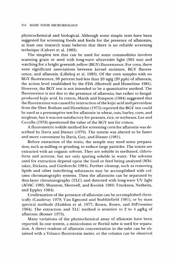
314 BASIC FOOD MICROBIOLOGY
physicochemical and biological. Although some simple tests have been suggested for screening foods and feeds for the presence of aflatoxins, at least one research team believes that there is no reliable screening technique (Calvert et al. 1983).
The simplest test that can be used for some commodities involves scanning grain or seed with long·wave ultraviolet light (365 nm) and watching for a bright greenish yellow (BGY) fluorescence. For corn, there were significant associations between kernel moisture, BGY fluores· cence, and aflatoxin (Lillehoj et al. 1983). Of the corn samples with no BGY fluorescence, 98 percent had less than 20 ng/g (20 ppb) of aflatoxin, the action level established by the FDA (Shotwell and Hesseltine 1981). However, the BGY test is not intended to be a quantitative method. The fluorescence is not due to the presence of aflatoxin, but rather to fungal· produced kojic acid. In cotton, Marsh and Simpson (1984) suggested that the fluorescence was caused by interaction ofthe kojic acid and peroxidase from the fiber. Bothast and Hesseltine (1975) reported the BGY test could be used as a presumptive test for aflatoxin in wheat, oats, barley, corn, and sorghum, but it was not satisfactory for peanuts, rice, or soybeans. Lee and Cucullu (1978) questioned the value of the BGY test for cotton.
A fluorometric·iodide method for screening corn for aflatoxin was de· scribed by Davis and Diener (1979). The system was altered to be faster and more convenient by Davis, Guy, and Diener (1981).
Before extraction of the toxin, the sample may need some prepara· tion, such as milling or grinding, to reduce large particles. The toxins are extracted with an organic solvent. They are soluble in methanol, chloro· form and acetone, but are only sparing soluble in water. The solvents used for extraction depend upon the food or feed being analyzed (Whi· taker, Dickens, and Giesbrecht 1984). Further cleanup, such as removing lipids and other interfering substances may be accomplished with col· umn chromatography systems. Then the aflatoxins can be separated by thin· layer chromatography (TLC) and detected with long·wave UV light (AOAC 1985; Shannon, Shotwell, and Kwolek 1983; Trucksess, Nesheim, and Eppley 1984).
Confirmation of the presence of aflatoxin can be accomplished chemically (Cauderay 1979; Van Egmond and Stubblefield 1981), or by mass spectral methods (Haddon et al. 1977; Rosen, Rosen, and DiProssimo 1984). The extraction and TLC method is sensitive to 2 to 4 J!g/kg of aflatoxin (Romer 1973).
Many variations of the physiochemical assay of aflatoxin have been reported. In one system, a minicolumn or florisil tube is used for separation. A direct readout of aflatoxin concentration in the tube can be obtained with a Velasco flu oro toxin meter, or the column can be observed
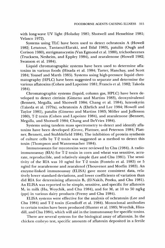
FOODBORNE AGENTS CAUSING ILLNESS 315
with long-wave UV light (Holaday 1981; Shotwell and Hesseltine 1981; Velasco 1972)_
Systems using TLC have been used to detect ochratoxin A (Howell 1982; Letutour, Tantaoui-Elaraki, and Ihlal 1983), patulin (Ough and Corison 1980), sterigmatocystin (Van Egmond et aL 1980), trichothecenes (Trucksess, Nesheim, and Eppley 1984), and zearalenone (Howell 1982; Swanson et aL 1984)_
Liquid chromatographic systems have been used to determine aflatoxins in various foods (Hisada et aL 1984; Tarter, Hanchay, and Scott 1984; Yousef and Marth 1985)_ Systems using high-pressure liquid chromatography (HPLC) have been suggested to separate and determine the various aflatoxins (Cohen and Lapointe 1981; Francis et aL 1982; Takeda 1984)_
Chromatographic systems (liquid, column gas, HPLC) have been developed to detect citrinin (Gimeno and Martins 1983), deoxynivalenol (Bennett, Megalla, and Shotwell 1984; Chang et aL 1984), luteoskyrin (Takeda et aL 1979a), ochratoxin A (Ehrlich and Lee 1984; Howell and Taylor 1981), patulin (Gimeno and Martins 1983; Moller and Joffesson 1980), T-2 toxin (Cohen and Lapointe 1984), and zearalenone (Bennett, Megalla, and Shotwell 1984; Chang and DeVries 1984)_
Systems using tandem mass spectrometry to detect and identify aflatoxins have been developed (Grove, Plattner, and Peterson 1984; Plattner, Bennett, and Stubblefield 1984)_ The inhibition of protein synthesis of culture cells by T-2 toxin was suggested as a method to detect this toxin (Thompson and Wannemacher 1984)_
Immunoassays for mycotoxins were reviewed by Chu (1984)_ A radioimmunoassay (RIA) for T-2 toxin in corn and wheat was sensitive, accurate, reproducible, and relatively simple (Lee and Chu 1981). The sensitivity of the RIA was 10 ng/ml for T-2 toxin (Fontelo et aL 1983) or 5 ng/ml for zearalenone and zearalenol (Thouvenot and Morfin 1983). An enzyme-linked immunoassay (ELISA) gave more consistent data, relatively lower standard deviations, and lower coefficients of variation than did RIA for determining aflatoxin BJ (EI-Nakib, Pestka, and Chu 1981). An ELISA was reported to be simple, sensitive, and specific for aflatoxin MJ in milk (Hu, Woychik, and Chu 1984), and for MJ at 10 to 50 ng/kg (ppt) in various dairy products (Fremy and Chu 1984).
ELISA systems were effective for the analysis of ochratoxin (Lee and Chu 1984) and T-2 toxin (Gendloff et aL 1984). Monoclonal antibodies to certain toxins have been produced (Hunter et aL 1985; Woychik, Hinsdill, and Chu 1984), which will aid in the immunoassay for specific toxins.
There are several systems for the biological assay of aflatoxin. In the chicken embryo test, specific amounts of aflatoxin deposited in a fertile
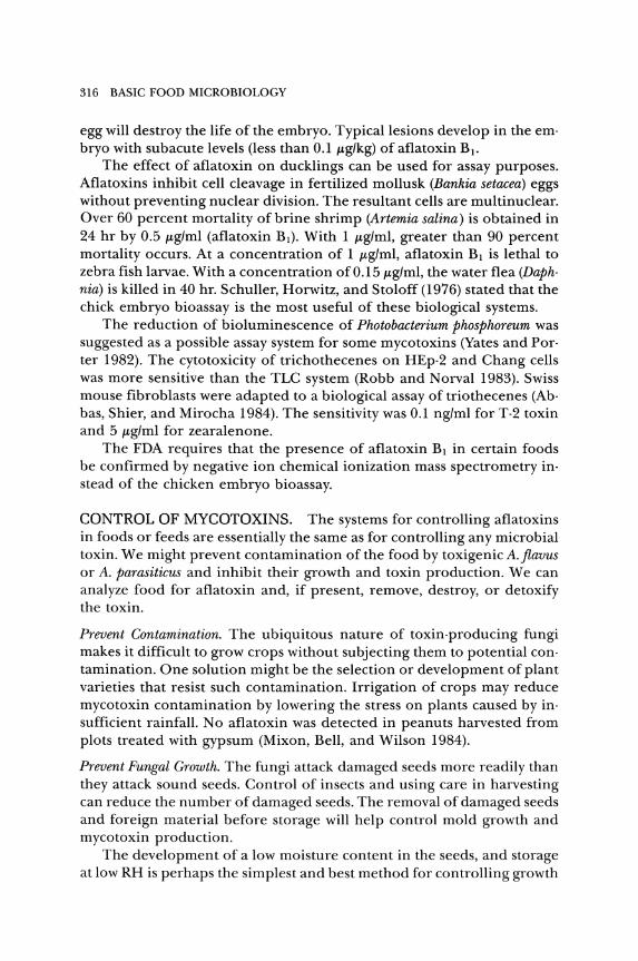
316 BASIC FOOD MICROBIOLOGY
egg will destroy the life of the embryo. Typical lesions develop in the em· bryo with subacute levels (less than 0.1 J.tg/kg) of aflatoxin B j.
The effect of aflatoxin on ducklings can be used for assay purposes. Aflatoxins inhibit cell cleavage in fertilized mollusk (Bankia setacea) eggs without preventing nuclear division. The resultant cells are multinuclear. Over 60 percent mortality of brine shrimp (Artemia salina) is obtained in 24 hr by 0.5 J.tg/ml (aflatoxin Bj). With 1 J.tg/ml, greater than 90 percent mortality occurs. At a concentration of 1 J.tg/ml, aflatoxin B j is lethal to zebra fish larvae. With a concentration of 0.15 J.tg/ml, the water flea (Daph· nia) is killed in 40 hr. Schuller, Horwitz, and Stoloff (1976) stated that the chick embryo bioassay is the most useful of these biological systems.
The reduction of bioluminescence of Photobacterium phosphoreum was suggested as a possible assay system for some mycotoxins (Yates and Por· ter 1982). The cytotoxicity of trichothecenes on HEp·2 and Chang cells was more sensitive than the TLC system (Robb and Norval 1983). Swiss mouse fibroblasts were adapted to a biological assay of triothecenes (Ab· bas, Shier, and Mirocha 1984). The sensitivity was 0.1 ng/ml for T-2 toxin and 5 J.tg/ml for zearalenone.
The FDA requires that the presence of aflatoxin Bj in certain foods be confirmed by negative ion chemical ionization mass spectrometry in· stead of the chicken embryo bioassay.
CONTROL OF MYCOTOXINS. The systems for controlling aflatoxins in foods or feeds are essentially the same as for controlling any microbial toxin. We might prevent contamination of the food by toxigenicA.flavus or A. parasiticus and inhibit their growth and toxin production. We can analyze food for aflatoxin and, if present, remove, destroy, or detoxify the toxin.
Prevent Contamination. The ubiquitous nature of toxin·producing fungi makes it difficult to grow crops without subjecting them to potential con· tamination. One solution might be the selection or development of plant varieties that resist such contamination. Irrigation of crops may reduce mycotoxin contamination by lowering the stress on plants caused by in· sufficient rainfall. No aflatoxin was detected in peanuts harvested from plots treated with gypsum (Mixon, Bell, and Wilson 1984).
Prevent Fungal Growth. The fungi attack damaged seeds more readily than they attack sound seeds. Control of insects and using care in harvesting can reduce the number of damaged seeds. The removal of damaged seeds and foreign material before storage will help control mold growth and mycotoxin production.
The development of a low moisture content in the seeds, and storage at low RH is perhaps the simplest and best method for controlling growth
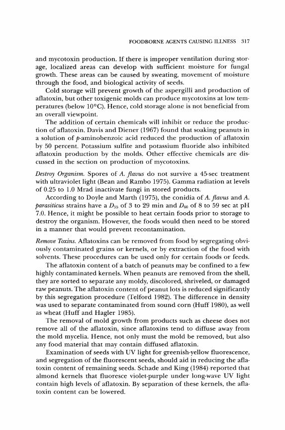
FOODBORNE AGENTS CAUSING ILLNESS 317
and mycotoxin production. If there is improper ventilation during stor· age, localized areas can develop with sufficient moisture for fungal growth. These areas can be caused by sweating, movement of moisture through the food, and biological activity of seeds.
Cold storage will prevent growth of the aspergilli and production of aflatoxin, but other toxigenic molds can produce mycotoxins at low tern· peratures (below lO°C). Hence, cold storage alone is not beneficial from an overall viewpoint.
The addition of certain chemicals will inhibit or reduce the produc· tion of aflatoxin. Davis and Diener (1967) found that soaking peanuts in a solution of p-aminobenzoic acid reduced the production of aflatoxin by 50 percent. Potassium sulfite and potassium fluoride also inhibited aflatoxin production by the molds. Other effective chemicals are dis· cussed in the section on production of mycotoxins.
Destroy Organism. Spores of A. flavus do not survive a 45-sec treatment with ultraviolet light (Bean and Rambo 1975). Gamma radiation at levels of 0.25 to 1.0 Mrad inactivate fungi in stored products.
According to Doyle and Marth (1975), the conidia of A.flavus and A. parasiticus strains have a D55 of 3 to 29 min and D60 of 8 to 59 sec at pH 7.0. Hence, it might be possible to heat certain foods prior to storage to destroy the organism. However, the foods would then need to be stored in a manner that would prevent recontamination.
Remove Toxins. Aflatoxins can be removed from food by segregating obviously contaminated grains or kernels, or by extraction of the food with solvents. These procedures can be used only for certain foods or feeds.
The aflatoxin content of a batch of peanuts may be confined to a few highly contaminated kernels. When peanuts are removed from the shell, they are sorted to separate any moldy, discolored, shriveled, or damaged raw peanuts. The aflatoxin content of peanut lots is reduced significantly by this segregation procedure (Telford 1982). The difference in density was used to separate contaminated from sound corn (Huff 1980), as well as wheat (Huff and Hagler 1985).
The removal of mold growth from products such as cheese does not remove all of the aflatoxin, since aflatoxins tend to diffuse away from the mold mycelia. Hence, not only must the mold be removed, but also any food material that may contain diffused aflatoxin.
Examination of seeds with UV light for greenish-yellow fluorescence, and segregation of the fluorescent seeds, should aid in reducing the aflatoxin content of remaining seeds. Schade and King (1984) reported that almond kernels that fluoresce violet-purple under long-wave UV light contain high levels of aflatoxin. By separation of these kernels, the afla· toxin content can be lowered.
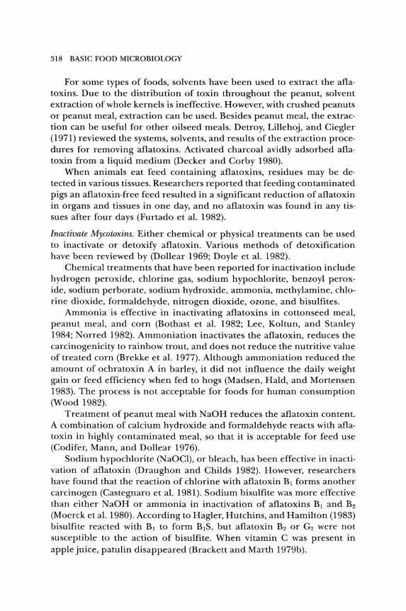
318 BASIC FOOD MICROBIOLOGY
For some types of foods, solvents have been used to extract the aflatoxins_ Due to the distribution of toxin throughout the peanut, solvent extraction of whole kernels is ineffective_ However, with crushed peanuts or peanut meal, extraction can be used. Besides peanut meal, the extraction can be useful for other oilseed meals. Detroy, Lillehoj, and Ciegler (1971) reviewed the systems, solvents, and results ofthe extraction proce· dures for removing aflatoxins. Activated charcoal avidly adsorbed aflatoxin from a liquid medium (Decker and Corby 1980).
When animals eat feed containing aflatoxins, residues may be de· tected in various tissues. Researchers reported that feeding contaminated pigs an aflatoxin-free feed resulted in a significant reduction of aflatoxin in organs and tissues in one day, and no aflatoxin was found in any tissues after four days (Furtado et al. 1982).
Inactivate Mycotoxins. Either chemical or physical treatments can be used to inactivate or detoxify aflatoxin. Various methods of detoxification have been reviewed by (Dollear 1969; Doyle et al. 1982).
Chemical treatments that have been reported for inactivation include hydrogen peroxide, chlorine gas, sodium hypochlorite, benzoyl peroxide, sodium perborate, sodium hydroxide, ammonia, methylamine, chlo· rine dioxide, formaldehyde, nitrogen dioxide, ozone, and bisulfites.
Ammonia is effective in inactivating aflatoxins in cottonseed meal, peanut meal, and corn (Bothast et al. 1982; Lee, Koltun, and Stanley 1984; Norred 1982). Ammoniation inactivates the aflatoxin, reduces the carcinogenicity to rainbow trout, and does not reduce the nutritive value of treated corn (Brekke et al. 1977). Although ammoniation reduced the amount of ochratoxin A in barley, it did not influence the daily weight gain or feed efficiency when fed to hogs (Madsen, Hald, and Mortensen 1983). The process is not acceptable for foods for human consumption (Wood 1982).
Treatment of peanut meal with NaOH reduces the aflatoxin content. A combination of calcium hydroxide and formaldehyde reacts with aflatoxin in highly contaminated meal, so that it is acceptable for feed use (Codifer, Mann, and Dollear 1976).
Sodium hypochlorite (NaOCI), or bleach, has been effective in inactivation of aflatoxin (Draughon and Childs 1982). However, researchers have found that the reaction of chlorine with aflatoxin BJ forms another carcinogen (Castegnaro et al. 1981). Sodium bisulfite was more effective than either NaOH or ammonia in inactivation of aflatoxins BJ and B2 (Moerck et al. 1980). According to Hagler, Hutchins, and Hamilton (1983) bisulfite reacted with BJ to form BJS, but aflatoxin B2 or G2 were not susceptible to the action of bisulfite. When vitamin C was present in apple juice, patulin disappeared (Brackett and Marth 1979b).
FOODBORNE AGENTS CAUSING ILLNESS 319
Oil, as extracted from oilseeds (peanuts and cottonseed) may contain small amounts of aflatoxins. The oil is treated with alkali and bleach to purify it. This processing reduces the aflatoxin content from 812 p.g/kg to less than 1 p.g/kg (Kensler and Natoli 1969).
The primary physical treatment used to reduce toxicity of aflatoxins is heat. Roasting peanuts at IS00C for Y2 hr reduces aflatoxin B1 about 80 percent and B2 about 60 percent (Lee, Cuculla, and Goldblatt 1968). Waltking (1971) roasted peanuts at 204°C and found an average loss of 40 to SO percent aflatoxin B1 and G1 and 20 to 40 percent of B2 and G2. Oil roasting of peanuts at 163°C for 3 min reduces B1 and G1 by 6S percent (Lee et al. 1969).
Roasting soybeans at 180°C reduced levels of aflatoxin from 40 to 73 percent (Hamada and Megalla 1982). Normal cooking of rice destroyed about 49 percent of the aflatoxin, while pressure cooking with-€xcess water reduced aflatoxins by 81 percent (Rehana, Basappa, and Murthy 1979). EI-Banna and Scott (1984) found that cooking fava beans or wheat reduced ochratoxin A by only 16 to 20 percent. About 20 percent of ochratoxin was lost by frying pork at IS00 to 160°C (Josefsson and Moller 1980). Aflatoxins and at least some other mycotoxins are considered to be heat stable.
Biological systems might be used to degrade aflatoxins (Mann and Rehm 1976). Corynebacterium rubrum, Aspergillus niger, Trichoderma viride, and Mucor ambiguus degrade aflatoxin B1 to aflatoxin Ro. Before such systems can be used in industry, much more research is needed, as well as approval by the FDA. Aflatoxin Ro reportedly is less toxic than B1 to ducklings, but is carcinogenic to trout.
Control by Regulation. The production of aflatoxin by toxigenic molds cannot be controlled by a regulation of a government agency. However, it is possible to regulate against the use of food or feed if it contains aflatoxin.
Aflatoxins are considered to be carcinogens. The Delaney clause to Section 409 of the Federal Food, Drug and Cosmetic Act prohibits establishment of a tolerance for additives that "induce cancer when ingested by man or animals."
Instead of tolerances, the FDA has used administrative guidelines, the acceptable level depending upon the ability to detect aflatoxin. In 1964, a control program was established requiring the analysis of shelled peanuts by a certified laboratory. Peanuts with more than 40 p.g/kg (ppb) could not be used in feed. In 1965, this was lowered to 30 p.g/kg, and in 1969, to 20 p.g/kg. Present methods enable the detection of aflatoxin at levels of 2.S p.g/kg or less. Ideally, there should be no aflatoxin, but realistically this would eliminate a large amount of food from an already overtaxed food supply. In 1975, the FDA proposed a formal tolerance of IS
320 BASIC FOOD MICROBIOLOGY
J1.g/kg for aflatoxin in peanuts and peanut products, but after more than ten years, this lower level has not been adopted. Dichter and Weinstein (1984) estimated that lowering the tolerance level to 15 J1.g/kg would be cost effective, and that lower levels of 10 or 5 ppb would not be.
Besides peanuts and peanut products, FDA surveillance activity is di· rected at any food that might contain aflatoxin and action is taken against any food or animal feed with 20 J1.g or more aflatoxin per kilogram. There is an administrative guideline of 0.5 ppb for aflatoxin Ml in fluid milk in interstate shipment. This lower level was set because milk is consumed in large quantities by children. For laboratory animals in toxicology studies, the FDA and Environmental Protection Agency specify that diets contain less than 5 ppb aflatoxin. Animal feed is included in the guidelines because aflatoxins consumed by animals may be found in the resultant food (milk, meat, eggs). However, during years when corn has contained levels of aflatoxin above 20 ppb, the FDA has made exceptions in certain cases (Hamilton 1984; Labuza 1983).
In Australia, the permitted levels of aflatoxin are 15 ppb for peanuts and 5 ppb for other foods (Pitt 1982). The only other restrictions are various tolerance levels of deoxynivalenol (vomitoxin) in wheat in Canada (Morrison 1982a, 1982b), and ochratoxin A in the kidney of hogs in Denmark.
The continual surveillance and the elimination of aflatoxincontaminated products from the food or feed supply constitutes a system of controlling these toxins.
VIRAL HEALTH HAZARDS
For most foodborne illnesses caused by bacteria, the organisms must proliferate in the food for illness to occur. Viruses that cause human illness cannot multiply in food. Thus, a virus that contaminates a food either survives or becomes inactivated. This means that viruses tend to be present only in very low concentration. This has made it difficult to detect viruses in foods. The isolation and identification of viruses require special techniques.
Bryan (1973) listed four viral diseases in which there is evidence of foodborne transmission. These diseases are infectious hepatitis, poliomyelitis, Bolivian hemorrhagic fever, and Russian spring-summer en· cephalitis. He also listed eight viral diseases that might be foodborne, but proof is lacking. These include reovirus infections, serum hepatitis, and nonbacterial gastroenteritis. Cukor and Blacklow (1984) stated that only two viruses, rotavirus and Norwalk virus, are important etiological agents of human gastroenteritis. Besides gastroenteritis, enteroviruses may in-
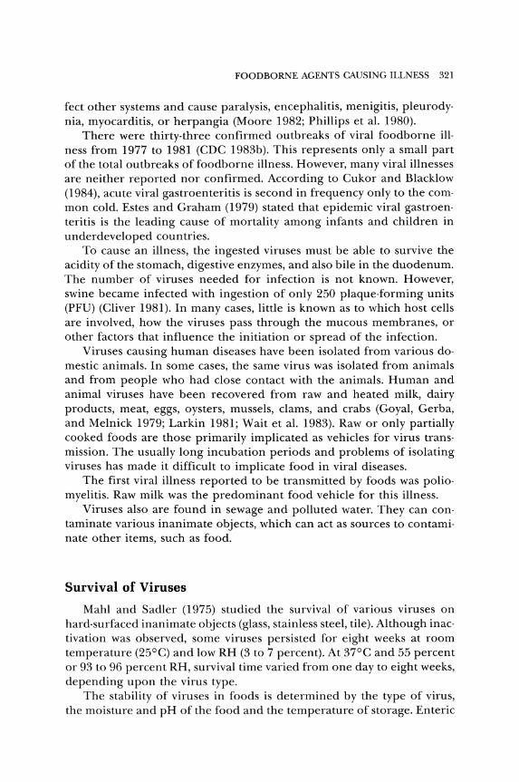
FOODBORNE AGENTS CAUSING ILLNESS 321
fect other systems and cause paralysis, encephalitis, menigitis, pleurodynia, myocarditis, or herpangia (Moore 1982; Phillips et aL 1980)_
There were thirty-three confirmed outbreaks of viral foodborne illness from 1977 to 1981 (CDC 1983b)_ This represents only a small part of the total outbreaks of foodborne illness. However, many viral illnesses are neither reported nor confirmed. According to Cukor and Blacklow (1984), acute viral gastroenteritis is second in frequency only to the common cold. Estes and Graham (1979) stated that epidemic viral gastroenteritis is the leading cause of mortality among infants and children in underdeveloped countries.
To cause an illness, the ingested viruses must be able to survive the acidity of the stomach, digestive enzymes, and also bile in the duodenum. The number of viruses needed for infection is not known. However, swine became infected with ingestion of only 250 plaque-forming units (PFU) (Cliver 1981)_ In many cases, little is known as to which host cells are involved, how the viruses pass through the mucous membranes, or other factors that influence the initiation or spread of the infection.
Viruses causing human diseases have been isolated from various domestic animals. In some cases, the same virus was isolated from animals and from people who had close contact with the animals. Human and animal viruses have been recovered from raw and heated milk, dairy products, meat, eggs, oysters, mussels, clams, and crabs (Goyal, Gerba, and Melnick 1979; Larkin 1981; Wait et aL 1983). Raw or only partially cooked foods are those primarily implicated as vehicles for virus transmission. The usually long incubation periods and problems of isolating viruses has made it difficult to implicate food in viral diseases.
The first viral illness reported to be transmitted by foods was poliomyelitis. Raw milk was the predominant food vehicle for this illness.
Viruses also are found in sewage and polluted water. They can contaminate various inanimate objects, which can act as sources to contaminate other items, such as food.
Survival of Viruses
Mahl and Sadler (1975) studied the survival of various viruses on hard-surfaced inanimate objects (glass, stainless steel, tile). Although inactivation was observed, some viruses persisted for eight weeks at room temperature (25°C) and low RH (3 to 7 percent). At 37°C and 55 percent or 93 to 96 percent RH, survival time varied from one day to eight weeks, depending upon the virus type.
The stability of viruses in foods is determined by the type of virus, the moisture and pH of the food and the temperature of storage. Enteric
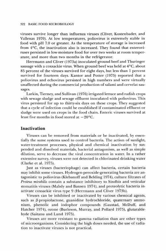
322 BASIC FOOD MICROBIOLOGY
viruses survive longer than influenza viruses (Cliver, Kostenbader, and Vallenas 1970). At low temperatures, poliovirus is extremely stable in food with pH 7.0 or greater. As the temperature of storage is increased from 4°C, the inactivation also is increased. They found that enteroviruses persisted in low-moisture food for over two weeks at room temperature, and more than two months in the refrigerator.
Herrmann and Cliver (1973a) inoculated ground beef and Thuringer sausage with a coxsackie virus_ When ground beef was held at 4°C, about 85 percent of the viruses survived for eight days, but less than 1 percent survived for fourteen days. Kantor and Potter (1975) reported that a poliovirus and echovirus persisted in high numbers and were virtually unaffected during the commercial production of salami and cervelat sausages.
Larkin, Tierney, and Sullivan (1976) irrigated lettuce and radish crops with sewage sludge and sewage effluent inoculated with poliovirus. This virus persisted for up to thirty-six days on these crops. They suggested that a cycle of infection could be established if contaminated effluent or sludge were used on crops in the food chain. Enteric viruses survived at least five months in food stored at - 20°C.
Inactivation
Viruses can be removed from materials or be inactivated, by essentially the same systems used to control bacteria. The action of sunlight, water-treatment processes, physical and chemical inactivation by suspended and dissolved materials, bacterial antagonism, as well as simple dilution, serve to decrease the viral concentration in water. In a rather extensive survey, viruses were not detected in chlorinated drinking water (Clarke et al. 1975).
Just as viruses (bacteriophage) can affect bacteria, certain bacteria may inhibit some viruses. Hydrogen-peroxide-generating bacteria are antagonistic to poliovirus (Klebanoff and Belding 1974), culture filtrates of Proteus mirabilis contain a substance inhibitory to Sindbis and vesicular stomatitis viruses (Mahdy and Bansen 1974), and proteolytic bacteria inactivate coxsackie virus type 9 (Herrmann and Cliver 1973b).
Viruses can be inhibited or inactivated by various chemical agents, such as !3-propiolactone, guanidine hydrochloride, quaternary ammonium, phenolic and iodophor compounds (Gaustad, McDuff, and Hatcher 1974), ozone (Burleson, Murray, and Pollard 1975), glutaraldehyde (Saitanu and Lund 1975)_
Viruses are more resistant to gamma radiation than are other types of microorganisms. Considering the high doses needed, the use of radiation to inactivate viruses is not practical.

FOODBORNE AGENTS CAUSING ILLNESS 323
Thermal inactivation seems to be an acceptable and practical method of controlling viruses in food. Many viruses are inactivated in 5 min when heated to 65°C. Therefore, it is sometimes assumed that properly cooked or pasteurized foods are not a concern of public health unless the prod· uct is recontaminated. However, researchers have reported that the time used to cook crabs may not be sufficient to inactivate viruses (DiGirolamo et al. 1972). Filppi and Banwart (1974) inoculated ground beef with poliovirus type 1 and heated the mixture at various temperatures. From their data, it is evident that meat, when highly contaminated with a potentially pathogenic virus, might be a source of infection, even if heated to 70°C. Other researchers found no surviving poliovirus 1 or coxsackie virus B-2 in hamburgers broiled to either 71°C or 76.7°C (Sullivan et al. 1975).
Cliver (1971) reported that inoculated polio 1 virus was not completely inactivated in oysters by stewing, frying, baking, or steaming. Pasteurization treatments, such as in egg products or milk, cannot be relied upon to destroy large numbers of heat-resistant viruses.
Methodology
The analysis of food for viruses requires techniques different from those used for bacterial enumeration. Viruses are determined by inoculation into host cells for enumeration, by observation with an electron microscope, or by immunological methods, either by determining viruses directly or by determining antibody produced by a host in response to a viral infection.
Since viruses, when present, are usually at a low level in food, a large quantity must be analyzed. After making a suspension of the food sample, the larger food particles are removed by filtering through glass wool or cheesecloth, or by slow centrifuging (Tierney"et al. 1973). After removal of the food, the viruses are concentrated. This is accomplished by membrane adsorption, adsorption to a precipitable salt, filtration, centrifugation, electrophoresis, or dialysis (Guttman-Bass and Nasser 1984; Sullivan et al. 1984).
To determine viruses in the concentrate, the plaque-forming units that develop on a susceptible tissue culture are counted. Bacteria present in food can interfere with the growth of cells in a tissue culture. Therefore, if they are not removed, they must be inhibited. This is accomplished by adding antibiotics that inactivate the bacteria but not the tissue culture or viruses.
Due to the cell specificity of viruses, the more types of tissue culture cells that are used, the more likely that most types of viruses can be detected and isolated.
324 BASIC FOOD MICROBIOLOGY
In some cases, procedures using the electron microscope have been used to detect viruses, viruslike particles, or virus-antibody complexes_
Serological systems for the detection and identification of viruses include fluorescent techniques, RIA, hemagglutination inhibition, immunoperoxidase, counterimmunoelectrophoresis, latex agglutination tests, enzyme immunoassay, immunocytochemical and ELISA (Angarano, Laddago, and Materi 1984; Herrmann, Hendry, and Collins 1979; Sambourg et aL 1985; Smith 1985)_
Diseases
Although several human illnesses are caused by viruses, only those that may be considered a foodborne illness are discussed_ These illnesses were reviewed by Gerba, Rose, and Singh (1984)_
HEPATITIS. There are three types of viral hepatitis: hepatitis A, hepatitis B, and hepatitis non-A, non-B. Hepatitis A, sometimes called infectious hepatitis, is caused by a picornavirus which is transmitted by the fecal-oral route. The virus can be grown in cell culture (Deinhart and Gust 1982). The illness is not chronic, and no chronic carriers are known. Hepatitis B (serum hepatitis) is transmitted through blood or blood products or close contact with a hepatitis B-positive person. Hepatitis B may result in a persistent or chronic infection. This virus has not been grown in cell culture (Deinhart and Gust 1982). In 1983, the total number of cases of hepatitis reported in the United States was 56,469. Of these cases, 38 percent were hepatitis A, 43 percent hepatitis B, 6 percent hepatitis non-A, non-B, and 13 percent unspecified (CDC 1985b). This was the first year that the incidence of hepatitis B was greater than that of he pat itis k
Since hepatitis A virus is spread by the fecal-oral route, waterborne and foodborne outbreaks can occur. People with the infection who handle food are a primary source for spreading the virus. Even then, only about 6 percent of the cases are foodborne or waterborne (CDC 1985b).
The eating of raw shellfish (oysters, mussels, or clams) is an important source of the hepatitis virus. This is due to the potential contamination of water with raw sewage and the collection and concentration of the virus by the shellfish. However, since infected food handlers are a primary source, various foods have been implicated as vehicles of the virus. These foods include dairy products, meats, salads, bakery goods, and fruits, which were contaminated during preparation and received no further cooking prior to serving.
A series of outbreaks of hepatitis A due to eating raw clams was described by CDC (l982d). Some 6 to 72 hr after consumption, gastrointes-
FOODBORNE AGENTS CAUSING ILLNESS 325
tinal distress (diarrhea, abdominal cramps, nausea, vomiting), which lasted from one to three days, was experienced. From twenty·one to thirty· seven days after ingestion of the clams, symptoms of hepatitis A were ev· ident. These symptoms include bile in urine (dark urine) and jaundice. In an outbreak resulting from a family reunion of forty· five people, four· teen cases of clinical hepatitis were identified from nineteen to forty·four days. The illness may be rather mild, allowing cases to be ambulatory. In other cases, liver injury occurs, resulting in cirrhosis. A severe infection can cause death.
During hepatitis, the level of serum glutamic-oxalacetic transaminase (SGOT) or serum glutamic· pyruvic transaminase (SGPT) is at least twice as high as the normal laboratory standard, and there is a total level of serum bilirubin over 2 mgllOO ml. Also, immunoglobin M hepatitis A antibody (IgM anti HAV) is produced (CDC 1981b). This antibody usually appears in the serum before or shortly after the onset of symptoms and persists for two to three months. Detection of the antibody can identify early and subclinical cases. A team of researchers developed an ELISA system to detect IgM anti HAV in serum (Locarnini et al. 1979).
The hepatitis A virus is present in the feces some two weeks prior to symptoms.
The disease can be attacked from four areas. First, people who have been exposed or might be exposed to the virus can be given immune serum globulin. This has been an effective deterrent for the secondary spread of the illness. Second, the beds from which seafoods, such as shell· fish, are gathered should be clean and not polluted with raw sewage. Harvested shellfish should not be eaten raw. However, there are some people who insist on consuming these animals in the uncooked state.
The third means of control is to inactivate the virus in a food product by heating, radiation, drying, or chemicals. Unfortunately, little is known about the exact treatment to inactivate hepatitis virus A. Clams added to a pot of boiling water, heated until they opened and then eaten, were incriminated in an outbreak of hepatitis. This virus evidently is not inactivated by limited heating. However, adequate cooking should inactivate the virus.
Inasmuch as most of the foodborne outbreaks seem to be caused by a food handler contaminating a food with no further cooking, the fourth area of control is the most important. The infected food handler must be controlled. This is not easily accomplished, since the virus may be excreted in stools during the incubation period, from seven to ten days prior to the onset of symptoms and by asymptomatic persons. However, those clinically ill persons should not be allowed to work with food until they are clearly convalescent. Employees can be screened by the analysis of the SGOT or SGPT levels of their serum. If the levels are more than

326 BASIC FOOD MICROBIOLOGY
twice the normal values, then the employee should not work with food. Better personal hygiene of food handlers, including more frequent hand washing and less handling of food and ingredients, would help prevent the spread of not only infectious hepatitis, but also other illnesses.
ROTA VIRUS. Human rotavirus is a major cause of gastroenteritis in infants and young children throughout the world (Albert, Bishop, and Shann 1983; Al·Nakib et al. 1980; Coiro et al. 1983; Mata et al. 1983; Oishi et al. 1979; Paniker, Mathew, and Mathan 1982; Rodriguez et al. 1980). The illness is most prevalent and severe in children from six months to two years of age. After the second year of life, rotavirus infections requir· ing hospitalization decline significantly. By age five, most children have acquired serum antibodies to rotavirus.
The incubation period is two to four days, and the symptoms include diarrhea, vomiting, fever, and abdominal pain. The duration of the ill· ness is two to ten days. Due to loss of fluids and severe dehydration, death can occur, especially in children less than one year old. In temper· ate climates, the incidence of the illness is highest during the winter and is virtually absent in the summer. This is the opposite of salmonellosis, which is more prevalent during the summer. Some possible reasons for wintertime prevalence have been suggested (Brandt et al. 1982). In tropi· cal areas, the incidence of rotavirus tends to be influenced by rainy or dry seasons.
The transmission of rotavirus appears to be by the fecal· oral route; however, the respiratory route also has been suggested (Foster et al. 1980; Oishi et al. 1979).
Adults that contact infected infants often become infected with rotavirus as evidenced by serological tests. Most adults have only mild symptoms or are asymptomatic; however, a few may have a severe illness.
NORWALK VIRUS. This name was given to the causative agent involved in an outbreak of nonbacterial gastroenteritis in Norwalk, Ohio, in 1968. Particles 27 nm in size were observed in stool filtrates with the aid of electron microscopy. Similar particles have been observed in other outbreaks. Other characteristics of the viruses were described by Cukor and Blacklow (1984). According to Dolin (1978), six agents (Norwalk, Hawaii, Montgomery County, W, Ditchling, Cockle) are all similar, but there are multiple antigenic types. At the present time, Norwalk virus is the most prominent. None of these agents has been grown on cell cultures.
Foodborne, waterborne, and person·to·person (both primary and secondary) modes of transmission occur (Goodman et al. 1982; Kaplan et al. 1982). The illness appears during all seasons and affects all age groups.
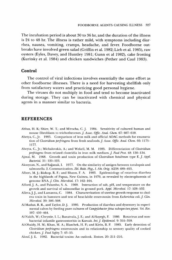
FOODBORNE AGENTS CAUSING ILLNESS 327
The incubation period is about 30 to 36 hr, and the duration of the illness is 24 to 48 hr. The illness is rather mild, with symptoms including diar· rhea, nausea, vomiting, cramps, headache, and fever. Foodborne out· breaks have involved green salad (Griffin et al. 1982; Lieb et al. 1985), raw oysters (Eyles, Davey, and Huntley 1981; Gunn et al 1982), cake frosting (Kuritsky et al. 1984) and chicken sandwiches (pether and Caul 1983).
Control
The control of viral infections involves essentially the same effort as other foodborne illnesses. There is a need for harvesting shellfish only from satisfactory waters and practicing good personal hygiene.
The viruses do not multiply in food and tend to become inactivated during storage. They can be inactivated with chemical and physical agents in a manner similar to bacteria.
REFERENCES
Abbas, H. K; Shier, W. T.; and Mirocha, C.]. 1984. Sensitivity of cultured human and mouse fibroblasts to trichothecenes.]. Assoc. Offic. Anal. Chem. 67: 607-610.
Abetya, C., Jr. 1983. Comparison of iron milk and official AOAC methods for enumera· tion of Clostridium perfringens from fresh seafoods.]. Assoc. Offic. Anal. Chem. 66: 1175-1177.
Abeyta, C., Jr.; Michalovskis, A.; and Wekell, M. M. 1985. Differentiation of Clostridium perfringens from related clostridia in iron milk medium.]. Food Prot. 48: 130-134.
Ajmal, M. 1968. Growth and toxin production of Clostridium botulinum type E.]. Appl.
Bacteriol. 31: 120-123. Aksoycan, N., and Saganak, I. 1977. On the similarity of antigen between torulopsis and
salmonella. 2. Communication. Zbl. Bakt. Hyg., I. Abt. Orig. A238: 489-493. Albert, M.].; Bishop, R. F.; and Shann, F. A. 1983. Epidemiology of rotavirus diarrhea
in the highlands of Papua, New Guinea, in 1979, as revealed by electrophoresis of genome RNA.]. Clin. Microbiol. 17: 162-164.
Alford,]. A., and Palumbo, S. A. 1969. Interaction of salt, pH, and temperature on the growth and survival of salmonellae in ground pork. Appl. Microbiol. 17: 528-532.
Alleva,].]., and Lamanna, C. 1984. Characterization of uterine growth response to chol· era toxin in hamsters and test of heat· labile enterotoxin from ~"scherichia coli.]. Clin. Microbiol. 20: 506-508.
AI·Mashat, R. R., and Taylor, D.]. 1980. Production of diarrhea and dysentery in experi· mental calves by feeding pure cultures of Campylobacter fetus subspecies jejuni. Vet. Rec. 107: 459-464.
AI·Nakib, W.; Chrystie, I. L.; Banatvala,]. E.; and AI-Sayegh, F. 1980. Rotavirus and nonbacterial infantile gastroenteritis in Kuwait. Int.]. Epidemiol. 9: 355-359.
AI·Obaidy, H. M.; Khan, M. A.; Blaschek, H. P.; and Klein, B. P. 1985. Early detection of Clostridium perfringens enterotoxin and its relationship to sensory quality of cooked chicken.]. Food Safety 7: 43-55.
Alouf,]. E. 1982. Bacterial toxins: An outlook. Toxicon. 20: 211-216.
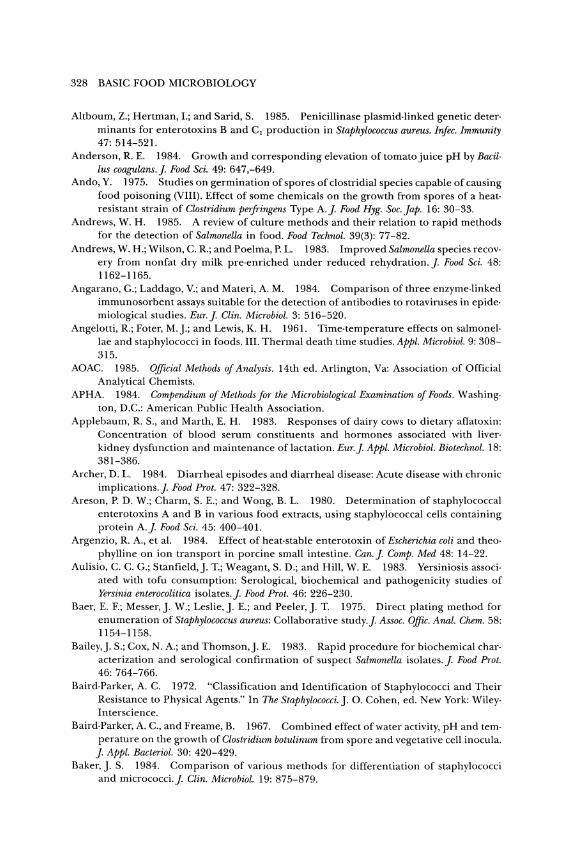
328 BASIC FOOD MICROBIOLOGY
Altboum, Z.; Hertman, I.; and Sarid, S. 1985. Penicillinase plasmid· linked genetic deter· minants for enterotoxins Band C 1 production in Staphylococcus aureus. lrifec. Immunity 47: 514-521.
Anderson, R E. 1984. Growth and corresponding elevation of tomato juice pH by Bacil· Ius coagulans.]. Food Sci. 49: 647,-649.
Ando, Y. 1975. Studies on germination of spores of clostridial species capable of causing food poisoning (VIII). Effect of some chemicals on the growth from spores of a heat· resistant strain of Clostridium perjTingens Type A.]. Food Hyg. Soc. lap. 16: 30-33.
Andrews, W. H. 1985. A review of culture methods and their relation to rapid methods for the detection of Salmonella in food. Food Technol. 39(3): 77-82.
Andrews, W. H.; Wilson, C. R; and Poelma, P. L. 1983. Improved Salmonella species recov· ery from nonfat dry milk pre· enriched under reduced rehydration.]. Food Sci. 48: 1162-1165.
Angarano, G.; Laddago, Y.; and Materi, A. M. 1984. Comparison of three enzyme·linked immunosorbent assays suitable for the detection of antibodies to rotaviruses in epide· miological studies. Eur.]. Clin. Microbiol. 3: 516-520.
Angelotti, R.; Foter, M.].; and Lewis, K. H. 1961. Time·temperature effects on salmonel· lae and staphylococci in foods. III. Thermal death time studies. Appl. Microbiol. 9: 308-315.
AOAC. 1985. Official Methods of Analysis. 14th ed. Arlington, Va: Association of Official Analytical Chemists.
APHA. 1984. Compendium of Methods for the Microbiological Examination of Foods. Washing· ton, D.C.: American Public Health Association.
Applebaum, R S., and Marth, E. H. 1983. Responses of dairy cows to dietary aflatoxin: Concentration of blood serum constituents and hormones associated with liver· kidney dysfunction and maintenance of lactation. Eur.]. Appl. Microbiol. Biotechnol. 18: 381-386.
Archer, D. L. 1984. Diarrheal episodes and diarrheal disease: Acute disease with chronic implications.]. Food Prot. 47: 322-328.
Areson, P. D. W.; Charm, S. E.; and Wong, B. L. 1980. Determination of staphylococcal enterotoxins A and B in various food extracts, using staphylococcal cells containing protein A.]. Food Sci. 45: 400-401.
Argenzio, R. A., et al. 1984. Effect of heat·stable enterotoxin of Escherichia coli and theo· phylline on ion transport in porcine small intestine. Can.]. Compo Med 48: 14-22.
Aulisio, C. C. G.; Stanfield,]. T.; Weagant, S. D.; and Hill, W. E. 1983. Yersiniosis associ· ated with tofu consumption: Serological, biochemical and pathogenicity studies of Yersinia enterocolitica isolates.]. Food Prot. 46: 226-230.
Baer, E. F.; Messer,]. W.; Leslie,]. E.; and Peeler,]. T. 1975. Direct plating method for enumeration of Staphylococcus aureus: Collaborative study.]. Assoc. OfJic. Anal. Chem. 58: 1154-1158.
Bailey,]. S.; Cox, N. A.; and Thomson,]. E. 1983. Rapid procedure for biochemical char· acterization and serological confirmation of suspect Salmonella isolates.]. Food Prot. 46: 764-766.
Baird·Parker, A. C. 1972. "Classification and Identification of Staphylococci and Their Resistance to Physical Agents." In The Staphylococci.]. O. Cohen, ed. New York: Wiley· Interscience.
Baird·Parker, A. C., and Freame, B. 1967. Combined effect of water activity, pH and tern· perature on the growth of Clostridium botulinum from spore and vegetative cell inocula.
]. Appl. Bacteriol. 30: 420-429. Baker,]. S. 1984. Comparison of various methods for differentiation of staphylococci
and micrococci.]. Clin. Microbiol. 19: 875-879.
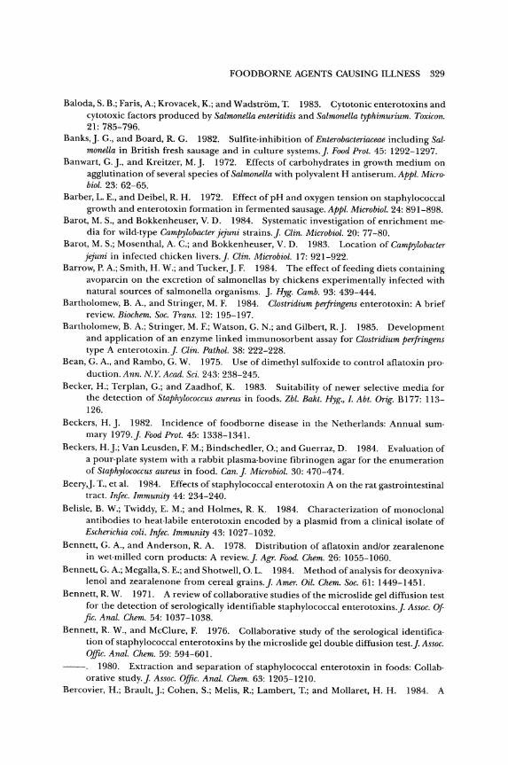
FOODBORNE AGENTS CAUSING ILLNESS 329
Baloda, S. B.; Faris, A.; Krovacek, K.; and Wadstrom, T. 1983. Cytotonic enterotoxins and cytotoxic factors produced by Salmonella enteritidis and Salmonella typhimurium. Toxicon. 21: 785-796.
Banks,]. G., and Board, R. G. 1982. Sulfite-inhibition of Enterobacteriaceae including Sal· monella in British fresh sausage and in culture systems.]. Food Prot. 45: 1292-1297.
Banwart, G.]., and Kreitzer, M.]. 1972. Effects of carbohydrates in growth medium on agglutination of several species of Salmonella with polyvalent H antiserum. Appl. Micro· bioi. 23: 62-65.
Barber, L. E., and Deibel, R H. 1972. Effect of pH and oxygen tension on staphylococcal growth and enterotoxin formation in fermented sausage. Appl. Microbiol. 24: 891-898.
Barot, M. S., and Bokkenheuser, V. D. 1984. Systematic investigation of enrichment me· dia for wild-type Campylobacter jejuni strains.]. Clin. Microbiol. 20: 77-80.
Barot, M. S.; Mosenthal, A. C.; and Bokkenheuser, V. D. 1983. Location ofCampylobacter jejuni in infected chicken livers.]. Clin. Microbiol. 17: 921-922.
Barrow, P. A.; Smith, H. W.; and Tucker,]. F. 1984. The effect of feeding diets containing avoparcin on the excretion of salmonellas by chickens experimentally infected with natural sources of salmonella organisms. ]. Hyg. Camb. 93: 439-444.
Bartholomew, B. A., and Stringer, M. F. 1984. Clostridium perfringens enterotoxin: A brief review. Biochem. Soc. Trans. 12: 195-197.
Bartholomew, B. A.; Stringer, M. F.; Watson, G. N.; and Gilbert, R.]. 1985. Development and application of an enzyme linked immunosorbent assay for Clostridium perfringens type A enterotoxin.]. Clin. Patkol. 38: 222-228.
Bean, G. A., and Rambo, G. W. 1975. Use of dimethyl sulfoxide to control aflatoxin pro· duction. Ann. N.Y. Acad. Sci. 243: 238-245.
Becker, H.; Terplan, G.; and Zaadhof, K. 1983. Suitability of newer selective media for the detection of Staphylococcus aureus in foods. Zbl. Bakt. Hyg., I. Abt. Orig. B 177: 113-126.
Beckers, H.]. 1982. Incidence of foodborne disease in the Netherlands: Annual sum· mary 1979.]. Food Prot. 45: 1338-1341.
Beckers, H.].; Van Leusden, F. M.; Bindschedler, 0.; and Guerraz, D. 1984. Evaluation of a pour· plate system with a rabbit plasma·bovine fibrinogen agar for the enumeration of Staphylococcus aureus in food. Can.]. Microbiol. 30: 470-474.
Beery,]. T., et al. 1984. Effects of staphylococcal enterotoxin A on the rat gastrointestinal tract. Injec. Immunity 44: 234-240.
Belisle, B. W.; Twiddy, E. M.; and Holmes, R K. 1984. Characterization of monoclonal antibodies to heat·labile enterotoxin encoded by a plasmid from a clinical isolate of Escherichia coli. Injec. Immunity 43: 1027-1032.
Bennett, G. A., and Anderson, R A. 1978. Distribution of aflatoxin and/or zearalenone in wet·milled corn products: A review.]. Agr. Food. Chem. 26: 1055-1060.
Bennett, G. A.; Megalla, S. E.; and Shotwell, O. L. 1984. Method of analysis for deoxyniva· lenol and zearalenone from cereal grains.]. Amer. Oil. Chem. Soc. 61: 1449-1451.
Bennett, R W. 1971. A review of collaborative studies of the microslide gel diffusion test for the detection of serologically identifiable staphylococcal enterotoxins.]. Assoc. Of fico Anal. Chem. 54: 1037-1038.
Bennett, R W., and McClure, F. 1976. Collaborative study of the serological identifica· tion of staphylococcal enterotoxins by the microslide gel double diffusion test.]. Assoc. OfflC. Anal. Chem. 59: 594-601.
1980. Extraction and separation of staphylococcal enterotoxin in foods: Collab· orative study.]. Assoc. Offic. Anal. Chem. 63: 1205-1210.
Bercovier, H.; Brault,].; Cohen, S.; Melis, R; Lambert, T.; and Mollaret, H. H. 1984. A
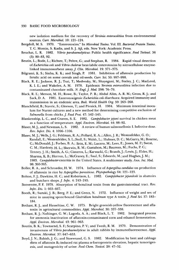
330 BASIC FOOD MICROBIOLOGY
new isolation medium for the recovery of Yersinia enterocolitica from environmental sources. Curro Microbiol. 10: 121-124.
Bergdoll, M. S. 1970. "Enterotoxins." In Microbial Toxins. Vol. III. Bacterial Protein Toxins. T. C. Montie, S. Kadis, and S.]. Ajl, eds. New York: Academic Press.
Beuchat, L. R. 1982. Vibrio parahaemolyticus: Public health significance. Food Technol. 36 (3): 80-83, 92.
Beutin, L.; Bode, L.; Richter, T.; Peltre, G.; and Stephan, R. 1984. Rapid visual detection of Escherichia coli and Vibrio cholerae heat· labile enterotoxins by nitrocellulose enzyme· linked immunosorbent assay.]. Clin. Microbial. 19: 371-375.
Bilgrami, K. S.; Sinha, K. K.; and Singh, P. 1981. Inhibition of aflatoxin production by ferulic acid on some cereals and oil· seeds. Curro Sci. 50: 997-998.
Black, R E.; Jackson, R ].; Tsai, T.; Medvesky, M.; Shayegani, M.; Feeley,]. C.; MacLeod, K 1. E.; and Wakelee, A. M. 1978. Epidemic Yersinia enterocolitica infection due to contaminated chocolate milk. N. Engl.]. Med. 298: 76-79.
Black, R E.; Merson, M. H.; Rowe, B.; Taylor, P. R; Abdul Alim, A. R. M.; Gross, R.].; and Sack, D. A. 1981. Enterotoxigenic Escherichia coli diarrhoea: Acquired immunity and transmission in an endemic area. Bull. World Health Org. 59: 263-268.
Blanchfield, B.; Stavric, S.; Gleeson, T.; and Pivnick, H. 1984. Minimum intestinal inocu· lum for Nurmi cultures and a new method for determining competitive exclusion of Salmonella from chicks.]. Food Prot. 47: 542-545.
Blankenship, L. C., and Craven, S. E. 1982. Campylobacter jejuni survival in chicken meat as a function of temperature. Appl. Environ. Microbial. 44: 88-92.
Blaser, M.]., and Newman, L. S. 1982. A review of human salmonellosis: 1. Infective dose. Rev. Infect. Dis. 4: 1096-1106.
Blaser, M.].; Wells,]. G.; Feldman, R A.; Pollard, R A..; Allen,]. R; Westenfelder, G. 0.; Randall, E.; Westenfelder, S. L.;Stoll, S.; Walsh, L.; Hubner, D. C.; McGarry, M.; Bourne, C.; McDonald,].; Forbes, B. A.; Britt, E. M.; Lucero, M.; Lees, E.;Jones, M. P.; Sweat, C. M.; Hardesty, H. L.; Sherwin, R. M.; Gustafson, M.; Bauman, M.; Fuchs, P. C.; Tenney,]. H.; Smith, A. G.; Cisneros, L.; Karwacki, G.; Brandt,].; Lewis,].; Elder, H.; Sharma, K B.; Herron, L.; McCraney, E.; Saul, S.; Edwards, M.; and Hughes,]. M.; 1983. Campylobacterenteritis in the United States. A multicenter study. Ann. Int. Med. 98: 360-365.
Boller, R A., and Schroeder, H. W. 1974. Influence of Aspergillus candidus on production of aflatoxin in rice by Aspergillus parasiticus. Phytopathology 64: 121-123.
Bolton, F.].; Dawkins, H. C.; and Robertson, L. 1982. Campylobacter jejunilcoli in abattoirs and butchers shops.]. Infec. 4: 243-245.
Bonventre, P. F. 1979. Absorption of botulinal toxin from the gastrointestinal tract. Rev. Infec. Dis. 1: 663-667.
Booth, R; Suzuki,]. B.; Berg, P. E.; and Grecz, N. 1972. Influence of weight and sex of mice in assaying spore·bound Clostridium botulinum type A toxin.]. Food Sci. 37: 183-184.
Bothast, R]., and Hesseltine, C. W. 1975. Bright greenish·yellow fluorescence and afla· toxin in agricultural commodities. Appl. Microbial. 30: 337-338.
Bothast, R.].; Nofsinger, G. W.; Lagoda, A. A.; and Black, L. T. 1982. Integrated process for ammonia inactivation of aflatoxin·contaminated corn and ethanol fermentation. Appl. Environ. Microbial. 43: 961-963.
Boutin, B. K; Townsend, S. F.; Scarpino, P. Y.; and Twedt, R M. 1979. Demonstration of invasiveness of Vibrio parahaemolyticus in adult rabbits by immunofluorescence. Appl. Environ. Microbial. 37: 647-653.
Boyd,]. N.; Babish,]. G.; and Stoewsand, G. S. 1982. Modification by beet and cabbage diets of aflatoxin B,·induced rat plasma a-foetoprotein elevation, hepatic tumorigenesis, and mutagenicity of urine. Food Chem. Toxicol. 20: 47-52.
FOODBORNE AGENTS CAUSING ILLNESS 331
Boyd,]. N., and Stoewsand, G. S. 1981. Blood {X.fetoprotein changes in rats fed aflatoxin B, and various levels of cabbage.]. Food Sci. 46: 1923-1926.
Boyd,]. W., and Southcott, B. A. 1971. Effects of sodium chloride on outgrowth and toxin production of Clostridium botulinum type E in cod homogenates.]. Fish. Res. Board Can. 28: 1071-1075.
Brackett, R E.; Applebaum, R. S.; Wiseman, D. W.; and Marth, E. H. 1982. Fate of afla· toxin M, in brick and Limburger.like cheese.]. Food Prot. 45: 553-556.
Brackett, R E.; and Marth, E. H. 1979a. Patulin in apple juice from roadside stands in Wisconsin.]. Food Prot. 42: 862-863.
1979. Ascorbic acid and ascorbate cause disappearance of patulin from buffer solutions and apple juice.]. Food Prot. 42: 864-866.
Bradbury, W. C.; Pearson, A. D.; Marko, M. A.; Congi, R. V.; and Penner, ]. L. 1984. Investigation of a Campylobacter jejuni outbreak by serotyping and chromosomal restric· tion endonuclease analysis.]. Clin. Microbiol. 19: 342-346.
Bradshaw,]. G.; Peeler,]. T.; and Twedt, R M. 1979. Thermal inactivation of Clostridium botulinum toxins types A and B in buffer, and beef and mushroom patties.]. Food Sci. 44: 1653-1657.
1981. Thermal inactivation of Clostridium botulinum toxin types F and G in buffer and in beef and mushroom patties.]. Food Sci. 46: 688-690, 696.
Bradshaw,]. G.; Shah, D. B.; Wehby, A.].; Peeler,]. T.; and Twedt, R M. 1984. Thermal inactivation of the Kanagawa hemolysin of Vibrio parahaemolyticus in buffer and shrimp.]. Food Sci. 49: 183-187.
Bradshaw,]. G.; Stelma, G. N.;Jones, V. I.; Peeler,]. T.; Wimsatt,]. c.; Corwin,].].; and Twedt, R M. 1982. Thermal inactivation of Clostridium perfringens enterotoxin in buffer and in chicken gravy.]. Food Sci. 47: 914-916.
Brandt, C. D.; Kim, H. W.; Rodriguez, W.].; Arrobio,]. 0.; Jeffries, B. C.; and Parrott, R. H. 1982. Rotavirus gastroenteritis and weather.]. Clin. Microbiol. 16: 478-482.
Brandwein, H.; Deutsch, A.; Thompson, M.; and Giannella, R 1985. Production ofneu· tralizing monoclonal antibodies to Escherichia coli heat·stable enterotoxin. Infec. Immu· nity 47: 242-246.
Brekke, O. L.; Sinnhuber, R. 0.; Peplinski, A.].; Wales,]. H.; Putnam, G. B.; Lee, D.].; and Ciegler, A. 1977. Aflatoxin in corn: Ammonia inactivation and bioassay with rainbow trout. Appl. Environ. Microbiol. 34: 34-37.
Brill, B. M.; Wasilauskas, B. L.; and Richardson, S. H. 1979. Adaptation of the staphylo· coccal coagglutination technique for detection of heat· labile enterotoxin of Escherichia coli.]. Clin. Microbiol. 9: 49-55.
Brooks,]. B.; Basta, M. T.; EI Kholy, A. M.; and Moss, C. W. 1984. Rapid differentiation of enterotoxigenic Escherichia coli that produce heat·stable and heat·labile toxins by frequency pulsed electron capture gas· liquid chromatography analysis of diarrheal stool specimens. J. Clin. Microbiol. 20: 1145-1153.
Brown, C. A. 1982. Aflatoxin M in milk. Food Technol. Aust. 34: 228-231. Brown, D. F., and Twedt, R. M. 1972. Assessment of the sanitary effectiveness of holding
temperatures on beef cooked at low temperature. Appl. Microbiol. 24: 599-603. Brown, J. E.; Griffin, D. E.; Rothman, S. W.; and Doctor, B. P. 1982. Purification and
biological characterization of shiga toxin from Shigella dysenteriae 1. Infec. Immunity 36: 996-1005.
Brunner, F.; Margadant, A.; Peduzzi, R; and Piffaretti,]. 1983. The plasmid pattern as an epidemiologic tool for Salmonella typhimurium epidemics: Comparison with the Iyso· type.]. Infec. Dis. 148: 7-11.
Bryan, F. L. 1968. What the sanitarian should know about staphylococci and salmonellae in non-dairy products. II. Salmonellae.]. Milk Food Technol. 31: 131-140.
332 BASIC FOOD MICROBIOLOGY
1973. Diseases Transmitted by Foods (a classification and summary). Publication No. (HSM) 73-8237. Atlanta, Ga.: U.S. Department Health, Education, and Welfare.
1982. Foodborne disease risk assessment of food service establishments in a com· munity.]. Food Prot. 45: 93-100.
1983. Epidemiology of milk·borne disease.]. Food Prot. 46: 637-649. Bryan, F. L.; Bartleson, C. A.; Sugi, M.; Sakai, B.; Miyashiro, L.; Tsutsumi, S.; and Chun, C.
1982. Hazard analysis of char siu and roast pork in Chinese restaurants and markets. ]. Food Prot. 45: 422-429.
Buchanan, R E., and Gibbons, N. E. 1974. Bergey's Manual of Determinative Bacteriology. 8th ed. Baltimore: Williams & Wilkins Co.
Buchanan, R L.,Jr., and Ayres,]. C. 1975. Effect of initi'll pH on aflatoxin production. Appl. Microbiol. 30: 1050-1051.
Buchanan, R. L., and Lewis, D. F. 1984. Caffeine inhibition of aflatoxin synthesis: Prob· able site of action. Appl. Environ. Microbiol. 47: 1216-1220.
Buchanan, R L., and Palumbo, S. A. 1985. Aeromonas hydrophilia and Aeromonas sobria as potential food poisoning species: A review.]. Food Safety 7: 15-29.
BulataoJayme,].; Almero, E. M.; Castro, C. A.; Jardeleza, T. R; and Salamat, L. A.1982. A case·control dietary study of primary liver cancer risk from aflatoxin exposure. Int.]. Epidemiol. 11: 112-118.
Bullerman, L. B. 1979. Significance of mycotoxins to food safety and human health.]. Food Prot. 42: 65-86.
1985. Effects of potassium sorbate on growth and ochratoxin produ.ction by Aspergillus ochraceus and Penicillium species.]. Food Prot. 48: 162-165, 168.
Bullerman, L. B.; Lieu, F. Y.; and Seier, S. A. 1977. Inhibition of growth and aflatoxin production by cinnamon and clove oils, cinnamic aldehyde and eugenol.]. Food Sci. 42: 1107-1109, 1116.
Bullerman, L. B.; Schroeder, L. L.; and Park, K. 1984. Formation and control of mycotox· ins in food.]. Food Prot. 47: 637-646.
Burdick, D.; Cox, N. A.; Thomson,]. E.; and Bailey,]. S. 1983. Heating by microwave, hot air, and flowing steam to eliminate inoculated Salmonella from poultry feed. Poultry Sci. 62: 1780-1785.
Burgess, M. N.; Mullan, N. A.; and Newsome, P. M. 1980. Heat·stable enterotoxins from Escherichia coli P16. Infec. Immunity 28: 1038-1040.
Burleson, G. R.; Murray, T. M.; and Pollard, M. 1975. Inactivation of viruses and bacteria by ozone, with and without sonication. Appl. Microbiol. 29: 340-344.
Calvert, O. H.; Lillehoj, E. B.; Kwolek, W. F.; Zuber, M. S.; and Lauver, E. L. 1983. Variabil· ity of bright, greenish-yellow fluorescent particles and aflatoxin in ground blends of Zea mays. Can.]. Microbiol. 29: 558-562.
Cann, D. C., and Taylor, L. Y. 1982. An outbreak of botulism in rainbow trout, Salmo gairdneri Richardson, farmed in Britain.]. Fish Dis. 5: 393-399.
Carpenter, D. F., and Silverman, G.]. 1974. Staphylococcal enterotoxin B and nuclease production under controlled dissolved oxygen conditions. Appl. Microbiol. 28: 628-637.
Carroll, B.]., and Ward, B. Q. 1967. Control of salmonellae in fish meal. Fish Ind. Res. 4 (1)29-36.
Carruthers, M. M. 1977. In vitro adherence of Kanagawa·positive Vibrio parahaemolyticus to epithelial cells.]. Infec. Dis. 136: 588-592.
Casman, E. P., and Bennett, R W. 1965. Detection of staphylococcal enterotoxin in food. Appl. Microbiol. 13: 181-189.
Castegnaro, M.; Friesen, M.; Michelon,].; and Walker, E. A. 1981. Problems related to the use of sodium hypochlorite in the detoxification of aflatoxin B,. Amer. Ind. Hyg. Assoc.]. 42: 398-401.
FOODBORNE AGENTS CAUSING ILLNESS 333
Cauderay, P. 1979. Rapid chemical confirmation method for aflatoxins B j and G j by di· rect acetylation on a thin layer plate before chromatography.]. Assoc. OJJic. Anal. Chem. 62: 197.
Cavanagh, P.; Ryden, A.; Pritchard, H.; and Kaldor, J. 1980. The public·health signifi· cance of campylobacter enteritis. Food Technol. Aust. 32: 98-100.
CDC. 1971. Salmonella Surveillance. Annual Summary, 1970. Atlanta, Ga.: Center for Disease Control.
1974a. Botulism in the United States, 1899-1973. Handbook for Epidemiologists, Clio nicians, and Laboratory Workers. Atlanta, Ga.: Center for Disease Control.
1974b. Follow·up on Salmonella eastbourne outbreak. Morbidity Mortality 23: 85-86. 1975. Salmonella Surveillance Report No. 125. Atlanta, Ga.: Center for Disease
Control. 1976a. Diarrheal illness on a cruise ship caused by enterotoxigenic Escherichia coli.
Morbidity Mortality 25: 229-230. 1976b. Staphylococcus Typing Phages. Atlanta, Ga.: Center for Disease Control. 1978a. Vibrio parahaemolyticus foodborne outbreak-Louisiana. Morbidity Mortality
27: 345-346. 1978b. Salmonella seminar. CDC Veterinary Public Health Notes, January 7-8.
Washington, D.C.: Centers for Disease Control. 1978c. Follow·up on Vibrio cholerae serotype Inaba infection·Louisiana. Morbidity
Mortality 27: 388-389. 1979a. Botulism-United States, 1978. Morbidity Mortality 28: 73-75. 1979b. Botulism in the United States, 1899-1977. Atlanta, Ga.: Centers for Disease
Control. 1981 a. Outbreak of Campylobaeter enteritis associated with raw milk-Kansas. Mar·
bidity Mortality 30: 218-220. 1981b. Suspected hepatitis A in a food handler-California. Morbidity Mortality
30: 596-597. 1981c. 1981d.
1981e. 90-92,97.
Botulism-United States, 1979-1980. Morbidity Mortality 30: 121-123. Cholera on a gulf coast oil rig-Texas. Morbidity Mortality 30: 589-590. Raw·milk·associated illness-Oregon, California. Morbidity Mortality 30:
1982a. Salmonella Surveillance. Annual Summary, 1980. Atlanta, Ga.: Centers for Disease Control.
1982b. A foodborne outbreak of streptococcal pharyngitis-Portland, Oregon. Morbidity Mortality 31: 3-5.
1982c. Multi·state outbreak of Yersiniosis. Morbidity Mortality 31: 505-506.
1982d. Enteric illness associated with raw clam consumption-New York. Morbid· ity Mortality 31: 449-451.
1983a. Group C streptococcal infections associated with eating homemade cheese-New Mexico. Morbidity MOTtality 32: 510, 515-516.
1983b. Foodborne Disease Surveillance. Annual Summary, 1981. Atlanta, Ga.: Cen· ters for Disease Control.
1984a. Salmonella dublin and raw milk consumption-California. Morbidity Mortality 33: 196-198.
1984b. Salmonellosis from inadequately pasteurized milk-Kentucky. Morbidity Mortality 33: 505-506.
1984c. Streptococcal foodborne outbreaks-Puerto Rico, Missouri. Morbidity Mol" tality 33: 669-672.
1984d. Campylobacter outbreak associated with certified raw milk products-Cal· ifornia. Morbidity Mortality 33: 562.
334 BASIC FOOD MICROBIOLOGY
1985a. Listeriosis outbreak associated with Mexican·style cheese-California. Morbidity Mortality 34: 357-359.
1985b. Hepatitis Surveillance. Report No. 49. Atlanta, Ga.: Centers for Disease Control.
Chan, F. T. H.; Stringel, G.; and Mackenzie, A. M. R 1983. Isolation of Campylobacter jejuni from an appendix.]. Clin. Microbiol. 18: 422-424.
Chandler, D. S., and Craven,]. A. 1981. A note on the persistence of Salmonella havana and faecal coliforms on a naturally contaminated piggery effiuent disposal site.]. Appl. Bacteriol. 51: 45-49.
Chang, H. L., and DeVries, ]. W. 1984. Short liquid chromatographic method for determination of zearalenone and alpha·zearalenol.]. Assoc. OfflC. Anal. Chern. 67: 741-744.
Chang, H. L.; DeVries,]. W.; Larson, P. A.; and Patel, H. H. 1984. Rapid determination of deoxynivalenol (vomitoxin) by liquid chromatography using modified Romer column cleanup.]. Assoc. OfflC. AnaL Chern. 67: 52-54.
Chang, K.; Kurtz, H.].; and Mirocha, C.]. 1979. Effects of the mycotoxin zearalenone on swine reproduction. Amer.]. Vet. Res. 40: 1260-1267.
Chang, P. C., and Dickie, N. 1971. Heterogeneity of staphylococcal enterotoxin B. Can. ]. Microbiol. 17: 1479-1481.
Chen, F.; Chen, C.; and Wei, R 1982. Acute toxicity ofPR toxin, a mycotoxin from Penicil· lium roque/orti. Toxicon. 20: 433-441.
Cherington, M.; Soyer, A.; and Greenberg, H. 1972. Effect of guanidine and germine on the neuromuscular block of botulism. Curro Ther. Res. 14: 91-94.
Cherry, W. B.; Davis, B. R; Edwards, P. R; and Hogan, R B. 1954. A simple procedure for identification of the genus Salmonella by means of a specific bacteriophage.]. Lab. Clin. Med. 44: 51-55.
Chopin, A.; Malcolm, S.;Jarvis, G.; Asperger, H.; Beckers, H.].; Bertona, A. M.; Cominazzini, C.; Carini, S.; Lodi, R.; Hahn, G.; Heeschen, W.; Jans,]. A.; Jervis, D. I.; Lanier,]. M.; O'Conner, F.; Rea, M.; Rossi,].; Seligmann, R; Tesone, S.; Waes, G.; Mocquot, G.; and Pivnick, H. 1985. ICMSF methods studies. XV. Comparison offour media and meth· ods for enumerating Staphylococcus aureu5 in powdered milk.]. Food Prot. 48: 21-27.
Chu, F. S. 1984. Immunoassays for analysis of mycotoxins.]. Food Prot. 47: 562-569. Ciegler, A. 1975. Mycotoxins: Occurrence, chemistry, biological activity. Lloydia 38: 21-
35. 1978. Fungi that produce mycotoxins: Conditions and occurrence. Mycopathologia
65: 5-11. Ciegler, A.; Beckwith, A. C.; and Jackson, L. K, 1976. Teratogenicity of patulin and pa·
tulin adducts formed with cysteine. Appl. Environ. Microbiol. 31: 664-667. Ciufecu, C.; Nacescu, N.; and Israil, A. 1983. The newly described pathogenic species
Vibrio mimicus isolated from human diarrhoeal stools and from a sea water sample. Zbl. Bakt. Hyg., I. Abt. Orig. A254: 89-94.
Clark,]. D.; Hatch, R. C.; Miller, D. M.; andJain, A. V. 1984. Caprine aflatoxicosis: Experi· mental disease and clinical pathologic changes. Amer.]. Vet. Res. 45: 1132-1135.
Clarke, N. A.; Akin, E. W.; Liu, O. C.; Hoff,]. C.; Hill, W. F.,Jr.; Brashear, D. A. ; and Jakubowski, W. 1975. Virus study for drinking·water supplies.]. Amer. Water Works Assoc. 67: 192-197.
Cleary, T. G.; Mathewson,].].; Faris, E.; and Pickering, L. K. 1985. Shiga· like cytotoxin production by enteropathogenic Escherichia coli serogroups. In/ec. Immunity 47: 335-337.
Clements,]. D., and Finkelstein, R A. 1979. Isolation and characterization of homoge· neous heat· labile enterotoxins with high specific activity from Escherichia coli cultures. In/ec. Immunity 24: 760-769.
FOODBORNE AGENTS CAUSING ILLNESS 335
Clevstrom, G.; Ljunggren, H.; Tegelstrom, S.; and Tideman, K. 1983. Production of afla· toxin by an Aspergillus flavus isolate cultured under a limited oxygen supply. Appl. Environ. Microbiol. 46: 400-405.
Clifford, W.].; and Anellis, A. 1975. Radiation resistance of spores of some Clostridium perfringens strains. Appl. Microbiol. 29: 861-863.
Cliver, D. O. 1971. Transmission of viruses through foods. Critical Reviews in Environmental Control. 1 (4): 551-579.
1981. Experimental infection by waterborne enteroviruses.]. Food Prot. 44: 861-865.
Cliver, D.O.; Kostenbader, K. D.,Jr.; and Vallenas, M. R. 1970. Stability of viruses in low moisture foods.]. Milk Food Techno!' 33: 484-491.
Codifer, L. P.,Jr.; Mann, G. E.; and Dollear, F. G. 1976. Aflatoxin inactivation: Treatment of peanut meal with formaldehyde and calcium hydroxide.]. Amer. Oil. Chem. Soc. 53: 204-206.
Cohen, H., and LaPointe, M. 1981. High pressure liquid chromotographic determination and fluorescence detection of atlatoxins in corn and dairy feeds.]. Assoc. Offic. Anal. Chem. 64: 1372-1376.
1984. Capillary gas chromatographic determination ofT-2 toxin, HT-2 toxin and diacetoxyscirpenol in cereal grains.]. Assoc. OfJic. Anal. Chem. 67: 1l05-1107.
Coiro, ]. R. R.; Bendati, M. M.; Neto, A. ].; Heuser, M. C. F.; and Vasconcellos, V. L. 1983. Rotavirus infection in Brazilian children with acute enteritis: A seasonal variation study. Amer.]. Trop. Med. Hyg. 32: 1186-1188.
Cole, R.].; Dorner,]. W.; Cox, R. H.; and Raymond, L. W. 1983. Two classes of alkaloid mycotoxins produced by Penicillium crustosum Thorn isolated from contaminated beer. ]. Agr. Food. Chem. 31: 655-657.
Colwell, R. R.; Seidler, R.].; Kaper,].; Joseph, S. W.; Garges, S.; Lockman, H.; Maneval, D.; Bradford, H.; Roberts, N.; Remmers, E.; Huq, I.; and Huq. A. 1981. Occurrence of Vibrio cholerae serotype 01 in Maryland and Louisiana estuaries. Appl. Environ. Microbioi. 41: 555-558.
Corbel, M.]. 1975. The serological relationship between Brucella spp., Yersina enterocolitica serotype IX and Salmonella serotypes of Kauffmann-White group N_]. Hyg. Camb. 75: 151-171.
Cote, L. M.; Reynolds,]. D.; Vesonder, R. F.; Buck, W. B.; Swanson, S. P.; Coffey, R. T.; and Brown, D_ C. 1984. Survey of vomitoxin-contaminated feed grains in midwestern United States, and associated health problems in swine.]. Amer. Vet. Med. Assoc. 184: 189-192.
Craig,]. P. 1972. The enterotoxic enteropathies. Symposia Soc. Gen. Microbiol. 22: 129-155. Crane, D. D.; Martin, L. D.; and Hirsh, D. C. 1984. Detection of Salmonella in feces by
using Felix-Ol bacteriophage and high-performance liquid chromatography.]. Microbioi. Methods 2: 251-256.
Craven, S. E.; Blankenship, L. C.; and McDonel,]. L. 1981. Relationship of sporulation, enterotoxin formation, and spoilage during growth of Clostridium perfringens type A in cooked chicken. Appl. Environ. Microbiol. 41: 1184-1191.
Crisley, F. D.; Peeler, ]. T.; Angelotti, R.; and Hall, H. E. 1968. Thermal resistance of spores of five strains of Clostridium botulinum type E in ground whitefish chubs.]. Food Sci. 33: 411-416.
Cross, W. R., and Nakamura, M. 1970. Analysis of the virulence of Shigellaflexneri by experimental infection of the rabbit eye.]. Infec. Dis. 122: 394-400.
Cukor, G., and Blacklow, N. R. 1984. Human viral gastroenteritis. Microbiol. Rev. 48: 157-179.
Daly, C.; Lachance, M.; Sandine, W. E.; and Elliker, P. R. 1973. Control of Staphylococcus aureus in sausage by starter cultures and chemical acidulation.]. Food Sci. 38: 426-430.
336 BASIC FOOD MICROBIOLOGY
Damoglou, A. P.; Downey, G. A.; and Shannon, W. 1984. The production of ochratoxin A and citrinin in barley.]. Sci. Food Agr. 35: 395-400.
D'Aoust,]. 1981. Update on preenrichment and selective enrichment conditions for de· tection of Salmonella in foods.]. Food Prot. 44: 369-374.
Davis, N. D. 1981. Sterigmatocystin and other mycotoxins produced by Aspergillus spe· cies.]. Food Prot. 44: 711-714.
Davis, N. D.; Dickens,]. W.; Freie, R. L.; Hamilton, P. B.; Shotwell, O. L.; Wyllie, T. D.; and Fulkerson,]. F. 1980. Protocols for surveys, sampling, post·collection handling, and analysis of grain samples involved in mycotoxin problems.]. Assoc. Offic. Anal. Chem. 63: 95-102.
Davis, N. D., and Diener, U. L. 1967. Inhibition of aflotoxin synthesis by p'aminobenzoic acid, potassium sulfite, and potassium fluoride. Appl. Microbiol. 15: 1517-1518.
1979. A fluorometric iodine rapid screen method for aflatoxin in corn.]. Appl. Biochem. 1: 123-126.
Davis, N. D.; Guy, M. L.; and Diener, U. L. 1981. Improved fluorometric·iodine method for determination of aflatoxin in corn.]. Assoc. Offic. Anal. Chem. 64: 1074-1076.
Day, D. W.; Mandai, B. K.; and Morson, B. C. 1978. The rectal biopsy appearances in salmonella colitis. Histopathology 2: 117-131.
Debevere,]. M. 1979. A simple method for the isolation and determination of Clostridium perfringens. Eur.]. Appl. Microbiol. Biotechnol. 6: 409-414.
DeBoer, E., and Boot, E. M. 1983. Comparison of methods for isolation and confirmation of Clostridium perfringens from spices and herbs.]. Food Prot. 46: 533-536.
DeBoer, E.; Hartog, B.].; and Borst, G. H. A. 1984. Milk as a source of Campylobacter jejuni. Neth. Milk Dairy]. 38: 183-194.
Decker, W.]., and Corby, D. G. 1980. Activated charcoal adsorbs aflatoxin B,. Vet. Human Toxicol. 22: 388-389.
Degre, R., and Sylvestre, M. 1983. Effect ofbutylated hydroxyanisole on the cytoplasmic membrane of Staphylococcus aureus Wood 46.]. Food Prot. 46: 206-209.
Deinhardt, F., and Gust, I. D. 1982. Viral hepatitis. Bull. World Health Org. 60: 661-691. Delmore, R. P., Jr., and Crisley, F. D. 1979. Thermal resistance of Vibrio parahaemolyticus
in clam homogenate.]. Food Prot. 42: 131-134. DePaola, A.; Presnell, M. W.; Becker, R. E.; Motes, M. L., Jr., Zywno, S. R.; Musselman.]. F.;
Taylor,].; and Williams, L. 1984. Distribution of Vibrio cholerae in the Apalachicola (Florida) Bay estuary.]. Food Prot. 47: 549-553.
Detroy, R. W.; Lillehoj, E.B.; and Ciegler, A. 1971. "Aflatoxin and Related Compounds." In Microbial Toxins. Vol. VI. Fungal Toxins. A. Ciegler, S. Kadis, and S.]. Ajl, eds. New York: Academic Press.
Devriese, L. A. 1980. Pathogenic staphylococci in poultry. World Poultry Sci.]. 36: 227-236.
DeWaart,]. 1973. Mycotoxins. Antonie van Leeuwenhoek]. Microbiol. Serol. 39: 361-366. Dezfulian, M., and Bartlett,]. G. 1984. Detection of Clostridium botulinum type A toxin by
enzyme· linked immunosorbent assay with antibodies produced in immunologically tolerant rabbits.]. Clin. Microbiol. 19: 645-648.
Dezfulian, M., and Dowell, V. R.,Jr. 1980. Cultural and physiological characteristics and antimicrobial susceptibility of Clostridium botulinum isolates from foodborne and in· fant botulism cases.]. Clin. Microbiol. 11: 604-609.
Dichter, C. R. 1984. Risk estimates of liver cancer due to aflatoxin exposure from pea· nuts and peanut products. Food Chem. Toxicol. 22: 431-437.
Dichter, C. R., and Weinstein, M. C. 1984. Cost·effectiveness of lowering the aflatoxin tolerance level. Food Chem. Toxicol. 22: 439-445.
Diener, U. L., and Davis, N. D. 1969. "Aflatoxin Formation by Aspergillus Jlavus." In AJla·
FOODBORNE AGENTS CAUSING ILLNESS 337
toxin Scientific Background, Control, and Implications. L. A. Goldblatt, ed. New York: Aca· demic Press.
1970. Limiting temperature and relative humidity for aflatoxin production by Aspergilllusflavus in stored peanuts.]. Amer. Oil. Chern. Soc. 47: 347-351.
DiGirolamo, R.; Wiczynski, L.; Daley, M.; Miranda, F.; and Viehweger, C. 1972. Uptake of bacteriophage and their subsequent survival in edible west coast crabs after process· ing. Appl. Microbiol. 23: 1073-1076.
Dillon, C. E.; Edwards, G. C.; Runte, V. E.; and Brandon, G. R 1969. Botulism-Pen· dleton, Oregon. Morbidity Mortality 18: 69-70.
Doern, G. V. 1982. Evaluation of a commercial latex agglutination test for identification of Staphylococcus aureus.]. Clin. Microbiol. 15: 416-418.
Dolin, R 1978. Norwalk agent·like particles associated with gastroenteritis in human be· ings.]. Amer. Vet. Med. Assoc. 173: 615-619.
Dollear, F. G. 1969. "Detoxification of Aflatoxins in Foods and Feeds." In Aflatoxin Scien· tiflC Background, Control, and Implications. L. A. Goldblatt, ed. New York: Academic Press.
Donta, S. T.; Sack, D. A.; Wallace, R B.; DuPont, H. L.; and Sack, R B. 1974. Tissue· culture assay of antibodies to heat·labile Escherichia coli enterotoxins. N. Engl.]. Med. 291: 117-121.
Doyle, M. P. 1983. Effect of carbon dioxide on toxin production by Clostridium botulinum. Eur.]. Appl. Microbiol. Biotechnol. 17: 53-56.
Doyle, M. P.; Applebaum, R. S.; Brackett, R E.; and Marth, E. H. 1982. Physical, chemical and biological degradation of mycotoxins in foods and agricultural commodities.]. Food Prot. 45: 964-971.
Doyle, M. P., and Marth, E. H. 1975. Thermal inactivation of conidia from Aspergillus flavus and Aspergillus parasiticus. I. Effects of moist heat, age of conidia, and sporulation medium.]. Milk Food Technol. 38: 678-682.
Doyle, M. P., and Roman, D.]. 1981. Growth and survival of Campylobacter fetus subsp. jejuni as a function of temperature and pH.]. Food Prot. 44: 596-601.
1982a. Response of Campylobacter jejuni to sodium chloride. Appl. Environ. Microbiol. 43: 561-565.
1982b. Recovery of Campylobacter jejuni and Campylobacter coli from inoculated foods by selective enrichment. Appl. Environ. Microbiol. 43: 1343-1353.
1982c. Prevalence and survival of Campylobacter jejuni in unpasteurized milk. Appl. Environ. Microbiol. 44: 1154-1158.
Drake, A. A.; Gilchrist, M.]. R; Washington,]. A., II; Huizenga, K. A.; and Van Scoy, R E. 1981. Diarrhea due to Campylobacter fetus subspecies jejuni. A clinical review of 63 cases. Mayo Clin. Proc, 56: 414-423.
Draughon, F. A., and Childs, E. A. 1982. Chemical and biological evaluation of aflatoxin after treatment with sodium hypochlorite, sodium hydroxide and ammonium hydrox· ide.]. Food Prot. 45: 703-706.
Dreyfus, L. A.; Frantz,]. C.; and Robertson, D. C. 1983. Chemical properties of heat· stable enterotoxins produced by enterotoxigenic Escherichia coli of different host origins. Infec. Immunity 42: 539-548.
Dreyfus, L. A.; Jaso·Friedmann, L.; and Robertson, D. C. 1984. Characterization of the mechanism of action of Escherichia coli heat· stable enterotoxin. Infec. Immunity 44: 493-501.
Duncan, C. L., and Strong, D. H. 1969. IJealloop fluid accumulation and production of diarrhea in rabbits by cell·free products of Clostridium perfringens.]. Bacteriol. 100: 86-94.
Duncan, M. S., and Adams, A. W. 1972. Effects of a chemical additive and of formalde· hyde·gas fumigation on Salmonella in poultry feeds. Poultry Sci. 51: 797-802.
338 BASIC FOOD MICROBIOLOGY
DuPont, H. L. 1978. Enteropathogenic organisms. New etiologic agents and concepts of disease. Med. Clinics No. Amer. 62: 945-960.
1981. Modern views on travellers' diarrhoea (emporiatric enteritis). Trans. Roy. Soc. Trop. Med. Hyg. 75: 137-140.
DuPont, H. L.; Sullivan, P.; Evans, D. G.; Pickering, L. K.; Evans, D.]"Jr.; Vollet,].].; Ericsson, C. D.; Ackerman, P. B.; and Tjoa, W. S. 1980. Prevention of traveler's diarrhea (em· poriatric enteritis).]. Amer. Med. Assoc. 243: 237-241.
Dyer, D. W., and landolo,].]. 1981. Plasmid·chromosomal transition of genes important in staphylococcal enterotoxin B expression. Infec. Immunity. 33: 450-458.
Echeverria, P.; Q)rskov, F.; Q)rskov, I.; and Plianbangchang, D. 1982. Serotypes of enter· otoxigenic Escherichia coli in Thailand and the Philippines. Infec. Immunity 36: 851-856.
Echeverria, P.; Seriwatana,].; Taylor, D. N.; Tirapat, C.; and Rowe, B. 1985. Escherichia coli contains plasm ids coding for heat·stable b, other enterotoxins, and antibiotic resist· ance. Infec. Immunity 48: 843-846.
Echeverria, P.; Verhaert, L.; Basaca·Sevilla, Y.; Banson, T,; Cross,].; Q)rskov, F.; and Q)rskov. I. 1978. Search for heat· labile enterotoxigenic Escherichia coli in humans, livestock, food, and water in a community in the Philippines.]. Infec. Dis. 138: 87-90.
Edwin, C.; Tatini, S. R.; Strobel, R S.; and Maheswaran, S. K. 1984. Production of mono· clonal antibodies to staphylococcal enterotoxin A. Appl. Environ. Microbiol. 48: 1171-1175.
Ehrlich, K. C., and Lee, L. S. 1984;' Mycotoxins in grain dust: Method for analysis of aflatoxins, ochratoxin A, zearalenone, vomitoxin, and secalonic acid D.]. Assoc. Offic. Anal. Chem. 67: 963-967.
Eidels, L.; Proia, R. L.; and Hart, D. A. 1983. Membrane receptors for bacterial toxins. Microbiol. Rev. 47: 596-620.
Eklund, M. W.; Peterson, M. E.; Poysky, F. T.; Peck, L. W.; and Conrad,]. F. 1982. Botulism in juvenile coho salmon (Oncorhynchus kisutch) in the United States. Aquaculture 27: Ill.
Eklund, M. W.; Poysky, F. T.; Reed, S. M.; and Smith, C. A. 1970. Bacteriophage and the toxigenicity of Clostridium botulinum type C. Science 172: 480-482.
Eklund, M. W.; Wieler, D. I.; and Poysky, F. T. 1967. Outgrowth and toxin production of non proteolytic type B Clostridium botulinum at 3.30 to 5.6°C.j. Bacteriol. 93: 1461-1462.
EI·Banna, A. A., and Scott, P. M. 1984. Fate of mycotoxins during processing of food· stuffs. III. Ochratoxin A during cooking of faba beans (Viciafaba) and polished wheat. ]. Food Prot. 47: 189-192.
EI·Nakib, 0.; Pestka,].].; and Chu, F. S. 1981. Determination of aflatoxin B, in corn, wheat, and peanut butter by enzyme· linked immunosorbent assay and solid phase radioimmunoassay.]. Assoc. OjJic. Anal. Chem. 64: 1077-1082.
Elwell, M. R; Liu, C. T.; Spertzel, R. 0.; and Beisel, W. R 1975. Mechanisms of oral staph· ylococcal enterotoxin B·induced emesis in the monkey. Proc. Soc. Exp. BioI. Med. 148: 424-427.
Entis, P., and Boleszczuk, P. 1983. Overnight enumeration of Vibrio parahaemolyticus in seafood by hydrophobic grid membrane filtration.]. Food Prot. 46: 783-786.
Entis, P.; Brodsky, M. H.; Sharpe, A. N.; and Jarvis, G. A. 1982. Rapid detection of Salmo· nella spp. in food by use of the ISO·GRID hydrophobic grid membrane filter. Appl. Environ. Microbiol. 43: 261-268.
Ercolani, G. L. 1976. Bacteriological quality assessment of fresh marketed lettuce and fennel. Appl. Environ. Microbiol. 31: 847-852.
Erickson, A., and Deibel, R H. 1973. Production and heat stability of staphylococcal nuclease. Appl. Microbiol. 25: 332-336.
Erickson,]. E., and Deibel, R H. 1978. New medium for i-apid screening and enumeration of Clostridium perf ring ens in foods. Appl. Environ. Microbiol. 36: 567-571.
FOOD BORNE AGENTS CAUSING ILLNESS 339
Esser, K., and Duvell, A. 1984. Biotechnological exploitation of the ergot fungus (Clavi· ceps purpurea). Process Biochem. 19(8): 142-149.
Essers, L., and Radebold, K. 1980. Rapid and reliable identification of Staphylococcus aureus by a latex agglutination test.]. Clin. Microbiol. 12: 641-643.
Estes, M. K., and Graham, D. Y. 1979. Epidemic viral gastroenteritis. Amer.]. Med. 66: 1001-1007.
Exarchos, C. C., and Gentry, R. F. 1982. Effect of aflatoxin Bl on egg production. Avian Dis. 26: 191-195.
Eyles, M.].; Davey, G. R.; and Huntley, E.]. 1981. Demonstration of viral contamination of oysters responsible for an outbreak of viral gastroenteritis.]. Food Prot. 44: 294-296.
Eyles, M.]., and Warth, A. D. 1981. Assessment of the risk of botulism from vacuum· packed raw fish: A review. Food Technol. Aust. 33: 574-580.
Fabbri, A. A.; Panfili, G.; Fanelli, C.; and Visconti, A. 1984. Effect of T·2 toxin on afla· toxin production. Trans. Brit. Mycol. Soc. 83: 150-152.
Fanelli, C.; Fabbri, A. A.; Finotti, E.; Fasella, P; and Passi, S. 1984. Free radicals and afla· toxin biosynthesis. Experimentia 40: 191-193.
Fanelli, C,; Fabbri, A. A.; Finotti, E.; and Passi, S. 1983. Stimulation of aflatoxin biosyn· thesis by lipophilic epoxides.]. Gen. Microbiol. 129: 1721-1723.
Fantasia, L. D.; Mestrandrea, L.; Schrade,]. P.; and Yager,]. 1975. Detection and growth of enteropathogenic Escherichia coli in soft ripened cheese. Appl. Microbiol. 29: 179-185.
Farmer,].]., III.; Fanning, G. R.; Huntley·Carter, G. P.; Holmes, B.; Hickman, F. W.; Richard, C.; and Brenner, D.]. 1981. Kluyvera, a new (redefined) genus in the family Enter· obacteriaceae: Identification of Kluyvera ascorbata sp. nov. and Kluyvera cryocrescens sp. nov. in clinical specimens.]. Clin. Microbiol. 13: 919-933.
Farrar, W. E., Jr. 1983. Molecular analysis of plasmids in epidemiologic investigation. ]. Infec. Dis. 148: 1-6.
FDA. 1978. Bacteriological Analytical Manualfor Foods. 5th ed. Washington, D.C.: U.S. Food and Drug Administration.
Feachem, R. G. 1984. Interventions for the control of diarrhoeal diseases among young children: Promotion of personal and domestic hygiene. Bull. World Health Org. 62: 467-476.
Fernandez, A.; Sninsky, C. A.; O'Brien, A. D.; Clench, M. H.; and Mathias,]. R. 1984. Purified Shigella enterotoxin does not alter intestinal motility. Infec. Immunity 43: 477-481.
Fey, H.; Pfister, H.; and Ruegg, O. 1984. Comparative evaluation of different enzyme· linked immunosorbent assay systems for the detection of staphylococcal enterotoxins A, B, C, and D.]. Clin. Microbiol. 19: 34-38.
Filppi,]. A., and Banwart, G.]. 1974. Effect of the fat content of ground beef on the heat inactivation of poliovirus.]. Food Sci. 39: 865-868.
Findlay, C. R. 1972. The persistence of Salmonella dublin in slurry in tanks and on pasture. Vet. Rec. 91: 233-235.
Fitts, R. 1985. Development of a DNA·DNA hybridization test for the presence of Salmo· nella in foods. Food Technol. 39(3): 95-102.
Fleming, D. W.; Cochi, S. L.; MacDonald, K. L.; Brondum,].; Hayes, P. S.; Plikaytis, B. D.; Holmes, M. B.; Audurier, A.; Broome, C. v.; and Reingold, A. L. 1985. Pasteurized milk as a vehicle of infection in an outbreak of listeriosis. N. Engl.]. Med. 312: 404-407.
Fontelo, P. A.; Beheler,].; Bunner, D. L.; and Chu, F. S. 1983. Detection of T-2 toxin by an improved radioimmunoassay. Appl. Environ. Microbiol. 45: 640-643.
Formal, S. B.; Hale, T. L.; and Sansonetti, P.]. 1983. Invasive enteric pathogens. Rev. Infect. Dis. 5: S702-S707.
Foster, E. M. 1969. The problem of salmonellae in foods. Food Technol. 23(9): 74-78.
340 BASIC FOOD MICROBIOLOGY
1971. The control of salmonellae in processed foods: A classification system and sampling plan.]. Assoc. Offic. Anal. Chem. 54: 259-266.
Foster, E. M; Bodily, H. L.; Brachman, P. S.; Ewing, W. H.; Lewis, K. H.; Neter, E.; Pomeroy, B. S.; Silliker,]. H.; and Smith, F. R 1970. An evaluation of the Salmonella problem: Summary and recommendations.]. Milk Food Technol. 33: 42-51.
Foster, S. 0.; Palmer, E. L.; Gary, G. W., Jr.; Martin, M. L.; Herrmann, K. L.; Beasley, P.; and Sampson,]. 1980. Gastroenteritis due to rotavirus in an isolated Pacific Island group: An epidemic of 3,439 cases.]. Infec. Dis. 141: 32-39.
Fraiser, M. B., and Koburger,]. A. 1984. Incidence of salmonellae in clams, oysters, crabs and mullet.]. Food Prot. 47: 343-345.
Francis, 0.]., Jr.; Lipinski, L.].; Gaul,]. A.; and Campbell, A. D. 1982. High pressure liquid chromatographic determination of aflatoxins in peanut butter using a silica gel· packed flowcell for fluorescence detection.]. Assoc. Offic. Anal. Chern. 65: 672-676.
Frantz,]. C., and Robertson, D. C. 1981. Immunological properties of Escherichia coli heat· stable enterotoxins: Development of a radioimmunoassay specific for heat·stable enterotoxins with suckling mouse activity. Infec. Immunity 33: 193-198.
Freed, R. C.; Evenson, M. L.; Reiser, R F.; and Bergdoll, M. S. 1982. Enzyme·linked immu· nosorbent assay for detection of staphylococcal enterotoxins in foods. Appl. Environ. Microbiol. 44: 1349-1355.
Fremy,]. M., and Chu, F. S.. 1984. Direct enzyme·linked immunosorbent assay for deter· mining aflatoxin MI at picogram levels in dairy products.]. Assoc. OfflC. Anal. Chern. 67: 1098-1101.
Fricker, C. R. 1985. A note on the effect of different storage procedures on the ability of Preston medium to recover campylobacters.]. Appl. Bacteriol. 58: 57-62.
Fricker, C. R., and Girwood, R W. A. 1985. A note on the isolation of salmonellas from environmental samples using three formulations of Rappaport's broth.]. Appl. Bacte· riol. 58: 343-346.
Frieben, W. R, and Duncan, C. L. 1973. Homology between enterotoxin protein and spore structural protein in C. perfringer>-s type A. Eur.]. Biochem. 39: 393-401.
Fukushima, H.; Ito, Y.; Saito, K.; Kuroda, K.; Inoue, ].; Tsubokura, M.; and Otsuki, K. 1981. Intrafamilial cases of Yersinia enterocolitica appendicitis. Microbiol. Immunol. 25: 71-73.
Fukushima, H.; Nakamura, R; Iitsuka, S.; Tsubokura, M.; Otsuki, K.; and Kawaoka, Y. 1984. Prospective systematic study of Yersinia spp. in dogs.]. Clin. Microbiol. 19: 616-622.
Fukushima, H.; Nakamura, R.; Ito, Y.; Saito, K.; Tsubokura, M.; and Otsuki, K. 1983. Ecological studies of Yersinia enterocolitica 1. Dissemination of Y. enterocolitica in pigs. Vet. Microbiol. 8: 469-483.
Furtado, R M.; Pearson, A. M.; Hogberg, M. G.; Miller, E. R.; Gray, ]. I.; and Aust, S. D. 1982. Withdrawal time required for clearance of aflatoxins from pig tissues.]. Agr.
Food. Chern. 30: 101-106. Gareis, M.; Bauer,].; von Montgelas, A.; and Gedek, B. 1984. Stimulation of aflatoxin BI
and T·2 toxin production by sorbic acid. Appl. Environ. Microbiol. 47: 416-418. Gaustad,]. W.; McDuff, C. R; and Hatcher, H.]. 1974. Test method for the evaluation of
virucidal efficacy of three common liquid surface disinfectants on a simulated envi· ronmental surface. Appl. Microbiol. 28: 748-752.
Gendloff, E. H.; Pestka,].].; Swanson, S. P.; and Hart, L. P. 1984. Detection ofT2 toxin in Fusarium sporotrichioides·infected corn by enzyme·linked immunosorbent assay. Appl. Environ. Microbiol. 47: 1161-1163.
Gentry, M. K., and Dalrymple,]. M. 1980. Quantitative microtiter cytotoxicity assay for Shigella toxin.]. Clin. Microbiol. 12: 361-366.
FOODBORNE AGENTS CAUSING ILLNESS 341
Gerba, C. P.; Rose,]. B.; and Singh, S. N. 1984. Waterborne gastroenteritis and viral hepa· titis. CRC Crit. Rev. Environ. Cont. 15(3): 213-236.
Giannella, R A. 1979. Importance of the intestinal inflammatory reaction in salmonella· mediated intestinal secretion. Infec. Immunity 23: 140-145.
Giannella, R A.; Broitman, S. A.; and Zamcheck, N. 1971. Salmonella enteritis. I. Role of redued gastric secretion in pathogenesis. Amer.]. Dig. Dis. 16; 1000-1006.
Giannella, R. A., and Drake, K. W. 1979. Effect of purified Escherichia coli heat·stable enterotoxin on intestinal cyclic nucleotide metabolism and fluid secretion. Infec. Immu·
nity 24: 19-23. Giannella, R A.; Drake, K. W.; and Luttrell, M. 1981. Development of a radioimmunoas·
say for Escherichia coli heat·stable enterotoxin: Comparison with the suckling mouse bioassay. Infec. Immunity 33: 186-192.
Giannella, R A.; Gots, R E.; Charney, A. N.; Greenough, W. B., III; and Formal, S. B. 1975. Pathogenesis of salmonella·mediated intestinal fluid secretion. Activation of adenyl ate cyclase and inhibition by indomethacin. Gastroenterology 69: 1238-1245.
Gilbert, G. L.; Davoren, R A.; Cole, M. E.; and Radford, N.]. 1981. Midtrimester abortion associated with septicaemia caused by Campylobacter jejuni. Med.]. Aust. 1: 585-586.
Gilbert, R]. 1974. Staphylococcal food poisoning and botulism. Postgrad. Med.]. 50: 603-611.
Gilbert, R]., and Kramer,]. M. 1984. Bacillus cereus enterotoxins: Present status. Biochem. Soc. Trans. 12: 198-200.
Gill, C. 0., and Harris, L. M. 1983. Limiting conditions of temperature and pH for growth of "thermphilic" campylobacters on solid media.]. Food Prot. 46: 767-768,770.
1984. Hamburgers and broiler chickens as potential sources of human Campylo· bacter enteritis.]. Food Prot. 47: 96-99.
Gill, D. M., and Richardson, S. H. 1980. Adenosine diphosphate·ribosylation of adenyl· ate cyclase catalyzed by heat·labile enterotoxin of Escherichia coli: Comparison with cholera toxin.]. Infec. Dis. 141: 64-70.
Gilliland, S. E., and Speck, M. L. 1972. Interactions of food starter cultures and food· borne pathogens: Lactic streptococci versus staphylococci and salmonellae.]. Milk Food Technol. 35: 307-310.
Gimeno, A., and Martins, M. L. 1983. Rapid thin layer chromatographic determination of patulin, citrinin, and aflatoxin in apples, pears, and their juices and jams.]. Assoc. OjJic. Anal. Chem. 66: 85-91.
Gingras, S. P., and Howard, L. V. 1980. Adherence of Vibrio parahaemolyticus to human epithelial cell lines. Appl. Environ. Microbiol. 39: 369-371.
Giugliano, L. G.; Mann, G. F.; and Drasar, B. S. 1982. Response of mammalian cell lines to the toxins of Escherichia coli.]. Med. Microbiol. 15: 531-539.
Giugliano, L. G.; Stringer, M. F.; and Drasar, B. S. 1983. Detection of Clostridium per· fringens enterotoxin by tissue culture and double·gel diffusion methods.]. Med. Micro· bioi. 16: 233-237.
Goodfellow, S.]., and Brown, W. L. 1978. Fate of Salmonella inoculated into beeffor cook· ing.]. Food Prot. 41: 598-605.
Goodman, R A.; Buehler,]. W.; Greenberg, H. B.; McKinley, T. W.; and Smith,]. D. 1982. Norwalk gastroenteritis associated with a water system in a rural Georgia community. Arch. Environ. Health 37: 358-360.
Goodwin, C. S.; Harper, W. E. S.; Stewart,]. K.; Gracey, M.; Burke, V.; and Robinson,]. 1983. Enterotoxigenic Aeromonas hydrophila and diarrhoea in adults. Med.]. Aust. 1: 25-26.
Gopalan, C.; Tulpule, P. G.; and Krishnamurthi, D. 1972. Induction of hepatic carcinoma with aflatoxin in the Rhesus monkey. Food Cosmet. Toxicol. 10: 519-521.
342 BASIC FOOD MICROBIOLOGY
Gorbach, S. L., and Khurana, C. M. 1972. Toxigenic Escherichia coli. A cause of infantile diarrhea in Chicago. N. Engl.]. Med. 287: 791-795.
Goren, E.; deJong, W. A.; Doornenbal, P.; Koopman,]. P.; and Kennis, H. M. 1984. Protec· tion of chicks against Salmonella infantis infection induced by strict anaerobically cui· tured intestinal microflora. Vet. Q:ulrt. 6: 22-26.
Gough, B.]., and Alford,]. A. 1965. Effect of curing agents on the growth and survival of food.poisoning strains of Clostridium perfringens.]. Food Sci. 30: 1025-1028.
Goyal, S. M.; Gerba, C. P.; and Melnick,]. L. 1979. Human enteroviruses in oysters and their overlying waters. Appl. Environ. Microbiol. 37: 572-581.
Graham,]. 1982. The occurrence of aflatoxin in peanuts in relation to soil type and pod splitting. Food Technol. Aust. 34: 208-212 ..
Granum, P. E.; Telle, S.; Olsvik, 0.; and Stavn, A. 1984. Enterotoxin formation by Clostrid· ium perfringens during sporulation and vegetative growth. Int.]. Food Microbiol. 1: 43-49.
Greenwood, M. H., and Hooper, W. L. 1983. Chocolate bars contaminated with Salmo· nella napoli: An infectivity study. Brit. Med.]. 286: 1394-1395.
Griffin, M. R; Surowiec,].].; McCloskey, D.I.; Capuano, B.; Pierzynski, B.; Quinn, M.; Woj· narski, R; Parkin, W. E.; Greenberg, H.; and Gary, G. W. 1982. Foodborne Norwalk virus. Amer.]. Epidemiol. 115: 178-184.
Grove, M. D.; Plattner, R. D.; and Peterson, R E. 1984. Detection of aflatoxin DI in am· moniated corn by mass spectrometry· mass spectrometry. Appl. Environ. Microbiol. 48: 887-889.
Gudding, R 1983. Differentiation of staphylococci on the basis of nuclease properties. ]. Clin. Microbiol. 18: 1098-1101.
Glidel, K., and Fey, H. 1981. Improvement ofthe polyvalent salmonella phage's 0·1 diag· nostic value by addition of a phage specific for the 0 groups E1-E •. Zbl. Bakt, Hyg., 1. Abt. Orig. A 249: 220-224.
Gunn, R A.;Janowski, H. T.; Lieb, S.; Prather, E. C.; and Greenberg, H. B. 1982. Norwalk virus gastroenteritis following raw oyster consumption. Amer.]. Epidemiol. 115: 348-4351.
Gunn, R. A., and Terranova, W. A. 1979. Botulism in the United States-1977. Rev. Infect. Dis. 1: 722-725.
Gunnarsson, A.; Hurvell, B.; and Thai, E. 1977. Recent experiences with the salmonella· O·I·phage in routine diagnostic work. Zbl. Bakt. Hyg., 1. Abt. Orig. A237: 222-227.
Gurwith, M. 1977. Rapid screening method for enterotoxigenic Escherichia coli. J. Clin. Microbiol. 6: 314-316.
Guttman-Bass, N., and Nasser, A. 1984. Simultaneous concentration of four enteroviruses from tap, waste, and natural waters. Appl. Environ. Microbiol. 47: 1311-1315.
Haddon, W. F.; Masri, M. S.; Randall, V. G.; Elsken, R H.; and Meneghelli, B.]. 1977. Mass spectral confirmation of aflatoxins. J. Assoc. Offic. Anal. Chem. 60: 107-113.
Haggblom, P. E., and Ghosh,]. 1985. Postharvest production of ochratoxin A by Aspergillus ochraceus and Penicillium viridicatum in barley with different protein levels. Appl. Environ. Microbiol. 49: 787-790.
Hagler, W. M,Jr.; Hutchins,]. E.; and Hamilton, P. B. 1983. Destruction of aflatoxin BI with sodium bisulfite: isolation of the major product aflatoxin B1S.J. Food Prot. 46:
295-300. Hamada, A. S., and Megalla, S. E. 1982. Effect of malting and roasting on reduction of
aflatoxin in contaminated soybeans. Myeopathologia 79: 3-6. Hamilton, P. B. 1984. Determining safe levels of mycotoxins.J. Food Prot. 47: 570-575. Hanchalay, S.; Seriwatana,].; Echeverria, P.; Holmgren,].; Tirapat, C.; Moseley, S. L.; and
Taylor, D. N. 1985. Non-Ol Vibrio ehalerae in Thailand: Homology with cloned chol· era toxin genes. J. CUn. Microbiol. 21: 288-289.
FOODBORNE AGENTS CAUSING ILLNESS 343
Hariharan, H., and Mitchell, W. R 1976. Observation on bacteriophages of Clostridium botulinum type C isolates from different sources and the role of certain phages in toxigenicity. Appl. Environ. Microbiol. 32: 145-158.
Harmon, S. M. 1982. New method for differentiating members of the Bacillus cereus group: Collaborative study.]. Assoc. Olftc. Anal. Chern. 65: 1134-1139.
Harmon, S. M.; Kautter, D. A.; and McClure, F. D. 1984. Comparison of selective plating media for enumeration of Bacillus cereus in foods.]. Food Prot. 47: 65-67.
Harmon, S. M.; Kautter, D. A.; and Peeler,]. T. 1971. Improved medium for enumeration of Clostridium perfringrms. Appl. Microbiol. 22: 688-692.
Harmon, S. M., and Placencia, A. M. 1978. Method for maintaining viability of Clostrid· ium perfringrms in foods during shipment and storage: Collaborative study.]. Assoc. Offic. Anal. Chern. 61: 785-788.
Harvey,].; Patterson,]. T.; and Gibbs, P. A. 1982. Enterotoxigenicity of Staphylococcus aureus strains isolated from poultry: Raw poultry carcasses as a potential food· poison· ing hazard.]' Appl. Bacteriol. 52: 251-258.
Harvey, S. M., and Greenwood,]. R 1983. Relationships among catalase· positive campy· lobacters determined by deoxyribonucleic acid-Deoxyribonucleic acid hybridiza· tion. Int.]. Syst. Bacteriol. 33: 275-284.
Hauschild, A. H. W. 1970. Erythemal activity of the cellular enteropathogenic factor of Clostridium perfringrms type A. Can.]. Microbiol. 16: 651-654.
1971. Clostridium perfringens enterotoxin.]. Milk Food Technol. 34: 596-599. 1973. Food poisoning by Clostridium perfringens. Can. Inst. Food Sci. Technol.]. 6:
106-110. Hauschild, A. H. W., and Hilsheimer, R 1971. Purification and characteristics of the
enterotoxin of Clostridium perfringens type A. Can]. Microbiol. 17: 1425-1433. 1974. Evaluation and modification of media for enumeration of Clostridium per·
fringens. Appl. Microbiol. 27: 78-82. 1979. Effect of salt content and pH on toxigenesis by Clostridium botulinum in cav·
iar.]. Food Prot. 42: 245-248. 1983. Prevalence of Clostridium botulinum in commercial liver sausage.]. Food Prot.
46: 242-244. Hauschild, A. H. W.; Hilsheimer, R; and Rogers, C. G. 1971. Rapid detection of Clostrid·
ium perfringens enterotoxin by a modified ligated intestinal loop technique in rabbits. Can.]. Microbiol. 17: 1475-1476.
Hawa, S. G.; Morrison, G.].; and Fleet, G. H. 1984. Method to rapidly enumerate Salmo· nella on chicken carcasses.]. Food Prot. 47: 932-936.
Hayes, A. W. 1978. Biological activities of mycotoxins. Mycopathologia 65: 29-41. Hensarling, T. P.; Jacks, T.].; Lee, L. S.; and Ciegler, A. 1983. Production of aflatoxins on
soybean and cottonseed meals. Mycopathologia 83: 125-127. Herrmann,]. E., and Cliver, D. O. 1973a. Enterovirus persistence in sausage and ground
beef.]. Milk Food Technol. 36: 426-428. 1973b. Degradation of coxsackievirus type A9 by proteolytic enzymes. Infec. Immu·
nity 7: 513-517. Herrmann,]. E.; Hendry, R M.; and Collins, M. F. 1979. Factors involved in enzyme·
linked immunoassay of viruses and evaluation of the method for identification of enteroviruses.]. Clin. Microbiol. 10: 210-217.
Hilker,]. S.;Heilman,W. R.; Tan, P. L.; Denny, C. B.;and Bohrer,C.W. 1968. Heat inactivation of enterotoxin A from Staphylococcus aureus in veronal buffer. Appl. Microbiol. 16: 308-310.
Hill, R H. 1985. Prevention of adhesion by indigenous bacteria to rabbit cecum epithe· Iium by a barrier of microvesicles. Infee. Immunity 47: 540-543.
Hill, W. E., and Payne, W. L. 1984. Genetic methods for the detection of microbial path·
344 BASIC FOOD MICROBIOLOGY
ogens. Identification of enterotoxigenic Escherichia coli by DNA colony hybridization: Collaborative study.]. Assoc. Offic. Anal. Chem. 67: 801-807.
Hill, W. E.; Payne, W. L; and Aulisio, C. C. G. 1983. Detection and enumeration ofviru· lent Yersinia enterocolitica in food by DNA colony hybridization. Appl. Environ. Microbwl. 46: 636-641.
Hirsh, D. C., and Martin, L. D. 1983a. Rapid detection of Salmonella spp. by using Felix· 01 bacteriophage and high· performance liquid chromatography. Appl. Environ. Micro· bioi. 45: 260-264.
1983b. Detection of Salmonella spp. in milk by using Felix·Ol bacteriophage and high·pressure liquid chromatography. Appl. Environ. Microbiol. 46: 1243-1245.
1984. Rapid detection of Salmonella in certified raw milk using charge· modified filters and Felix·Ol bacteriophage.]. Food Prot. 47: 388-390.
Hisada, K.; Terada, H.; Yamamoto, K.; Tsubouchi,H.; and Sakabe, Y. 1984. Reverse phase liquid chromatographic determination and confirmation of aflatoxin M, in cheese.]. Assoc. Offic. Anal. Chem. 67: 601-606.
Hitchins, A. D.; Wells, P.; McDonough, F. E.; and Wong, N. P. 1985a. A gastrointestinal and non·systemic dietary effect of yogurt in the alleviation of rat salmonellosis. Nutr. Rep. Int. 31: 601-608.
1985b. Amelioration of the adverse effect of a gastrointestina, challenge with Sal· monella enteritidis on weanling rats by a yogurt diet. Amer.]. Clin. Nutr. 41: 92-100.
Hitokoto, H.; Morozumi, S.; Wauke, T.; Sakai,S.; and Kurata, H. 1980. Inhibitory effects of spices on growth and toxin production of toxigenic fungi. Appl. Environ. Microbwl. 39: 818-822.
Hobbs, B. C. 1982. Observations on public health microbiology including experience in India. Food Technol. Aust. 34: 501-507.
Hoerr, F. j., et al. 1982. Experimental trichothecene mycotoxicosis produced in broiler chickens by Fusarium sporotrichiella var. sporotrichioides. Avian Pathol. II: 385-405.
Holaday, C. E. 1981. Rapid screening method for aflatoxin M, in milk.]. Assoc. Olfic. Anal. Chem. 64: 1064-1066.
Holmberg, S. D., and Blake, P. A. 1984. Staphylococcal food poisoning in the United States. New facts and old misconceptions.]. A mer. Med. Assoc. 251: 487-489.
Holmes, R. K., and Twiddy, E. M. 1983. Characterization of monoclonal antibodies that react with unique and cross· reacting determinants of cholera enterotoxin and its sub· units. Infec. Immunity 42: 914-923.
Honda, T; Samakoses, R.; Sornchai, C.; Takeda, Y.; and Miwatani, T 1983. Detection by a staphylococcal coagglutination test of heat· labile enterotoxin· producing enterotoxi· genic Escherichia coli.]. Clin. Microbiol. 17: 592-595.
Honda, T.; Sato, M.; and Miwatani, T. 1984. Differential detection of cholera enterotoxin and Escherichia coli heat·labile enterotoxin by enzyme· linked immunosorbent assays with antibodies specific to the two toxins.]. Clin. Microbiol. 20: 664-667.
Honda, T.; Taga, S.; Takeda, Y.; and Miwatani, T 1981. Modified Elek test for detection of heat· labile enterotoxin of enterotoxigenic Escherichia coli.]. Clin. Microbiol. 13: 1-5.
Honda, T; Takeda, Y.; Miwatani, T.; and Nakahara, N. 1983. Failure of antisera to thermostable direct hemolysin and cholera enterotoxin to prevent accumulation of fluid caused by Vibrio parahaemolyticus.]. Infec. Dis. 147: 779.
Hood, M. A.; Ness, G. E.; Rodrick, G. E.; and Blake, N. j. 1983. Distribution of Vibrio cholerae in two Florida estuaries. Microb. Ecol. 9: 65-75.
Hopkins, R. S.; Olmsted, R.; and Istre, G. R. 1984. Endemic Campylobacter jejuni infection in Colorado: Identified risk factors. Amer.]. Public Health 74: 249-250.
Howell, M. V. 1982. The detection and determination of mycotoxins in food and feedings tuffs. ]. Sci. Food Agr. 33: 590-591.
Howell, M. v., and Taylor, P. W. 1981. Determination of aflatoxins, ochratoxin A, and
FOODBORNE AGENTS CAUSING ILLNESS 345
zearalenone in mixed feeds, with detection by thin layer chromatography or high performance liquid chromatography.]. Assoc. Offic. Anal. Chem. 64: 1356-1363.
Hu, W. j.; Woychik, N.; and Chu, F. S. 1984. ELISA of picogram quantities of aflatoxin M, in urine and milk.]. Food Prot. 47: 126-127.
Huang, K C.; Chen, T. S. T.; and Rout, W. R. 1974. Effect of staphylococcal enterotoxins A, B, and C, on ion transport and permeability across the flounder intestine. Proc. Soc.
Exp. BioI. Med. 147: 250-254. Hudson, P. j.; Vogt, R. L.; Brondum,j.; and Patton, C. M. 1984. Isolation of Campylobacter
jejuni from milk during an outbreak of campylobacteriosis.]. Infec. Dis. 150: 789. Huff, W. E. 1980. A physical method for the segregation of aflatoxin·contaminated corn.
Cereal Chem. 57: 236-238. Huff, W. E.; Doerr, j. A.; Wabeck, C. j.; Chaloupka, G. W.; May, j. D.; and Merkley, j. W.
1983. Individual and combined effects of aflatoxin and ochratoxin A on bruising in broiler chickens. Poultry Sci. 62: 1764-1771.
Huff, W. E., and Hagler, W. M.,Jr. 1985. Density segregation of corn and wheat naturally contaminated with aflatoxin, deoxynivalenol and zearalenone.]. Food Prot. 48: 416-420.
Huhtanen, C. N.; Knox, D.; and Shimanuki, H. 1981. Incidence and origin of Clostridium botulinum spores in honey.]. Food Prot. 44: 812-814.
Huhtanen, C. N.; Naghski,j.; Custer, C. S.; and Russell, R. W. 1976. Growth and toxin production by Clostridium botulinum in moldy tomato juice. Appl. EnviTOn. Microbiol. 32: 711-715.
Humphrey, C. D.; Montag, D. M.; and Pittman, F. E. 1985. Experimental infection of hamsters with Campylobacter jejuni.]. Infec. Dis. 151: 485-493.
Hunter, K.W., Jr.; Brimfield, A. A.; Miller, M. A.; Finkelman, F. D.; and Chu, S. F. 1985. Preparation and characterization of monoclonal antibodies to the tricothecene myco· toxin T2. Appl. Environ. MicTObiol. 49: 168-172.
Hurst, A., and Hughes, A. 1983. The protective effect of some food ingredients on Staphy· lococcus aureus MF 31.]. Appl. Bacteriol. 55: 81-88.
Ibrahim, G. F. 1978. Inhibition of Staphylococcus aureus under simulated cheddar cheese· making conditions. Aust.]. Dairy Technol. 33: 102-108.
1981. A simple sensitive method for determining staphylococcal thermo nuclease in cheese.]' Appl. Bacteriol. 51: 307-312.
Ibrahim, G. F., and Baldock, A. K 1981. Thermostable deoxyribonuclease content and enterotoxigenicity of Cheddar cheese made with sub· normal starter activity.]. Food Prot. 44: 655-660.
Idziak, E. S., and Mossel, D. A. A. 1980. Enumeration of vital and thermally stressed Staphylococcus aureus in foods using Baird·Parker pig plasma agar (BPP).]. Appl. Bacte· riol. 48: 101-113.
Inoue, K., and !ida, H. 1970. Conversion of toxigenicity in Clostridium botulinum type C. Jap.]. Ivlicrobiol. 14: 87-89.
Inoue, T.; Okamota, K; Moriyama, T.; Takahashi, T.; Shimizu, K; and Miyama, A. 1983. Effect of Yersinia enterocolitica on cyclic guanosine 3', 5'·monophosphate levels in mouse intestines and cultured cells. Microbial. Immunol. 27: 159-166.
Ito, K A.; Chen,j. K; Lerke, P. A.; Seeger, M. L.; and Unverferth,j. A. 1976. Effect of acid and salt concentration in fresh· pack pickles on the growth of Clostridium botulinum spores. Appl. Environ. Microbiol. 32: 121-124.
Ito, T.; Kuwahara, S.; and Yokota, T. 1983. Automatic and manual latex agglutination tests for measurement of cholera toxin and heat·labile enterotoxin of Escherichia coli. ]. Clin. Microbiol. 17: 7-12.
Iwasaki, M.; Ohishi, 1.; and Sakaguchi, G. 1980. Evidence that botulinum C2 toxin has two dissimilar components. Infec. Immunity 29: 390-394.
346 BASIC FOOD MICROBIOLOGY
jarvis, A. W., and Harding, S. 1972. Staphylococcal food·poisoning in New Zealand. N. Z. Med.]. 76: 182-183.
jarvis, B. 1971. Factors affecting the production of mycotoxins.]. Appl. Bacteriol. 34: 199-213.
jiwa, S. F. H. 1983. Enterotoxigenicity, hemagglutination and cell·surface hydrophobicity in Aeromonas hydrophila, A. sobria, and A. salmonicida. Vet. Microbiol. 8: 17-34.
jiwa, S. F. H.; Krovacek, K.; and Wadstrom, T. 1981. Enterotoxigenic bacteria in food and water from an Ethiopian community. Appl. Environ. Microbiol. 41: 1010-1019.
jiwa, S. F. H., and Mi'msson. 1. 1983. Hemagglutinating and hydrophobic surface proper· ties of salmonellae producing enterotoxin neutralized by cholera anti·toxin. Vet. Micro· bioI. 8: 443-458.
johnston,]. M.; Martin, D. L.; Perdue,].; McFarland, L. M.; Caraway, C. T.; Lippy, E. C.; and Blake, P. A. 1983. Cholera on a gulf coast oil rig. N. ~ngl.]. Med. 309: 523-526.
jones, F. T.; Hagler, W. M.,jr.; and Hamilton, P. B. 1984. Correlation of aflatoxin contami· nation with zinc content of chicken feed. Appl. Environ. Microbiol. 47: 478-480.
jones, P. W.; Smith, G. S.; and Bew,]. 1977. The effect of the microflora in cattle slurry on the survival of Salmonella dublin. Brit. Vet.]. 133: 1-8.
josefsson, B. G. E., and Moller, T. E. 1980. Heat stability of ochratoxin A in pig products. ]. Sci. Food Agr. 31: 1313-1315.
Kafel, S. 1981. Effects of preenrichment media and their incubation conditions on iso· lating salmonellae from fish meal.]. Food Prot. 44: 268-270.
Kandelaki, N. V. 1964. Determination of dysentery and typhoid bacteria in soil by the phage titer increase test. Hyg. Sanitation 29( 11): 135-136.
Kang, C. K.; Woodburn, M.; Pagenkopf, A.; and Cheney, R. 1969. Growth, sporulation, and germination of Clostridium perfringens in media of controlled water activity. Appl. Microbiol. 18: 798-805.
Kangsadalampai, K.; Salunkhe, D. K.; and Sharma, R. P. 1981. Patulin and rubratoxin B: Interactions of toxic and hepatic effects and mutagenic potential.]. Food Prot. 44: 39-42.
Kantor, M. A., and Potter, N. N. 1975. Persistence of echovirus and poliovirus in fer· mented sausages. Effects of sodium nitrite and processing variables.]. Food Sci. 40: 968-972.
Kaplan,]. E.; Schonberger, L. B.; Varano, G.; jackman, N.; Bied,].; and Gary, G. W. 1982. An outbreak of acute nonbacterial gastroenteritis in a nursing home. Amer.]. Epidemiol. 116: 940-948.
Kapral, F. A.; O'Brien, A. D.; Ruff, P. D.; and Drugan, W.].,jr. 1976. Inhibition of water absorption in the intestine by Staphylococcus aureus delta·toxin. Infec.lmmunity 13: 140-145.
Kauffman, P. E. 1980. Enzyme immunoassay for staphylococcal enterotoxin A.]. Assoc. OiJic. Anal. Chem. 63: 1138-1143.
Kauffmann, F. 1972. Serological Diagnosis of Salmonella· species Kauffmann·White· Schema. Baltimore: The Williams and Wilkins Co.
Kautter, D. A.; Lilly, T., jr.; Solomon, H. M.; and Lynt, R. K. 1982. Clostridium botulinum spores in infant foods: A survey.]. food Prot. 45: 1028-1029.
Kay, B. A.; Wachsmuth, K.; and Gemski, P. 1982. New virulence·associated plasmid in Yersinia enterocolitica.]. Clin. Microbiol. 15: 1161-1163.
Kendrick, B., and Shimizu, A. 1984. Mushroom poisoning-analysis of two cases, and a possible new treatment, plasmapheresis. Mycologia 76: 448-453.
Kennedy, D.].; Greenberg, R. N.; Dunn,]. A.; Abernathy, R.; Ryerse,]. S.; and Guerrant, R. L. 1984. Effects of Escherichia coli heat·stable enterotoxin ST b on intestines of mice, rats, rabbits, and piglets. Infec. Immunity 46: 639-643.
Kensler, C.]., and Natoli, D.]. 1969. "Processing to Ensure Wholesome Products." In
FOODBORNE AGENTS CAUSING ILLNESS 347
Aflatoxin, ScientifIC Background, Control, and Implications. L. A. Goldbalt, ed. New York: Academic Press.
Kenyon,]. E.; Piexoto, D. R; Austin,B.; and Gillies, D. C. 1984. Seasonal variation in numbers of Vibrio cholerae (Non·Ol) isolated from California coastal waters. Appl. En· viron. Microbiol. 47: 1243-1245.
Ketterer, P.].; Blaney, B.].; Moore, C.].; McInnes, I. S.; and Cook, P. W. 1982. Field cases of aflatoxicoses in pigs. Aust. Vet.]. 59: 113-117.
Keusch, G. T., andJacewicz, M. 1977. The pathogenesis of shigella diarrhea. VI. Toxin and antitoxin in Shigella flexneri and Shigella sonnei infections in humans.]. Infec. Dis. 135: 552-556.
Kibenge, F. S. B.; Wilcox, G. E.; and Perret, D. 1982. Staphylococcus aureus isolated from poultry in Australia, I. Phage typing and cultural characteristics. Vet. Microbiol. 7: 471-483.
Kinde, H.; Genigeorj~is, C. A.; and Pappaioanou, M. 1983. Prevalence of Campylobacter jejuni in chicken wings. Appl. Environ. Microbiol. 45: 1116-1118.
Klebanoff, S.]., and Belding, M. E. 1974. Virucidal activity of HzOz generating bacteria: Requirement for peroxidase and a halide.]. Infec. Dis. 129: 345-348.
Klipstein, F.A., and Engert, R. F. 1978. Reversal of jejunal water secretion by glucose in rats exposed to coliform enterotoxins. Gastroenterology 75: 255-262.
1984. Properties of crude Campylobacter jejuni heat·labile enterotoxin. Infec. Immu· nity 45: 314-319.
1985. Immunological relationship of the B subunits of Campylobacter jejuni and Escherichia coli heat· labile enterotoxins. Infec. Immunity 48: 629-633.
Klipstein, F. A.; Engert, R. F.; Houghten, R A.; and Rowe, B. 1984. Enzyme·linked immu· nosorbent assay for Escherichia coli heat·stable enterotoxin.]. Clin. Microbiol. 19: 798-803.
Klipstein, F. A.; Engert, R F.; and Short, H. B. 1977. Relative enterotoxigenicity of col· iform bacteria.]. Infec. Dis. 136: 205-215.
Klipstein, F. A.; Rowe, B.; Engert, R F.; Short, H. B.; and Gross, R]. 1978. Entero· toxigenicity of enteropathogenic serotypes of Escherichia coli isolated from infants with epidemic diarrhea. Infec. Immunity 21: 171-178.
Koenig, S., and Marth, E. H. 1982. Behavior of Staphylococcus aureus in cheddar cheese made with sodium chloride or a mixture of sodium chloride and potassium chloride. ]. Food Prot. 45: 996-1002.
Koidis, P., and Doyle, M. P. 1983. Survival of Campylobacter jejuni in fresh and heated red meat.]. Food Prot. 46: 771-774.
Komorowski, R A., and Hensley, G. T. 1973. Epizootic salmonellosis in an open zoo avi· ary. Arch. Environ. Health 27: 110-111.
Kosunen, T. U.; Bang, B. E.; and Hurme, M. 1984. Analysis of Campylobacter jejuni antigens with monoclonal antibodies.]. Clin. Microbiol. 19: 129-133.
Kourany, M. 1983. Medium for isolation and differentiation of Vibrio parahaemolyticus and Vibrio alginolyticus. Appl. Environ. Microbiol. 45: 310-312.
Kourany, M., and Vasquez, M. A. 1975. The first reported case from Panama of acute gastroenteritis caused by Vibrio parahaemolyticus. Amer.]. Trop. Med. Hyg. 24: 638-640.
Kozaki, S.; Dufrenne,].; Hagenaars, A. M.; and Notermans, S. 1979. Enzyme linked immunosorbent assay (ELISA) for detection of Clostridium botulinum type B toxin.lap.]. Med. Sci. Bioi. 32: 199-205.
Krieg, N. R., and Holt,]. G. 1984. Bergey's Manual of Systematic Bacteriology. Vol. 1. Baltimore, Md.: Williams and Wilkins.
Krishnamachari, K. A. Y. R; Bhat, R Y.; Nagarajan, Y.; and Tilak, T. B. G. 1975. Hepatitis due to aflatoxicosis. Lancet 1 (May 10): 1061-1063.
348 BASIC FOOD MICROBIOLOGY
Kuo,]. K. S., and Silverman, G.]. 1980. Application of enzyme· linked immunosorbent assay for detection of staphylococcal enterotoxins in food.]. Food Prot. 43: 404-407.
Kuritsky,]. N.; Osterholm, M. T.; Greenberg, H. B.; Korlath,]. A.; Godes,]. R.; Hedberg, C. W.; Forfang,]. C.; Kapikian, A. Z.; McCullough,]. C.; and White, K. E. 1984. Norwalk gastroenteritis: A community outbreak associated with bakery product con· sumption. Ann. Int. Med. 100: 519-521.
Kuroki, T. 1981. Induction by cholera toxin of synchronous divisions in vivo in the epi· dermis resulting in hyperplasia. Proc. Natl. Acad. Sci. U.S. 78: 6958-6962.
Labbe, R. G., and Duncan, C. L. 1974. Sporulation and enterotoxin production by Clos· tridium perfringens type A under conditions of controlled pH and temperature. Can.]. Microbiol. 20: 1493-150l.
1977. Spore coat protein and enterotoxin synthesis in Clostridium perfringens.]. Bacteriol. 131: 713-715.
Labuza, T. P. 1983. Regulation of mycotoxins in food.]. Food Prot. 46: 260-265. Lachica, R. V. F.; Genigeorgis, C.; and Hoeprich, P. D. 1971. Metachromatic agar·diffusion
methods for detecting staphylococcal nuclease activity. Appl. Microbiol. 21: 585-587. Lachica, R. V. F.; jang, S. S.; and Hoeprich, P. D. 1979. Thermonuclease seroinhibition
test for distinguishing Staphylococcus aureus from other coagulase· positive staphylo· cocci.]. Clin. Microbiol. 9: 141-143.
Lahellec, C.; Fung, D. Y. C.; and Cunningham, F. E. 1981. Growth effect of sorbate and selected antioxidants on toxigenic strains of Staphylococcus aureus.]. Food Prot. 44: 531-534.
Lallier, R.; Lariviere, S.; and St. Pierre, S. 1980. Escherichia coli heat·stable enterotoxin: Rapid method of purification and some characteristics of the toxin. Infec. Immunity 28: 469-474.
Lamanna, C. 1968. "The Concept of Toxins." In The Anaerobic Bacteria. V. Fredette, ed. Montreal, Canada: Institute of Microbiology and Hygiene.
Lamanna, C.; Hillowalla, R. A.; and Alling, C. C. 1967. Buccal exposure to botulinal toxin.]. Infec. Dis. 117: 327-331.
Lambert, ].D., and Maxcy, R. B. 1984. Effect of gamma radiation on Campylobacter jejuni. ]. Food Sci. 49: 665-667, 674.
Lamm, S. H.; Taylor, A.,jr.; Gangarosa, E.].; Anderson, H. W.; Young, W.; Clark, M. H.; and Bruce, A. R. 1972. Turtle·associated salmonellosis. I. An estimation of the magni· tude of the problem in the United States, 1970-1971. Amer.]. Epidemiol. 95: 511-517.
Lamprecht, E. C., and Elliot, M. C. 1974. Death rate of Salmonella oranienburg in fish meals as influenced by autoxidation treatment.]. Sci. Food Agr. 25: 1329-1338.
Lange, S.; Hansson, H. A.; and Lonnroth, I. 1983. Influence of bile acids on cholera tox· in·induced secretion in mouse jejunum. Acta. Path. Microbiol. Immunol. Scand. Sect. B 91: 215-220.
Lansden,]. A., and Davidson,]. I. 1983. Occurrence of cyclopiazonic acid in peanuts. Appl. Environ. Microbiol. 45: 766-769.
Larkin, E. P. 1981. Food contaminants-Viruses.]. Food Prot. 44: 320-325. Larkin, E. P.; Tierney,]. T.; and Sullivan, R. 1976. Persistence of virus on sewage·irrigated
vegetables.]. Environ. Eng. Div. ASCE. 102 (EEl): 29-35. Latham, R. H., and Schable, C. A. 1982. Foodborne hepatitis A at a family reunion. Use
of IgM·specific hepatitis A serologic testing. A mer. ]. Epidemiol. 115: 640-645. Le Bars,]. 1979. Cyclopiazonic acid production by Penicillium camemberti Thorn and natu·
ral occurrence of this mycotoxin in cheese. Appl. Environ. Microbiol. 38: 1052-1055. Lee,]. S. 1973. What seafood processors should know about Vibrio parahaemolyticus.].
Milk Food Technol. 36: 405-408. Lee, L. S., and Cucullu, A. F. 1978. Aflatoxin·negative cottonseeds in bright, greenish
yellow fluorescent seed locks.]. Amer. Oil Chern. Soc. 55: 591.
FOOD BORNE AGENTS CAUSING ILLNESS 349
Lee, L. S.; Cucullu, A. F.; Franz, A. O.,Jr.; and Pons, W. A., Jr. 1969. Destruction of aflatox· ins in peanuts during dry and oil roasting.]. Agr. Food. Chem. 17: 451.
Lee, L. S.; Cucullu, A. F.; and Goldblatt, L. A. 1968. Appearance and aflatoxin content of raw and dry roasted peanut kernels. Food Techrwl. 22: 1131-1134.
Lee, L. S.; Koltun, S. P.; and Stanley,]. B. 1984. Ammoniation of aflatoxin B I in a pressure chamber used to decontaminate toxin·containing cottonseed meal.]. Amer. Oil. Chern. Soc. 61: 1607-1608.
Lee, R Y.; Silverman, G.].; and Munsey, D. T. 1981. Growth and enterotoxin A produc· tion by Staphylococcus aureus in precooked bacon in the intermeqiate moisture range. ]. Food Sci. 46: 1687-1692,1700.
Lee, S., and Chu, F. S. 1981. Radioimmunoassay of T·2 toxin in corn and wheat. ]. Assoc. Offic. Anal. Chern. 64: 156-161.
Lee, S.C., and Chu, F.S. 1984. Enzyme·linked immunosorbent assay of ochratoxin A in wheat.]. Assoc. OJflC. Anal. Chem. 67: 45-49.
Lenz, W.; Thelen, R; Pickenhahn, P.; and Brandis, H. 1983. Detection of enterotoxin in cultures of Staphylococcus aureus by the enzyme linked immunosorbent assay (ELISA) and the microslide immunodiffusion. Zbl. Bakt. Hyg., I. Abt. Orig. A253: 466-475.
Letutour. B.; Tantaoui·Elaraki, A.; and Ihlal, L. 1983. Simultaneous detection of aflatoxin BI and ochratoxin A in olive oil.]. Amer. Oil. Chern. Soc. 60: 835-837.
Levine, M. M.; Bergquist, E.].; Nalin, D. R; Waterman, D. H.; Hornick, R. B.; Young, C. R; and Sotman, S. 1978. Escherichia coli strains that cause diarrhoea but do not pro· duce heat·labile or heat·stable enterotoxins and are non· invasive. Lancet 1 (May27): 1119-1122.
Levine, M. M.; DuPont, H. L.; Formal, S. B.; Hornick, R B.; Takeuchi, A.; Gangarosa, E.].; Snyder, M.].; and Libonati,]. P. 1973. Pathogenesis of Shigella dysenteriae 1 (Shiga) dysentery.]. InJec. Dis. 127: 261-270.
Levine, M. M., and Edelman, R. 1984. Enteropathogenic Escherichia coli of classic sero· types associated with infant diarrhea: Epidemiology and pathogenesis. Epidemiol. Rev. 6: 31-51.
Lewis, G. E., Jr.; Kulinski, S. S.; Reichard, D. W.; and Metzger,]. F. 1981. Detection of Clostridium botulinum type G toxin by enzyme· linked immunosorbent assay. Appl. Environ. Microbiol. 42: 1018-1022.
Ley, F.J.; Kennedy, T. S.; Kawashima, K.; Roberts, D.; and Hobbs, B. C.1970. The use of gamma radiation for the elimination of Salmonella from frozen meat.]. Hyg. 68: 293-311.
Lieb, 5.; Gunn, R A.; Medina, R; Singh, N.; May, R D.; Janowski, H. T.; and Woodward, W. E. 1985. Norwalk virus gastroenteritis. An outbreak associated with a cafeteria at a college. Amer.]. Epidemiol. 121: 259-268.
Lillehoj, E. B.; Kwolek, W. F.; Horner, E. S.; Widstrom, N. W.;Josephson, L. M.; Franz, A. 0.; and Catalano, E. A. 1980. Aflatoxin contamination of pre harvest corn: Role of Aspergillus jlavtis inoculum and insect damage. Cereal Chern. 57: 255-257.
Lillehoj, E. B.; Lagoda, A.; and Maisch, W. F. 1978. The fate of aflatoxin in naturally contaminated corn during the ethanol fermentation. Can.]. Microbiol. 25: 91I-914.
Lillehoj, E. B.; McMillian, W. W.; Widstrom, N. W.; Guthrie, W. D.; Jarvis,]. L.; Barry, D.; and Kwolek, W. F. 1984. Aflatoxin contamination of maize kernels before harvest. Mycopathologia 86: 77-81.
Lillehoj, E. B.; Manwiller, A.; Whitaker, T. B.; and Zuber, M. S. 1983. Hybrid differences in estimation of preharvest occurrence of bright greenish·yellow fluorescence and aflatoxin in corn.]. Environ. QJtal. 12: 216-219.
Lin, C. C. S., and Fung, D. Y. C. 1983. Effect of BHA, BHT, TBHQ and PG on growth and toxigenesis of selected aspergilli.]. Food Sci. 48: 576-580.
Lior, H. 1984. New, extended biotyping scheme for Campylobacter jejuni, Campylobacter coli, and "Campylobacter laridis."J. Clin. Microbiol. 20: 636-640.
Liu, T. S.; Snoeyenbos, G. H.; and Carlson, V. L.. 1969. The effect of moisture and storage
350 BASIC FOOD MICROBIOLOGY
temperature on a Salmonella senftenberg 775W population in meat and bone meal. Poultry §ci. 48: 1628-1633.
Ljungh, A.; Eneroth, Po; and Wadstrom, T. 1982. Cytotonic enterotoxin from Aeromonas hydrophila. Toxicon 20: 787-794.
Llewellyn, G. C.; Burkett, M. L.; and Eadie, T. 1981. Potential mold growth, aflatoxin production, and antimycotic activity of selected natural spices and herbs.]. Assoc. OfficAnal. Chern. 64: 955-960.
Locarnini, S. A.; Coulepis, A_ G.; Stratton, A. M.; Kaldor, J; and Gust, I. D. 1979. Solidphase enzyme-linked immunosorbent assay for detection of hepatitis A-specific immunoglobulin M_]. Clin. Microbial. 9: 459-465.
Lotter, L. P., and Genigeorgis, C. A. 1975. Deoxyribonucleic acid base composition and biochemical properties of certain coagulase-negative enterotoxigenic cocci. Appl. Microbiol. 29: 152-158.
LIIIvborg, U. 1984. Monoclonal antibodies. A short review_ Amer. CUn_ Prod. Rev. 3(1): 26-28.
Lovett,J; Francis, D. W_; and Hunt,J M. 1983. Isolation of Campylobacter jejuni from raw milk_ Appl. Environ. Microbiol. 46: 459-462.
Lovett,J, and Peeler,J T_ 1984. Detection of Escherichia coli enterotoxins by using mouse adrenal cell and suckling mouse assays: Collaborative study.]. Assoc_ Offic. Anal. Chem_ 67: 946-949.
Luechtefeld, N. W., and Wang, W. L. 1981. Campylobacter fetus subsp.jejuni in a turkey processing plant.]. Clin. Microbiol. 13: 266-268.
Luiten, L. S.; Marchello,J A.; and Dryden, F. D. 1982. Growth of Staphylococcus aureU5 on beef steaks as influenced by type of packaging.]. Food Prot. 45: 268-270.
Lund, B. M., and Wyatt, G. M. 1984. The effect of redox potential, and its interaction with sodium chloride concentration, on the probability of growth of Clostridium botulinum type E from spore inocula. Food Microbial. 1: 49-65.
Lynt, R. K.; Kautter, D. A.; and Solomon, H. M. 1982. Differences and similarities among proteolytic and non proteolytic strains of Clostridium botulinum types A, B, E and F: A review.]. Food Prot. 45: 466-474.
McCardell, B. A.; Madden, J M.; and Lee, E. C. 1984. Campylobacter jejuni and Campylo· bacter coli production of cytotonic toxin immunologically similar to cholera toxin.]. Food Prot. 47: 943-949.
McClane, B. A., and Strouse, R. J 1984. Rapid detection of Clostridium perfringens type A enterotoxin by enzyme-linked immunosorbent assay.]. Clin. Microbial. 19: 112-115.
McDonel,J L. 1980. Mechanism of action of Clostridium perfringens enterotoxin. Food Technolo 34(4): 91-95.
McDonel,J L.; Chang, L. W.; Pounds,J G.; and Duncan, C. L. 1978. The effects of Clostridium perfringens enterotoxin on rat and rabbit ileum. An electron microscopic study. Lab Invest. 39: 210-218.
McDonel, J L., and Duncan, C. L. 1977. Regional localization of activity of Clostridium perfringens type A enterotoxin in the rabbit ileum, jejunum, and duodenum,]. Irifec. Dis. 136: 661-666.
McDonel,J L., and McClane, B. A. 1981. Highly sensitive assay for Clostridium perfringens enterotoxin that uses inhibition of plating efficiency of Vero cells grown in culture. ]. Clin. Microbial. 13: 940-946.
McKelvie, P. 1980. Q fever in a Queensland meatworks. Med.]. Aust. 1: 590-593. McLean, R. A.; Lilly, H. D.; and Alford,J A. 1968. Effects of meat·curing salts and temper·
ature on production of staphylococcal enterotoxin B.J. Bacteriol. 95: 1207-1211. Madsen, A.; Hald, B.; and Mortensen, H. P. 1983. Feeding experiments with ochratoxin
A contaminated barley for bacon pigs. 3. Detoxification by ammoniation heating + NaOH, or autoclaving. Acta Agr. Scand. 33: 171-175.
FOODBORNE AGENTS CAUSING ILLNESS 351
Madsen, A.; Mortensen, H. P.; and Hald, B. 1982. Feeding experiments with ochratoxin A contaminated barley for bacon pigs. 1. Influence on pig performance and residues. Acta Agr. Scand. 32: 225-239.
Magan, N.; Cayley, G. R; and Lacey,]. 1984. Effect of water activity and temperature on mycotoxin production of Alternaria alternata in culture and on wheat grain. Appl. En· viTon. Microbiol. 47: 1113-1117.
Magrini, R C.; Chirife,].; and Parada,]. L. 1983. A study of Staphylococcus aureus growth in model systems and processed cheese.]. Food Sci. 48: 882-885.
Mahdy, M. S., and Bansen, E. 1974. A viral inhibitory substance in culture filtrates of Proteus mirabilis. I. Inhibition of Sin db is virus plaque formation without a parallel anti· viral action in fluid medium. Can.]. Microbiol. 20: 1195-1203.
Mahl. M.C., and Sadler, C. 1975. Virus survival on inanimate surfaces. Can.]. Microbiol. 21: 819-823.
Maier, B. R, and Hentges, D.J. 1972. Experimental Shigella infections in laboratory ani· mals. I. Antagonism by human normal flora components in gnotobiotic mice. Infec. Immunity 6: 168-173.
Ma·Lin, C. F. A., and Beuchat, L. R. 1980. Recovery of chill·stressed Vibrio parahaemolyticus from oysters with enrichment broths supplemented with magnesium and iron salts. Appl. Environ. Microbiol. 39: 179-185.
Mallonee, D. H.; Glatz, B. A.; and Pattee, P. A. 1982. Chromosomal mapping of a gene affecting enterotoxin A production in Staphylococcus aureus. Appl. Environ. Microbiol. 43: 397-402.
Mann, R., and Rehm, H.]. 1976. Degradation products from aflatoxin BI by Corynebacte· rium rubrum, Aspergillus niger, Trichoderma viride and Mucor ambiguus. Eur.]. Appl. Micro· bioi. 2: 297-306.
Marasas, W. F. 0.; van Rensburg, S.].; and Mirocha, C.]. 1979. Incidence of Fusarium species and the mycotoxins, deoxynivalenol, and zearalenone, in corn produced in esophageal cancer areas in Transkei.]. Agr. Food. Chem. 27: 1108-1112.
Marier, R; Wells,]. G.; Swanson, R C.; Callahan, W.; and Mehlman, I.]. 1973. An out·
break of enteropathogenic Escherichia coli foodborne disease traced to imported French cheese. Lancet 2: (Dec. 15) 1376-1378.
Markus, Z., and Silverman, G.]. 1969. Enterotoxin B synthesis by replicating and non· replicating cells of Staphylococcus aureus.]. Bacteriol. 97: 506-512.
Marsh, P. B., and Simpson, M. E. 1984. Detection of Aspergillus flavus and aflatoxins in cotton and corn by ultraviolet fluorescence.]. Environ. Qual. 13: 8-17.
Marsh, P. B.; Simpson, M. E.; and Trucksess, M. W. 1975. Effects of trace metals on the production of aflatoxins by Aspergillus parasiticus. Appl. Microbiol. 30: 52-57.
Mata, L.; Simhon, A.; Padilla, R; del Mar Gamboa, M.; Vargas, G.; Hernandez, F.; Mohs, E.; and Lizano, C. 1983. Diarrhea associated with rotaviruses, enterotoxigenic nscher· ichia coli, Campylobacter, and other agents in Costa Rican children, 1976-1981. Amer.]. Trop. Med. Hyg. 32: 146-153.
Mathias,]. R.; Carlson, G. M.; Martin,]. L.; Shields, R. P.; and Formal, S. 1980. Shigella dysenteriae I enterotoxin: Proposed role in pa[hogenesis of shigellosis. Amer. J. Physiol. 239: G382-G386.
Mattingly,]. A., and Gehle, W. D. 1984. An improved enzyme immunoassay for the detec· tion of Salmonella.]. Food Sci. 49: 807-809.
Mattingly,]. A.; Robison, B.].; Boehm, A.; and Gehle, W. D. 1985. Use of monoclonal antibodies for the detection of Salmonella in foods. Food Technol. 39(3): 90-94.
Mayer, V. W., and Legator, M. S. 1969. Production of petite mutants of Saccharomyces cere· visiae by patulin.]. Agr. Food. Chem. 17: 454-456.
Mead, G. C.. 1969. Growth and sporulation of Clostridium welchii in breast and leg muscle of poultry.]. Appl. Bacteriol. 32: 86-95.
352 BASIC FOOD MICROBIOLOGY
Medina, M.; Gaya, P.; and Nunez, M. 1982. Behavior of salmonellae during manufacture and ripening of Manchego cheese.]. Food Prot. 45: 1091-1095.
Medwid, R. D., and Grant, D. W. 1980. Inactivation of staphylococcal thermo nuclease by an enzyme· like factor produced by Streptococcus Jaecalis subsp. liquefaciens.]. Food Prol. 43: 201-203.
Mehdi, N. A. Q.; Carlton, W. W.; and Tuite,]. 1984. Mycotoxicoses produced in ducklings and turkeys by dietary and multiple doses of citrinin. Avian Palhol. 13: 37-50.
Mehlman, I.].; Romero, A.; Atkinson,]. C.; Aulisio, C.; and Sanders, A. C. 1982. Detection of invasiveness of mammalian cells by Escherichia coli: Collaborative study.]. Assoc. OjJzc. Anal. Chem. 65: 602-607.
Merson, M. H.; Yolken, R. H.; Sack, R. B.; Froehlich,]. L.; Greenberg, H. B.; Huq, I.; and Black, R. W. 1980. Detection of Escherichia coli enterotoxins in stools. InJec. Immunity 29: 108-113.
Meyer, G.; Hinterberger, ].; and Korte, R. 1977. Detoxification of staphylococcal. enterotoxin B in water. Zbl. Bakt. Hyg.,1. Abt. Orig. B164: 352-359.
Meyer, R. F.; Miller, L.; Bennett, R. W.; and Macmillan,]. D. 1984. Development of a monoclonal antibody capable of interacting with five serotypes of Staphylococcus aureus enterotoxin. Appl. Environ. Microbiol. 47: 283-287.
Meyer, R. F., and Palmieri, M.]. 1980. Single radial immunodiffusion method for screen· ing staphylococcal isolates for enterotoxin. Appl. Environ. Microbiol. 40: 1080-1085.
Middlebrook,]. L., and Dorland, R. B. 1984. Bacterial toxins: Cellular mechanisms of action. Microbiol. Rev. 48: 199-221.
Midura, T. F.; Snowden, S.; Wood, R. M.; and Arnon, S. S. 1979. Isolation of Clostridium botulinum from honey.]. Clin. Microbiol. 9: 282-283.
Miller, D. M.; Clark,]. D.; Hatch, R. C.; and Jain, A. V. 1984. Caprine aflatoxicosis: Serum electrophoresis and pathologic changes. Amer.]. Vet. Res. 45: 1136-1141.
Miller, M. L., and Koburger,]. A. 1984. Evaluation of direct enrichment at elevated tern· perature for recovery of salmonellae from oysters.]. Food Prot. 47: 267-269.
Mills, S. D., and Bradbury, W. C. 1984. Human antibody response to outer membrane proteins of Campylobacter jejuni during infection. InJec. Immunity 43: 739-743.
Mirocha, C.].; Schauerhamer, B.; Christensen, C. M.; Niku·Paavola, M. L.; and Nummi, M. 1979. Incidence of zearalenol (Fusarium mycotoxin) in animal feed. Appl. Environ. Microbiol. 38: 749-750.
Mislivec, P. B. 1981. Toxic species of Penicillium common in food.]. Food Prot. 44: 723-726.
Misra, R. S.; Sinha, K. K.; and Singh, P. 1981. Aflatoxin production by Aspergillus parasiti· cus (NRRL·3240) on maize seeds in competitive environment. Nat. Acad. Sci. Letters 4: 123-124.
Mixon, A. C.; Bell, D. K.; and Wilson, D. M. 1984. Effect of chemical and biological agents on the incidence of Aspergillus flavus and aflatoxin contamination of peanut seed. Phytopathology 74: 1440-1444.
Miyamoto, Y.; Kato, T.; Obara, Y.; Akiyama, S.: Takizawa, K.; and Yamai, S. 1969. In vitro hemolytic characteristic of Vibrio parahaemolyticus: Its close correlation with human pathogenicity.]. Bacteriol. 100: 1147-1149.
Moerck, K. E.; McElfresh, P.; Woh1man, A.; and Hilton, B. W. 1980. Aflatoxin destruction in corn using sodium bisulfite, sodium hydroxide and aqueous ammonia.]. Food Prot. 43: 571-574.
Moller, T. E., and Josefsson, E. 1980. Rapid high pressure liquid chromatography of pa· tulin in apple juice.]. Assoc. OJJic. Anal. Chem. 63: 1055-1056.
Montville, T.]. 1982. Metabiotic effect of Bacillus licheniformis on Clostridium botulinum: Implications for home·canned tomatoes. Appl. Environ. Microbiol. 44: 334-338.
FOODBORNE AGENTS CAUSING ILLNESS 353
Moore, M. 1982. Enteroviral disease in the United States, 1970-1979.]. Infec. Dis. 146: 103-108.
Morgan, D. R.;Johnson, P. C.; West, A. H.; Wood, L. V.; Ericsson, C. D.; and DuPont, H. L. 1984. Isolation of enteric pathogens from patients with traveler's diarrhea using fe· cal transport media. FEMS Microbiol. Letters. 23: 59-63.
Morris,]. G., Jr., and Hatheway, C. L. 1980. Botulism in the United States, 1979. ]. Infec. Dis. 142: 302-305.
Morrison, A. B. 1982a. Revised Policy Statement on Vomitoxin, October 15, 1982. Ontario, Canada: Health Protection Branch.
1982b. Supplement to the Revised Policy Statement on Vomitoxin. November 9, 1982. Ontario, Canada: Health Protection Branch.
Moseley, S. L.; Huq, 1.; Alim, A. R. M. A.; So, M.; Samadpour·Motalebi, M.; and Falkow, S. 1980. Detection of enterotoxigenic Escherichia coli by DNA colony hybridization.]. Infec. Dis. 142: 892-898.
Moskowitz, L. B., and Chester, B. 1982. Growth of non·Campylobacter, oxidase· positive bacteria on selective Campylobacter agar.]. Clin. Microbiol. 15: 1144-1147.
Moss,].; Burns, D. L.; Hsia,]. A.; Hewlett, E. L.; Guerrant, R. L.; and Vaughan, M. 1984. Cyclic nucleotides: Mediators of bacterial toxin action in disease. Ann. Int. Med. 101:
653-666. Mulder, R. W. A. W. 1982. The use of low temperatures and radiation to destroy enter·
obacteriaceae and salmonellae in broiler carcasses.]. Food Technol. 17: 461-466. Mulder, R. W. A. W.; Notermans, S.; and Kampelmacher, E. H. 1977. Inactivation of sal·
monellae on chilled and deep frozen broiler carcasses by irradiation.]. Appl. Bacteriol. 42: 179-185.
Mulligan, M. E. 1984. Epidemiology of Clostridium difficile·induced intestinal disease. Rev. Infect. Dis. 6: S222-S228.
Naik, H. S., and Duncan, C. L. 1977. Rapid detection and quantitation of Clostridium perfringens enterotoxin by counterimmunoelectrophoresis. Appl. Environ. Microbiol. 34: 125-128.
1978. Thermal inactivation of Clostridium perfringens enterotoxin.]. Food Prot. 41: 100-103.
Nakagawa, M., and Nishida, S. 1969. Heat resistance and a·toxigenicity of Clostridium perfringens strains in normal intestines ofJapanese.]ap.]. Microbiol. 13: 133-137.
Nalin, D. R.; Levine, M. M.; Young, C. R.; Bergquist, E.].; and McLaughlin,]. C. 1978. Increased Escherichia coli enterotoxin detection after concentrating culture superna· tants: Possible new enterotoxin detectable in dogs but not in infant mice.]. Clin. Micro· bioi. 8: 700-703.
Neill, R.].; Twiddy, E. M.; and Holmes, R. K. 1983. Synthesis of plasmid· coded heat·labile enterotoxin in wild· type and hypertoxinogenic strains of Escherichia coli and in other genera of Enterobacteriaceae. Infec. Immunity 41: 1056-1061.
Nelson,]. D., and Haltalin, K. C. 1972. Effect of neonatal folic acid deprivation on later growth and susceptibility to Shigella infection in the guinea pig. Amer.]. Clin. Nutr. 25: 992-996.
Newberne, P. M. 1974. The new world of mycotoxins-Animal and human health. Clin. Toxicol. 7: 161-177.
Ng, L.; Stiles, M. E.; and Taylor, D. E. 1985. Comparison of basal media for culturing Campylobacter jejuni and Campylobacter coli.]. Clin. Microbial. 21: 226-230.
Nichols, B. L., and Soriano, H. A. 1977. A critique of oral therapy of dehydration due to diarrheal syndromes. Amer.]. Clin. Nutr. 30: 1457-1472.
Niilo, L. 1971. Mechanism of action of the enteropathogenic factor of Clostridium per· fringens type A. Infec. Immunity 3: 100-106.

354 BASIC FOOD MICROBIOLOGY
1974. Response of ligated intestinal loops in chickens to the enterotoxin of Clos· tridium perJringens. Appl. Microbiol. 28: 889-891.
1975. Measurement of biological activities of purified and crude enterotoxin of Clostridium perJringens. InJec. Immunity 12: 440-442.
1979. Identification of enterotoxigenic Clostridium perJringens type A in mixed cui· tures. Can]. Compo Med. 43: 98-101.
Niles, E. v.; Norman,]. A.; and Pimbley, D. 1985. Growth and aflatoxin production of Aspergillus jlavus in wheat and barley. Trans. Brit. Mycol. Soc. 84: 259-266.
N orkrans, G., and Svedhem, A. 1982. Epidemiological aspects of Campylobacter jejuni enteritis.]. Hyg. Camb. 89: 163-170.
Norred, W. P. 1982. Ammonia treatment to destroy aflatoxins in corn.]. Food Prot. 45: 972-976.
Northolt, M. D.; Van Egmond, H. P.; and Paulsch, W. E. 1979a. Penicillic acid production by some fungal species in relation to water activity and temperature.j. Food Prot. 42: 476-484.
1979b. Ochratoxin A production by some fungal species in relation to water activity and temperature.]. Food Prot. 42: 485-490.
Northolt, M.D.; Van Egmond, H. P.; Soentoro, P.; and Deijll, E. 1980. Fungal growth and the presence of sterigmatocystin in hard cheese_]. Assoc. Offic. Anal. Chem. 63: 115-119.
Notermans, S.; Boot, R.; Tips, P. D.; and DeNooij, M. P. 1983. Extraction of staphylococcal enterotoxins (SE) from minced meat and subsequent detection of SE with enzymelinked immunosorbent assay (ELISA).]. Food Prot. 46: 238-241.
Notermans, S.; Dufrenne,].; and Keijbets, M.]. H. 1981. Vacuum-packed cooked potatoes: Toxin production by Clostridium botulinum and shelf life.]. Food Prot. 44: 572-575.
Notermans, S.; Dufrenne, J.; and Kozaki, S. 1979. Enzyme-linked immunosorbent assay for detection of Clostridium botulinum type E toxin. Appl. Environ. Microbiol. 37: 1173-1175.
Notermans, S.; Dufrenne,].; and van Leeuwen, W.]. 1982. Contamination of broiler chickens by Staphylococcus aureus during processing; incidence and origin.]. Appl. Bacteriol. 52: 275-280.
Notermans, S.; Dufrenne,].; and van Schothorst, M. 1978. Enzyme-linked immunosor· bent assay for detection of Clostridium botulinum toxin type A. jap.]. Med. Sci. Bioi. 31: 81-85.
Notermans, S., and Heuvelman, C.]. 1983. Combined effect of water activity, pH and sub-optimal temperature on growth and enterotoxin production of Staphylococcus aureus.]. Food Sci. 48: 1832-1835, 1840.
Nozawa, R. T.; Yokota, T.; and Kuwahara, S. 1978. Assay method for Vibrio cholerae and Escherichia coli enterotoxins by automated counting of floating Chinese hamster ovary cells in culture medium.]. Clin. Microbiol. 7: 479-485.
O'Brien, A. D.; Thompson, M. R.; Gemski, P.;Doctor, B. P.; and Formal, S. B. 1977. Biological properties of Shigella flexneri 2A toxin and its serological relationship to Shigella dysenteriae 1 toxin. InJee. Immunity 15: 796-798.
Odlaug, T. E., and Pflug, 1.]. 1979. Clostridium botulinum growth and toxin production in tomato juice containing Aspergillus gracilis. Appl. Environ. Microbiol. 37: 496-504.
Oguma, K. 1976. The stability of toxigenicity in Clostridium botulinum types C and D.]. Gen. Microbiol. 92: 67-75.
Oguma, K.; Iida, H.; and Inoue, K. 1975. Observations on nonconverting phage, C-n71, obtained from a non toxigenic strain of Clostridium botulinum type C. jap.]. Microbiol. 19: 167-172.
Oguma, K.; Murayama, S.; Syuto, B.; Iida, H.; and Kubo, S. 1984. Analysis of antigenicity
FOODBORNE AGENTS CAUSING ILLNESS 355
of Clostridium botulinum type C, and D toxins by polyclonal and monoclonal anti· bodies. Infec. Immunity 43: 584-588.
Ohishi, I. 1983a. Absorption of Clostridium botulism type B toxins of different molecular sizes from different regions of rat intestine. FEMS Microbiol. Letters 16: 257-260.
1983b. Lethal and vascular permeability activities of botulinum C2 toxin induced by separate injections of the two toxin components. Infec. Immunity 40: 336-339.
Ohishi, I.; Yamamoto, K.; and Sakaguchi, G. 1981. Evidence for intestinal absorption of Clostridium perfringens enterotoxin. FEMS Microbiol. Letters 12: 369-372.
Oishi, I.; Maeda, A.; Kurimura, T.; and Kimura, M. 1979. Epidemiological and virologi· cal studies on outbreaks of acute gastroenteritis associated with rotavirus in primary schools in Osaka. Biken]. 22: 61-69.
Olsiewski, P.].; Thalenfeld, B. E.; and Engelhardt, D. L. 1985. Applications of nonradio· active DNA probe technology. Amer. Biotechnol. Lab.3(1): 47-49.
Olson,]. C., Jr. 1975. Development and present status of FDA Salmonella sampling and testing plans.]. Milk Food Technol. 38: 369-371.
Oosterom,].; Notermans, S.; Karman, H.; and Engels, G. B. 1983. Origin and preva· lence of Campylobaeter jejuni in poultry processing.]. Food Prot. 46: 339-344.
Oosterom,].; Van Erne, E. H. W.; and Van Schothorst, M. 1982. Epidemiological studies on salmonella in a certain area ("Walcheren Project"). V. Studies into the possibility of fattening pigs free from salmonella. Zbl. Bakt. Hyg., l. Abt. Orig. A252: 490-506.
Oosterom,].; DeWilde, G.]. A.; DeBoer, E.; DeBlaauw, L. H.; and Karman, H. 1983. Survival of Campylobaeter jejuni during poultry processing and pig slaughtering.]. Food Prot. 46: 702-706.
Osborne, B. G., and Willis, K. H. 1984. Studies into the occurrences of some trichothe· cene mycotoxins in UK home·grown wheat and in imported wheat.]. Sci. FoodAgr. 35: 579-583.
Ostovar, K.; Pereira, R R; and Gallop, R A. 1971. Effects of gamma irradiation on Salmo· nella spp. in smoked lake whitefish (Coregonus clupeaformis).]. Fish. Res. Board Can. 28: 643-646.
Ouchterlony, O. 1968. Handbook of Immunodiffusion and Immunoelectrophoresis. Ann Arbor, Mich.: Ann Arbor Science Publishers.
Ough, C. S., and Corison, C. A. 1980. Measurement of patulin in grapes and wines.]. Food Sci. 45: 476-478.
Pace, P.]., and Krumbiegel, E. R. 1973. Clostridium botulinum and smoked fish production: 1963-1972.]. Milk Food Technol. 36: 42-49.
Palumbo, S. A.; Huhtanen, C. N.; and Smith,]. L. 1974. Microbiology of the frankfurter process: Salmonella and natural aerobic flora. Appl. Microbiol. 27: 724-732.
Paniker, C. K.].; Mathew, S.; and Mathan, M. 1982. Rotavirus and acute diarrhoeal dis· ease in children in a southern Indian coastal town. Bull. World Health Org. 60: 123-127.
Parada,]. L.; Chirife,]., and Magrini, R. C. 1982. Effect of BHA, BHT and potassium sorbate on growth of Staphylococcus aureus in a model system and process cheese.]. Food Prot. 45: 11 08-1111.
Park, H. S.; Marth, E. H.; and Olson, N. F. 1970. Survival of Salmonella typhimurium in cold·pack cheese food during refrigerated storage.]. Milk Food Technol. 33: 383-388.
Parker, M. T. 1972. "Phage· typing of Staphylococcus aureus." In Methods in Microbiology. Vol. 7B.]. R Norris and D. W. Ribbons, eds. London and New York: Academic Press.
Paster, N.; Lisker, N.; and Chet, I. 1983. Ochratoxin A production by Aspergillus ochraeeus Wilhelm grown under controlled atmospheres. Appl. Environ. Microbiol. 45: 1136-1139.
Patamaroj, U.; Seriwatana,].; and Echeverria, P. 1983. Identification of enterotoxigenic Escherichia coli isolated from swine with diarrhea in Thailand by colony hybridization, using three enterotoxin gene probes.]. CUn. Microbiol. 18: 1429-1431.
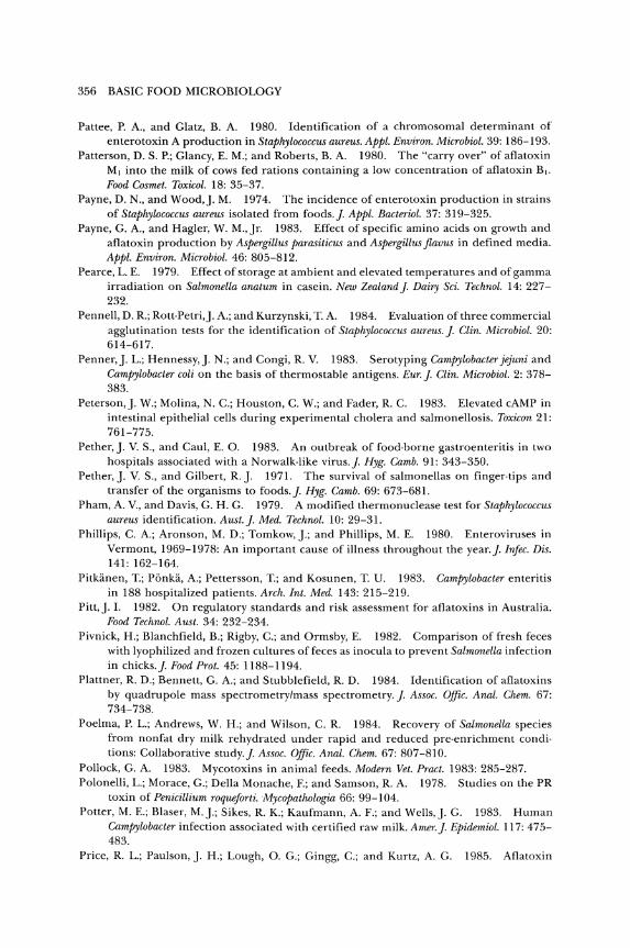
356 BASIC FOOD MICROBIOLOGY
Pattee, P. A., and Glatz, B. A. 1980. Identification of a chromosomal determinant of enterotoxin A production in Staphylococcus aureus. Appl. Environ. Microbiol. 39: 186-193.
Patterson, D. S. P.; Glancy, E. M.; and Roberts, B. A. 1980. The "carryover" of aflatoxin M, into the milk of cows fed rations containing a low concentration of aflatoxin B,. Food Cosmet. Toxieol. 18: 35-37.
Payne, D. N., and Wood,]. M. 1974. The incidence of enterotoxin production in strains of Staphylococcus aureus isolated from foods.]. Appl. Bacteriol. 37: 319-325.
Payne, G. A., and Hagler, W. M., Jr. 1983. Effect of specific amino acids on growth and aflatoxin production by Aspergillus parasiticus and Aspergillus flavus in defined media. Appl. Environ. Microbiol. 46: 805-812.
Pearce, L. E. 1979. Effect of storage at ambient and elevated temperatures and of gamma irradiation on Salmonella anatum in casein. New Zealand]. Dairy Sci. Technol. 14: 227-232.
Pennell, D. R.; Rott·Petri,]. A.; and Kurzynski, T. A. 1984. Evaluation of three commercial agglutination tests for the identification of Staphylococcus aureus.]. Clin. Microbiol. 20: 614-617.
Penner,]. L.; Hennessy,]. N.; and Congi, R. V. 1983. Serotyping Campylobacter jejuni and Campylobacter coli on the basis of thermostable antigens. Eur.]. Clin. Microbiol. 2: 378-383.
Peterson,]. W.; Molina, N. C.; Houston, C. W.; and Fader, R. C. 1983. Elevated cAMP in intestinal epithelial cells during experimental cholera and salmonellosis. Toxicon 21: 761-775.
Pether,]. V. S., and Caul, E. O. 1983. An outbreak of food·borne gastroenteritis in two hospitals associated with a Norwalk·like virus.]. Hyg. Camb. 91: 343-350.
Pether,]. V. S., and Gilbert, R.]. 1971. The survival of salmonellas on finger·tips and transfer of the organisms to foods.]. Hyg. Camb. 69: 673-681.
Pham, A. V., and Davis, G. H. G. 1979. A modified thermonuclease test for Staphylococcus aureus identification. Aust.]. Med. Technol. 10: 29-31.
Phillips, C. A.; Aronson, M. D.; Tomkow,].; and Phillips, M. E. 1980. Enteroviruses in Vermont, 1969-1978: An important cause of illness throughout the year.]. Intec. Dis. 141: 162-164.
Pitkanen, T.; Ponka, A.; Pettersson, T.; and Kosunen, T. U. 1983. Campylobacter enteritis in 188 hospitalized patients. Arch. Int. Med. 143: 215-219.
Pitt,]. 1. 1982. On regulatory standards and risk assessment for af1atoxins in Australia. Food Technol. Aust. 34: 232-234.
Pivnick, H.; Blanchfield, B.; Rigby, C.; and Ormsby, E. 1982. Comparison of fresh feces with lyophilized and frozen cultures of feces as inocula to prevent Salmonella infection in chicks.]. Food Prot. 45: 1188-1194.
Plattner, R. D.; Bennett, G. A.; and Stubblefield, R. D. 1984. Identification of aflatoxins by quadrupole mass spectrometry/mass spectrometry.]. Assoc. Offie. Anal. Chem. 67: 734-738.
Poelma, P. L.; Andrews, W. H.; and Wilson, C. R. 1984. Recovery of Salmonella species from nonfat dry milk rehydrated under rapid and reduced pre-enrichment conditions: Collaborative study.]. Assoc. OfJic. Anal. Chem. 67: 807-810.
Pollock, G. A. 1983. Mycotoxins in animal feeds. Modern Vet. Pract. 1983: 285-287. Polonelli, L.; Morace, G.; Della Monache, F.; and Samson, R. A. 1978. Studies on the PR
toxin of Penicillium roqueforti. Myeopathologia 66: 99-104. Potter, M. E.; Blaser, M.].; Sikes, R. K.; Kaufmann, A. F.; and Wells,]. G. 1983. Human
Campylobaeter infection associated with certified raw milk. Amer.]. ~pidemiol. 117: 475-483.
Price, R. L.; Paulson,]. H.; Lough, O. G.; Gingg, C.; and Kurtz, A. G. 1985. Aflatoxin
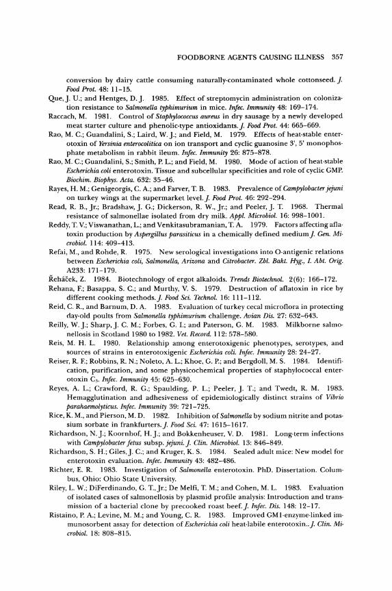
FOODBORNE AGENTS CAUSING ILLNESS 357
conversion by dairy cattle consuming naturally.contaminated whole cottonseed.]. Food Prot. 48: 11-15.
Que,]. U.; and Hentges, D.]. 1985. Effect of streptomycin administration on coloniza· tion resistance to Salmonella typhimurium in mice. Infec. Immunity 48: 169-174.
Raccach, M. 1981. Control of Staphylococcus aureus in dry sausage by a newly developed meat starter culture and phenolic·type antioxidants.]. Food Prot. 44: 665-669.
Rao, M. C.; Guandalini, S.; Laird, W.].; and Field, M. 1979. Effects of heat·stable enter· otoxin of Yersinia enterocolitica on ion transport and cyclic guanosine 3', 5' monophos· phate metabolism in rabbit ileum. Infec. Immunity 26: 875-878.
Rao, M. C.; Guandalini, S.; Smith, P. L.; and Field, M. 1980. Mode of action of heat·stable Escherichia coli enterotoxin. Tissue and subcellular specificities and role of cyclic GMP. Biochim. Biophys. Acta. 632: 35-46.
Rayes, H. M.; Genigeorgis, C. A.; and Farver, T. B. 1983. Prevalence of Gampylobacter jejuni on turkey wings at the supermarket level.]. Food Prot. 46: 292-294.
Read, R. B., Jr.; Bradshaw,]. G.; Dickerson, R. W., Jr.; and Peeler,]. T. 1968. Thermal resistance of salmonellae isolated from dry milk. Appl. Microbiol. 16: 998-1001.
Reddy, T. Y.; Viswanathan, L.; and Venkitasubramanian, T. A. 1979. Factors affecting afla· toxin production by Aspergillus parasiticus in a chemically defined medium]. Gen. Mi· crobiol. 114: 409-413.
Refai, M., and Rohde, R. 1975. New serological investigations into O·antigenic relations between Escherichia coli, Salmonella, Arizona and Gitrobacter. Zbl. Bakt. Hyg., l. Abt. Orig. A233: 171-179.
Rehacek, Z. 1984. Biotechnology of ergot alkaloids. Trends Biotechnol. 2 (6): 166-172. Rehana, F.; Basappa, S. C.; and Murthy, V. S. 1979. Destruction of aflatoxin in rice by
different cooking methods.]. Food Sci. Technol. 16: 111-112. Reid, C. R., and Barnum, D. A. 1983. Evaluation of turkey cecal microflora in protecting
clay·old poults from Salmonella typhimurium challenge. Avian Dis. 27: 632-643. Reilly, W.].; Sharp,]. C. M.; Forbes, G. L; and Paterson, G. M. 1983. Milkborne salmo·
nellosis in Scotland 1980 to 1982. Vet. Record. 112: 578-580. Reis, M. H. L. 1980. Relationship among enterotoxigenic phenotypes, serotypes, and
sources of strains in enterotoxigenic Escherichia coli. Infec. Immunity 28: 24-27. Reiser, R. F.; Robbins, R. N.; Noleto, A. L.; Khoe, G. P.; and Bergdoll, M. S. 1984. Identifi·
cation, purification, and some physicochemical properties of staphylococcal enter· otoxin Ca. Infec. Immunity 45: 625-630.
Reyes, A. L.; Crawford, R. G.; Spaulding, P. L.; Peeler, ]. T.; and Twedt, R. M. 1983. Hemagglutination and adhesiveness of epidemiologically distinct strains of Vibrio parahaemolyticus. Infec. Immunity 39: 721-725.
Rice, K. M., and Pierson, M. D. 1982. Inhibition of Salmonella by sodium nitrite and potas· sium sorbate in frankfurters.]. Food Sci. 47: 1615-1617.
Richardson, N.].; Koornhof, H.].; and Bokkenheuser, V. D. 1981. Long·term infections with Gampylobacter fetus subsp.jejuni.]. Glin. Microbiol. 13: 846-849.
Richardson, S. H.; Giles,]. C.; and Kruger, K. S. 1984. Sealed adult mice: New model for enterotoxin evaluation. Infec. Immunity 43: 482-486.
Richter, E. R. 1983. Investigation of Salmonella enterotoxin. PhD. Dissertation. Colum· bus, Ohio: Ohio State University.
Riley, L. W.; DiFerdinando, G. T.,Jr.; De Melfi, T. M.; and Cohen, M. L. 1983. Evaluation of isolated cases of salmonellosis by plasmid profile analysis: Introduction and trans· mission of a bacterial clone by precooked roast beef.]. Infec. Dis. 148: 12-17.
Ristaino, P. A.; Levine, M. M.; and Young, C. R. 1983. Improved GMl·enzyme·linked immunosorbent assay for detection of Escherichia coli heat-labile enterotoxin . .]. Glin. Microbiol. 18: 808-815.
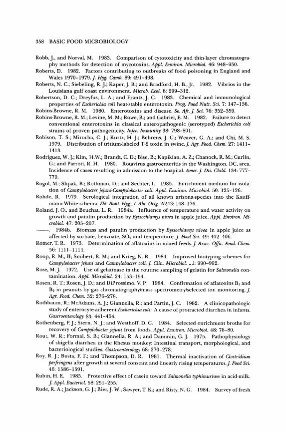
358 BASIC FOOD MICROBIOLOGY
Robb,]., and Norval, M. 1983. Comparison of cytotoxicity and thin· layer chromatogra· phy methods for detection of mycotoxins. Appl. Environ. Microbiol. 46: 948-950.
Roberts, D. 1982. Factors contributing to outbreaks of food poisoning in England and Wales 1970-1979.J. Hyg. Camb. 89: 491-498.
Roberts, N. C.; Siebeling, R.].; Kaper,]. B.; and Bradford, H. B.,jr. 1982. Vibrios in the Louisiana gulf coast environment. Microb. Ecol. 8: 299-312.
Robertson, D. C.; Dreyfus, L. A.; and Frantz,]. C. 1983. Chemical and immunological properties of Escherichia coli heat·stable enterotoxin. Prog. Food Nutr. Sci. 7: 147-156.
Robins·Browne, R. M. 1980. Enterotoxins and disease. So. Afr.J. Sci. 76: 352-359. Robins·Browne, R. M.; Levine, M. M.; Rowe, B.; and Gabriel, E. M. 1982. Failure to detect
conventional enterotoxins in classical enteropathogenic (serotyped) Escherichia coli strains of proven pathogenicity. Infec. Immunity 38: 798-801.
Robison, T. S.; Mirocha, C.].; Kurtz, H.].; Behrens,]. C.; Weaver, G. A.; and Chi, M. S. 1979. Distribution of tritium·labeled T·2 toxin in swine.]. Agr. Food. Chem. 27: 1411-1413.
Rodriguez, W.].; Kim, H.W.; Brandt, C. D.; Bise, B.; Kapikian, A. Z.; Chanock, R. M.; Curlin, G.; and Parrott, R. H. 1980. Rotavirus gastroenteritis in the Washington, DC, area. Incidence of cases resulting in admission to the hospital. Amer.]. Dis. Child. 134: 777-779.
Rogol, M.; Shpak, B.; Rothman, D.; and Sechter, 1. 1985. Enrichment medium for isola· tion of Campylobacter jejuni·Campylobacter coli. Appl. Environ. Microbiol. 50: 125-126.
Rohde, R. 1979. Serological integration of all known arizona·species into the Kauff· mann·White schema. Zbl. Bakt. Hyg., l. Abt. Orig. A243: 148-176.
Roland,]. 0., and Beuchat, L. R. 1984a. Influence of temperature and water activity on growth and patulin production by Byssochlamys nivea in apple juice. Appl. Environ. Mi· crobiol. 47: 205-207.
1984b. Biomass and patulin production by Byssochlamys nivea in apple juice as affected by sorbate, benzoate, S02 and temperature.]. Food Sci. 49: 402-406.
Romer, T. R. 1973. Determination of aflatoxins in mixed feeds.]. Assoc. Offic. Anal. Chem. 56: 1111-1114.
Roop, R. M., II; Smibert, R. M.; and Krieg, N. R. 1984. Improved biotyping schemes for Campylobacter jejuni and Campylobacter coli.]. Clin. Microbiol. _J: 990-992.
Rose, M.]. 1972. Use of gelatinase in the routine sampling of gelatin for Salmonella con· tamination. Appl. Microbiol. 24: 153-154.
Rosen, R. T.; Rosen,]. D.; and DiProssimo, V. P. 1984. Confirmation of aflatoxins BJ and B2 in peanuts by gas chromatography/mass spectrometry/selected ion monitoring.]. Agr. Food. Chem. 32: 276-278.
Rothbaum, R.; McAdams, A.].; Giannella, R.; and Partin,]. C. 1982. A clinicopathologic study of enterocyte·adherent nscherichia coli: A cause of protracted diarrhea in infants. Gastroenterology 83: 441-454.
Rothenberg, P.].; Stern, N.].; and Westhoff, D. C. 1984. Selected enrichment broths for recovery of Campylobacter jejuni from foods. Appl. Environ. Microbiol. 48: 78-80.
Rout, W. R.; Formal, S. B.; Giannella, R. A.; and Dammin, G.]. 1975. Pathophysiology of shigella diarrhea in the Rhesus monkey: Intestinal transport, morphological, and bacteriological studies. Gastroenterology 68: 270-278.
Roy, R.].; Busta, F. F.; and Thompson, D. R. 1981. Thermal inactivation of Clostridium perfringens after growth at several constant and linearly rising temperatures.]. Food Sci. 46: 1586-1591.
Rubin, H. E. 1985. Protective effect of casein toward Salmonella typhimurium in acid·milk. ]. Appl. Bacteriol. 58: 251-255.
Rude, R. A.;jackson, G.].; Bier,]. W.; Sawyer, T. K.; and Risty, N. G. 1984. Survey of fresh
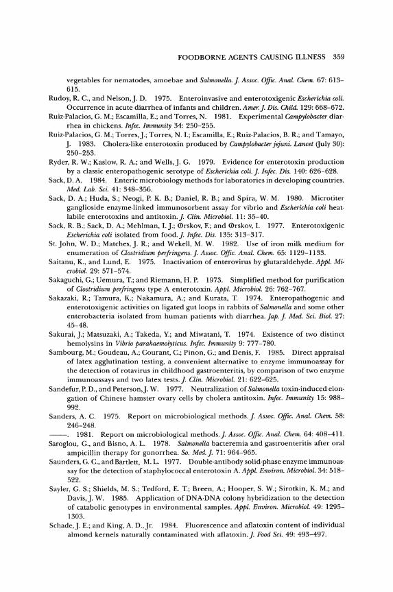
FOODBORNE AGENTS CAUSING ILLNESS 359
vegetables for nematodes, amoebae and Salmonella.]. Assoc. Olfic. Anal. Chem. 67: 613-615.
Rudoy, R C., and Nelson,]. D. 1975. Enteroinvasive and enterotoxigenic Escherichia coli. Occurrence in acute diarrhea of infants and children. Amer.]. Dis. Child. 129: 668-672.
Ruiz·Palacios, G. M.; Escamilla, E.; and Torres, N. 1981. Experimental Campylobacter diar· rhea in chickens. Infec. Immunity 34: 250-255.
Ruiz·Palacios, G. M.; Torres,].; Torres, N.I.; Escamilla, E.; Ruiz·Palacios, B. R; and Tamayo, ]. 1983. Cholera·like enterotoxin produced by Campylobacter jejuni. Lancet (July 30): 250-253.
Ryder, R W.; Kaslow, R A.; and Wells,]. G. 1979. Evidence for enterotoxin production by a classic enteropathogenic serotype of Escherichia coli.]. Irifec. Dis. 140: 626-628.
Sack, D. A. 1984. Enteric microbiology methods for laboratories in developing countries. Med. Lab. Sci. 41: 348-356.
Sack, D. A.; Huda, S.; Neogi, P. K. B.; Daniel, R B.; and Spira, W. M. 1980. Microtiter ganglioside enzyme·linked immunosorbent assay for vibrio and Escherichia coli heat· labile enterotoxins and antitoxin.]. Clin. Microbial. 11: 35-40.
Sack, R. B.; Sack, D. A.; Mehlman, I.].; <Drskov, F.; and <Drskov, I. 1977. Enterotoxigenic Escherichia coli isolated from food.]. Infec. Dis. 135: 313-317.
St.John, W. D.; Matches,]. R.; and Wekell, M. W. 1982. Use of iron milk medium for enumeration of Clostridium perfringens.]. Assoc. OfJic. Anal. Chem. 65: 1129-1133.
Saitanu, K., and Lund, E. 1975. Inactivation of enterovirus by glutaraldehyde. Appl. Mi· crobiol. 29: 571-574.
Sakaguchi, G.; Uemura, T.; and Riemann, H. P. 1973. Simplified method for purification of Clostridium perfringens type A enterotoxin. Appl. Microbial. 26: 762-767.
Sakazaki, R.; Tamura, K.; Nakamura, A.; and Kurata, T. 1974. Enteropathogenic and enterotoxigenic activities on ligated gut loops in rabbits of Salmonella and some other enterobacteria isolated from human patients with diarrhea. Jap.]. Med. Sci. Bioi. 27: 45-48.
Sakurai,].; Matsuzaki, A.; Takeda, Y.; and Miwatani, T. 1974. Existence of two distinct hemolysins in Vibrio parahaemolyticus. Infec. Immunity 9: 777-780.
Sambourg, M.; Goudeau, A.; Courant, C.; Pinon, G.; and Denis, F. 1985. Direct appraisal of latex agglutination testing, a convenient alternative to enzyme immunoassay for the detection of rotavirus in childhood gastroenteritis, by comparison of two enzyme immunoassays and two latex tests.]. Clin. Microbial. 21: 622-625.
Sandefur, P. D., and Peterson,]. W. 1977. Neutralization of Salmonella toxin· induced elon· gation of Chinese hamster ovary cells by cholera antitoxin. Infec. Immunity 15: 988-992.
Sanders, A. C. 1975. Report on microbiological methods.]. Assoc. Olfic. Anal. Chern. 58: 246-248.
1981. Report on microbiological methods.]. Assoc. Olfic. Anal. Chern. 64: 408-411. Saroglou, G., and Bisno, A. L. 1978. Salmonella bacteremia and gastroenteritis after oral
ampicillin therapy for gonorrhea. So. Med.]. 71: 964-965. Saunders, G. C., and Bartlett, M. L. 1977. Double·antibody solid· phase enzyme immunoas·
say for the detection of staphylococcal enterotoxin A. Appl. Environ. Microbial. 34: 518-522.
Sayler, G. S.; Shields, M. S.; Tedford, E. T.; Breen, A.; Hooper, S. W.; Sirotkin, K. M.; and Davis,]. W. 1985. Application of DNA-DNA colony hybridization to the detection of catabolic genotypes in environmental samples. Appl. Environ. Microbial. 49: 1295-1303.
Schade,]. E.; and King, A. D.,Jr. 1984. Fluorescence and aflatoxin content of individual almond kernels naturally contaminated with aflatoxin.]. Food Sci. 49: 493-497.
360 BASIC FOOD MICROBIOLOGY
Schaffner, C. P.; Mosbach, K.; Bibil, V. C.; and Watson, C. H. 1967. Coconut and Salmonella infection. Appl. Microbiol. 15: 471-475.
Scheuber, P. H.; Mossmann, H.; Beck, G.; and Hammer, D. K 1983. Direct skin test in highly sensitized guinea pigs for rapid and sensitive determination of staphylococcal enterotoxin B. Appl. Environ. Microbiol. 46: 1351-1356.
Schiemann, D. A. 1981. An enterotoxin· negative strain of Yersinia enterocolitica serotype 0:3 is capable of producing diarrhea in mice. Infec. Immunity 32: 571-574.
Schneitz, C.; Seuna, E.; and Rizzo, A. 1981. The anaerobically cultured cecal flora of adult fowls that protects chickens from Salmonella infections. Acta Pathol. Microbiol. Scand. Sect. B 89: 109-116.
Schroeder, S. A. 1967. What the sanitarian should know about salmonellae and staphylo· cocci in milk and milk products.]. Milk Food Technol. 30: 376-380.
Schuller, P. L.; Horwitz, W.; and Stoloff, L. 1976. A review of sampling plans and collab· oratively studied methods for analysis of aflatoxins.]. Assoc. Offic. Anal. Chem. 59: 1315-1343.
Scott, P. M. 1981. Toxins of Penicillium species used in cheese manufacture.]. Food Prot. 44: 702-710.
Scott, P. M., and Kennedy, B.P.C. 1976. Analysis of blue cheese for roque fortine and other alkaloids from Penicillium roque forti. ]. Agr. Food. Chem. 24: 865-868.
Scott, R. E.; Jones, A.; and Gaucher, G. M. 1984. A manganese requirement for patulin biosynthesis by culture of Penicillium urticae. Biotechnol. Letters 6: 231-236.
Scott, W. T., and Bullerman, L. B. 1975. Patulin: A mycotoxin of potential concern in foods.]. Milk Food Technol. 38: 695-705.
Sedlock, D. M., and Deibel, R. H. 1978. Detection of Salmonella enterotoxin using rabbit ileal loops. Can.]. Microbiol. 24: 268-273.
Segner, W. P.; Schmidt, C. F.; and Boltz,]. K. 1971. Minimal growth temperature, sodium chloride tolerance, pH sensitivity, and toxin production of marine and terrestrial strains of Clostridium botulinum type C. Appl. Microbiol. 22: 1025-1029.
Sellin, L. C. 1981. The action of botulinum toxin at the neuromuscular junction. Med. BioI. 59: 11-20.
Sellin, L. C.; Thesleff, S.; and Dasgupta, B. R. 1983. Different effects of types A and B botulinum toxin on transmitter release at the rat neuromuscular junction. Acta Physiol. Scand. 119: 127-133.
Shafer, W. M., and Iandolo,].]. 1978a. Chromosomal locus for staphylococcal enter· otoxin B. Infec. Immunity 20: 273-278.
1978b. Staphylococcal enterotoxin A: A chromosomal gene product. Appl. En· viron. Microbiol. 36: 389-391.
1979. Genetics of staphylococcal enterotoxin B in methicillin·resistant isolates of Staphylococcus aUreus. Infec. Immunity 25: 902-911.
Shah, D. B.; Kauffman, P. E.; Boutin, B. K.; and Johnson, C. H. 1982. Detection of heat· labile·enterotoxin·producing colonies of Escherichia coli and Vibrio cholerae by solid· phase sandwich radioimmunoassays.]. Clin. Microbiol. 16: 504-508.
Shandera, W. X.; HatKin, B.; Martin, D. L.; Taylor,]. P.; Maserang, D. L.; Wells,]. G.; Kelly, M.; Ghandi, K; Kaper,]. B.; Lee,]. V.; and Blake, P. A. 1983. Persistence of cholera in the United States. Amer.]. Trop. Med. Hyg. 32: 812-817.
Shank, R. C.; Bourgeois, C. H.; Keschamras, N.; and Chandavimol, P. 1971. Aflatoxins in autopsy specimens from Thai children with an acute disease of unknown aetiology. Food Cosmet. Toxicol. 9: 501-507.
Shanker, S.; Rosenfield,]. A.; Davey, G. R.; and Sorrell, T. C. 1982. Campylobacter jejuni: Incidence in processed broilers and biotype distribution in human and broiler iso· lates. Appl. Environ. Microbiol. 43: 1219-1220.

FOOD BORNE AGENTS CAUSING ILLNESS 361
Shannon, G. M.; Shotwell, O. L.; and Kwolek, W. F. 1983. Extraction and thin layer chro· matography of aflatoxin B\ in mixed feeds.J. Assoc. OfflC. Anal. Chern. 66: 582-586.
Sharma, V. D.; Singh,l. P.; and Singh, S. P. 1983. Lethal and enterotoxic effects of Salmo· neUa saintpaul toxin. Ind.]. Exp. Biol. 21: 639-640.
Shayegani, M.; Morse, D.; DeForge, I.; Root, T.; Parsons, L. M.; and Maupin, P. S. 1983. Microbiology of a major foodborne outbreak of gastroenteritis caused by Yersinia enter·
ocolitica serogroup 0:8J. Clin. Microbiol. 17: 35-40. Sherman, D. M 1984. Genetic engineering-3: monoclonal antibodies for veterinary
medicine. Vet. Med. 10: 959-962, 964. Shinoda, S.; Nakahara, N.; Ninomiya, Y.; Itoh, K.; and Kane, H. 1983. Serological method
for identification of Vibrio parahaemolyticus from marine samples. Appl. Environ. Micro· bioi. 45: 148-152.
Shotwell, O. L., and Hesseltine, C. W. 1981. Use of bright greenish yellow fluorescence as a presumptive test for aflatoxin in corn. Cereal Chem. 58: 124-127.
1983. Five·year study of my cot ox ins in Virginia wheat and dent corn.]. Assoc. Offic. Anal. Chern. 66: 1466-1469.
Skirrow, M. B. 1977. CamfrYlobacter enteritis: A 'new' disease. Br. Med.]. 2: 9-12. Skjelkvale, R., and Duncan, C. L. 1975. Enterotoxin formation by different toxigenic
types of Clostridium perfringens. Infec. Immunity 11: 563-575. Skjelkvale, R., and Uemura, T. 1977. Detection of enterotoxin in faeces and anti·enter·
otoxin in serum after Clostridium perfringens food·poisoning.]. Appl. Bacteriol. 42: 355-363.
Smelt,]. P. P. M., and Haas, H. 1978. Behaviour of proteolytic Clostridium botulinum types A and B near the lower temperature limits of growth. Eur.]. Appl. Microbiol. 5: 143-154.
Smelt,]. P. P. M.; Raa1Jes, G.]. M.; Crowther,]. S.; and Verrips, C. T. 1982. Growth and toxin formation by Clostridium botulinum at low pH values.]. Appl. Bacteriol. 52: 75-82.
Smith, G. S., and Blaser, M.]. 1985. Fatalities associated with Campylobacter jejuni infec· tions.]. Amer. Med. Assoc. 253: 2873-2875.
Smith, H. 1977. Microbial surfaces in relation to pathogenicity. Bacteriol. Rev. 41: 475-500.
1984. The biochemical challenge of microbial pathogenicity.]. Appl. Bacteriol. 47: 395-404.
Smith,]. L.; Buchanan, R. L.; and Palumbo, S. A. 1983. Effect of food environment on staphylococcal enterotoxin synthesis: A review.]. Food Prot. 46: 545-555.
Smith,]. L.; Huhtanen, C. N.; Kissinger,]. C.; and Palumbo, S. A. 1975. Survival of sal· monellae during pepperoni manufacture. Appl. Microbiol. 30: 759-763.
Smith,]. L.; Palumbo, S. A.; Kissinger,]. C.; and Huhtanen, C. N. 1975. Survival of Salmo· nella dublin and Salmonella typhimurium in Lebanon bologna.]. Milk Food Technol. 38: 150-154.
Smith, L. D.; Davis,]. W.; and Libke, K. G. 1971. Experimentally induced botulism in weanling pigs. Amer.]. Vet. Res. 32: 1327-1330.
Smith, M. R. 1971. Vibrio parahemolyticus. Clin. Med. 78: 22-25. Smith, P. B. 1972. "Bacteriophage Typing of Staphylococcus aureus." In The Staphylococci.
]. O. Cohen, ed. New York: Wiley·Interscience. Smith, T. F. 1985. Epidemic viral gastroenteritis: Detection of rota virus by enzyme immu·
noassay. Mayo Clin. Proc. 60: 204-205. Snyder,]. D.; Wells,]. G.; Yashuk,].; Puhr, N.; and Blake, P. A. 1984. Outbreak of invasive
Escherichia coli gastroenteritis on a cruise ship. Amer.]. Trop. Med. Hyg. 33: 281-284. Soerjadi·Liem, A. S.; Snoeyenbos, G. H.; and Weinack, O. M. 1984. Comparative studies
on competitive exclusion of three isolates of campylobacter fetus subsp. jejuni in chickens by native gut microflora. Avian Dis. 28: 139-146.
362 BASIC FOOD MICROBIOLOGY
Solomon, H. M.; Kautter, D. A.; and Lynt, R. K. 1982. Effect of low temperatures on growth of nonproteolytic Clostridium botulinum types Band F and proteolytic type G in crabmeat and broth.]. Food Prot. 45: 516-518.
Solomon, H. M.; Lynt, R. K.,jr.; Kautter, D. A.; and Lilly, T.,jr. 1969. Serological studies of Clostridium botulinum type E and related organisms. II. Serology of spores.]. Bacteriol.
98: 407-414. Somerville, P. C.; Nhlapo, W. M.; and Alberts, M. 1983. Differentiation of Salmonella typhi
using resistotyping.]. Appl. Bacteriol. 54: 171-176. Son, N. T., and Fleet, G. H. 1980. Behavior of pathogenic bacteria in the oyster, Crassostrea
commercialis, during depuration, re·laying, and storage. Appl. Environ. Microbiol. 40: 994-1002.
Sonnabend, 0.; Sonnabend, W.; Heinzle, R.; Sigrist, T.; Dirnhofer, R.; and Krech, U. 1981. Isolation of Clostridium botulinum type G and identification of type G botulinal toxin in humans: Report of five sudden unexpected deaths.]. Infec. Dis. 143: 22-27.
Sorenson, W.G.; Tucker,]. D.; and Simpson,]. P. 1984. Mutagenicity ofthe tetramic myco· toxin cydopiazonic acid. Appl. Environ. Microbiol. 47: 1355-1357.
Speelman, P.; Kabir, 1.; and Islam, M. 1984. Distribution and spread of colonic lesions in shigellosis: a colonoscopic study.]. Infec. Dis. 150: 899-903.
Speirs,]. 1.; Stavrk S.; and Konowalchuck,]. 1977. Assay of Escherichia coli heat·labile enterotoxin with Vero cells. Infec. Immunity 16: 617-622.
Spira, W. M.; Sack, R. B.; and Froehlich,]. L. 1981. Simple adult rabbit model for Vibrio cholerae and enterotoxigenic Escherichia coli diarrhea. Irifec. Immunity 32: 739-747.
Stadhouders,].; Hassing, F.; and Galesloot, T. E. 1980. A rapid and simple method for the detection of Staphylococcus aureus thermo nuclease in cheese. Neth. Milk Dairy]. 34:
199-204. Stamper, W.]., and Banwart, G.]. 1974. Effect of various peptones in the growth medium
on the agglutination of ten Salmonella species with pooled Spicer·Edwards antisera.]. Food Sci. 39: 80-82.
Stark, A. 1980. Mutagenicity and carcinogenicity of mycotoxins: DNA binding as a possi· ble mode of action. Ann. Rev. Microbiol. 34: 235-262.
Stark, A. A., and Demain, A. L. 1980. Genetic activity and hazards of mycotoxins. ASM News 46: 80-83.
Stelma, G. N.,jr.; Wimsatt,]. C.; Kauffman, P. E.; and Shah, D. B. 1983. Radioimmunoas· say for Clostridium perfringens enterotoxin and its use in screening isolates implicated in food-poisoning outbreaks.]. Food Prot. 46: 1069-1073.
Stern, N.]. 1982. Methods for recovery of Campylobacter jejuni from foods.]. Food Prot. 45: 1332-1337.
Stern, N.].; Hernandez, M. P.; Blankenship, L.; Deibel, K. E.; Doores, S.; Doyle, M. P.; Ng, H.; Pierson, M. D.; Sofos,]. N.; Sveum, W. H.; and Westhoff, D. C. 1985. Prevalence and distribution of Campylobacter jejuni and Campylobacter coli in retail meats.]. Food Prot. 48: 595-599.
Stern, N.].; Pierson, M. D.; and Kotula, A. W. 1980. Effects of pH and sodium chloride on Yersinia enterocolitica growth at room and refrigeration temperatures.]. Food Sci. 45: 64-67.
Stier, R. F.; Bell, L.; Ito, K. A.; Shafer, B.D.; Brown, L. A.; Seeger, M. L.; Allen, B. H.; Porcuna, M. N.; and Lerke, P. A. 1981. Effect of modified atmosphere storage on C. botulinum toxigenesis and the spoilage microflora of salmon fillets.]. Food Sci. 46: 1639-1642.
Stiffler·Rosenberg, G., and Fey, H. 1978. Simple assay for staphylococcal enterotoxins A, B, and C: Modification of enzyme· linked immunosorbent assay.]. Clin. Microbiol. 8: 473-479.
Stinson, E. E.; Osman, S. F.; and Bills, D. D. 1979. Water·soluble products from patulin during alcoholic fermentation of apple juice.]. Food Sci. 44: 788-789, 796.
FOODBORNE AGENTS CAUSING ILLNESS 363
Stora, C.; Dvorackova, I.; and Ayraud, N. 1983. Aflatoxin and Reye's syndrome.]. Med. 14: 47-54.
Stringer, M. F.; Turnbull, P. C. B.; and Gilbert, R.]. 1980. Application of serological typing to the investigation of outbreaks of Clostridium perfringens food poisoning, 1970-1978.]. Hyg. Camb. 84: 443-456.
Strong, D. H.; Duncan, C. L.; and Perna, G. 1971. Clostridium perfringens type A food poi· soning. II. Response of the rabbit ileum as an indication of enteropathogenicity of strains of Clostridium perfringens in human beings. Infec. Immunity 3: 171-178.
Stryker, W. S.; Fraser, D. W.; and Facklam, R R 1982. Foodborne outbreak of group G streptococcal pharyngitis. Amer.]. Epidemiol. 116: 533-540.
Stubblefield, R. D.; Pier, A. C.; Richard,]. L.; and Shotwell, O. L. 1983. Fate ofaflatoxins in tissues, fluids, and excrements from cows dosed orally with aflatoxin B1• Amer.]. Vet. Res. 44: 1750-1752.
Sugii, S.; Ohishi, I.; and Sakaguchi, G. 1977. Intestinal absorption of botulinum toxins of different molecular sizes in rats. Infec. Immunity 17: 491-496.
Sugiyama, H. 1980. Clostridium botulinum neurotoxin. Microbiol. Rev. 44: 419-448. Sugiyama, H., and Yang, K. H. 1975. Growth potential of Clostridium botulinum in fresh
mushrooms packaged in semipermeable plastic film. Appl. Microbiol. 30: 964-969. Sullivan, R.; Marnell, R. M.; Larkin, E. P.; and Read, R B.,jr. 1975. Inactivation of polio·
virus I and coxsackievirus B·2 in broiled hamburgers.]. Milk Food Technol. 38: 473-475.
Sullivan, R.; Peeler,]. T.; Tierney,]. T.; and Larkin, E. P. 1984. Evaluation of a method for recovering poliovirus I from 100·gram oyster samples.]. Food Prot. 47: 108-110.
Sutherland,]. P., and Varnam, A. H. 1977. Methods of isolation and potential importance of Yersinia enterocolitica in foods stored at low temperatuers.]. Appl. Bacteriol.43 (3): xiii-xiv.
Svedhem, A., and Kaijser, B. 1980. Campylobacter fetus subspeciesjejuni: A common cause of diarrhea in Sweden.]. Infec. Dis. 142: 353-359.
1981. Isolation of Campylobacter jejuni from domestic animals and pets: Probable origin of human infection.]. Infec. 3: 37-40.
Svedhem, A; Kaijser, B.; and Sjogren, E. 1981. The occurrence of Campylobacter jejuni in fresh food and survival under different conditions.]. Hyg. Camb. 87: 421-425.
Svennerholm, A., and Wiklund, G. 1983. Rapid GMI·enzyme·linked immunosorbent assay with visual reading for identification of Escherichia coli heat· labile enterotoxin. ]. Clin. Microbiol. 17: 596-600.
Swaminathan, B.; Aleixo,]. A. G.; and Minnich, S. A. 1985. Enzyme immunoassays for Salmonella: One·day testing is now a reality. Food Technol. 39(3): 83-89.
Swanson, S. P.; Corley, R A.; White, D. G.; and Buck, W. B. 1984. Rapid thin layer chro· matographic method for determination of zearalenone and zearalenol in grains and animal feeds.]. Assoc. Offic. Anal. Chem. 67: 580-582.
Tacket, C. 0.; Dominguez, L. B.; Fisher, H.].; and Cohen, M. L. 1985a. An outbreak of multiple·drug·resistant Salmonella enteritis from raw milk.]. Amer. Med. Assoc. 252: 2058-2060.
Tacket, C. 0.; Hickman, F.; Pierce, G. V.; and Mendoza, L. F. 1982. Diarrhea associated with Vibrio jluvialis in the United States.]. Clin. Microbiol. 16: 991-992.
Tacket, C. 0.; Narain,]. P.; Sattin, R; Lofgren,]. P.; Konigsberg, C.; Rendtorff, R C.; Rausa, A.; Davis, B. R; and Cohen, M. L. 1984. A multi state outbreak of infections caused by Yersinia enterocolitica transmitted by pasteurized milk.]. Amer. Med. Assoc. 251: 483-486.
Tackett, C. 0., et al. 1985b. An outbreak of Yersinia enterocolitica infections caused by con· taminated tofu (soybean curd). Amer.]. Epidemiol. 121: 705-711.
Tager, M. 1974. Current views on the mechanisms of coagulase action in blood clotting. Ann. N. Y. Acad. Sci. 236: 277-291.
364 BASIC FOOD MICROBIOLOGY
Takahashi, H.; Yasaki, H.; Nanayama, U.; Manabe, M.; and Matsuura, S. 1984. Distribu· tion of sterigmatocystin and fungal mycelium in individual brown rice kernels natu· rally infected by Aspergillus versicolor. Cereal Chem. 61: 48-52.
Takeda, N. 1984. Determination of aflatoxin M, in milk by reversed· phase high· performance liquid chromatography.]. Chromatogr. 288: 484-488.
Takeda, T.; Kitamura, M.; Takeda, Y.; and Miwatani, T. 1983. In vitro transfer of Ent plasmid from Escherichia coli to Shigella dysenteriae and Salmonella. FEMS Microbiol. Letters 18: 253-256.
Takeda, T.; Takeda, Y.; Miwatani, T.; Gregory, P.; Morita, T.; and Matsushiro, A. 1981. Genetic labeling of an Ent plasmid that encodes heat·stable enterotoxin of enterotoxigenic Escherichia coli isolated from patients. Biken.]. 24: 127-135.
Takeda, Y.; Honda, T.; Taga, S.; and Miwatani, T. 1981. In vitro formation of hybrid tox· ins between subunits of Escherichia coli heat·labile enterotoxin and those of cholera enterotoxin. Infec. Immunity 34: 341-346.
Takeda, Y.; Isohata, E.; Amano, R.; ;and Uchiyama, M. 1979. Simultaneous extraction and fractionation and thin layer chromatographic determination of 14 mycotoxins in grains.]. Assoc. OIfIC. Anal. Chem. 62: 573-578.
Takeda, Y.; Okamoto, K.; and Miwatani, T. 1979. Mitomycin C stimulates production of a toxin in Shigella species that causes morphological changes in Chinese hamster ovary cells. Infec. Immunity 23: 178-180.
Tamminga, S. K.; Beumer, R. R.; and Kampelmacher, E. H. 1978. The hygienic quality of vegetables grown in or imported into the Netherlands: a tentative survey.]. Hyg. Camb. 80: 143-154.
Tamminga, S. K.; Beumer, R. R.; Kampelmacher, E. H.; and van Leudsen, F. M. 1976. Survival of Salmonella eastbourne and Salmonella typhimurium in chocolate.]. Hyg. Camb. 76: 41-47.
1977. Survival of Salmonella eastbourne and Salmonella typhimurium in milk chocolate prepared with artificially contaminated milk powder.]. Hyg. Camb. 79: 333-337.
Tamminga, S. K.; Beumer, R. R.; Keijbets, M.]. H.; and Kampelmacher, E. H. 1978. Micro· bial spoilage and development of food poisoning bacteria in peeled, completely or partly cooked vacuum packed potatoes. Arch. Lebensmittelhyg. 29: 215-219.
Tanaka, N. 1982. Toxin production by Clostridium botulinum in media at pH lower than 4.6.J. Food Prot. 45: 234-237.
Tarter, E.].; Hanchay,].; and Scott, P. M. 1984. Improved liquid chromatographic method for determination of aflatoxins in peanut butter and other commodities.]. Assoc. Offic. Anal. Chem. 67: 597-600.
Tatini, S. R. 1976. Thermal stability of enterotoxins in food.]. Milk Food Technol. 39: 432-438.
Tatini, S. R.; Cords, B. R.; and Gramoli,]. 1976. Screening for staphylococcal enterotox· ins in food. Food Technol. 30(4): 64, 66, 70, 72-74.
Tatini, S. R.; Soo, H. M.; Cords, B. R.; and Bennett, R. W. 1975. Heat·stable nuclease for assessment of staphylococcal growth and likely presence of enterotoxins in foods.]. Food Sci. 40: 352-356.
Taylor, R.].; and Burrows, M. R. 1971. The survival of Escherichia coli and Salmonella dublin in slurry on pasture and the infectivity of S. dublin for grazing calves. Brit. Vet.]. 127: 536-543.
Taylor, W. R.; Schell,W. L.; Wells,]. G.; Choi, K.; Kinnunen, D. E.; Heiser, P. T.; and Helstad, A. G. 1982. A foodborne outbreak of enterotoxigenic Escherichia coli diarrhea. N. Engl.]. Med. 306: 1093-1095.
Teich, A. H., and Hamilton,]. R. 1985. Effect of cultural practices, soil phosphorus, po· tassium, and pH on the incidence of fusarium head blight and deoxynivalenol levels in wheat. Appl. Environ. Microbiol. 49: 1429-1431.
FOODBORNE AGENTS CAUSING ILLNESS 365
Telford, A. P. 1982. Control of aflatoxins in peanuts by segregation and sorting. Food Technol. Aust. 34: 214-215.
Tenover, F. C.; Knapp, J. S.; Patton, C.; and Plorde, J. J. 1985. Use of auxotyping for epidemiological studies of Campylobacter jejuni and Campylobacter coli infections. Infec. Immunity 48: 384-388.
Tenover, F. C.; Williams, S.; Gordon, K. P.; Harris, N.; Nolan, C.; and Plorde, J. J. 1984. Utility of plasmid fingerprinting for epidemiological studies of Campylobacter jejuni infections.]. Infec. Dis. 149: 279.
Terao, K. 1983. Sterigmatocystin-A masked potent carcinogenic mycotoxin. ]. Toxicol.· Toxin Rev. 2: 77-110.
Tesone, S.; Hughes, A.; and Hurst, A. 1981. Salt extends the upper temperature limit for growth of food·poisoning bacteria. Can.]. Microbiol. 27: 970-972.
Thelestam, M., and Florin, I. 1984. Cytopathogenic action of Clostridium difficile toxins.]. Toxicol.-Toxin Rev. 3: 139-180.
Thomas, M. E. M., and Mogford, H. E. 1970. Salmonellosis in general practice. Observa· tions of cases and their households in Enfield.]. Hyg. Camb. 68: 663-671.
Thompson, D. G., and Malagelada,J. R. 1982. Vomiting and the small intestine. Dig. Dis. Sci. 27: 1121-1125.
Thompson, M. R.; Brandwein, H.; Labine·Racke, M.; and Giannella, R. A. 1984. Simple and reliable enzyme-linked immunosorbent assay with monoclonal antibodies for detection of Escherichia coli heat-stable enterotoxins.]. Clin. Microbiol. 20: 59-64.
Thompson, N.E.; Ketterhagen, M.J.; and Bergdoll, M. S. 1984. Monoclonal antibodies to staphylococcal enterotoxins Band C: Cross-reactivity and localization of epitopes on tryptic fragments. Infec. Immunity 45: 281-285.
Thompson, W.L., and Wannemacher, R. W.,Jr. 1984. Detection and quantitation ofT-2 mycotoxin with a simplified protein synthesis inhibition assay. Appl. Environ. Microbiol. 48: 1176-1180.
Thorne, G. M., and Gorbach, S. L. 1978. Enterotoxigenic Escherichia coli: Detection and importance in diarrheal disease of children.]. Amer. Vet. Med. Assoc. 173: 592-595.
Thornley, M.J. 1963. "Microbiological Aspects of the Use of Radiation for the Elimination of Salmonellae from Foods and Feeding Stuffs." In Radiation Control of Salmonellae in Food and Feed Products. Tech. Report Series No. 22. Vienna, Austria: International Atomic Energy Agency.
Thouvenot, D., and Morfin, R. F. 1983. Radioimmunoassay for zearalenone and zearalanol in human serum: Production, properties, and use of porcine antibodies. Appl_ Environ. Microbiol. 45: 16-23.
Tice, G., and Buchanan, R. L. 1981. Regulation of aflatoxin biosynthesis: Effect of ex· ogenously supplied cyclic nucleotides.]. Food Sci. 47: 153-157.
Tierney, J. T.; Sullivan, R.; Larkin, E. P.; and Peeler, J. T. 1973. Comparison of methods for the recovery of virus inoculated into ground beef. Appl. Microbiol. 26: 497-501.
To, E. C., and Robach, M. C. 1980. Potassium sorbate dip as a method of extending shelf life and inhibiting the growth of Salmonella and Staphylococcus aureus on fresh, whole broilers. Poultry Sci. 59: 726-730.
Todd, E. C. D. 1983a. Foodborne disease in Cahada-A 5-year summary.]. Food Prot. 46: 650-657.
1983b. Factors that contributed to foodborne disease in Canada, 1973-1977.]. Food Prot. 46: 737-747_
1985. Economic loss from foodborne disease outbreaks associated with foodservice establishments.]. Food Prot. 48: 169-180.
Toledo, M. R. F.; Alvariza, D. C. B.; Murahovschi,J.; Ramos, S. R. T. S.; and Trabulsi, L. R. 1983. Enteropathogenic Escherichia coli serotypes and endemic diarrhea in infants. Infec. Immunity 39: 586-589.
366 BASIC FOOD MICROBIOLOGY
Tompkin, R. B., and Stozek, S. K. 1974. Direct addition of liquid propylene oxide to dried materials for destruction of salmonellae. Appl. Microbiol. 27: 276-277.
Tongpim, S.; Beumer, R. R.; Tamminga, S. K.; and Kampelmacher, E. H. 1984. Comparison of modi fed Rappaport's medium (RV) and Muller-Kauffmann medium (MK-ISO) for the detection of Salmonella in meat products. Int.]. Food Microbiol. 1: 33-42.
Townsend, C.T.; Yee, L.; and Mercer, W. A. 1954. Inhibition of the growth of Clostridium botulinum by acidification. Food Res. 19: 536-542.
Trenholm, H. L.; Hamilton, R. M. G.; Friend, D. W.; Thompson, B. K.; and Hartin, K. E. 1984. Feeding trials with vomitoxin (deoxynivalenol)-contaminated wheat: Effects on swine, poultry, and dairy cattle.]. A mer. Vet. Med. Assoc. 185: 527-531.
Troller,]. A. 1976. Staphylococcal growth and enterotoxin production-factors for contro!.]. Milk Food Technol. 39: 499-503.
Trucksess, M. W.; Nesheim, S.; and Eppley, R. M. 1984. Thin layer chromatographic determination of deoxynivalenol in wheat and corn-J. Assoc. Offic. Anal. Chern. 67: 40-43.
Trucksess, M. W.; Stoloff, L.; Young, K.; Wyatt, R. D.; and Miller, B. L. 1983. Aflatoxicol and aflatoxins B J and M J in eggs and tissues of laying hens consuming aflatoxincontaminated feed. Poultry Sci. 62: 2176-2182.
Trust, T.]., and Bartlett, K. H. 1979. Aquarium pets as a source of antibiotic·resistant salmonellae. Can.]. Microbiol. 25: 535-541.
Tsukamoto, T.; Kinoshita, Y.; Taga, S.; Takeda, Y.; and Miwatani, T. 1980. Value of passive immune hemolysis for detection of heat-labile enterotoxin produced by enterotoxigenic Escherichia coli.]. Clin. Microbiol. 12: 768-771.
Tulloch, E. F.,Jr.; Ryan, K.].; Formal, S. B.; and Franklin, F. A. 1973. Invasive enteropathic Escherichia coli dysentery. An outbreak in 28 adults. Ann. Int. Med. 79: 13-17.
Turnbull, P. C. B. 1981. Bacillus cereus toxins. Pharmac. Ther. 13: 453-505. Turnbull, P. C. B., and Rose, P. 1982. Campylobacter jejuni and salmonella in raw red meats.
]. Hyg. Camb. 88: 29-37. Twedt, R. M; Peeler,]. T.; and Spaulding, P. L. 1980. Effective ileal loop dose of Kanaga
wa-positive Vibrio parahaemolyticus. Appl. Environ. Microbiol. 40: 1012-1016. Twedt, R. M.; Spaulding, P. L.; and Hall, H. E. 1969. Morphological, cultural, biochemical,
and serological comparison of Japanese strains of Vibrio parahaemolyticus with related cultures isolated in the United States.]. Bacteriol. 98: 511-518.
Tweten, R. K., and Iandolo,].]. 1983. Transport and processing of staphylococcal enterotoxin B.]. Bacteriol. 153: 297-303.
Uemura, T.; Genigeorgis,C.; Riemann, H. P.; and Franti, C. E. 1974. Antibody against Clostridium perfringens type A enterotoxin in human sera. Infec. Immunity 9: 470-471.
Uemura, T., and Skjelkvale, R. 1976. An enterotoxin produced by Clostridium perfringens type D. Purification by affinity chromatography. Acta Path. Microbiol. Scand. Sect. B 84: 414-420.
Ulshen, M. H., and Rollo, ]. L. 1980. Pathogenesis of Escherichia coli gastroenteritis in man-Another mechanism. N. Engl.]. Med. 302: 99-101.
Underdal, B., and Rossebo, L. 1972. Inactivation of strains of Salmonella senftenberg by gamma irradiation.]. Appl. Bacteriol. 35: 371-377.
USDA. 1971. Recommended Procedure for the Isolation of Salmonella Organisms from Animal Feeds and Feed Ingredients. ARS 91-68-1. Washington, D.C.: U.S. Department of Agriculture.
1978. The Official United States Standards for Grains. U.S. Department of Agriculture, Federal Grain Inspection Service, Inspection Division, Washington, D.C.
1983. Production requirements for cooked beef, roast beef and cooked corned beef. Fed. Register 48(106): 24314-24318.
VanCauwenberge,]. E.; Bothast, R.].; and Kwolek, W. F. 1981. Thermal inactivation of eight Salmonella serotypes on dry corn flour. Appl. Environ. Microbial. 42: 688-691.
FOODBORNE AGENTS CAUSING ILLNESS 367
Van den Broek, M.]. M., and Mossel, D. A. A. 1977. Sublethal cold shock in Vibrio para· haernolyticus. Appl. Environ. Microbiol. 34: 97-98.
Vanderzant, C., and Nickelson, R 1972. Survival of Vibrio parahaemolyticus in shrimp tis· sue under various environmental conditions. Appl. Microbiol. 23: 34-37.
van Dijk, H.].; O'Dell, G. D.; and Bodine, A. B. 1984. Effects of aflatoxin MJ intake at physiologic levels on newborn dairy calves. Amer.]. Vet. Res. 45: 1994-1997.
Van Egmond, H. P.; Paulsch, W. E.; Deijll, E.; and Schuller, P. L. 1980. Thin layer chromat· ographic method for analysis and chemical confirmation of sterigmatocystin in cheese.]. Assoc. Offic. Anal. Chem. 63: 110-114.
Van Egmond, H. P., and Stubblefield, R D. 1981. Improved method for confirmation of identity of aflatoxins BJ and Ml in dairy products and animal tissue extracts.]. Assoc. OjJic. Anal. Chem. 64: 152-155.
van Hell, H., and Helmich,]. 1984. The evolution of pregnancy testing. Applications of monoclonal technology. Amer. Clin. Prod. Rev. 3(3): 38-45.
van Miert, A. S.]. P. A. M.; van Duin, C. T. M.; Verheijden,]. H. M.; and Schotman, A.]. H. 1983. Staphylococcal enterotoxin B and Escherichia coli endotoxin; Comparative ob· servations in goats on fever and associated clinical hematologic and blood biochemical changes after intravenous and intramammary administratIOn. Amer.]. Vet. Res. 44: 955-963.
Vassiliadis, P.; Kalapothaki, Y.; Mavrommati, c.; and Trichopoulos, D. 1984. A compari· son of the original Rappaport medium (R medium) and the Rappaport·Vassiliadis medium (RV medium) in the isolation of salmonellae from meat products.]. Hyg. Camb. 93: 51-58.
Velasco,]. 1972. Detection of aflatoxin using small columns of florisil.]. Amer. Oil. Chern. Soc. 49: 141-142.
Velaudapillai,T.; Niles, G. R; and Nagaratnam, W. 1969. Salmonellas, shigellas, and en· teropathogenic Escherichia coli in uncooked food.]. Hyg. Camb. 67: 187-191.
Vesonder, R F., and Horn, B. W. 1985. Sterigmatocystin in dairy cattle feed contami· nated with Aspergillus versicolor. Appl. Environ. Microbiol. 49: 234-235.
Vlajinac, H., and Krajinovic, S. 1983. Colicine production as an epidemiological marker for Shigella sonnei.]. Hyg. Camb. 91: 273-276.
Wadstrom, T., et al. 1976. Enterotoxin·producing bacteria and parasites in stools of Ethi· opian children with diarrhoeal disease. Arch. Dis. Child. 51: 865-870.
Wait, D. A.; Hackney, C.R.; Carrick, R].; Lovelace, G.; and Sobsey, M.D. 1983. Enteric bacterial and viral pathogens and indicator bacteria in hard shell clams.]. Food Prot. 46: 493-496.
Waldman, S. A.; O'Hanley, P.; Falkow, S.; Schoolnik, G.; and Murad, F. 1984. A simple, sensitive, and specific assay for the heat·stable enterotoxin of Escherichia coli.]. Infec. Dis. 149: 83-89.
Walker, P.]., and Grimes, D.]. 1985. A note on Yersinia enterocolitica in a swine farm watershed.]. Appl. Bacteriol. 58: 139-143.
Waltking, A. E. 1971. Fate of aflatoxin during roasting and storage of contaminated pea· nut products.]. Assoc. Offic. Anal. Chem. 54: 533-539.
1980. Sampling and preparation of samples of peanut butter for aflatoxin analy· sis.]. Assoc. OjJic. Anal. Chem. 63: 103-106.
Watson, G. N. 1985. The assessment and application of a bacteriocin typing scheme for Clostridium perjringens.]. Hyg. Camb. 94: 69-70.
Welkos, S. L. 1984. Experimental gastroenteritis in newly·hatched chicks infected with Campylobacter jejuni.]. Med. Microbiol. 18: 233-248.
Welkos, S.; Schreiber, M.; and Baer, H. 1974. Identification of Salmonella with the 0·1 bacteriophage. Appl. Microbiol. 28: 618-622.
Wempe,]. M.; Genigeorgis, C. A.; Farver, T. B.; and Yusufu, H. 1. 1983. Prevalence of

368 BASIC FOOD MICROBIOLOGY
Campylobacter jejuni in two California chicken processing plants. Appl. Environ. Micro· biol. 45: 355-359.
Whitaker, T. B.; Dickens,]. W.; and Giesbrecht, F. G. 1984. Effects of methanol concentra· tion and solvent: Peanut ratio on extraction of aflatoxin from raw peanuts.]. Assoc. OfflC. Anal. Chem. 67: 35-36.
Widstrom, N. W. 1979. The role of insects and other plant pests in aflatoxin contamina· tion of corn, cotton, and peanuts-A review. J. Environ. Qual. 8: 5-11.
Wieneke, A. A. 1974. Enterotoxin production by strains of Staphylococcus aureus isolated from foods and human beings.]. Hyg. Camb. 73: 255-262.
Wilcock, B. P. 1979. Experimental Klebsiella and Salmonella infection in neonatal swine. Can.]. Compo Med. 43: 200-206.
Wilson, C. R; Andrews, W. H.; Poelma, P. L.; and Wagner, D. E. 1985. Recovery of Salmo· nella species from dried foods rehydrated by the soak method.]. Food Prot. 48: 505-508.
Wiseman, D. W., and Marth, E. H. 1983. Behavior of aflatoxin MI in yogurt, buttermilk and kefir.J. Food Prot. 46: 115-11S.
Wong, K. H., et al. 1985. Typing of heat·stable and heat·labile antigens of Campylobacter jejuni and Campylobacter coli by coagglutination.]. Clin. MicrobioL. 21: 702-707.
Wood, G. M. 1982. Effects of processing on mycotoxins in maize. Chem. Ind. 1982: 972-974.
Woodburn, M.].; Somers, E.; Rodriguez,].; and Schantz, E.]. 1979. Heat inactivation rates of botulinum toxins A, B, E and F in some foods and buffers.]. Food Sci. 44: 1658-1661.
Woychik, N. A.; Hinsdill, R D.; and Chu, F. S. 1984. Production and characterization of monoclonal antibodies against aflatoxin MI. AppL. Environ. MicrobioL. 48: 1096-1099.
Wray, B. B.; and Hayes, A. W. 1980. Aflatoxin Bl in the serum of a patient with primary hepatic carcinoma. Environ. Res. 22: 400-403.
Wray, C., and Sojka, W.]. 1978. Experimental Salmonella typhimurium infection in calves. Res. Vet. Sci. 25: 139-143.
Wright, E. P.; Tillett, H. E.; Hague,]. T.; Clegg, F. G.; Darnell, R; Culshaw,]. A.; and Sorrell, ]. A. 1983. Milk·borne campylobacter enteritis in a rural area.]. Hyg. Camb. 91: 227-233.
Wyatt, C.]., and Guy, V. H. 1981. Growth of Salmonella typhimurium and Staphylococcus aureus in retail pumpkin pies.]. Food Prot. 44: 418-421.
Wyatt, R 1980. A scientist answers some questions about mycotoxins. Ga. Agr. Res. 21(3): 28-30.
Wyatt, R D.; Neathery, M. W.; Moos, W. H.; Miller, W.].; Gentry, R. P.; and Ware, G. O. 1985. Effects of dietary aflatoxin and zinc on enzymes and other blood constituents in dairy calves.]. Dairy Sci. 68: 437-442.
Yamada, S.; Igarashi, H.; and Terayama, T. 1977. Improved reverse passive hemagglutina· tion for simple and rapid detection of staphylococcal enterotoxins A-E in food. Micro· bioI. Immunol. 21: 675-682.
Yamagishi, T.; Serikawa, T.; Morita, R; Nakamura, S.; and Nishida, S. 1976. Persistent high numbers of Clostridium perfringens in the intestines of Japanese aged adults.Jap. ]. Microbiol. 20: 397-403.
Yamakawa, K.; Nishida, S., and Nakamura, S. 1983. C2 toxicity in extract of Clostridium botulinum type C spores. Infec. Immunity 41: 858-860.
Yamakawa, K.; Tanabe, N.; Okada, Y.; Nishida, S.; and Nakamura, S. 1983. Rabbit ileal loop responses to Clostridiumsordellii strains. Microbiol. ImmunoL. 27:807-809.
Yano, T.; Oliveira, M. S.; Fontes, C. F.; de Almeida, A. C. P.; and de Castro, A. F. P. 1982. Detection of heat·labile (LT) enterotoxin of enterotoxigenic Escherichia coli by the ra·

FOODBORNE AGENTS CAUSING ILLNESS 369
dial immune hemolysis test: A modification for clinical use. Med. Microbiol. Immunol. 171: 171-178.
Yates, 1. E., and Porter, J. K. 1982. Bacterial bioluminescence as a bioassay for mycotox· ins. Appl. nnviron. Microbiol. 44: 1072-1075.
Yousef, A.E., and Marth, E. H. 1985. Rapid reverse phase liquid chromatographic deter· mination of aflatoxin Ml in milk.]. Assoc. Offic. Anal. Chem. 68: 462-465.
Zibilske, L. M., and Weaver, R. W. 1978. Effect of environmental factors on survival of Salmonella typhimurium in soil.]. Environ. Qual. 7: 593-597.