had superfamily phosphotransferase substrate ...dd39/126.pdf · had superfamily phosphotransferase...
TRANSCRIPT

HAD Superfamily Phosphotransferase Substrate Diversification: Structure andFunction Analysis of HAD Subclass IIB Sugar Phosphatase BT4131†,‡
Zhibing Lu,§ Debra Dunaway-Mariano,*,| and Karen N. Allen*,§
Department of Physiology and Biophysics, Boston UniVersity School of Medicine, Boston, Massachusetts 02118-2394, andDepartment of Chemistry, UniVersity of New Mexico, Albuquerque, New Mexico 87131
ReceiVed January 3, 2005; ReVised Manuscript ReceiVed March 21, 2005
ABSTRACT: The BT4131 gene from the bacteriumBacteroides thetaiotaomicronVPI-5482 has been clonedand overexpressed inEscherichia coli. The protein, a member of the haloalkanoate dehalogenasesuperfamily (subfamily IIB), was purified to homogeneity, and its X-ray crystal structure was determinedto1.9 Å resolution using the molecular replacement phasing method. BT4131 was shown by an extensivesubstrate screen to be a broad-range sugar phosphate phosphatase. On the basis of substrate specificityand gene context, the physiological function of BT4131 in chitin metabolism has been tentatively assigned.Comparison of the BT4131 structureR/â cap domain structure with those of other type IIB enzymes(phosphoglycolate phosphatase, trehalose-6-phosphate phosphatase, and proteins of unknown functionknown as PDB entries 1NF2, 1NRW, and 1RKQ) identified two conserved loops (BT4131 residues172-182 and 118-130) in theRââ(RâRâ)Rââ type caps and one conserved loop in theRââRââ typecaps, which contribute residues for contact with the substrate leaving group. In BT4131, the two loopscontribute one polar and two nonpolar residues to encase the displaced sugar. This finding is consistentwith the lax specificity BT4131 has for the ring size and stereochemistry of the sugar phosphate. Incontrast, substrate docking showed that the high-specificity phosphoglycolate phosphatase (PDB entry1L6R) uses a single substrate specificity loop to position three polar residues for interaction with theglycolate leaving group. We show how active site “solvent cages” derived from analysis of the structuresof the type IIB HAD phosphatases could be used in conjunction with the identity of the residues stationedalong the cap domain substrate specificity loops, as a means of substrate identification.
The haloalkanoic acid dehalogenase (HAD)1 superfamily(1) consists of more than 3000 members in organisms rangingfrom prokaryotes to humans [for a recent review, see thework of Allen and Dunaway-Mariano (2)]. Although thesuperfamily was named after the first family memberstructurally characterized, 2-haloacid dehalogenase (3, 4), thevast majority of known catalytic activities carried out byfamily members are directed at phosphoryl transfer. Thephosphohydrolase activities of the ATPases and the phos-phomonoesterases are the most prevalent. Diversification ofthe HAD superfamily (HADSF) catalytic scaffold to include
phosphonate (P-C bond) hydrolysis [phosphonoacetaldehydehydrolase (5)] and the transfer of phosphoryl groups betweenhexose hydroxyl substituents [phosphomutases (6)] hasoccurred less frequently but is, nonetheless, remarkable.Because the HAD fold is found in many different phospho-hydrolases within a given organism [with 58 HAD homo-logues inHomo sapiensfor instance (J. Selengut, personalcommunication)], it is evident that substrate diversificationis the focus of the evolutionary expansion of this largesuperfamily. Identification of the structural features of theHAD members that underlie substrate specificity is a key tothe assignment of function in this superfamily and a majorgoal of our work.
How then do HAD phosphohydrolases distinguish theirsubstrates? All members of the HADSF possess anR,â-coredomain consisting of a modified Rossmann fold, one faceof which houses the active site. The core domain catalyzesthe transfer of the phosphoryl group from a specificphosphate ester or anhydride to an active site Asp, and thento a water molecule (Figure 1A). The catalytic scaffold ofthe active site is formed by four loops (Figure 1B) comprisedof the four consensus motifs (motifs I-IV) by which familymembers are recognized. The Asp nucleophile is located onloop 1. Loop 2 positions a conserved Ser/Thr that binds thesubstrate phosphoryl group, whereas loop 3 positions aconserved Arg/Lys that orients and shields charge in the Asp
† This work was supported by NIH Grant GM61099 to K.N.A. andD.D.-M.
‡ The coordinates of the refined structure have been deposited withthe Protein Data Bank as entry 1YMQ.
* To whom correspondence should be addressed. K.N.A.: Depart-ment of Physiology and Biophysics, Boston University School ofMedicine, 80 East Concord St., Boston, MA 02118-2394; telephone,(617) 638-4398; fax, (617) 638-4273; e-mail, [email protected].: Department of Chemistry, University of New Mexico,Albuquerque, NM 87131; telephone, (505) 277-3383; fax,(505) 277-6202; e-mail, [email protected].
§ Boston University School of Medicine.| University of New Mexico.1 Abbreviations: â-PGM, â-phosphoglucomutase; HAD, halo-
alkanoate dehalogenase; HADSF, haloalkanoate dehalogenase super-family; Me, metal ion; NAG6P,N-acetylglucosamine 6-phosphate;PNPP,p-nitrophenyl phosphate; phosphonatase, phosphonoacetaldehydehydrolase; PSP, phosphoserine phosphatase.
8684 Biochemistry2005,44, 8684-8696
10.1021/bi050009j CCC: $30.25 © 2005 American Chemical SocietyPublished on Web 05/27/2005

nucleophile and the phosphoryl group. Finally, loop 4positions two or three Asp residues that bind the Mg2+
cofactor (in phosphohydrolases). The main roles of the coredomain are thus binding, orienting, and activating thesubstrate phosphoryl group for transfer and the phosphorylgroup acceptor for nucleophilic attack. The conserved “coreresidues” form the standard equipment for catalysis foundin all HAD phosphotransferases examined to date.
Substrate recognition derives from structural accessorizing,which for a majority of the HAD members involves theaddition of a second domain [for a discussion of substraterecognition in single-domain HAD members, see the workof Peisach et al. (7)]. Typically, this second domain is asmaller cap domain, the nature and location of which areused to divide the HADSF into subfamilies (8) (Figure 2A).Subfamily I has a smallR-helical bundle cap domain locatedbetween motif I and motif II in the primary sequence;subfamily II has a mixedR/â domain located between motifII and motif III (Figure 2B), whereas subfamily III has nocap domain.
Studies of cap function have focused on the subfamily Ienzymes, such as phosphoserine phosphatase (PSP) (9, 10),phosphonoacetaldehyde hydrolase (phosphonatase) (11, 12),and â-phosphoglucomutase (â-PGM) (13). X-ray crystal-lographic studies of these three enzymes have revealed twofunctionally important conformational states (Figure 3) thatdiffer in the disposition of the cap domain and the coredomain. In the “cap-closed” conformation, the cap domainis interfaced with the active site of the core domain. Residuesfrom a specific cap segment termed the “substrate specificityloop” (12) enter the active site where they participate insubstrate binding and catalysis. For instance, the substratespecificity loop of phosphonatase pictured in Figure 3
contributes the catalytic Lys53 residue that forms a Schiffbase with the phosphonoacetaldehyde substrate as well asthe Met49 residue, which contributes to the hydrophobicenvironment proposed to modulate Lys53 ionization (11, 14).The cap-open conformation allows the active site access tosolvent, thus facilitating substrate binding and productrelease. Interconversion between conformational states isbased on hinge motion in the solvated domain linkers and isregulated by substrate binding (14).
Cap function among members of HAD subfamily II, thetopic of this paper, has not been previously described. Herein,we report the structure and biochemical function of BT4131from the human gut bacteriumBacteroides thetaiotaomicron.On the basis of an analysis of this enzyme, and the recentlyreported structures of several other HAD subfamily IIenzymes [PDB entries 1NF2 (15), 1L6R (16), 1NRW,1RKQ, and 1U02], we identify the cap-open and cap-closedconformational states, and the two substrate specificity loops,used in substrate recognition. BT4131 serves as an exampleof how residues stationed on substrate specificity loops canbe used in conjunction with the active site solvent cage andthe genome context (17) of the encoding gene as an inroadto the identification of biochemical function in “unknown”HAD subfamily II members.
EXPERIMENTAL PROCEDURES
Materials. Except where indicated, all chemicals wereobtained from Sigma-Aldrich. The Biolmol green phosphateassay kit was purchased from Biolmol Research LaboratoriesInc. Primers, T4 DNA ligase, and restriction enzymes werefrom Invitrogen.Pfu, Pfu Turbo polymerases, and the pET3vector kit were from Stratagene. The Geneclean Spin Kitand the Qiaprep Spin Miniprep Kit were from Qiagen. Hostcells were purchased from Novagen. Genomic DNA fromB. thetaiotaomicronVPI-5482 (ATCC 29148) was a kindgift from J. Gordon (Washington University, St. Louis, MO).
Cloning, Expression, and Purification.The cDNA encod-ing the BT4131 gene fromB. thetaiotaomicronVPI-5482was amplified by PCR using the genomic DNA fromB.thetaiotaomicronVPI-5482 andPfuTurbo DNA polymerase.Oligonucleotide primers (5′-GGACTAAAAGGAACATAT-GACGAAAG and 5′-CAGATGTGCTGACCGGATCCAT-GAA), containing restriction endonuclease cleavage sitesNdeI andBamHI, were used in the PCRs. The pET-3a vector,cut by restriction enzymesNdeI andBamHI, was ligated tothe PCR product that had been isolated and digestedwith the same restriction enzymes. The ligation product(pET-3A-042) was used to transformEscherichia coliJM109competent cells (Stratagene) to prepare plasmid DNA forsequencing. Plasmid DNA was purified using a Qiaprep SpinMiniprep Kit. The gene sequence was confirmed by DNAsequencing carried out by the Molecular Genetics CoreFacility at the Boston University School of Medicine.
The recombinant plasmid DNA, pET-3A-042, was usedto transform competentE. coli BL21(DE3)pLysS cells. Thetransformed cells (4 L) were grown at 37°C with agitationat 250 rpm in Luria broth containing 50µg/mL ampicillinfor 4-6 h to an OD600 of ∼0.6-1.0 and then induced for4 h at 37°C at a final concentration of 0.4 mM isopropylâ-D-thiogalactopyranoside (IPTG). The cells were harvestedby centrifugation [6500 rpm (7350g)] for 15 min at 4°C to
FIGURE 1: (A) Two-step reaction catalyzed by HADSF phospho-hydrolases and (B) the supporting catalytic scaffold of the prototypephosphoserine phosphatase (PSP) (PDB entry 1L8L). The nucleo-philic Asp (side chain shown) is located on loop 1 (red). Loop 2(green) positions a Ser/Thr to bind substrate (phosphate groupcolored magenta); loop 3 (cyan) positions an Arg/Lys to orient thenucleophile, and loop 4 (gold) positions an Asp residue thatcoordinates the Mg2+ cofactor (gray sphere).
BT4131 Is a Monosaccharide Phosphatase Biochemistry, Vol. 44, No. 24, 20058685

yield 2 g/L of culture medium. The cell pellet (8 g) wassuspended in 80 mL of ice-cold lysis buffer consisting of50 mM Na+-Hepes (pH 7.5 at 25°C) and 5 mM DTT. Thecells were lysed by sonication (6× 6 min at 50% powerand 30% duty cycle) with a Branson 250 sonifier (VWRScientific Inc.). The cell lysate was centrifuged at 4°C for120 min at 38384g. The supernatant was fractionated byammonium sulfate-induced protein precipitation. The proteinprecipitated with 75-100% ammonium sulfate was harvestedby centrifugation at 26895g for 15 min. Following dissolutionin 25 mL of lysis buffer, the protein was loaded onto a50 mL Butyl-Sepharose (Amersham Biosciences) columnpre-equilibrated with 1 M ammonium sulfate in buffer A[50 mM Hepes (pH 7.5 and 25°C) and 0.5 mM DTT]. Thecolumn was first washed with 200 mL of 1 M ammoniumsulfate in buffer A and then eluted with a 1 Llinear gradientof 1 to 0 M ammonium sulfate in buffer A. The columnfractions were analyzed by SDS-PAGE. The desired proteineluted at∼0.1 M ammonium sulfate. The fractions werecombined and concentrated at 4°C using a 10K AmiconUltra Centrifugal filter (Millipore) and then stored at-80 °C. The protein, shown to be homogeneous by SDS-PAGE, was obtained in a yield of 10 mg of protein/g wetcells.
Kinetic Constant Determinations.The purified recombi-nant enzyme was concentrated with an Amicon Ultrafiltrationapparatus (PM10) or Centricon-10 (Millipore), and dialyzedagainst buffer A before being used in kinetic studies. Thesteady-state kinetic parameters (Km and kcat) of phosphor-ylated substrates were determined from initial reactionvelocities measured at varying substrate concentrations(ranging from 0.5Km to 5Km). The initial velocities weremeasured for reaction mixtures containing 5 mM MgCl2 in50 mM Hepes buffer (pH 7.0) at 25 or 37°C. The assaymethods used for the various substrates are described below.Protein concentrations were determined by the Bradfordmethod (18), and absorbance measurements were performedwith a Perkin-Elmerλ25 UV-vis spectrophotometer. Datawere fitted to eq 1 with KinetAsystI
whereV0 is the initial velocity,Vmax the maximum velocity,[S] the substrate concentration, andKm the Michaelis-Menten constant for the substrate. Thekcat value wascalculated fromVmax and [E] according to the equationkcat ) Vmax/[E], where [E] is the protein subunit concentrationin the assay reaction.
FIGURE 2: Representative structures of subfamily I (â-PGM), subfamily II type A (NagD), subfamily II type B (phosphoglycolate phosphatase),and subfamily III (magnesium-dependent phosphatase 1) (shown left to right). Subfamily members share a common core domain (cyan) butare distinguished by the presence and location of cap domains with differing topologies (magenta). (A) The entire structures are depictedas ribbon diagrams, and the cap domains of only (B) type I, (C) type IIA, and (D) type IIB are depicted as topology diagrams.
V0 ) Vmax[S]/(Km + [S]) (1)
8686 Biochemistry, Vol. 44, No. 24, 2005 Lu et al.

The sulfate inhibition constant was determined from initialvelocity data measured in the presence and absence of sulfateand fitted to eq 2 (for competitive inhibition)
where I is the concentration of the inhibitor andKi is theinhibition constant.
ActiVity Assays.The rate of p-nitrophenyl phosphate(PNPP) hydrolysis was determined by monitoring theincrease in absorbance at 410 nm (∆ε ) 18.4 mM-1 cm-1)(15) at 25 or 37°C. The 0.5 mL assay mixtures contained50 mM Hepes buffer (pH 7.0), 5 mM MgCl2, and variousconcentrations of PNPP.
The rate of â-D-glucose 6-phosphate hydrolysis wasdetermined by monitoring the rate of NADPH (340 nm;∆ε ) 6.22 mM-1 cm-1) formation in a 0.5 mL coupled assaysolution initially containing 50 mM Hepes buffer (pH 7.0and 25°C), 5 mM MgCl2, 0.2 mM NADP+, and 1 unit ofglucose dehydrogenase (EC 1.1.1.119). The kinetic constantsdetermined with this assay agreed with those determinedusing the fixed time phosphate assay described below.
Phosphate Assays.Phosphate ester hydrolysis for allsubstrates except PNPP was monitored using the Biolmolgreen kit to detect the total level of phosphate release. The1 mL assay mixture, containing 50 mM Hepes buffer(pH 7.0), 5 mM MgCl2, 1 mM substrate, and 1.2µM BT4131
was incubated at 37°C for 10 min. In parallel, thebackground level of phosphate was measured using a controlreaction mixture, which excluded the BT4131. For analysis,100µL of the mixture was added to 1 mL of Biomol greenreagent. After a 30 min incubation at room temperature, theabsorbance of the solution at 620 nm was measured. Steady-state kinetic constant determinations were carried out usingreaction solutions containing lower BT4131 concentrations(from 0.1 to 1.2µM depending on the reactant) and varyingconcentrations of phosphate ester (0.5-5Km).
Metal ActiVation. The metal cofactor specificity wasexamined using PNPP as a substrate. The BT4131 was firstdialyzed at 4°C against 1 L of 10 mM EDTA in 50 mMHepes (pH 7.5) and then against 1 L of 1 mM Hepes(pH 7.0). Reaction solutions contained∼0.5 µM BT4131,1 mM PNPP, and varying concentrations of MgCl2, MnCl2,CoCl2, NiCl2, FeSO4, ZnCl2, CuBr2, and CaCl2 in 50 mMHepes (pH 7.0 and 25°C). The initial velocity data werefitted with eq 1 using KinetAsystI.
Molecular Mass Determination.The theoretical molecularmass was calculated from the amino acid composition, whichwas derived from the gene sequence, by using the EXPASYMolecular Biology Server program Compute pI/MW (19).The molecular mass was measured by MALDI mass spec-trometry (Dana Farber Cancer Institute, Boston, MA) andby SDS-PAGE (4% stacking gel and 12% separating gel).The molecular mass of native BT4131 was estimated byFPLC gel filtration column chromatography against proteinstandards (25-232 kDa from Amersham Pharmacia Biotech).The 2.5 cm × 120 cm Amersham Pharmacia BiotechSephacryl S-200 column was eluted at 25°C with 50 mMHepes and 100 mM NaCl (pH 7.5). The native mass wascalculated from a plot of the logarithm of the molecular massversus elution volume from the column.
Crystallization and Diffraction Data Collection.Thepurified protein (∼17 mg/mL), exchanged into 1 mM Hepesbuffer (pH 7.5), was screened for crystallization by sparsematrix screening (20) with Crystal Screen Kits I and II(Hampton Research). After 10 days at room temperature(∼25 °C), large orthorhombic crystals with overall dimen-sions of 0.5 mm× 0.4 mm × 0.4 mm appeared in thepresence of 30% (w/v) polyethelene glycol 1500.
Crystals were frozen for data collection by passing themthrough 100% Paratone-N (Hampton Research) and thenplacing them directly in a stream of nitrogen gas cooled byliquid nitrogen. Diffraction data were collected at-180 °Cto 1.6 Å resolution using Cu KR radiation from a RigakuRU-300 generator equipped with an R-Axis IV++ image platelocated at the Boston University School of Medicine. Datawere indexed and scaled using DENZO and SCALEPACK(21). Data collection statistics are summarized in Table 1.The crystals are orthorhombic, belonging to space groupP212121, with the following unit cell dimensions:a ) 49.35 Å,b ) 55.74 Å, andc ) 94.80 Å. The unit cellvolume is consistent with the presence of a monomer in theasymmetric unit assuming a Matthews coefficient of 2.3.
Phase Determination, Refinement, and the Final Model.The phase problem was solved using the molecular replace-ment method. The monomeric structure of a hypotheticalphosphatase from the HADSF member whose sequence is22.5% identical and 42.5% similar to that of BT4131[PDB entry 1NRW (deposited as to be published)] was used
FIGURE 3: Overlay of the cap-open (gray) and cap-closed (cyan)structures of phosphonoacetaldehyde hydrolase. The Schiff base-forming Lys and essential Met contributed to the active site fromthe substrate specificity loop on the cap domain are depicted assticks.
V0 ) Vmax[S]/[Km(1 + I/Ki) + [S]] (2)
BT4131 Is a Monosaccharide Phosphatase Biochemistry, Vol. 44, No. 24, 20058687
as the search model. Side chains except Gly were mutatedto Ala, and ligands were omitted. MOLREP in the CCP4program suite was used to solve the rotation and translationfunctions, yielding a correct solution with a correlationcoefficient of 31.9% and anR-factor 53.8% at 3.5 Åresolution. Although theR-factor was high, the differencein the correlation coefficient between this solution and thenext best solution was large, and the resulting model gaveno overlap between symmetry mates. The model obtainedfrom Molrep was further refined using CNS (22).
Successive rounds of manual rebuilding were performedusing the molecular graphics program O (23) followed byminimization and simulated annealing in CNS with data to1.9 Å resolution. Because the initial model had 27 moreamino acid residues than BT4131, these extra residues weregradually removed from the model. To avoid model bias,ligand molecules were added whenRfree (24) was less than30%. Waters were also added at this stage. The N-terminalmethionine did not have clear electron density, and wasremoved from the model. The final model comprises 260amino acids, two magnesium ions, one sulfate ion, and 183water molecules, with anRfree of 21.0% and anRwork of17.2%. Analysis of the Ramachadran plot as defined byPROCHECK (25) showed that 90.0% of residues fall in themost favored regions with 10.0% in the additionally allowedregions and no residues falling in the generously allowed ordisallowed regions. Refinement statistics are summarized inTable 1.
Generation of an ActiVe Site CaVity Plot and ModelDocking.VOIDOO (26) and O were used to calculate theBT4131 active site cavity plot and to dock the potential
BT4131 substrate in the calculated active site volume. Forthe calculation of the cavity, a probe radius of 1.4 Å wasutilized. The models of the substrates used for docking wereobtained from the Protein Data Bank (PDB entries 1HOT,1FXQ, 1C9C, and 1JXA), and docking was manuallyperformed in O. Allowed torsions were made to the ligandsto give the conformer that best fit the shape of the activesite volume.
RESULTS AND DISCUSSION
The goal of this work was to develop a strategy that couldbe used by investigators in the identification of biochemicalfunction in unknown members of the HADSF subfamily IIphosphohydrolases. The first step was to determine thestructure of a subfamily II member and analyze this structurein the context of its known physiological substrate. HADSFsubfamily II member BT4131 fromB. thetaiotaomicronVPI-5482 was selected for structure-function analysisbecause its function as a phosphosugar phosphatase couldbe inferred from the genome context of its encoding gene(27) (see below). Thus, we had an inroad to the determinationof the BT4131 physiological substrate and hence to theidentification of the cap domain residues conferring substratespecificity. Several structures sharing the same cap topology(RââRâRâRââ) as BT4131 had been recently deposited inthe Protein Data Bank [PDB entries 1NF2 (15), 1L6R (16),1NRW, 1RKQ, and 1U02]. Following structure-functionanalysis of BT4131, the structures of these homologues wereanalyzed to define substrate specificity determinants in thetype II subfamily. Finally, the structures of the homologueswere employed to explore the use of the active site solventcage in the identification of substrate structure.
Cloning and Expression of BT4131.The BT4131 genefrom B. thetaiotaomicronVPI-5482 was cloned and ex-pressed inE. coli BL21(DE3)pLysS competent cells usingthe pET-3A vector. The protein was purified to homogeneityin a yield of 10 mg/g wet cells using a column chromatog-raphy-based protocol. The subunit mass of BT4131 wasdetermined by MALDI mass spectroscopy to be 28 726 Da.The theoretical mass of the native protein was calculated as28 856 Da, while that of the native protein without theN-terminal Met residue, 28 725 Da, agrees well with theexperimental mass. The SDS-PAGE analysis gives anestimated subunit mass of 29 kDa, while the native massdetermined by gel filtration analysis is 30 kDa. Thus,recombinant BT4131 is monomeric, and its N-terminal Methas been removed by post-translational modification.
OVerall Structure and Fold.The final model of BT4131,including all residues except the first Met residue, was refinedat 1.9 Å resolution to anRwork of 17.2% and anRfree of 21.0%.All residues are well-defined except cap domain residues121-131, which have a highB-factor (57.5) compared tothe averageB-factor of the entire protein (26.0), indicativeof conformational flexibility. There is one molecule in theasymmetric unit. There are two bound divalent metal cations,one which occupies the cofactor binding site found in allHAD phosphotransferases (28) and the other which is boundoutside of the active site near the C-terminus (Figure 4A).The latter is coordinated to one oxygen atom of the Asp240side chain, the backbone oxygen atoms of Leu219, Arg220,and Ala220, and two water molecules.
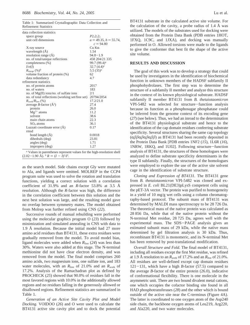
Table 1: Summarized Crystallographic Data Collection andRefinement Statistics
data collection statisticsspace group P212121
unit cell dimensions a ) 49.35,b ) 55.74,c ) 94.80
X-ray source Cu KRwavelength (Å) 1.54resolution range (Å) 94.8-1.9no. of total/unique reflections 458 204/21 335completeness (%) 99.7 (99.4)a
I/σ(I) 32.7 (4.4)a
Rmerge(%)b 5.5 (35)a
volume fraction of protein (%) 62data redundancy 4.7
refinement statisticsno. of protein atoms 2017no. of waters 183no. of Mg(II) ions/no. of sulfate ions 2/1no. of total reflections (working set/free set) 20794/2054Rwork/Rfree (%) 17.2/21.0averageB-factor (Å2) 27.4
protein 26.0Mg2+ atoms 11.2solvent 38.6main chain atoms 22.3SO4 atoms 36.1
Luzzati coordinate error (Å) 0.17rmsd
bond length (Å) 0.0018dihedrals (deg) 23.81angles (deg) 1.71impropers (deg) 1.27
a Values in parentheses represent values for the high-resolution shell(2.02-1.90 Å). b R ) (I - I)2/I2.
8688 Biochemistry, Vol. 44, No. 24, 2005 Lu et al.

BT4131 is composed of two distinct domains connectedby two linker regions (Figure 4A,B). The larger domain isthe “conserved core” domain that houses the active site. Theslightly smaller domain is the cap domain, which docks onthe core domain to cover its active site. The core domain isa modified Rossmann fold comprised of a six-strandedparallelâ sheet (â1-â3 andâ11-â13) surrounded by sixhelices (R1, R2, and R7-R10). Two additionalâ sheets(â4 andâ5) following â3 are part of the connection betweenthe core domain and cap domain. The cap domain topologyis of theRââ(RâRâ)Rââ type (see a further description inthe section below).
The disposition of the cap domain relative to the coredomain reflects a cap-closed conformation, wherein the coredomain active site is sequestered from bulk solvent. InBT4131, the closed conformation might be attributed to thepresence of the anionic (probably sulfate) ligand in the activesite (see below). In other HAD enzymes, bound sulfate,phosphate, and tungstate ligands have been correlated with
closed enzyme structures (5, 10, 29), whereas both open andclosed conformations have been observed with the un-liganded enzymes (5). It has been suggested that the anionmimics the phosphoryl group of the substrate and byshielding the positive charge of the bound Mg2+ facilitatescap domain-core domain association (5).
The R/â core domain is similar in structure to the coredomain common to all other HADSF members. The capdomain topology places BT4131 in the HAD subfamily IItype B category (subclass IIB). A search of the DALIdatabase (30) identified 19 HADSF members withZ scoresgreater than 6.0. Because HADSF members share the coredomain, the “hits” represent all three subfamilies. Thesestructures share 4-24% overall amino acid sequence identityand rms deviations of 1.9-3.8 Å compared to BT4131. TheHAD structures having the highestZ scores, however, arethose which share cap topology with BT4131 and assumethe same domain-domain conformation (i.e., open vsclosed). The most closely related fold belongs to 1NF2
FIGURE 4: (A) Stereoview of the structure of BT4131 colored from the N-terminus (blue) to the C-terminus (red). Mg2+ ions are shownas magenta spheres. Sulfate ion bound in the active site is shown as a ball and stick. (B) Topological diagram of the secondary structureof BT4131. The red area represents the BT4131 core domain, the pink area the linker, and the blue area the cap domain. There is anextended segment inserted betweenR5 andR6 (residues 133-137) which in other subfamily II type B enzymes is aâ strand. (C) BT4131active site with conserved catalytic segments colored as in Figure 1B.
BT4131 Is a Monosaccharide Phosphatase Biochemistry, Vol. 44, No. 24, 20058689

(Z ) 26), which is a subclass II type B HAD phosphatasewith unknown function (15). The next most related structureis 1L6R (Z ) 20), another subclass IIB HAD enzyme, whichfunctions as a phosphoglycolate phosphatase (16). Bothstructures are, like BT4131, observed in the cap-closedconformation (see further discussion below). From the DALIsearch results, we anticipated that BT4131 is a phosphatase,a prediction that was confirmed by the observed catalyzedhydrolysis of PNPP (Table 2).
BT4131 Core Domain.The four-loop catalytic platformof the BT4131 core domain is shown in Figure 4C, and theresidues that closely surround the bound metal ion and sulfateligands are shown in Figure 5. In general, the structure ofthe core domain active site in BT4131 is essentially the sameas the core domain active sites that have been reported fornumerous other HADSF phosphotransferases (for example,compare the BT4131 structure in Figure 4C to the PSPstructure in Figure 1B). However, there are two features ofthe BT4131 active site that are of special note that will bediscussed here.
The first point of interest is the metal cofactor bindingsite. BT4131-catalyzed hydrolysis of PNPP was assessed inthe presence and absence of Mg2+. Whereas no activity abovebackground was observed for metal-free BT4131 (preparedusing EDTA; see Experimental Procedures) in the absenceof added Mg2+, the apparentkcat (PNPP concentration∼ Km)and Km values (hereKm is equivalent to the dissociationconstant for binding to the free enzyme in the presence ofPNPP) for Mg2+-activated BT4131 are 0.05 s-1 and 111µM,respectively (Table 2). The purified enzyme which was nottreated with EDTA retained a small amount of activity(6% of the activity measured in the presence of Mg2+),indicating metal ion contamination in the enzyme preparationand/or assay solution. In fact, although divalent metal ionswere not added to the crystallization buffer, electron densitycorresponding to a metal ion appeared in the BT4131structure at a contour level of 11σ in an Fo - Fc electrondensity map. The density was tentatively assigned as Mg2+
for two reasons. First, the anomalous peak at the metal ionbinding site should show a very low-magnitude signal formagnesium (0.18 anomalous electron) and a significant peakfor a transition metal such as Mn2+ (2.79 anomalouselectrons) compared to the anomalous signal from a S atomin the protein (0.56 anomalous electron). The peak at themetal binding site is visible when the anomalous electrondensity map is contoured to 12.5σ, whereas the peak fromthe S atom of a Met residue with a lowB-factor (14 Å2) isvisible up to 7.6σ. We assume a high total occupancy of thesite (close to 100%) because of the lowB-factors (all lessthan the averageB-factor of the protein) of the liganding
carboxylates (i.e., there is no evidence of a mix of occupiedand unoccupied sites). Thus, from a weighted average, theamount of anomalous scattering at the metal site is consistentwith 75% occupancy by a metal with the scattering propertiesof Mg2+ and 25% occupancy by a metal with the scatteringproperties of Mn2+. Indeed, little residual density appearedafter refinement of this metal in that position. Second, thekinetic constants derived from the metal ion specificity studyreported in Table 2 suggested that Mg2+ is the physiologicalactivator (i.e., it yields the highest activity). Of the otherdivalent metal cations which are activators and bind withhigher affinity than Mg2+ (see Table 2), only Ni2+ has similaranomalous scattering to Mg2+ (but it results in one-third ofthe activity). Thus, although we cannot completely rule outNi2+, the metal cofactor is probably Mg2+.
Regardless of the true metal ion identity, the position ofthe metal defines the core domain residues that bind it (Figure5). As expected for enzyme-bound Mg2+, there are sixligands with octahedral geometry. One of these ligands isassigned as a sulfate ion, and the basis for this assignmentis described below. We suggest that by analogy to other HADphosphotransferase structures, the sulfate ligand that weobserve in the BT4131 active site is replaced with thephosphoryl group of the substrate in the enzyme-substratecomplex (9, 14, 31). Thus, as with other HAD phospho-transferases, the Mg2+ cofactor serves to bind the transferringphosphoryl group. The observed coordination of the metalion to the carboxylate of the loop 1 Asp8 nucleophile andto the backbone carbonyl of the loop 1 Asp10 acid/baseresidue is also common to the other HAD phosphotrans-ferases. It is the manner in which loop 4 binds to the Mg2+
that makes the BT4131 catalytic site unique. In a previousanalysis of loop 4-mediated metal ion binding in HADphosphotransferases (28), a seven-station template thatfunctions to position metal binding residues was described.The HAD phosphotransferases differ in the number andposition of binding residues along this template as well inthe type of binding (inner sphere vs outer sphere) that isinvolved. In BT4131, Asp211 at station 3 provides one metalligand, Asn214 at station 6 binds the metal ion via formationof a hydrogen bond with one water ligand, and Asp215 atstation 7 binds the metal via formation of a hydrogen bondwith the second water ligand. This configuration is uniqueamong those previously described (28) and serves as furtherevidence of the plasticity in configuration of the metal-binding loop of the HADSF phosphotransferases.
The assignment of the above-mentioned sulfate ligand isbased on the scattering properties of sulfate and the presenceof sulfate in the crystallization milieu. Specifically, electrondensity at a 2.5σ contour level in anFo - Fc map appearedin the enzyme active site in a region overlapping the regionidentified as the phosphate-binding site in other HADSFmembers (5-7, 10, 29). An anomalous difference mapcalculated with the Cu KR data (1.54 Å) showed the intensityof the peak corresponding to the sulfate sulfur position isequivalent to that of a nearby sulfur atom in Met. Becauseno phosphate ion had been used in BT4131 purification andcrystallization, but ammonium sulfate had been used in thepurification, we suspected that the electron density derivedfrom a bound sulfate ion. Sulfate ion was tested as acompetitive inhibitor versus PNPP and found to have aKi
of 1.1 ( 0.1 mM, indicative of modest affinity for the
Table 2: Steady-State Kinetic Constants Measured forBT4131-Catalyzed Cleavage of PNPP at pH 7.0 and 25°C in thePresence of Various Divalent Metal Ions
metal ion (Me) kcat (s-1) Km (µM) kcat(Me)/kcat(Mg2+)
Mg2+ 0.051( 0.001 111( 8 1.0Mn2+ 0.0284( 0.0008 9( 1 0.56Co2+ 0.0077( 0.001 6.2( 0.3 0.15Zn2+ 0.0044( 0.0003 13( 2 0.086Ca2+ 0.00104( 0.00003 85( 7 0.02Ni2+ 0.0017( 0.00003 1.5( 0.1 0.33Cu2+ no activationFe2+ no activation
8690 Biochemistry, Vol. 44, No. 24, 2005 Lu et al.

phosphate-binding site. The nearest significant neighbors tothe assigned sulfate ion are the Mg2+, loop 1 Asp8 (thenucleophile), loop 1 Asp10 (the acid/base), loop 2 Thr43(phosphate-binding residue), loop 3 Lys188 (phosphate-binding residue), and loop 4 Asn214 (metal-binding residue)(Figure 5). Note that Asp8 and Asp10 are proximal to thesulfate but do not participate in hydrogen bonds. This sulfateposition is identical to that of sulfate or phosphate ligandsin other HAD phosphotransferases (6, 10, 29).
The second feature of the BT4131 active site of specialinterest relates to the Asp10 acid/base residue. This residueis conserved among HAD phosphate ester hydrolases toprotonate the alkoxide leaving group formed in the firstpartial reaction (Figure 1A). In the second partial reaction,involving the hydrolysis of the aspartyl-phosphate inter-mediate, it may also serve to deprotonate the water nucleo-phile. In the HAD subfamily I phosphotransferases examinedto date, the Asp10 acid/base is positioned by the substrate(6, 9, 29). In contrast, Asp10 of BT4131 is positioned forits role in acid/base catalysis by loop 2 residue Arg45 (Figure5). Inspection of all reported HAD phosphotransferasestructures revealed that in subfamily IIB and III members,the Asp acid/base residue is bound by an electropositiveresidue which occupies the same spatial position as the Arg45of BT4131 but can vary in sequence position. At present, it
is not known why the Asp acid/base is “pinned down” byan active site residue in these subfamilies, but not insubfamily I, but this appears to be a general phenomenonthat warrants future investigation.
Substrate Specificity and Biochemical Function.BT4131activated with Mg2+ was found to catalyze the hydrolysisof PNPP with reasonable efficiency [kcat/Km ) 0.11 ×10³ M-1 s-1 (Table 3)]. The physiological substrate ofBT4131 was, however, unknown. On the basis of the locationof the encoding gene near the chitobiase (â-N-acetylhexo-saminidase) gene (32), we thought that BT4131 might beinvolved in sugar catabolism and thus might be a sugarphosphate phosphatase. To test this proposal, we examinedthe size and shape of the substrate-binding site in BT4131as defined by the active site cavity in the catalytically active,domain-domain closed conformation of the enzyme. Byusing VOIDOO (26) to generate a surface plot of the cavityavailable to a probe the size of a water molecule (1.4 Åradius), a solvent cage was created. Common phosphorylatedmetabolites, including pyridoxal 5-phosphate and the sugarphosphates glucose 6-phosphate, arabinose 5-phosphate, andN-acetylglucosamine 6-phosphate (NAG6P), were evaluatedfor “goodness of fit” by manually docking their structuresinto the solvent cage. Of the metabolites screened, NAG6Pgave the best fit (Figure 6) in that it filled the cage without
FIGURE 5: Sulfate binding site and hydrogen bond network in the BT4131 active site (A) and the 2(Fo - Fc) electron density map shownin stereo (B) contoured at 1.2σ. Mg2+ is colored magenta, and waters are colored red [one water ligand to Mg2+ (held by Asn214) is notshown for clarity].
BT4131 Is a Monosaccharide Phosphatase Biochemistry, Vol. 44, No. 24, 20058691

extending beyond it. The glucose 6-phosphate and arabinose5-phosphate were also good matches but left small regionsof the cage empty. We note that in the BT4131 structure,four water molecules occupy the space filled by the sugarmotif of the docked NAG6P.
Using the results of our “virtual” substrate screen as aguide, a focused activity screen was devised. On the basisof the shape of the solvent cage, we predicted that hexose6-phosphates or pentose 5-phosphates would be goodsubstrates whereas ring isomers such as glucose 1-phosphatewould not be. This is because direct attachment of the ringto the transferring phosphoryl group prevents the latter fromextending through the narrow pocket to the Asp8 nucleophile(see Figure 6). Consistent with this prediction, we found thatthe cyclic sugarsâ-D-glucose 6-phosphate, 2-deoxy-D-glucose6-phosphate,N-acetylglucosamine 6-phosphate,D-fructose6-phosphate,D-mannose 6-phosphate,D-arabinose 5-phos-phate,D-ribose 5-phosphate, and deoxyribose 5-phosphateare active substrates (kcat ) 1-26 s-1; kcat/Km ∼ 1 ×10³ M-1 s-1) and that the cyclic sugarsR-D-galactose1-phosphate,R-D-glucose 1-phosphate, andR-D-(+)-mannose1-phosphate are not (kcat is under the detection limit of0.01 s-1) (Table 3). Similarly, pyridoxal phosphate, in which
the phosphoryl group is extended from the planar ring by amethylene group, is significantly more reactive thanPNPP, in which the phosphate group is attached directly tothe ring (kcat/Km ) 2 × 10³ M-1 s-1 vs kcat/Km ) 0.11 ×10³ M-1 s-1).
A second obvious feature of the substrate binding site inBT4131 is the hydrophobic nature of the residues (most ofwhich derive from the cap domain) surrounding the sugarunit of the docked sugar-phosphate ligand (Figure 6).Because these nonpolar residues cannot offer hydrogenbonding interactions with the substrate hydroxyl groups, theinteraction between the charged phosphoryl group and theelectropositive groups of the core domain will be theprinciple source of favorable ligand binding energy (seeFigure 5A). Thus, as long as the transferring phosphorylgroup can access the Asp8 in the absence of a steric clash,catalysis is anticipated. Consequently, the ability of BT4131to distinguish between most (monosaccharide) sugar phos-phate substrates will be limited. Indeed, the values of thesubstrate specificity constantkcat/Km for D-glucose 6-phos-phate,D-fructose 6-phosphate,D-mannose 6-phosphate,D-arabinose 5-phosphate,D-ribose 5-phosphate,DL-glycerol3-phosphate, and sorbitol 6-phosphate are within a factor
Table 3: Phosphorylated Compounds Tested as Substrates for BT4131 (50 mM Hepes buffer, pH 7.0, 5 mM MgCl2, and 37°C)
substrate kcat (s-1) Km (mM) kcat/Km (M-1 s-1)
p-nitrophenyl phosphate (PNPP) 0.083( 0.002 0.77( 0.05 0.11× 10³
â-D-glucose 6-phosphate 3.6( 0.2 10.7( 1.7 0.34× 10³
2-deoxy-D-glucose 6-phosphate 26( 1 3.9( 0.4 6.7× 10³
trehalose 6-phosphate ∼0.06D-glucosamine 6-phosphate 0.27( 0.02 6.0( 0.9 0.045× 10³
N-acetyl-D-glucosamine 6-phosphate 3.6( 0.5 17( 5 0.21× 10³
D-fructose 6-phosphate 0.9( 0.1 4( 1 0.27× 10³
D-fructose 1,6-(bis)phopsphate 0.014( 0.002 4( 1 0.004× 10³
sucrose 6′-monophosphate 0.013( 0.001 3.1( 0.1 0.004× 10³
D-mannose 6-phosphate 1.3( 0.1 1.9( 0.3 0.7× 10³
D-arabinose 5-phosphate 9.6( 0.7 3.5( 0.5 3.2× 10³
D-ribose 5-phosphate 8.7( 0.4 4.9( 0.6 1.8× 10³
deoxyribose 5-phophate 1.45( 0.03 3.0( 0.1 0.48× 10³
ADP 0.016( 0.001 1.6( 0.4 0.01× 10³
DL-R-glycerol 3-phosphate 10.4( 0.2 8.7( 0.4 1.2× 10³
glyceraldehyde 3-phosphate ∼1.0pyridoxal 5′-phosphate 1.75( 0.06 0.88( 0.08 2× 10³
sorbitol 6-phosphate 5.4( 0.2 6.8( 0.5 0.8× 10³
D-gluconate 6-phosphate 0.040( 0.005 2.2( 0.7 0.018× 10³
FIGURE 6: Stereodiagram of the active site of BT4131 with the substrateN-acetylglucosamine 6-phosphate (NAG6P, green) docked in acage representing the accessible surface of the active site [calculated with VOIDOO (34)].
8692 Biochemistry, Vol. 44, No. 24, 2005 Lu et al.
of 10 in magnitude, with a meankcat/Km value equal to1 × 103 M-1 s-1 (Table 3).
A second corollary to the spatially confined, hydrophobicsugar-binding site is that phosphate esters that have chargedsubstituents or that are simply too large to fit in the activesite of the enzyme in the catalytically active, closedconformation will not be efficient substrates. Indeed,L-tyrosineO-phosphate,L-serineO-phosphate, ethanolamineO-phosphate, AMP, ATP,D-fructose 2,6-(bis)phosphate, andpyrophosphate are not active substrates, andD-gluconate6-phosphate,D-glucosamine 6-phosphate, andD-fructose 1,6-(bis)phosphate are very poor substrates as are the disaccha-rides trehalose 6-phosphate and sucrose 6′-monophosphate.
On the basis of the substrate activity profile and the size,shape, and hydrophobicity of the substrate-binding site, weconclude that BT4131 might function in the catalysis ofrelease of phosphate from hexose 6-phosphates and pentose5-phosphates. The catalytic efficiency is low:kcat/Km
∼ 1 × 10³ M-1 s-1 compared to the 1× 10⁶ to 1 ×10⁸ M-1 s-1 “gold-standard” for single-substrate enzymesinvolved in primary metabolism (33). The modest catalyticefficiency and the broad substrate range are strong indicatorsthat BT4131 functions in a secondary pathway.
Indeed, the (annotated) gene context of BT4131 inB. thetaiotaomicronas well as that of the ortholog BF0989(69% identical sequences) inBacteroides fragilisis sugges-tive of its role in recycling nitrogen and carbon from thecell wall polymer chitin. InB. thetaiotaomicron, neighborgene BT4132 encodes chitobiase, an enzyme that catalyzesthe hydrolysis of chitin-derived chitobiose (N-acetylgluco-samine disaccharide) toN-acetylglucosamine. Another neigh-bor, BT4127, encodes glucosamine-6-phosphate isomerase.This enzyme recycles “NH3” via the catalyzed conversionof glucosamine 6-phosphate andL-glutamate to fructose6-phosphate andL-glutamine, respectively. The gene contextprompts the speculation that BT4131 might function asfructose-6-phosphate phosphatase for chitin catabolism.
Enzymes that function in secondary pathways are notsubjected to the selection pressure for catalytic perfection,and theirkcat/Km values often reflect this. Moreover, broadsubstrate range is not compatible with catalytic perfectionbecause the enzyme must accommodate a variety of substratestructures. There is in fact some evidence to suggest thatthe BT4131 active site accommodates rather than comple-ments its intended sugar phosphate substrate. Whereas theoptimal fit is observed with NAG6P (kcat/Km ) 0.2 ×10³ M-1 s-1), the best substrate activity is observed with2-deoxyglucose 6-phosphate (kcat/Km ) 7 × 10³ M-1 s-1).Because 2-deoxyglucose 6-phosphate is not a known bacterialmetabolite, it cannot be the physiological substrate and the10-fold higher kcat that is observed for 2-deoxyglucose6-phosphate is not predicted by interaction between the ligandand the active site. Together, these observations suggest thatthe catalytic site of BT4131 is not specially tailored or honedfor substrate specificity or catalytic efficiency. Thus, BT4131appears to be an all-purpose sugar phosphate phosphatasethat has been recruited for the purpose of recycling carbonand nitrogen from the cell wall polysaccharide chitin. In thiscapacity, exceptional speed and specificity are not requiredand are not features that will be cultivated by naturalselection. Alternatively, it is always possible that in the panelof substrates that were tested in vitro, the true substrate has
been missed. Such an omission might also be expected toresult in the measurement of a relatively lowkcat/Km for anumber of the “nearest neighbor” substrates. However, iffructose 6-phosphate is not the “correct substrate”, then it isat least a close structural analogue, and the specificitydeterminants found from interactions of the enzyme with thissubstrate model will still be valid. In the following section,we compare the structure and function of BT4131 with thoseof other HAD subfamily II type B enzymes to identifyfeatures of the type B cap domain that underlie catalyticefficiency and substrate specificity.
Comparison of Cap Domain Structure and Function inHAD Subfamily II Type B Phosphatases.HAD subfamily IIenzymes display two topologically different cap domainsindicative of two separate pathways of evolution. SubfamilyII type A has the cap domain topologyRâRââRâRâ, andtype B has the cap domain topologyRââ(RâRâ)Rââ (seepanels A and B of Figure 2). BT4131 is type B. There isonly one type A structure currently reported (PDB entry1PW5, NagD gene product fromThermotoga maritima),whereas several subfamily II type B structures [phospho-glycolate phosphatase, TA0175 fromThermotoga acido-philium (PDB entry 1L6R) (16), trehalose-6-phosphatephosphatase fromT. acidophilium (PDB entry 1U02),“hypothetical phosphatase” TM0651 fromT. maritima(PDB entry 1NF2) (15), “HAD-like hydrolase” fromBacillussubtilis (PDB entry 1NRW), and NYSGRC Target T1436from E. coli (PDB entry 1RKQ)] have been reported. Thetype B cap domains, compared in Figure 2B, are the focusof this structure-function analysis.
As outlined in the previous discussion, the comparison ofcap-open and cap-closed structures of the HADSF type Ienzymes painted a picture of the catalytic cycle, starting withsubstrate binding (cap-open), proceeding through the chemi-cal steps (cap-closed), and ending with product release (cap-open). Although we do not at present have a structure ofBT4131 in the cap-open conformation, by overlaying the cap-closed BT4131 structure with the cap-open structure of theunknown phosphatase 1NF2, we can infer the cap domainmotion required for a catalytic cycle in the type IIB HADenzymes (Figure 7A). This analysis is allowed by theconservation of the fold in the core domains and in the capdomains of the type IIB enzymes (i.e., the gross differencesbetween the structures are due to differences in the relativepositions of the cap and core domains). TheR/â fold of thecap domain of the type IIB members shows little variationin the relative orientation of structural elements within thedomain (unlike the type I HADSF members where there issignificant variation in the angles between theR helices inthe helical bundle cap). Accordingly, cap domain movementin BT4131 is likely to occur through backbone rotation inthe two solvated peptide linkers at or near residues 83-85(linker 1, sequence SAI) and residues 184-186 (linker 2,sequence KGD). The caveat exists that a bulky residue inone structure may change the relative orientation of the coreto cap and thus the analysis of the changes when comparingtwo different proteins. However, it is probable such changeswould be more subtle than entire domain movements. Indeed,the fact that the active site in the closed form of BT4131 iscompletely solvent inaccessible necessitates domain-domainmovement. In a more general sense, we anticipate that thesame type of “hinge motion” in the interdomain linkers which
BT4131 Is a Monosaccharide Phosphatase Biochemistry, Vol. 44, No. 24, 20058693

mediates cap association and dissociation in the HADsubfamily I enzymes (9, 11) will also operate in the subfamilyIIB enzymes.
The HAD subfamily IIB cap domains can be subclassifiedinto the small RââRââ domain group or the largeRââ(RâRâ)Rââ domain group. The small domain lacks the(RâRâ) insert of the large domain and as a consequence usesa single loop to cover the core domain active site, while thelarger domain uses two loops (Figure 7B). The smallRââRââ domain group includes phosphoglycolate phos-phatase (PDB entry 1LR6) and trehalose-6-phosphate phos-phatase (PDB entry 1U02). The largeRââ(RâRâ)Rââdomain group includes BT4131 and unknown phosphatases1NF2, 1NRW, and 1RKQ. These structures have beenanalyzed with two goals in mind: (1) to identify the structuralmotifs of the cap domains that contribute to the substrate-binding site of the enzyme in the cap-closed conformationand (2) to link the sequences of these motifs to substratestructure and biochemical function.
We started first with phosphoglycolate phosphatase inwhich high catalytic efficiency and substrate specificity areoperative [kcat/Km ) 2.2× 10⁵ M-1 s-1 (16)]. The unligandedphosphoglycolate phosphatase structure reveals the cap-closed conformation. In this study, we generated a solventcage and docked the phosphoglycolate ligand into the cage,thereby demonstrating a very good fit and identifying theresidues that might, on the basis of this model of the ligandedenzyme, function in substrate binding. In addition to thestandard core domain interactions with the phosphorylmoiety, an interaction between the carboxylate substituentof the phosphoglycolate ligand and a cap domain His143 isevident (see Figure 8). We anticipate that His143 plays akey role in phosphoglycolate recognition. Significantly,
His143 is located on the substrate specificity loop spanningresidues 136-145 in the phosphoglycolate phosphatase. Thisloop, formed by residues 172-182 in BT4131, is present inall subfamily II type B enzymes (i.e., a structural motifcommon to the small and large cap domains). Notably, thespecificity loop His143 of phosphoglycolate phosphatase isreplaced with an Asp or Glu in all other subfamily II type Bmembers of known structure. In the case of BT4131, thecorresponding Asp180 may form a hydrogen bond with theC(1)OH group of the sugar phosphate substrate (see Figure6). Other residues originating from the substrate specificityloop of phosphoglycolate phosphatase are Ser138 and Ser141[corresponding to Trp174 and Phe178, respectively, inBT4131 (Figure 6)]. These polar residues contribute to thepolar phosphoglycolate binding site (as Trp174 and Phe178contribute to the nonpolar sugar phosphate binding site inBT4131).
Of the largeRââ(RâRâ)Rââ domain group, BT4131 andunknown phosphatases 1NRW and 1RKQ exist in a cap-closed conformation, which provides a “snapshot” of thecomposite active site formed by cap and core domainresidues. Comparison of the active site solvent cagesgenerated for 1NRW and 1RKQ shows that while 1NRWhas a small cavity, 1RKQ has a cavity similar in size to thatof BT4131. Both structures contain the previously notedsubstrate specificity loop (residues 172-182 in BT4131numbering), hereafter termed the “primary substrate specific-ity loop”. In addition, it is evident that a second loop(residues 118-130 in BT4131) contained within theRâRâinsert of all large domain enzymes also contributes to theactive site. We denote this loop as the “secondary substratespecificity loop”. 1NRW positions small polar residuesSer200 and Asn204 on the primary substrate specificity loopand Arg125 on the secondary loop (attaining the sameposition relative to the substrate as Phe125 in BT4131). Incontrast to BT4131, 1NRW is structured to interact with aphosphate monoester that has a polar leaving group. In1RKQ, the residue contributed to the binding site by thesecondary substrate specificity loop is His129, whereas theresidues contributed by the common substrate specificity loopare Ser184 and Phe188. In BT4131, the correspondingsubstrate specificity loop residues are Trp174 and Phe178.As with 1NRW, 1RKQ is compatible with a polar substrate.The ligand size and shape suggested by the solvent cage andthe placement of the residues from the specificity loopssuggest likely substrates for 1NRW and 1RKQ, which willbe evaluated with a focused substrate screen.
Summary.BT4131, a representative of the HAD subfamilyIIB class of phosphatases, was analyzed in terms of activesite structure and substrate specificity. The potential applica-tion of the active site solvent cage in conjunction with theidentity of the residues stationed along the cap domainsubstrate specificity loop(s) in predicting substrate structurewas discovered. This “solvent cage”-based method wasapplied to virtual substrate screening in the known enzymephosphoglycolate phosphatase to verify predictive power. Infuture studies, the method will be used to predict thesubstrates of unknown HAD subfamily IIB class phos-phatases and their functions confirmed by applying focusedsubstrate screens to the purified proteins.
FIGURE 7: (A) Superposition of BT4131 (red and magenta for thecore and cap, respectively) and 1NF2 (green and blue for the coreand cap, respectively) to compare the cap-closed and -open forms,respectively, of this subgroup. (B) Superposition of the backboneof BT4131 (red) and phosphoglycolate phosphatase (1L6R, blue)with the proposed substrate specificity loops depicted in gold andgreen, respectively. In BT4131, the secondary substrate specificityloop originating from the cap domain (labeled 118-133) alsocontributes some residues to the active site and excludes bulksolvent. The Mg2+ cofactor and sulfate ligand from BT4131 aredepicted as a ball and stick. This overlay of BT4131 andphosphoglycolate phosphatase also serves to compare the large capand small cap topologies within subfamily II type B.
8694 Biochemistry, Vol. 44, No. 24, 2005 Lu et al.

ACKNOWLEDGMENT
We thank Dr. Jeffrey Gordon (Washington University) forkindly providing the genomic DNA fromB. thetaiotaomicronVPI-5482. We thank Dr. Tracy Arakaki for her generousassistance with data processing, molecular replacement, andrefinement. We also gratefully acknowledge Dr. Ezra Peisachfor assistance with figure preparation and careful reading ofthe manuscript.
REFERENCES
1. Koonin, E. V., and Tatusov, R. L. (1994) Computer Analysis ofBacterial Haloacid Dehalogenases Defines a Large Superfamilyof Hydrolases with Diverse Specificity: Application of an IterativeApproach to Database Search,J. Mol. Biol. 244, 125-132.
2. Allen, K. N., and Dunaway-Mariano, D. (2004) Phosphoryl grouptransfer: Evolution of a catalytic scaffold,Trends Biochem. Sci.29, 495-503.
3. Hisano, T., Hata, Y., Fujii, T., Liu, J. Q., Kurihara, T., Esaki, N.,and Soda, K. (1996) Crystal structure ofL-2-haloacid dehalogenasefrom Pseudomonassp. YL. An R/â hydrolase structure that isdifferent from theR/â hydrolase fold,J. Biol. Chem. 271, 20322-20330.
4. Ridder, I. S., Rozeboom, H. J., Kalk, K. H., Janssen, D. B., andDijkstra, B. W. (1997) Three-dimensional structure ofL-2-haloaciddehalogenase fromXanthobacter autotrophicusGJ10 complexedwith the substrate-analogue formate,J. Biol. Chem. 272, 33015-33022.
5. Morais, M. C., Zhang, W., Baker, A. S., Zhang, G., Dunaway-Mariano, D., and Allen, K. N. (2000) The crystal structure ofBacillus cereusphosphonoacetaldehyde hydrolase: Insight intocatalysis of phosphorus bond cleavage and catalytic diversificationwithin the HAD enzyme superfamily,Biochemistry 39, 10385-10396.
6. Lahiri, S. D., Zhang, G., Dunaway-Mariano, D., and Allen, K. N.(2002) Caught in the act: The structure of phosphorylatedâ-phosphoglucomutase fromLactococcus lactis, Biochemistry 41,8351-8359.
7. Peisach, E., Selengut, J., Dunaway-Mariano, D., and Allen, K. N.(2004) Structure of the Magnesium-Dependent Protein TyrosinePhosphatase, MDP-1,Biochemistry 43, 12770-12779.
8. Selengut, J. D. (2001) MDP-1 is a new and distinct member ofthe haloacid dehalogenase family of aspartate-dependent phos-phohydrolases,Biochemistry 40, 12704-12711.
9. Wang, W., Cho, H. S., Kim, R., Jancarik, J., Yokota, H., Nguyen,H. H., Grigoriev, I. V., Wemmer, D. E., and Kim, S. H. (2002)Structural characterization of the reaction pathway in phospho-serine phosphatase: Crystallographic “snapshots” of intermediatestates,J. Mol. Biol. 319, 421-431.
10. Wang, W., Kim, R., Jancarik, J., Yokota, H., and Kim, S. H. (2001)Crystal structure of phosphoserine phosphatase fromMethano-
coccus jannaschii, a hyperthermophile, at 1.8 Å resolution,Structure 9, 65-71.
11. Zhang, G., Mazurkie, A. S., Dunaway-Mariano, D., and Allen,K. N. (2002) Kinetic Evidence for a Substrate-Induced Fit inPhosphonoacetaldehyde Hydrolase Catalysis,Biochemistry 41,13370-13377.
12. Lahiri, S. D., Zhang, G., Dai, J., Dunaway-Mariano, D., and Allen,K. N. (2004) Analysis of the substrate specificity loop of the HADsuperfamily cap domain,Biochemistry 43, 2812-2820.
13. Lahiri, S. D., Zhang, G., Dunaway-Mariano, D., and Allen, K. N.(2003) The Pentacovalent Phosphorus Intermediate of a Phos-phoryl Transfer Reaction,Science 299, 2067-2071.
14. Morais, M. C., Zhang, G., Zhang, W., Olsen, D. B., Dunaway-Mariano, D., and Allen, K. N. (2004) X-ray crystallographic andsite-directed mutagenesis analysis of the mechanism of Schiff-base formation in phosphonoacetaldehyde hydrolase catalysis,J.Biol. Chem. 279, 9353-9361.
15. Shin, D. H., Roberts, A., Jancarik, J., Yokota, H., Kim, R.,Wemmer, D. E., and Kim, S. H. (2003) Crystal structure of aphosphatase with a unique substrate binding domain fromTher-motoga maritima, Protein Sci. 12, 1464-1472.
16. Kim, Y., Yakunin, A. F., Kuznetsova, E., Xu, X., Pennycooke,M., Gu, J., Cheung, F., Proudfoot, M., Arrowsmith, C. H.,Joachimiak, A., Edwards, A., and Christendat, D. (2004) Structureand function-based characterization of a new phosphoglycolatephosphatase fromThermoplasma acidophilum, J. Biol. Chem. 279,517-526.
17. Aravind, L. (2000) Guilt by association: Contextual informationin genome analysis,Genome Res. 10, 1074-1077.
18. Bradford, M. M. (1976) A rapid and sensitive method for thequantitation of microgram quantities of protein utilizing theprinciple of protein-dye binding,Anal. Biochem. 72, 248-254.
19. Wilkins, M. R., Gasteiger, E., Bairoch, A., Sanchez, J.-C.,Williams, K. L., Appel, R. D., and Hochstrasser, D. F. (1998)Protein Identification and Analysis Tools in the ExPASy Server,in 2-D Proteome Analysis Protocols(Link, A. J., Ed.) HumanaPress, Totowa, NJ.
20. Jancerik, J., and Kim, S. H. (1991) Sparse matrix sampling: Ascreening method for crystallization of proteins,J. Appl. Crys-tallogr. 24, 409-411.
21. Otwinowski, Z., and Minor, W. (1997) Processing of X-rayDiffraction Data Collected in Oscillation Mode,Methods Enzymol.276, 307-326.
22. Brunger, A. T., Adams, P. D., Clore, G. M., DeLano, W. L., Gros,P., Grosse-Kunstleve, R. W., Jiang, J. S., Kuszewski, J., Nilges,M., Pannu, N. S., Read, R. J., Rice, L. M., Simonson, T., andWarren, G. L. (1998) Crystallography & NMR system: A newsoftware suite for macromolecular structure determination,ActaCrystallogr. D54(Part 5), 905-921.
23. Jones, T. A., Zou, J. Y., Cowan, S. W., and Kjeldgaard, G. J.(1991) Improved methods for building protein models in electrondensity maps and the location of errors in these models,ActaCrystallogr. A47(Part 2), 110-119.
FIGURE 8: Stereoview of the active site of phosphoglycolate phosphatase (1L6R) with the substrate phosphoglycolate (yellow backbone)docked in a cage (green) representing the accessible surface of the active site [calculated with VOIDOO (34)].
BT4131 Is a Monosaccharide Phosphatase Biochemistry, Vol. 44, No. 24, 20058695

24. Brunger, A. T. (1992) The Free R Value: a Novel StatisticalQuantity for Assessing the Accuracy of Crystal Structures,Nature355, 472-474.
25. Laskowski, R. A., MacArthur, M. W., Moss, D. S., and Thornton,J. M. (1993) PROCHECK: A program to check the stereochem-ical quality of protein structures,J. Appl. Crystallogr. 26, 283-291.
26. Kleywegt, G. J., and Jones, T. A. (1994) Detection, delineation,measurement and display of cavities in macromolecular structures,Acta Crystallogr. D50, 178-185.
27. Xu, J., Bjursell, M. K., Himrod, J., Deng, S., Carmichael, L. K.,Chiang, H. C., Hooper, L. V., and Gordon, J. I. (2003) A genomicview of the human-Bacteroides thetaiotaomicronsymbiosis,Science 299, 2074-2076.
28. Zhang, G., Morais, M. C., Dai, J., Zhang, W., Dunaway-Mariano,D., and Allen, K. N. (2004) Investigation of Metal Ion Bindingin Phosphonoacetaldehyde Hydrolase Identifies Sequence Markersfor Metal-Activated Enzymes of the HAD Enzyme Superfamily,Biochemistry 43, 4990-4997.
29. Rinaldo-Matthis, A., Rampazzo, C., Reichard, P., Bianchi, V., andNordlund, P. (2002) Crystal structure of a human mitochondrialdeoxyribonucleotidase,Nat. Struct. Biol. 10, 779-787.
30. Holm, L., and Sander, C. (1997) Dali/FSSP classification of three-dimensional protein folds,Nucleic Acids Res. 25, 231-234.
31. Rinaldo-Matthis, A., Rampazzo, C., Balzarini, J., Reichard, P.,Bianchi, V., and Nordlund, P. (2004) Crystal Structures of theMitochondrial Deoxyribonucleotidase in Complex with TwoSpecific Inhibitors,Mol. Pharmacol. 65, 860-867.
32. Xu, J., Bjursell, M. K., Himrod, J., Deng, S., Carmichael, L. K.,Chiang, H. C., Hooper, L. V., and Gordon, J. I. (2003)Science299, 2074-2076.
33. Fersht, A. (1999)Structure and Mechanism in Protein Science,W. H. Freeman and Co., New York.
34. Kleywegt, G. J., and Jones, T. A. (1994) Detection, delineation,measurement and display of cavities in macromolecular structures,Acta Crystallogr. D50, 178-185.
BI050009J
8696 Biochemistry, Vol. 44, No. 24, 2005 Lu et al.