high-risk births & obstetric emergencies chapter 36
TRANSCRIPT
High-Risk Births & Obstetric Emergencies
Chapter 36

INTRAPARTAL COMPLICATIONS
• Interference with normal processes & patterns of labor/birth resulting in maternal or fetal jeopardy.
• Preterm labor; dysfunctional labor patterns; prolonged labor; hemorrhage – uterine ruputure/inversion; amniotic-fluid embolus.

Dysfunctional Labor:• Possible Causes:
• Catecholamines (response to anxiety/fear), increase physical/psychological stress, leads to myometrial dysfunction; painful & ineffective labor.
• Premature or excessive analgesia, particularly during latent phase.
• Maternal factors.• Fetal factors.• Placental factors.• Physical restrictions (position in bed).

ASSESSMENT:• Antepartal history.• Emotional status.• Vital signs, FHR.• Contraction pattern (frequency,
duration, intensity).• Vaginal discharge.
GOAL = to minimize physical/ psychological stress during labor/birth.
Emotional support.

Preterm Labor:
• Occurs after 20 weeks gestation and before 38 weeks.
• Causes may be from maternal, fetal, or placental factors.
• Prevention:• Primary: close observation and eduction in
S&S of labor.• Secondary: prompt, effective Rx of
associated disorders.• Tertiary: suppression of preterm labor.

Tertiary: suppression of preterm labor• Bedrest.• Position: side-lying – to promote placental
perfusion.• Hydration.• Pharmacological: betaadrenergic agents to
reduce sensitivity of uterine myometrium to oxytocic & prostaglandin stimulation; increase bld flow to uterus.
• Pt may be maintained at home with adequate follow-up & health teaching.

CONTRAINDICATIONS: for suppression of labor• Placenta previa or abruptio placenta.• Chorioamnionitis.• Erythroblastosis fetalis.• Severe preeclampsia.• Severe diabetes (brittle).• Increasing placental insufficiency.• Cervical dilation of 4 cm or more.• ROM (depends on cause & if sepsis
exists).

Nursing Assessment: PTL• Maternal VS. Response to medication:
• Hypotension• Tachycardia, arrhythmia• Dyspnea, chest pain• Nausea & vomiting
• Signs of infection:• Increased temperature• Tachycardia• Diaphoresis• Malaise

• Emotional status: denial, guilt, anxiety, exhaustion.
• Signs of continuing & progressing labor:• Effacement• Dilation• Station
(vaginal exam ONLY if indicated by other signs of continuing labor progress)
• Status of membranes.
• FHR, activity (continuous monitoring).
• Ctx: frequency, duration, strength.

Report PROMPTLY to MD:
• Maternal pulse of 110 or more.
• Diastolic pressure of 60 mmHg or less.
• Increase in maternal temperature.
• Respirations of 24 or more; crackles (rales).
• Complaint of dyspnes.
• Contractions: increasing frequency, strength, duration, or cessation of ctx.

• Intermittent back and thigh pain.
• Rupture of membranes.
• Vaginal bleeding.
• Fetal distress.
IF LABOR CONTINUES:
• GOAL = facilitate infant survival; emotional support; support comfort measures; health teaching.

Dysfunctional Labor Pattern
• Hypertonic labor
• Hypotonic labor
• Precipitate labor level
Chapter 26

HYPERTONIC DYSFUNCTION:• Increased resting tone of uterine
myometrium; diminished refractory period; prolonged latent phase.• Nullipara: more than 20 hours.• Multipara: more than 14 hours.
• Etiology: unknown. Theory – ectopic initiation of incoordiante uterine ctx.
• Assessment:• Onset (early labor)

• Contractions:• Continuous fundal tension, incomplete
relaxation.• Painful.• Ineffectual – no effacement or dilation.
• Signs of fetal distress:• Meconium-stained fluid.• FHR irregularities.
• Maternal VS.• Emotional status.• Medical evaluation: to rule out CPD.
• Vaginal examination, x-ray pelvimetry, ultrasonography.

Interventions with Hypertonic Dysfunction:• Short-acting barbiturates (to encourage
rest, relaxation).• IV fluids (to restore / maintain hydration
& fluid-electrolyte balance).• If CPD – c/s.• Provide emotional support.• Provide comfort measures.• Prevent infection (strict aseptic
technique).• Prepare patient for c/s if needed.

HYPOTONIC DYSFUNCTION:• After normal labor at onset, ctx diminish
in frequency, duration, & strength.• Lowered uterine resting tone; cervical
effacement & dilation slow / cease.• Etiology:
• Premature or excessive analgesia / anesthesia (epidural, spinal block).
• CPD.• Overdistention (hydramnios, fetal
macrosomia, multifetal pregnancy).• Fetal malposition / malpresentation.• Maternal fear / anxiety.

• Assessment:• Onset (latent phase & most common in
active phase).• Contractions - normal previously, will
demonstrate:• Decreased frequency.• Shorter duration.• Diminished intensity (mild to moderate).• Less uncomfortable.
• Cervical changes – slow or cease.• Signs of fetal distress – rare.
• Usually late in labor d/t infection secondary to prolonged ROM.
• Tachycardia.

• Maternal VS (elevated temperature) – may indicate infection.
• Medical diagnosis – procedures: vaginal examination, x-ray pelvimetry, ultrasonography. To rule out CPD (most common cause).
• Management:• Amniotomy (artificial ROM).• Oxytocin augmentation of labor.• If CPD, prepare for c/s.• Emotional support, comfort measures,
prevent infection.

Precipitate Labor
• Labor that progresses rapidly and ends with the delivery occurring less than 3 hours after the onset of uterine activity.
• Rapid labor and delivery.

Fetal Malpresentation and Malposition
• Breech presentation
• Shoulder presentation
• Face presentation
• Malpositions
Chapter 26

Breech Presentations• Fetal descent in which the fetal
buttocks, legs, feet, or combination of these parts is found first in the maternal pelvis.
• Labor tends to be longer and more difficult due to a softer presenting part, that does not fill the birth canal completely.
• Increase risks for fetal outcome.

Shoulder Presentation
• Fetal descent in which the shoulder precedes the fetal head in the maternal pelvis alone or along with the ftal arm and hand.
• Vaginally undeliverable.

Face Presentation
• Fetal descent in which hyperextension of the fetal head and neck allows the fetal face to descend into the maternal pelvis, as opposed to flexion that results in fetal vertex presentation.
• Brow presentation = occurs when the area between the anterior fontanelle and the fetal eyes descends first.

Malpositions• Persistent occipitoposterior position.• Persistent occipitotransverse position.• Result from fetal rotation as the fetus
descends through the pelvis.• Possible precipitating factors are
macrosomia and pelvic abnormalities.• Results in increased discomfort
(particularly back labor), prolonged, abnormal labor, soft tissue injury, lacerations, or an extensive episiotomy incision.

Maternal and Fetal Structural Abnormalities
• Cephalopelvic disproportion (CPD)
• Macrosomia
Chapter 26

DYSTOCIA:• Difficult labor.• Causes:
• “3 Ps” for mother: Psych, Placenta, Position.• “3Ps” for fetus: Power, Passageway, Passenger.
• POWER: forces of labor (uterine contractions, use of abdominal muscles).• Premature analgesia / anesthesia.• Uterine overdistension (multifetal pregnancy, fetal
macrosomia)• Uterine myomas.

• PASSAGEWAY: Resistance of cervix, pelvic structures.• Rigid cervix.• Distended bladder.• Distended rectum.• Dimensions of the bony pelvis: oelvic
contractures.
• PASSENGER: accommodation of the presenting part to pelvic diameters.• Fetal malposition / malpresentation.• Fetal anomalies.• Fetal size.

Hazards with Dystocia:• MATERNAL:
1. Fatigue, exhaustion, dehydration.
2. Lowered pain threshold, loss of control.
3. Intrauterine infection.
4. Uterine rupture.
5. Cervical, vaginal, perineal lacerations.
6. Postpartum hemorrhage.
• FETAL:1. Hypoxia, anoxia, demise.
2. Intracranial hemorrhage.
Placental Abnormalities
• Placenta previa
• Abruptio placentae
• Other placental abnormalities
Chapter 26

PLACENTA PREVIA• Abnormal placement of placenta so that
it partially covers the cervix; dilatation results in bleeding, which can be of hemorrhagic proportions.
• The placenta is located over or very near the internal cervical os.
• Severe hemorrhage can result from digital palpation of the internal os.
• Previa is a serious but uncommon complication, occurring in .3-.5% of pregnancies.

• Advanced maternal age and multiparity increase the risk.
• Painless hemorrhage is symptomatic of previa, often around the end of the 2nd trimester.
• Clinical diagnosis is reached through ultrasound examination in which the placenta is localized in relationship to the cervix.
• Manual examination is contraindicated!• Management of pregnancy depends on
gestational age.
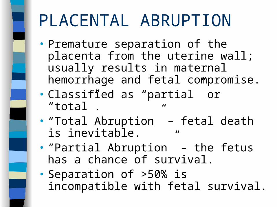
PLACENTAL ABRUPTION• Premature separation of the placenta
from the uterine wall; usually results in maternal hemorrhage and fetal compromise.
• Classified as “partial” or “total”.• “Total Abruption” – fetal death is
inevitable.• “Partial Abruption” – the fetus has a
chance of survival.• Separation of >50% is incompatible with
fetal survival.

Grading of Placental Abruptions:
• Grade I: Slight vag.bleeding & some uterine irritability. Maternal BP is unaffected & there are normal fibrinogen levels. FHR has a normal pattern.
• Grade II: External bleeding is mild to moderate. The uterus is irritable. Tetanic ctx may be present. Maternal BP is maintained. FHR shows signs of distress. Maternal fibrinogen level is decreased.

• Grade III: The bleeding may be severe & may be concealed in some instances. Uterine ctx are tetanic and painful. Maternal hypotension may be present. The fibrinogen level is greatly decreased & there are coagulation problems.
Diagnosis: may be made by ultrasound, but frequently the diagnosis is made and confirmed at delivery, by inspection of the placenta.
Umbilical Cord Abnormalities
• Velamentous insertion of the cord
• Umbilical cord compression
• Umbilical cord prolapse
Chapter 26

Velamentous Insertion of the Cord
• Condition where the umbilical cord joins the placenta at the edge, rather than the typical insertion in the center.
• Can result in chronic altered fetal perfusion. Can lead to trauma and compression during L&D, resulting in rupture and hemorrhage.

PROLAPSED UMBILICAL CORD:• Cord descent in advance of presenting
part; compression interrupts blood flow, exchange of fetal / maternal gases. Leads to fetal hypoxia, anoxia, death (if unrelieved).
• Etiology:• SROM or AROM.• Excessive force of escaping fluid
(hydramnios).• Malposition (breech, compound
presentation, transverse lie).• Preterm or SGA fetus – allows space for
cord descent.

• Assessment:• Visualization of cord outside (or inside)
vagina.• Palpation of pulsating mass on vaginal
exam.• Fetal distress – variable deceleration and
persistent bradycardia.
• Nursing interventions:• Reduce pressure on cord. • Increase maternal / fetal oxygenation (O2
per mask @ 8-10 liters).• Protect exposed cord (continuous pressure
on presenting part to keep pressure off cord).

• Identify fetal response to these measures, reduce threat to fetal survival: moniotr FHR continuously.
• Expedite termination of threat to fetus (prepare for immediate vaginal or c/s).
• Support mother and significant other (try to explain things while mobilizing delivery team).

Amniotic Fluid Abnormalities
• Polyhydramnios
• Oligohydramnios
• Amniotic fluid embolism
Chapter 26

Summary of Danger Signs During Labor:
• Contractions: strong, every 2 min. or less, lasting 90 sec. or more; poor relaxation between ctx.
• Sudden sharp abdominal pain followed by boardlike abdomen and shock (abruptio placenta or uterine rupture).
• Marked vaginal bleeding.
• FHR periodic pattern decelerations – late; variable; absent.

• Baseline FHR:• Bradycardia (<100 bpm)• Tachycardia (>160 bpm)
• Amniotic fluid:• Amount: excessive; diminished.• Odor• Color: meconium stained or particulate;
port-wine; yellow.• 24 hr or more since ROM.
• Maternal hypotension.
POSTPARTUM COMPLICATIONS
Chapter 37

Postpartum Hemorrhage:
• Definition:• More than 500cc of blood loss after vaginal
birth.• More than 1000cc of blood loss after C/S.
• Blood loss is often underestimated by up to 50% (ACOG, 1998). Subjective.
• #1 cause of PP Hemorrhage = Uterine Atony.

Risk Factors for PP Hemorrhage:
• Uterine Atony: Marked hypotonia of the uterus• Overdistended uterus• Anesthesia and analgesia• Previous history of uterine atony• High parity• Prolonged labor, oxytocin-induced labor• Trauma during labor and birth

Risk Factors for PP Hemorrhage:
• Lacerations of the birth canal
• Retained placental fragments
• Ruptured uterus
• Inversion of the uterus
• Placenta accreta
• Coagulation disorders
• Placental abruption

Risk Factors for PP Hemorrhage:
• Placenta previa
• Manual removal of a retained placenta
• Magnesium sulfate administration during labor or postpartum period
• Endometritis
• Uterine subinvolution

Lacerations:
• Cervix, vagina, perineum.• Suspected when bleeding continues despite
a firm, contracted uterine fundus.• Characteristics: bleeding can be a slow
trickle, an oozing, or frank hemorrhage.• Influencing factors: structural, maternal, fetal• Lacerations = the most common cause of
injuries in the lower portion of the genital tract.

Retained Placenta:Causes:
• Partial separation of normal placenta• Entrapment of the partially or completely
separated placenta by uterine constriction ring
• Mismanagement of the 3rd stage of labor• Abnormal adherence of the entire placenta
or a portion of placenta to the uterine wall
Types:• Nonadherent retained placenta• Adherent retained placenta

Inversion of the Uterus• Rare, but life threatening. (1 in 2000-
2500 births). May recur with additional births.
• Contributing factors:• Fundal implantation of placenta• Vigorous fundal pressure• Excessive traction applied to cord• Uterine atony• Leiomyomas• Abnormally adherent placental tissue

Uterine Subinvolution
• Causes:• Retained placental fragments• Pelvic infection
• Signs and symptoms:• Prolonged lochial discharge• Irregular or excessive bleeding• Hemorrhage • Pelvic exam reveals a uterus that is larger
than normal and may be boggy

Assessing Cardiac Output: PPH
NURSING ASSESSMENTS:
• Palpation of pulses (rate,quality, equality)
• Auscultation
• Inspection
• Observation
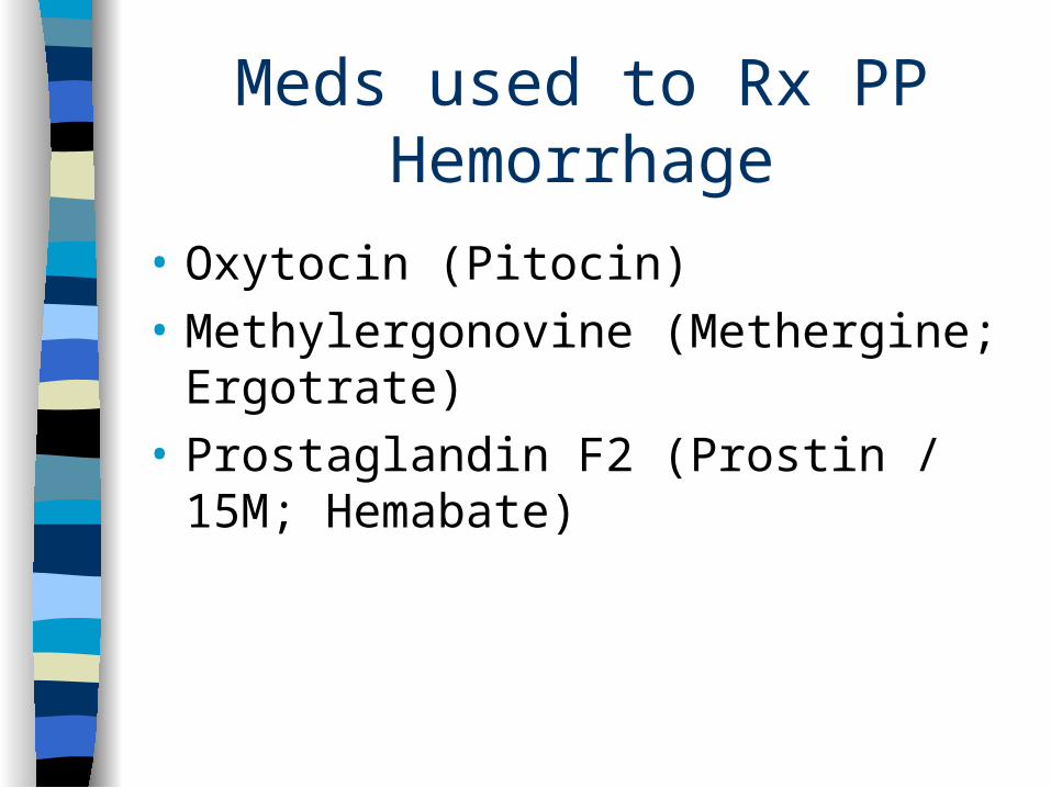
Meds used to Rx PP Hemorrhage
• Oxytocin (Pitocin)
• Methylergonovine (Methergine; Ergotrate)
• Prostaglandin F2 (Prostin / 15M; Hemabate)

Emergency: Hemorrhagic ShockAssessments:
• Respirations = rapid and shallow• Pulse = rapid, weak, irregular• BP = decreasing (late sign)• Skin = cool, pale, clammy• Urinary Output = decreasing• Level of Consciousness = lethargy to coma• Mental status = anxiety to coma• Central venous pressure = decreased
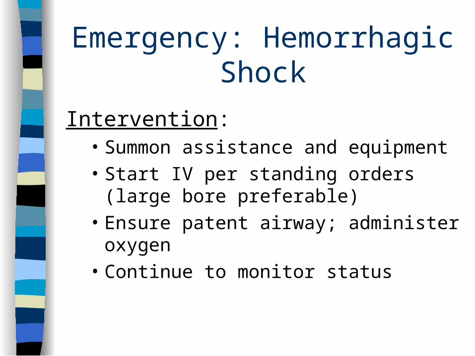
Emergency: Hemorrhagic Shock
Intervention:• Summon assistance and equipment• Start IV per standing orders (large bore
preferable)• Ensure patent airway; administer oxygen• Continue to monitor status

Coagulopathies• Idiopathic Thrombocytopenic Purpura: (ITP)• von Willebrand Disease: a type of
hemophilia, factor VIII deficiency, most common congenital clotting defect of women in childbearing years.
• Disseminated Intravascular Coagulation: (DIC) a pathologic form of clotting, diffuse. Includes platelets, fibrinogen, prothrombin, and factors V and VII.
• Thromboembolic Disease: formation of clot(s) in blood vessels caused by inflammation or partial obstruction of the vessel.

Postpartum Infection
• Antepartal factors:• Hx of previous venous trhombosis, UTI,
mastitis, pneumonia• Diabetes mellitus• Alcoholism• Drug abuse• Immunosuppression• Anemia• Malnutrition

Intrapartal Factors
• Cesarean birth
• PROM
• Chorioamnionitis
• Prolonged labor
• Bladder catheterization
• Internal fetal or uterine pressure monitor
• Multiple vaginal exams after ROM

Intrapartal Factors (continued)
• Epidural anesthesia
• Retained placental fragments
• PP hemorrhage
• Episiotomy or lacerations
• Hematomas
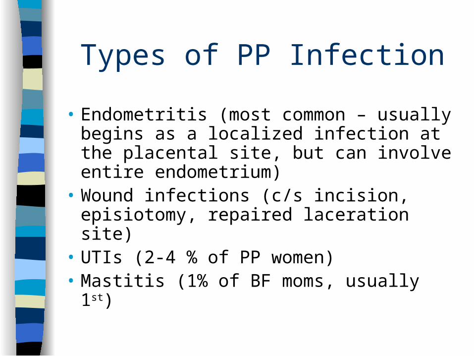
Types of PP Infection
• Endometritis (most common – usually begins as a localized infection at the placental site, but can involve entire endometrium)
• Wound infections (c/s incision, episiotomy, repaired laceration site)
• UTIs (2-4 % of PP women)• Mastitis (1% of BF moms, usually 1st)

Sequelae of Childbirth Trauma
• Uterine Displacement & prolapse
• Cystocele and Rectocele
• Urinary Incontinence
• Genital Fistulas

PP Psychologic Complications
• Mood Disorders: with or without psychotic features, if the onset occurs within 4 weeks of childbirth.• “Baby Blues” – occurs in up to 70% of PP
moms• Postpartum Depression • Postpartum Psychosis

High-Risk Newborn & Family
Chapter 26

Infants With Special Needs• Priorities
• Initiation & maintenance of respirations• Establishment of extrauterine circulation• Control of body temperature• Intake of adequate nourishment• Establishment of waste elimination• Establishment of an infant-parent
relationship• Prevention of infection• Provision of developmental care for mental
& social development
High-Risk Infants• May need resuscitation at birth.
• Most institutions require AHA Certification in Neonatal Resuscitation of all personnel at deliveries
• Requirements may include:• Warmth• Oxygen• Intubation• Suctioning

Small for Gestational Age (SGA)
• Definition: birth weight is below the 10th percentile on an intrauterine growth curve for that age infant.
• Infant could be preterm, term, or postterm.
• Have difficulty maintaining body warmth d/t low fat stores; may develop hypoglycemia from low glucose stores.

Large for Gestational Age (LGA)
• Definition: birthweight is above the 90th percentile on an intrauterine growth chart for that gestational age.
• Infant could be preterm, term, or postterm.
• Often are IDM (infants of diabetic mothers), and particularly prone to hypoglycemia or birth trauma.

Preterm Infants
• Definition: born before 37 weeks of gestation.
• Particular problems: respiratory function, anemia, jaundice, persistent patent ductus arteriosus, & intracranial hemorrhage.
• Low-birthweight infants = those weighting 1500-2500 grams.

• Very-low-birthweight infants = those weighing 1000-1500 grams.
• Extremely-very-low-birthweight infants = those weighing between 500-1000 grams.
• All such infants need intensive care from the moment of birth.
• Risks: neurologic after-effects caused by being so critically close to the age of viability.

Postterm Infants
• Definition: born after 42 weeks gestation.
• Particular problems: establishing respirations, meconium aspiration, hypoglycemia, temperature regulation, and polycythemia.

Respiratory Distress Syndrome• Commonly occurs in preterm infants
from a deficiency or lack of surfactant in the alveoli.
• Without surfactant the alveoli collapse on expiration & require extreme force for reinflation.
• Primary Rx: synthetic surfactant replacement at birth by ET tube insufflation, followed by oxygen and ventilatory support.
Transient Tachypnea
• A temporary condition caused by slow absorption of lung fluid at birth.
• Close observation of the infant is necessary until the fluid is absorbed and respirations slow to a normal rate.

Meconium Aspiration Syndrome
• Occurs when infant inhales meconium-stained amniotic fluid during birth.
• Results in irritation to the airway (from meconium) & may lead to both airway spasm and pneumonia.
• Infant needs: oxygen, ventilatory support, & possibly antibiotic until the effects of the airway subside.

Apnea:
• Definition: a pause in respirations longer than 20 seconds, with accompanying bradycardia.
• Occurs in preterm infants who have secondary stresses such as: infection, hyperbilirubinemia, hypoglycemia, or hypothermia.

Sudden Infant Death Syndrome
• Definition: the sudden, unexplained death of an infant.
• Associated with infants sleeping on their stomachs (prone) and infants who were born premature.
• Nursing prevention: advising parents to position their infant on the back for sleeping.

Hyperbilirubinemia
• Results from: destruction of RBCs, due either to a normal physiologic response or an abnormal destruction of the RBCs.
• Hemolytic disease of the newborn is destruction of RBCs from Rh or ABO incompatibility.
• Phototherapy or exchange transfusion is used to prevent kernicterus.

Neonatal Hemorrhagic Disease
• Definition: a lack of clotting ability resulting from a deficiency of vitamin K at birth.
• Prevention is by injection of vitamin K to all infants at birth.

Retinopathy of Prematurity
• Definition: destruction of the retina due to exposure of immature retinal capillaries to oxygen.
• Monitoring oxygen saturation via arterial blood gases is an important prevention measure.

Infections of the Newborn
• Streptococcal Group B pneumonia: from maternal GBBS.
• Hepatitis B infection
• Ophthalmia neonatorum: from gonococcal and chlamydial conjunctivitis.
• Herpes Virus infection.

Other Neonatal Risks:
• Infants of diabetic mothers (IDM)
• Infants of drug abusing women
NOTE: respiratory distress, hypoglycemia, hypo/hyperthermia are common S&S of neonatal infection.