histozoic myxosporeans infecting the stomach wall of
TRANSCRIPT
RESEARCH Open Access
Histozoic myxosporeans infecting thestomach wall of elopiform fishes representa novel lineage, the GastromyxidaeMark A. Freeman1,2* and Árni Kristmundsson3
Abstract
Background: Traditional studies on myxosporeans have used myxospore morphology as the main criterion foridentification and taxonomic classification, and it remains important as the fundamental diagnostic feature used toconfirm myxosporean infections in fish and other vertebrate taxa. However, its use as the primary feature in systematicshas led to numerous genera becoming polyphyletic in subsequent molecular phylogenetic analyses. It is now known thatother features, such as the site and type of infection, can offer a higher degree of congruence with molecular data, albeitwith its own inconsistencies, than basic myxospore morphology can reliably provide.
Methods: Histozoic gastrointestinal myxosporeans from two elopiform fish from Malaysia, the Pacific tarpon Megalopscyprinoides and the ten pounder Elops machnata were identified and described using morphological, histological andmolecular methodologies.
Results: The myxospore morphology of both species corresponds to the generally accepted Myxidium morphotype, butboth had a single nucleus in the sporoplasm and lacked valvular striations. In phylogenetic analyses they were robustlygrouped in a discrete clade basal to myxosporeans, with similar shaped myxospores, described from gill monogeneans,which are located at the base of the multivalvulid clade. New genera Gastromyxum and Monomyxum are erected toaccommodate these myxosporean taxa from fish and gill monogeneans respectively. Each are placed in a new family,the Gastromyxidae with Gastromyxum as the type genus and Monomyxidae with Monomyxum as the type genus.
Conclusions: To improve modern systematics of the myxosporeans it is clear that a combination of biological, ecological,morphological and molecular data should be used in descriptive studies, and the naming and redistribution of taxa andgenera is going to be necessary to achieve this. Here we demonstrate why some Myxidium-shaped myxospores shouldnot be included in the family Myxidiidae, and create two new families to accommodate them based on their site ofinfection, host biology / ecology, DNA sequence data and morphological observations. Subsequent descriptive worksneed to follow a similar course if we are going to create a prevailing and workable systematic structure for theMyxosporea.
Keywords: Gastromyxum, Monomyxidae, Monomyxum, Elopiformes, Monogenea, Histozoic, Hyperparasite
* Correspondence: [email protected] University School of Veterinary Medicine, Basseterre, St. Kitts, West Indies2Institute of Ocean and Earth Sciences, University of Malaya, Kuala Lumpur,MalaysiaFull list of author information is available at the end of the article
© 2015 Freeman and Kristmundsson. Open Access This article is distributed under the terms of the Creative CommonsAttribution 4.0 International License (http://creativecommons.org/licenses/by/4.0/), which permits unrestricted use, distribution,and reproduction in any medium, provided you give appropriate credit to the original author(s) and the source, provide a linkto the Creative Commons license, and indicate if changes were made. The Creative Commons Public Domain Dedicationwaiver (http://creativecommons.org/publicdomain/zero/1.0/) applies to the data made available in this article, unless otherwisestated.
Freeman and Kristmundsson Parasites & Vectors (2015) 8:517 DOI 10.1186/s13071-015-1140-7

BackgroundMyxosporeans are common parasites of fish and in re-cent molecular taxonomic studies they form two maingroups, being mainly from hosts that inhabit eitherfreshwater or marine environments [1, 2]. Known myx-osporean life cycles involve two hosts, with infections infish resulting in the production of myxospores, whilstmorphologically dissimilar actinospores are produced inan annelid invertebrate host. Traditional studies on myx-osporeans have used myxospore morphology as the maincriterion for identification and taxonomic classification.This has led to numerous genera becoming polyphyleticin subsequent molecular phylogenetic analyses and it isnow known that other features, such as the site of infec-tion in fish, can, for some clades of myxosporeans, offera far higher degree of congruence with molecular data,than basic myxospore morphology can provide [1, 3–5].However, myxospore morphology remains important, asit is the fundamental diagnostic feature that veterinar-ians and scientists use for identification and it remainsthe distinguishing characteristic of myxosporean infec-tions in fish and other vertebrate taxa.Myxidium incomptavermi was described from Myxi-
dium-shaped myxospores found infecting monogeneansfrom the gills of Megalops cyprinoides in Malaysia [6].However, monogeneans are not typical hosts for myxos-poreans and myxospores are not normally found in theinvertebrate host. In that study, no myxospores of M.incomptavermi were found in the fish that were host tothe infected monogeneans and multiple fish were foundto have monogeneans that were infected with myxos-pores. But, a specific PCR was able to detect the DNA ofM. incomptavermi in numerous tissues of the fish host,in particular the stomach and intestine, suggesting thatthe fish might be involved in the life cycle or the trans-mission of M. incomptavermi to gill monogeneans [6].The aim of the present study was to screen for the
presence of gastrointestinal myxosporeans from twoelopiform fishes (order: Elopiformes), from Malaysianmangrove systems, the Pacific tarpon M. cyprinoides andthe ten pounder Elops machnata. Both fish species arefound in shallow coastal marine and brackish waterenvironments, entering the mangroves during the night.The ten pounders or ladyfish (Elopiformes: Elopidae),and tarpons (Elopiformes: Megalopidae) together withtheir sister group the eels (Anguilliformes) form theElopomorpha which are one of the oldest major extantteleost lineages [7, 8].
MethodsElops machnata were purchased from the main fishmarket in Kuah, Langkawi. Megalops cyprinoides, werebought from the same market and also captured whilstnight fishing with gill nets in Kilim mangroves, northeast
Langkawi, and kept alive prior to examination. In addition,M. cyprinoides were purchased from the fish market atPangkor Island, Perak and captured live at the BachokMarine Research Station, Kelantan. All fish were exam-ined for the presence of histozoic myxosporeans in thegastrointestinal tract by scraping the stomach and intes-tine lining with a scalpel blade and viewing with an Olym-pus BX-41 compound microscope. Images of freshmyxospores were taken using a Leica DMLB digital cam-era using Cell imaging software. The dimensions of at least20 myxospores from each host fish were calculated usingImage J 1.42q [9] following the accepted guidelines of Lom& Arthur [10]. Elops machnata were also examined for thepresence of gill monogeneans, by removing each gill arch,placing it in seawater and observing between the primaryfilaments using a dissecting microscope.Tissue samples that were shown to be positive for
myxosporeans were fixed in 10 % buffered formalin forhistological analysis. After fixation, tissues were preparedfor standard wax histology and 4 μm sections weredewaxed, stained with Giemsa and examined using acompound microscope. Positive tissue samples were alsofixed for transmission electron microscopy (TEM) aspreviously described [11]. Fish that were found to haveheavy infections were used to prepare samples for scan-ning electron microscopy (SEM). For the SEM examin-ation, myxospores were removed from heavily infectedstomach wall tissues by scraping with a scalpel andplacing in a tube containing PBS. Tubes were then cen-trifuged at 1500g for 5 min, the supernatant removedand the pellet fixed in 2.5 % glutaraldehyde for 4h. Afterfixation, the spores were prepared for SEM and viewedfollowing the methods described by Kristmundsson andFreeman [12]. In brief, washed spores were syringedonto a polycarbonate membrane, fixed with 1 % osmiumtetroxide and dehydrated through an ethanol series. Mem-branes were dried, mounted on stubs, sputter-coated withgold and viewed, between 5 and 10 kV, with a FEI Quanta450 FEG FE-SEM.Infected gastrointestinal tissue or myxospore-positive
intestinal scrapings were fixed in 95 % ethanol or placeddirectly into DNA lysis buffer for molecular analyses.Total DNA was extracted using a GeneMATRIX DNAisolation kit (EURx Poland) following the tissue protocoland used as templates in subsequent PCR reactions.Parasite small subunit ribosomal DNA (SSU rDNA) wasamplified using the general myxosporean primers andmethodology described by Freeman et al. [4] and theKudoa-specific primer Kud-80f actgcgaagcgctcagta [13]with the new reverse primer Kud-790r cgcctgctttgagcactgtg, utilising the same PCR conditions. PCRs wereconducted on parasite DNA from 4 fish for each species.A specific PCR was designed and optimised for the newmyxosporean from M. cyprinoides, in order to be able to
Freeman and Kristmundsson Parasites & Vectors (2015) 8:517 Page 2 of 13
differentiate it from Myxidium incomptavermi in fish withdual infections or those with both DNA present. Theprimers Mi2-470f taccggagttgaccttcacg and Mi2-1040ractgatcccgtggtggcat amplified a 570bp region of SSU rDNAof this novel myxosporean, using the same PCR conditionsexcept utilising an annealing temperature of 65C. Two add-itional forward primers were designed to match the newmyxosporeans described in this study but not to anneal toM. incomptavermi. Gastro-250f actgtgcatatcgaatgggct andGastro-1100fagtacggtcgcaaggctga were used with the gen-eral reverse primers (1430r and 18gM respectively) andPCR conditions described by Freeman et al. [4]. All DNAsamples taken from M. cyprinoides were screened for M.incomptavermi using the specific PCR described by Free-man & Shinn [6] and spores measurements were only takenfrom PCR negative fish and those shown to be positive withthe specific PCR (Mi2-470f / Mi2-1040r).All PCRs were performed in triplicate and PCR prod-
ucts of the expected sizes were recovered using a Gene-MATRIX PCR products extraction kit (EURx Poland).Sequencing reactions were performed using BigDyeTMTerminator cycle sequencing chemistry utilising the sameoligonucleotide primers that were used for the originalPCRs. DNA sequencing was performed in both forwardand reverse directions for all PCR products and nucleotideBLAST searches performed for each sequence read toconfirm a myxosporean origin [14]. The contiguous se-quences were obtained manually using CLUSTAL X andBioEdit [15, 16]. CLUSTAL X was used for the initial SSUrDNA sequence alignments of the novel sequences and 31other histozoic marine myxosporeans. Percentage diver-gence matrices were constructed from selected alignedtaxa in CLUSTAL X using the neighbour-joining methodbased on the Kimura 2-parameter model [17].Phylogenetic analyses were performed using the max-
imum likelihood methodology in PhyML [18] with thegeneral time-reversible substitution model selected and1000 bootstrap repeats, and Bayesian inference (BI) ana-lysis using MrBayes v. 3.2 [19]. For the BI analysis, modelsof nucleotide substitution were first evaluated for thealignment using MrModeltest v. 2.2 [20]. The mostparameter-rich evolutionary model based on the AIC wasthe general time-reversible, GTR + I + G model of evolu-tion. Therefore, the settings used for the analysis were nst= 6, with the gamma-distributed rate variation across sitesand a proportion of invariable sites (rates = invgamma).The priors on state frequency were left at the defaultsetting (Prsetstatefreqpr = dirichlet (1,1,1,1)). Posteriorprobability distributions were generated using the MarkovChain Monte Carlo (MCMC) method with four chainsbeing run simultaneously for 1,000,000 generations. Burnin was set at 2500 and trees were sampled every 100 gen-erations making a total of 7500 trees used to compile themajority rule consensus trees.
Results and discussionSix Elops machnata ranging in size from 39–81 cm infork length (FL) and a total of 38 Megalops cyprinoides,26 from Langkawi (FL 19–32 cm), 9 from Pangkor (FL17–30) and 3 from Bachok (FL 20–25) were dissectedand checked for the presence on myxospores under themicroscope. All six E. machnata (100 % prevalence) hada histozoic myxosporean infecting the stomach wall. Atotal of 17 M. cyprinoides (45 % prevalence) also had amyxosporean in the stomach wall, 10 from Langkawi(38 % prevalence), seven from Pangkor (78 % preva-lence) and none from Bachok (0 % prevalence). No mor-phologically different myxosporeans were found lower inthe intestinal tract in either fish species and spore dens-ity was highest from the stomach tissue. No monoge-neans were found on the gills of E. machnata, the gillsof M. cyprinoides were not examined in this study.In E. machnata the myxospores were somewhat rem-
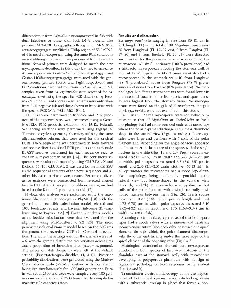
iniscent to that of Myxidium or Zschokkella in basicmorphology but had more rounded ends with raised tipswhere the polar capsules discharge and a clear rhomboidshape in the sutural view (Figs. 1a and 2a). Polar cap-sules were large and pyriform with 6 coils of the polarfilament and, depending on the angle of view, appearedto almost meet in the centre of the spore, with the singlenucleus to one side (Figs. 1a and 2a). Fresh spores mea-sured 7.92 (7.1–8.5) μm in length and 5.42 (4.9–5.9) μmin width, polar capsules measured 3.3 (3.0–3.5) μm inlength and 2.36 (2.1–2.5) μmin width n = 60 (3 fish). InM. cyprinoides the myxospores had a more Myxidium-like morphology, being moderately sigmoidal in thesutural view but lemon-shaped in the valvular view(Figs. 1b,c and 2b). Polar capsules were pyriform with 4coils of the polar filament with a single centrally posi-tioned nucleus between them (Fig. 2b). Fresh sporesmeasured 10.29 (7.84–11.56) μm in length and 5.64(4.72–6.78) μm in width, polar capsules measured 3.40(2.63–4.32) μm in length and 2.75 (1.69–3.87) μm inwidth n = 138 (5 fish).Scanning electron micrographs revealed that both spore
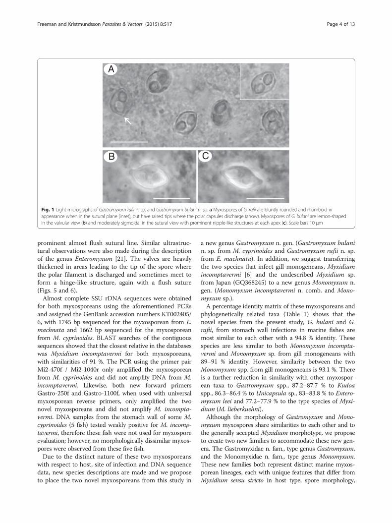
types had smooth valves with a sinuous and relativelyinconspicuous sutural line, each valve possessed one apicalelement, through which the polar filament discharges,with the other end tucking under the valve edge of theapical element of the opposing valve (Fig. 3 a-d).Histological examination showed that myxosporean
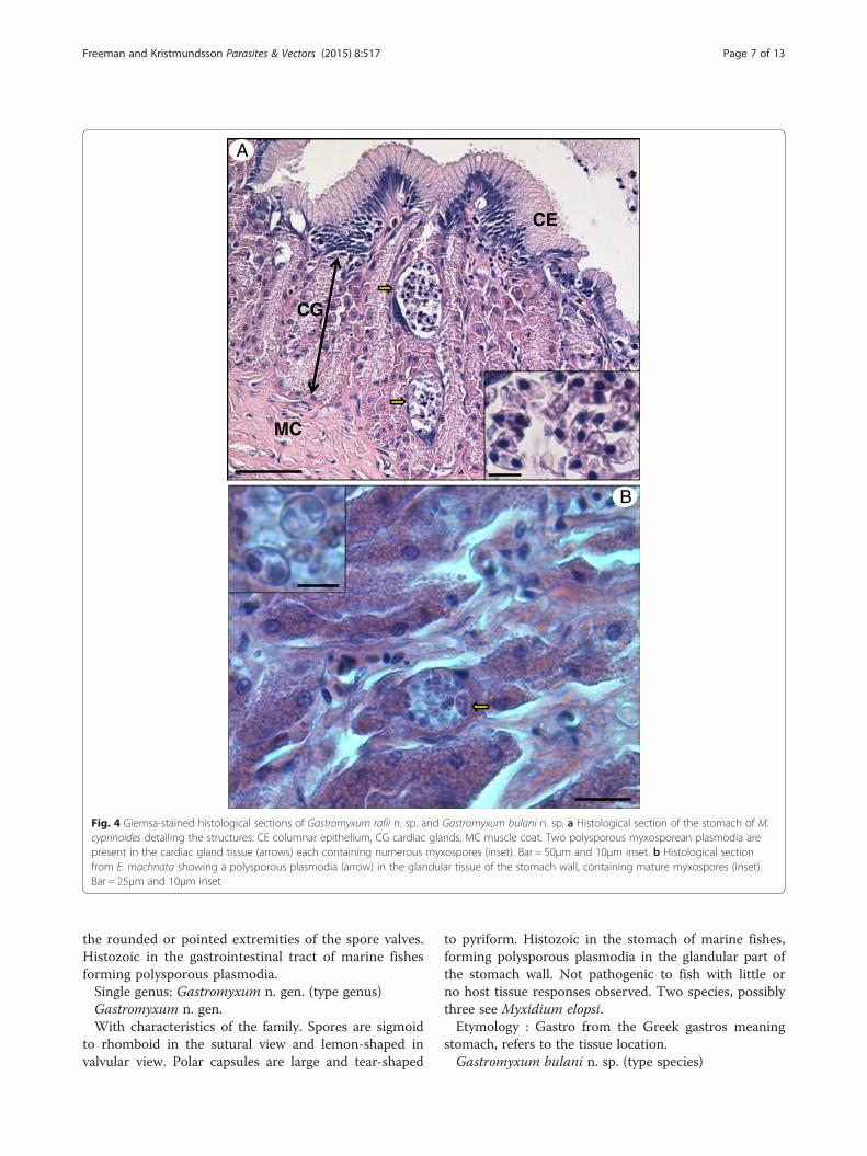
infections in both species of fish were histozoic in theglandular part of the stomach wall, with myxosporesdeveloping in polysporous plasmodia with no sign ofsignificant pathology or host responses being evident(Fig. 4 a and b).Transmission electron microscopy of mature myxos-
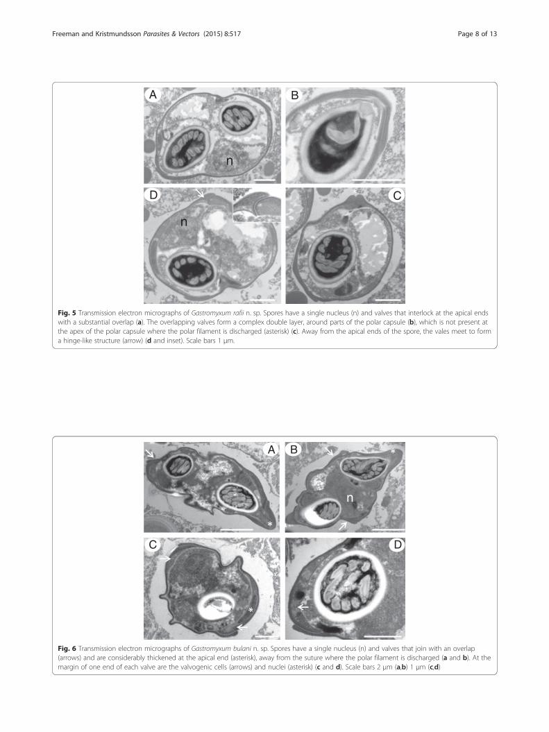
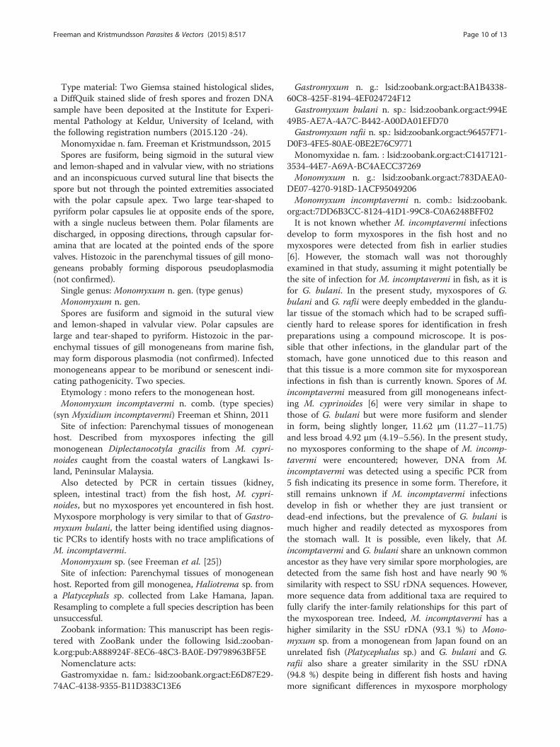
pores of both novel species reveal interlocking valveswith a substantial overlap in places that forms a non-
Freeman and Kristmundsson Parasites & Vectors (2015) 8:517 Page 3 of 13
prominent almost flush sutural line. Similar ultrastruc-tural observations were also made during the descriptionof the genus Enteromyxum [21]. The valves are heavilythickened in areas leading to the tip of the spore wherethe polar filament is discharged and sometimes meet toform a hinge-like structure, again with a flush suture(Figs. 5 and 6).Almost complete SSU rDNA sequences were obtained
for both myxosporeans using the aforementioned PCRsand assigned the GenBank accession numbers KT002405/6, with 1745 bp sequenced for the myxosporean from E.machnata and 1662 bp sequenced for the myxosporeanfrom M. cyprinoides. BLAST searches of the contiguoussequences showed that the closest relative in the databaseswas Myxidium incomptavermi for both myxosporeans,with similarities of 91 %. The PCR using the primer pairMi2-470f / Mi2-1040r only amplified the myxosporeanfrom M. cyprinoides and did not amplify DNA from M.incomptavermi. Likewise, both new forward primersGastro-250f and Gastro-1100f, when used with universalmyxosporean reverse primers, only amplified the twonovel myxosporeans and did not amplify M. incompta-vermi. DNA samples from the stomach wall of some M.cyprinoides (5 fish) tested weakly positive for M. incomp-tavermi, therefore these fish were not used for myxosporeevaluation; however, no morphologically dissimilar myxos-pores were observed from these five fish.Due to the distinct nature of these two myxosporeans
with respect to host, site of infection and DNA sequencedata, new species descriptions are made and we proposeto place the two novel myxosporeans from this study in
a new genus Gastromyxum n. gen. (Gastromyxum bulanin. sp. from M. cyprinoides and Gastromyxum rafii n. sp.from E. machnata). In addition, we suggest transferringthe two species that infect gill monogeneans, Myxidiumincomptavermi [6] and the undescribed Myxidium sp.from Japan (GQ368245) to a new genus Monomyxum n.gen. (Monomyxum incomptavermi n. comb. and Mono-myxum sp.).A percentage identity matrix of these myxosporeans and
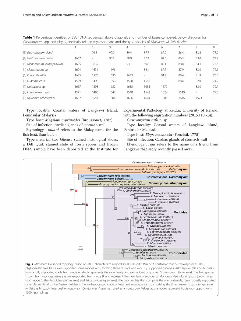
phylogenetically related taxa (Table 1) shows that thenovel species from the present study, G. bulani and G.rafii, from stomach wall infections in marine fishes aremost similar to each other with a 94.8 % identity. Thesespecies are less similar to both Monomyxum incompta-vermi and Monomyxum sp. from gill monogeneans with89–91 % identity. However, similarity between the twoMonomyxum spp. from gill monogeneans is 93.1 %. Thereis a further reduction in similarity with other myxospor-ean taxa to Gastromyxum spp., 87.2–87.7 % to Kudoaspp., 86.3–86.4 % to Unicapsula sp., 83–83.8 % to Entero-myxum leei and 77.2–77.9 % to the type species of Myxi-dium (M. lieberkuehni).Although the morphology of Gastromyxum and Mono-
myxum myxospores share similarities to each other and tothe generally accepted Myxidium morphotype, we proposeto create two new families to accommodate these new gen-era. The Gastromyxidae n. fam., type genus Gastromyxum,and the Monomyxidae n. fam., type genus Monomyxum.These new families both represent distinct marine myxos-porean lineages, each with unique features that differ fromMyxidium sensu stricto in host type, spore morphology,
A
B C
Fig. 1 Light micrographs of Gastromyxum rafii n. sp. and Gastromyxum bulani n. sp. a Myxospores of G. rafii are bluntly rounded and rhomboid inappearance when in the sutural plane (inset), but have raised tips where the polar capsules discharge (arrow). Myxospores of G. bulani are lemon-shapedin the valvular view (b) and moderately sigmoidal in the sutural view with prominent nipple-like structures at each apex (c). Scale bars 10 μm
Freeman and Kristmundsson Parasites & Vectors (2015) 8:517 Page 4 of 13

tissue location and development and DNA sequence data.The type species of Myxidium, M. lieberkuehni is com-monly found as coelozoic in the urinary system of thefreshwater pike, Esox lucius, in the holarctic region of thenorthern hemisphere, it has elongated and striated myxos-pores with a sporoplasm containing two nuclei unlikethose in the current description that have a single nucleusand smooth spore valves. In addition, M. lieberkuehni is
robustly phylogenetically placed away from marine myx-osporeans in a discrete clade with other coelozoic speciesinfecting freshwater fishes [1, 12, 22].Phylogenetic analyses of some of the marine histozoic
myxosporeans consistently and robustly place membersof the Gastromyxidae and Monomyxidae at the base ofthe Multivalvulida (Kudoidae and Trilosporidae) (Fig. 7).With the currently available DNA data, members of the
B
A
Fig. 2 Line drawings of myxospores of (a) Gastromyxum rafii n. sp. and (b) Gastromyxum bulani n. sp. Spores in sutural view on left hand side, theposition of the suture is shown with a dashed line. Spores shown in valvular view on the right hand side. Scale bar 5 μm
Freeman and Kristmundsson Parasites & Vectors (2015) 8:517 Page 5 of 13
Enteromyxum, Gastromyxum and Monomyxum generaform separate but discrete and well-supported clades inthis histozoic group (Fig. 7). The two Monomyxum spp.have not always convincingly formed a discrete clade,and in the analyses by Freeman and Shinn [6] they wereunresolved but adjacent taxa in the tree. However, theaddition of the two Gastromyxum sequences has addedstability to this part of the phylogenetic tree. To confirmthis we aligned the two novel Gastromyxum sequencesto the original alignment file used in the Freeman andShinn publication [6]. With the Gastromyxum sequencesadded to the original alignment, a maximum likelihoodanalysis (performed as previously described [6]) alsoplaced the two Monomyxum sequences in a discreteclade with a support of 82 % (data not shown). This con-firms the phylogenetic relationship, within Monomyxum,that we demonstrate in Fig. 7, which supports the cre-ation of the novel family Monomyxidae for Monomyxumspp. In addition, in other recent extensive phylogeneticstudies of the myxosporeans, even without the inclusionof the Gastromyxum sequences, it has also been shownthat the two Monomyxum sequences are placed togetherin a discrete clade, albeit only with moderate support[23]. In the same study, it was demonstrated that Cera-tonova spp. are related to the Enteromyxum and Kudoaclades [23], and their relationship to Enteromyxum andthe Gastromyxidae is further supported as they are alsohistozoic parasites in the intestine of fishes. However,the genus Ceratonova was recently placed in the family
Ceratomyxidae [24]. This reversion to myxospore morph-ology as the primary taxonomic criterion could be due tothe complications of the freshwater hosts for Ceratonovaspp., and the early inclusion of Ceratonova shasta inCeratomyxa, but is more likely due to the lack of a suit-able family placement for these enteric histozoic cerato-myxid forms. As the region of the phylogenetic tree forenteric myxosporeans is relatively sparse, we are unable toexpand the boundaries of the Gastromyxidae to includeother genera and create a pararphyletic family. However,the placement of Enteromyxum spp. in the Myxidiidaeand Ceratonova spp. in the Ceratomyxidae is incorrect,and the erection of new families to accommodate thesegenera is warranted in the future.
Taxonomic summaryClass Myxosporea Bütschli, 1881Order Bivalvulida Shulman, 1959Suborder Variisporina Lom et Noble, 1984Gastromyxidae n. fam. Freeman et Kristmundsson, 2015Spores are generally fusiform, being sigmoid to rhom-
boid in the sutural view and lemon to crescent-shaped andin valvular view, with no striations and an inconspicuouscurved sutural line that bisects the spore but not throughthe pointed extremities associated with the polar capsuleapex. Two large tear-shaped to pyriform polar capsules lieat opposite ends of the spore, with a single nucleusbetween them. Polar filaments are discharged, in opposingdirections, through capsular foramina that are located at
C
A B
D
Fig. 3 Scanning electron micrographs of Gastromyxum rafii n. sp. and Gastromyxum bulani n. sp. Gastromyxum rafii spores have rounded ends(a) and are rhomboid in the sutural view (b). Gastromyxum bulani spores are lightly fusiform in the valvular view (c), sigmoid in the sutural view(d) and have more pointed ends with an apical opening for the polar filament to be released through (inset d). Both species have smooth valvesand non-prominent sutural lines. Each valve has one rounded/pointed end which forms a nipple-like structure at its apex, with the other endconcealed beneath the apex of the opposing valve. Scale bars 3 μm (inset 1 μm).
Freeman and Kristmundsson Parasites & Vectors (2015) 8:517 Page 6 of 13
the rounded or pointed extremities of the spore valves.Histozoic in the gastrointestinal tract of marine fishesforming polysporous plasmodia.Single genus: Gastromyxum n. gen. (type genus)Gastromyxum n. gen.With characteristics of the family. Spores are sigmoid
to rhomboid in the sutural view and lemon-shaped invalvular view. Polar capsules are large and tear-shaped
to pyriform. Histozoic in the stomach of marine fishes,forming polysporous plasmodia in the glandular part ofthe stomach wall. Not pathogenic to fish with little orno host tissue responses observed. Two species, possiblythree see Myxidium elopsi.Etymology : Gastro from the Greek gastros meaning
stomach, refers to the tissue location.Gastromyxum bulani n. sp. (type species)
MC
CG
CE
A
B
Fig. 4 Giemsa-stained histological sections of Gastromyxum rafii n. sp. and Gastromyxum bulani n. sp. a Histological section of the stomach of M.cyprinoides detailing the structures: CE columnar epithelium, CG cardiac glands, MC muscle coat. Two polysporous myxosporean plasmodia arepresent in the cardiac gland tissue (arrows) each containing numerous myxospores (inset). Bar = 50μm and 10μm inset. b Histological sectionfrom E. machnata showing a polysporous plasmodia (arrow) in the glandular tissue of the stomach wall, containing mature myxospores (inset).Bar = 25μm and 10μm inset
Freeman and Kristmundsson Parasites & Vectors (2015) 8:517 Page 7 of 13
A B
CD
n
n
*
Fig. 5 Transmission electron micrographs of Gastromyxum rafii n. sp. Spores have a single nucleus (n) and valves that interlock at the apical endswith a substantial overlap (a). The overlapping valves form a complex double layer, around parts of the polar capsule (b), which is not present atthe apex of the polar capsule where the polar filament is discharged (asterisk) (c). Away from the apical ends of the spore, the vales meet to forma hinge-like structure (arrow) (d and inset). Scale bars 1 μm.
n
A B
C D
Fig. 6 Transmission electron micrographs of Gastromyxum bulani n. sp. Spores have a single nucleus (n) and valves that join with an overlap(arrows) and are considerably thickened at the apical end (asterisk), away from the suture where the polar filament is discharged (a and b). At themargin of one end of each valve are the valvogenic cells (arrows) and nuclei (asterisk) (c and d). Scale bars 2 μm (a,b) 1 μm (c,d)
Freeman and Kristmundsson Parasites & Vectors (2015) 8:517 Page 8 of 13
Type locality: Coastal waters of Langkawi Island,Peninsular MalaysiaType host: Megalops cyprinoides (Broussonet, 1782)Site of infection: cardiac glands of stomach wallEtymology : bulani refers to the Malay name for the
fish host, ikan bulanType material: two Giemsa stained histological slides,
a Diff Quik stained slide of fresh spores and frozenDNA sample have been deposited at the Institute for
Experimental Pathology at Keldur, University of Iceland,with the following registration numbers (2015.110 -14).Gastromyxum rafii n. sp.Type locality: Coastal waters of Langkawi Island,
Peninsular MalaysiaType host: Elops machnata (Forsskål, 1775)Site of infection: Cardiac glands of stomach wallEtymology : rafii refers to the name of a friend from
Langkawi that sadly recently passed away.
Table 1 Percentage identities of SSU rDNA sequences, above diagonal, and number of bases compared, below diagonal, forGastromyxum spp. and phylogenetically related myxosporeans and the type species of Myxidium, M. lieberkuehni
1 2 3 4 5 6 7 8 9
(1) Gastromyxum elopsi - 94.8 90.9 89.4 87.7 87.2 86.4 83.8 77.9
(2) Gastromyxum bulani 1657 - 90.8 88.9 87.3 87.6 86.3 83.0 77.2
(3) Monomyxum incomptavermi 1695 1635 - 93.1 89.6 89.1 88.8 84.1 77.3
(4) Monomyxum sp. 1694 1634 1696 - 88.1 87.7 87.9 83.0 76.1
(5) Kudoa thyrsites 1635 1576 1630 1633 - 91.2 88.4 81.9 75.0
(6) K. amamiensis 1559 1498 1556 1558 1558 - 88.4 82.0 74.2
(7) Unicapsula sp. 1657 1598 1652 1655 1635 1572 - 83.0 76.7
(8) Enteromyxum leei 1571 1490 1547 1548 1543 1522 1540 - 77.0
(9) Myxidium lieberkuehni 1632 1551 1604 1606 1604 1586 1616 1573 -
Fig. 7 Maximum likelihood topology based on 1851 characters of aligned small subunit rDNA of 33 histozoic marine myxosporeans. Thephylogenetic tree has a well-supported spine (nodes A-C), forming three distinct and robustly supported groups. Gastromyxum rafii and G. bulaniform a fully supported clade from node A which represents the new family and genus Gastromyxidae: Gastromyxum (blue area). The two speciesknown from monogeneans are well-supported from node B, and represent the new family and genus Monomyxidae: Monomyxum (brown area).From node C, the Kudoidae (purple area) and Trilosporidae (grey area), the two families that comprise the multivalvulida, form robustly supportedsister clades. Basal to the Gastromyxidae is the well-supported clade of intestinal myxosporeans comprising the Enteromyxum spp. (orange area),whilst the histozoic intestinal myxosporean Ceratonova shasta was used as an outgroup. Values at the nodes represent bootstrap support from1000 resamplings
Freeman and Kristmundsson Parasites & Vectors (2015) 8:517 Page 9 of 13
Type material: Two Giemsa stained histological slides,a DiffQuik stained slide of fresh spores and frozen DNAsample have been deposited at the Institute for Experi-mental Pathology at Keldur, University of Iceland, withthe following registration numbers (2015.120 -24).Monomyxidae n. fam. Freeman et Kristmundsson, 2015Spores are fusiform, being sigmoid in the sutural view
and lemon-shaped and in valvular view, with no striationsand an inconspicuous curved sutural line that bisects thespore but not through the pointed extremities associatedwith the polar capsule apex. Two large tear-shaped topyriform polar capsules lie at opposite ends of the spore,with a single nucleus between them. Polar filaments aredischarged, in opposing directions, through capsular for-amina that are located at the pointed ends of the sporevalves. Histozoic in the parenchymal tissues of gill mono-geneans probably forming disporous pseudoplasmodia(not confirmed).Single genus: Monomyxum n. gen. (type genus)Monomyxum n. gen.Spores are fusiform and sigmoid in the sutural view
and lemon-shaped in valvular view. Polar capsules arelarge and tear-shaped to pyriform. Histozoic in the par-enchymal tissues of gill monogeneans from marine fish,may form disporous plasmodia (not confirmed). Infectedmonogeneans appear to be moribund or senescent indi-cating pathogenicity. Two species.Etymology : mono refers to the monogenean host.Monomyxum incomptavermi n. comb. (type species)
(syn Myxidium incomptavermi) Freeman et Shinn, 2011Site of infection: Parenchymal tissues of monogenean
host. Described from myxospores infecting the gillmonogenean Diplectanocotyla gracilis from M. cypri-noides caught from the coastal waters of Langkawi Is-land, Peninsular Malaysia.Also detected by PCR in certain tissues (kidney,
spleen, intestinal tract) from the fish host, M. cypri-noides, but no myxospores yet encountered in fish host.Myxospore morphology is very similar to that of Gastro-myxum bulani, the latter being identified using diagnos-tic PCRs to identify hosts with no trace amplifications ofM. incomptavermi.Monomyxum sp. (see Freeman et al. [25])Site of infection: Parenchymal tissues of monogenean
host. Reported from gill monogenea, Haliotrema sp. froma Platycephals sp. collected from Lake Hamana, Japan.Resampling to complete a full species description has beenunsuccessful.Zoobank information: This manuscript has been regis-
tered with ZooBank under the following lsid.:zooban-k.org:pub:A888924F-8EC6-48C3-BA0E-D9798963BF5ENomenclature acts:Gastromyxidae n. fam.: lsid:zoobank.org:act:E6D87E29-
74AC-4138-9355-B11D383C13E6
Gastromyxum n. g.: lsid:zoobank.org:act:BA1B4338-60C8-425F-8194-4EF024724F12Gastromyxum bulani n. sp.: lsid:zoobank.org:act:994E
49B5-AE7A-4A7C-B442-A00DA01EFD70Gastromyxum rafii n. sp.: lsid:zoobank.org:act:96457F71-
D0F3-4FE5-80AE-0BE2E76C9771Monomyxidae n. fam. : lsid:zoobank.org:act:C1417121-
3534-44E7-A69A-BC4AECC37269Monomyxum n. g.: lsid:zoobank.org:act:783DAEA0-
DE07-4270-918D-1ACF95049206Monomyxum incomptavermi n. comb.: lsid:zoobank.
org:act:7DD6B3CC-8124-41D1-99C8-C0A6248BFF02It is not known whether M. incomptavermi infections
develop to form myxospores in the fish host and nomyxospores were detected from fish in earlier studies[6]. However, the stomach wall was not thoroughlyexamined in that study, assuming it might potentially bethe site of infection for M. incomptavermi in fish, as it isfor G. bulani. In the present study, myxospores of G.bulani and G. rafii were deeply embedded in the glandu-lar tissue of the stomach which had to be scraped suffi-ciently hard to release spores for identification in freshpreparations using a compound microscope. It is pos-sible that other infections, in the glandular part of thestomach, have gone unnoticed due to this reason andthat this tissue is a more common site for myxosporeaninfections in fish than is currently known. Spores of M.incomptavermi measured from gill monogeneans infect-ing M. cyprinoides [6] were very similar in shape tothose of G. bulani but were more fusiform and slenderin form, being slightly longer, 11.62 μm (11.27–11.75)and less broad 4.92 μm (4.19–5.56). In the present study,no myxospores conforming to the shape of M. incomp-tavermi were encountered; however, DNA from M.incomptavermi was detected using a specific PCR from5 fish indicating its presence in some form. Therefore, itstill remains unknown if M. incomptavermi infectionsdevelop in fish or whether they are just transient ordead-end infections, but the prevalence of G. bulani ismuch higher and readily detected as myxospores fromthe stomach wall. It is possible, even likely, that M.incomptavermi and G. bulani share an unknown commonancestor as they have very similar spore morphologies, aredetected from the same fish host and have nearly 90 %similarity with respect to SSU rDNA sequences. However,more sequence data from additional taxa are required tofully clarify the inter-family relationships for this part ofthe myxosporean tree. Indeed, M. incomptavermi has ahigher similarity in the SSU rDNA (93.1 %) to Mono-myxum sp. from a monogenean from Japan found on anunrelated fish (Platycephalus sp.) and G. bulani and G.rafii also share a greater similarity in the SSU rDNA(94.8 %) despite being in different fish hosts and havingmore significant differences in myxospore morphology
Freeman and Kristmundsson Parasites & Vectors (2015) 8:517 Page 10 of 13
(Figs. 1, 2, 3). These generic relationships are confirmed inthe phylogenetic analyses (Fig. 7) which support monoge-neans as being true hosts for myxosporeans and demon-strates that the two Monomyxum spp. share a more recentcommon ancestor than they do to either Gastromyxumsp. from fish and vica versa. However, this allows us tospeculate that myxospore morphology may be more con-served in certain host systems. Gastromyxum bulani andM. incomptavermi share very similar myxospore morph-ologies and are both found on the same species of fish,albeit one in a monogenean; however, they are less genet-ically related to each other than each is to another,different shaped, myxospore form in a different fish /monogenean host system. This might suggest that evo-lutionary changes in myxospore morphology couldoccur at a slower rate in one species of fish, even whenbecoming hyperparsitic in their gill monogeneans, thanit does when a switching of fish host has occurred, evenif the fish are found in the same environment, as is thecase for M. cyprinoides and E. machnata. Put simply, ifmyxospore morphology evolved evenly in all host sys-tems, we might expect the more closely related Gastro-myxum spp. to look more similar, when in fact, G.bulani looks remarkably similar to M. incomptavermi,known from the same fish host, despite being moredistantly related to it. It is also possible that it is acoincidence that these spores have near identicalmorphologies, however, we consider this unlikely as mor-phological variability among Myxidium-shaped spores iswide-ranging [26], and the similarities between these twospecies is very striking. It is also noteworthy that G. bulaniand G. rafii both infect elopiform fishes (order: Elopi-formes), from mangrove systems from the west coast ofpeninsular Malaysia, suggesting that elopiform fish maybe a more common host to Gastromyxum spp.. Interest-ingly, the myxosporean, Myxidium elopsi, described fromthe intestine of Elops senegalensis from the Atlantic coastof West Africa [27] has a very similar spore morphologyto G. rafii and is found as histozoic in the intestine of aclosely related elopiform fish. It is highly likely that M.elopsi belongs in the genus Gastromyxum, and its similarshape to G. rafii and elopiform host further support thetheories that myxospore morphology is potentially betterconserved in a single species or closely related fish species(same genus), and that elopiform fishes may be a commonhost for Gatromyxum spp.No monogeneans were found on the gills of E. mach-
nata in this study and PCR testing of gill monogeneanDNA from M. cyprinoides from a previous study [6],using specific PCRs designed in this study for Gastro-myxum, did not amplify G. bulani. This suggests thatGastromyxum spp. do not infect gill monogeneans, butthis will require further research effort to unambiguouslydemonstrate.
Traditional myxosporean taxonomy has been heavilydependent upon the morphology of myxospores todifferentiate between the numerous different taxa.More recently, multiple molecular phylogenetic studiesof the Myxosporea have repeatedly and robustly dem-onstrated that the generic assignment of taxa based onmyxospore morphology has resulted in a polyphyleticdistribution of many genera, which has led to somegenera such as Myxidium and Zschokkella becomingmisleadingly high in species number, many of which areincorrectly assigned. It is also clear that some myxos-pore morphotypes have formed or reformed on morethan one occasion during myxosporean evolution,which is probably due to comparable biological and en-vironmental constraints and pressures that needed tobe satisfied during their evolution, but does not neces-sarily mean they belong in the same family or genus.Many older studies provide excellent descriptions, butassign primary weight to myxospore morphology (noDNA data available) which unknowingly led to incor-rect taxonomic placements with respect to genus orfamily. However, surprisingly numerous recent descrip-tions, also provide good taxonomic data but still assignprimary weight to myxospore morphology, leading togeneric assignments that are inconsistent with knownmolecular data [6, 24].As myxospores remain our definitive confirmation of
myxosporean infections in vertebrates, we must findalternative ways in which to develop a more meaningfulsystematic approach for the nomenclature of myxos-porean taxa. As myxospore presence and morphology isoften our first point of reference it must remain part ofthe overall approach, but must be combined with, butcome second, to other potentially more meaningfuldata. However, it is not clear how this can be easilyachieved and readily applied across the whole group.The site of infection in the host (muscle, GB, urinarysystem etc.) and whether development is histozoic orcoelozoic is useful and gives strong congruence withmolecular data for some clades but it is not clear in all,for example the parvicapsulids. Histopathology can re-veal different developmental stages and important in-teractions with host cells, but if cross-referenced toinfections at a differing stage could be misleading andmay vary with both parasite and host strains. Host spe-cies and environmental data (marine, freshwater, geo-graphic location etc.) can show good compatibility tomolecular phylogenies, but again numerous exceptionsexist, such as the marine myxobolids.In short, if we want a new taxonomic framework for
the myxosporeans to reflect molecular phylogenies, it isimperative to provide good molecular data with everydescription. But, this must be augmented with as muchother relevant data as possible.
Freeman and Kristmundsson Parasites & Vectors (2015) 8:517 Page 11 of 13

ConclusionsDuring this study, it became clear that the discovery ofmyxosporean parasites, found as histozoic infections inthe stomach of elopiform fishes from Malaysia, were noveland that they required taxonomic description. However,when we evaluated all the taxonomic information gath-ered herein, no family placement or generic assignmentwas suitable. Therefore, we have placed less emphasis onmyxospore morphology and created a new family andgenus, the Gastromyxidae: Gastromyxum, to accommo-date them. During this process, it also became clear thatmyxosporean taxa infecting gill monogeneans were adistinct lineage, that have been incorrectly assigned toMyxidium due to the dogma of following myxosporemorphology as the primary taxonomic criterion [6], andthat a new generic and family assignment, the Monomyxi-dae: Monomyxum, was called for. In many cases, myxos-pore morphology will be able to provide useful characterswithin certain groups/clades, which may correspond tocertain myxosporean genera, such as in the Gastromyxum;however, it must be used carefully and in combinationwith other important features for that particular group orclade of myxosporeans.In order to resolve the perennial taxonomic issues
surrounding this group, deeper taxonomic assignmentand resolution is going to be required. Here we demon-strate and give sound reasons why some species withMyxidium-shaped myxospores should not be included inthe family Myxidiidae, and subsequent descriptive workswill hopefully follow a similar course so we can collect-ively create a modern and workable phylogenetic frame-work for the myxosporeans.
Competing interestsThe authors declare that they have no competing interests.
Authors’ contributionsMF designed the study and ran the project. ÁK and MF sampled anddissected the fish. ÁK did the histopathology and MF did the SEM, TEM andmolecular work. MF and ÁK wrote the manuscript and both authorsapproved the final version.
AcknowledgementsThis project was financially supported by a University of Malaya ResearchGrant RP001L-13SUS. We would like to thank Hasbi from Kilim, Langkawiwhose knowledge as a local fisherman was vital for the collection of thelive fish needed for this study and for the memorable times on his boat.We would also like to thank the reviewers for their constructive and helpfulsuggestions that helped form the final version of this manuscript.
Author details1Ross University School of Veterinary Medicine, Basseterre, St. Kitts, West Indies.2Institute of Ocean and Earth Sciences, University of Malaya, Kuala Lumpur,Malaysia. 3Institute for Experimental Pathology, University of Iceland, Reykjavik,Iceland.
Received: 4 May 2015 Accepted: 3 October 2015
References1. Fiala I. The phylogeny of Myxosporea (Myxozoa) based on small subunit
ribosomal RNA gene analysis. Int J Parasitol. 2006;36:1521–34.2. Holzer AS, Wootten R, Sommerville C. The secondary structure of the
unusually long 18S ribosomal RNA of the myxozoan Sphaerospora truttaeand structural evolutionary trends in the Myxozoa. Int J Parasitol.2007;37:1281–95.
3. Holzer AS, Sommerville C, Wootten R. Molecular relationshipsand phylogeny in a community of myxosporeans and actinosporeans basedon their 18S rDNA sequences. Int J Parasitol. 2004;34:1099–111.
4. Freeman MA, Yokoyama H, Ogawa K. Description and phylogeny ofCeratomyxa anko sp. n. and Zschokkella lophii sp. n. from the Japaneseanglerfish, Lophius litulon (Jordan). J Fish Dis. 2008;31:921–30.
5. Bartošová P, Freeman MA, Yokoyama H, Caffara M, Fiala I. Phylogeneticposition of Sphaerospora testicularis and Latyspora scomberomori n. gen.n. sp.(Myxozoa) within the marine urinary clade. Parasitology. 2011;138:381–93.
6. Freeman MA, Shinn AP. Myxosporean hyperparasites of gill monogeneansare basal to the Multivalvulida. Parasit Vectors. 2011;4:220.
7. Chen JN, López JA, Lavoué S, Miya M, Chen WJ. Phylogeny of theElopomorpha (Teleostei): evidence from six nuclear and mitochondrialmarkers. Mol Phylogenet Evol. 2014;70:152–61.
8. Wiley EO, Johnson GD. A teleost classification based on monophyleticgroups. In: Nelson JS, Schultze H-P, Wilson MVH, editors. Origin andPhylogenetic Interrelationships of Teleosts. 2010. Verlag Dr. Friedrich Pfeil,pp. 123–182.
9. Schneider CA, Rasband WS, Eliceiri KW. NIH Image to ImageJ: 25 years ofimage analysis. Nat Methods. 2012;9:671–5.
10. Lom J, Arthur JR. A guideline for the preparation of species description onMyxosporea. J Fish Dis. 1989;12:151–6.
11. Freeman MA, Kristmundsson Á. Ultrastructure of Nucleospora cyclopteri, anintranuclear microsporidian infecting the Atlantic lumpfish (Cyclopteruslumpus L.). Bul Eur Ass Fish Pathol. 2013;33:194–8.
12. Kristmundsson Á, Freeman MA. Sphaeromyxids form part of a diverse groupof myxosporeans infecting the hepatic biliary systems of a wide range ofhost organisms. Parasit Vectors. 2013;6:51.
13. Kristmundsson Á, Freeman MA. Negative effects of Kudoa islandica n. sp.(Myxosporea: Kudoidae) on aquaculture and wild fisheries in Iceland. Int JParasitol Parasites Wildl. 2014;3:135–46.
14. Zhang Z, Schwartz S, Wagner L, Miller W. A greedy algorithm for aligningDNA sequences. J Comput Biol. 2000;7:203–14.
15. Thompson JD, Gibson TJ, Plewniak F, Jeanmougin F, Higgins DG. TheCLUSTAL-X windows interface: flexible strategies for multiple sequencealignment aided by quality analysis tools. Nucl Acids Res. 1997;24:4876–82.
16. Hall TA. BioEdit: a user-friendly biological sequence alignment editor andanalysis program for Windows 95/98/ NT. Nucleic Acids Symp Ser.1999;41:95–8.
17. Saitou N, Nei M. The neighbour-joining method: a new method forreconstructing phylogenetic trees. Mol Biol Evol. 1987;4:406–25.
18. Guindon S, Dufayard JF, Lefort V, Anisimov M, Hordijk W, Gascuel O.New algorithms and methods to estimate maximum-likelihoodphylogenies:assessing the performance of PhyML 3.0. Syst Biol. 2010;59:307–21.
19. Ronquist F, Huelsenbeck JP. MrBayes 3: Bayesian phylogenetic inferenceunder mixed models. Bioinformatics. 2003;19:1572–4.
20. Nylander JAA, Ronquist F, Huelsenbeck JP, Nieves-Aldrey JL. Bayesianphylogenetic analysis of combined data. Syst Biol. 2004;53:47–67.
21. Palenzuela O, Redondo MJ, Alvarez-Pellitero P. Description of Enteromyxumscophthalmi gen. nov., sp. nov. (Myxozoa), an intestinal parasite of turbot(Scophthalmus maximus L.) using morphological and ribosomal RNAsequence data. Parasitology. 2002;124:369–79.
22. Lom J, Dyková I, Feist S. Myxosporea induced xenoma formation in pikeEsox lucius L. renal corpuscles associated with Myxidium lieberkhuehniinfection. Eur J Protistol. 1989;24:271–80.
23. Fiala I, Hlavničková M, Kodádková A, Freeman MA, Bartošová-Sojková P,Atkinson SD. Evolutionary origin of Ceratonova shasta and phylogeny of themarine myxosporean lineage. Mol Phylogenet Evol. 2015;86:75–89.
24. Atkinson SD, Foott JS, Bartholomew JL. Erection of Ceratonova n. gen.(Myxosporea: Ceratomyxidae) to encompass freshwater species C.gasterostea n. sp. from threespine stickleback (Gasterosteus aculeatus) and C.shasta n. comb. from salmonid fishes. J Parasitol. 2014;100:640–5.
25. Freeman MA, Yoshinaga T, Ogawa K, Lim LHS. Myxidium-like myxosporeanhyperparasites of gill monogeneans are basal multivalvulidans. Proc 14th
Freeman and Kristmundsson Parasites & Vectors (2015) 8:517 Page 12 of 13
EAFP Meeting, Prague, September 14–19, 2009. http://myxozoa.org/files/posters/freeman_etal_EAFP2009-hyperparasitism.pdf
26. Eiras JC, Saraiva A, Cruz CF, Santos MJ, Fiala I. Synopsis of the species ofMyxidium Bütschli, 1882 (Myxozoa: Myxosporea: Bivalvulida). Syst Parasitol.2011;80:81–116.
27. Kpatcha TK, Diebakate C, Toguebaye BS. Myxosporidies (Myxozoa,Myxosporea) des genres Sphaeromyxa Thélohan, 1892, Myxidium Butschli,1882, Zschokkella Auerbach, 1910, Bipteria Kolvaljova, Zubtchenko et Krasin,1983 et Leptotheca Thélohan, 1895 parasites des poissons des côtessénégalaises Afrique de l’Ouest. J Afr Zool. 1996;110:309–17.
Submit your next manuscript to BioMed Centraland take full advantage of:
• Convenient online submission
• Thorough peer review
• No space constraints or color figure charges
• Immediate publication on acceptance
• Inclusion in PubMed, CAS, Scopus and Google Scholar
• Research which is freely available for redistribution
Submit your manuscript at www.biomedcentral.com/submit
Freeman and Kristmundsson Parasites & Vectors (2015) 8:517 Page 13 of 13