immunity 25, 559–570, october 2006 2006 elsevier inc. … the n-terminal igv and the c-terminal...
TRANSCRIPT

Immunity 25, 559–570, October 2006 ª2006 Elsevier Inc. DOI 10.1016/j.immuni.2006.06.020
NTB-A Receptor Crystal Structure: Insightsinto Homophilic Interactions in the SignalingLymphocytic Activation Molecule Receptor Family
Erhu Cao,1 Udupi A. Ramagopal,3 Alexander Fedorov,3
Elena Fedorov,3 Qingrong Yan,1 Jeffrey W. Lary,5
James L. Cole,5 Stanley G. Nathenson,1,2,*
and Steven C. Almo3,4,*1Department of Cell Biology2Department of Microbiology and Immunology3Department of Biochemistry4Department of Physiology and BiophysicsAlbert Einstein College of MedicineBronx, New York 104615National Analytical Ultracentrifugation FacilityUniversity of ConnecticutBiotechnology Bioservices Center Unit 3149Storrs, Connecticut 06269-3149
Summary
The signaling lymphocytic activation molecule
(SLAM) family includes homophilic and heterophilicreceptors that regulate both innate and adaptive im-
munity. The ectodomains of most SLAM family mem-bers are composed of an N-terminal IgV domain and
a C-terminal IgC2 domain. NK-T-B-antigen (NTB-A) isa homophilic receptor that stimulates cytotoxicity in
natural killer (NK) cells, regulates bactericidal activi-ties in neutrophils, and potentiates T helper 2 (Th2) re-
sponses. The 3.0 A crystal structure of the completeNTB-A ectodomain revealed a rod-like monomer that
self-associates to form a highly kinked dimer span-ning an end-to-end distance of w100 A. The NTB-A
homophilic and CD2-CD58 heterophilic dimers showoverall structural similarities but differ in detailed
organization and physicochemical properties of theirrespective interfaces. The NTB-A structure suggests
a mechanism responsible for binding specificitywithin the SLAM family and imposes physical con-
straints relevant to the colocalization of SLAM-familyproteins with other signaling molecules in the immu-
nological synapse.
Introduction
Signaling lymphocytic activation molecule (SLAM)-fam-ily receptors, SLAM, NK-T-B-antigen (NTB-A), CD84,CD2-like receptor activating cytotoxic cells (CRACC),Ly-9 and 2B4, are widely expressed on hematopoieticcell surfaces and regulate both innate and adaptiveimmunity (Engel et al., 2003; Veillette, 2006; Veilletteet al., 2006). The SLAM family is a subset of the greaterCD2 family that also includes CD2, CD48, and CD58.The ectodomains of the CD2-family receptors sharea common architecture, composed of a membranedistal IgV domain and a membrane proximal truncatedIgC2 domain. Ly-9 is the sole exception with two tandemrepeats of the fundamental IgV-IgC2 motif.
*Correspondence: [email protected] (S.C.A.), nathenso@aecom.
yu.edu (S.G.N.)
The cytoplasmic tails of the SLAM-family receptorscontain immunoreceptor tyrosine-based switch motifs(ITSMs), which are docking sites for the SH2 domain ofSLAM-associated protein (SAP) and the related Ewing’ssarcoma-associated transcript (EAT)-2 and EAT-2-re-lated transducer (ERT) proteins (Latour and Veillette,2004). SAP also binds to the SH3 domain of the Fyn ty-rosine kinase, and formation of the SLAM-SAP-Fyn ter-nary complex triggers a signaling cascade that results inphosphorylation of SH2-containing inositol 50-phospha-tase (SHIP), Downstream of tyrosine kinase (Dok)1,Dok2, and Shc and ultimately inhibits interferon (IFN)-g
secretion in T cells (Latour et al., 2001). The SAP-Fynpathway also operates in natural killer (NK) cells and en-hances cytotoxicity against tumor cells (Bloch-Queyratet al., 2005). The importance of SAP and the SLAM-family receptors in immune regulation is further under-scored by the finding that patients with defects in SAPexhibit X-linked lymphoproliferative (XLP) disease, animmunodeficiency characterized by the inability to con-trol Epstein-Barr Virus (EBV) infection. In contrast toSAP, EAT-2 and ERT both repress NK cell cytotoxicityand IFN-g secretion (Roncagalli et al., 2005).
NTB-A, also denoted as Ly108, is expressed on a widerange of immune cells and exerts a stimulatory effect onNK cells, resulting in augmented cytotoxicity. SAP ap-pears to be involved in NTB-A signaling in NK cellsbecause this stimulatory effect is impaired in NK cellsderived from XLP patients (Bottino et al., 2001). Furtherinsight into the immunoregulatory function of NTB-A hasbeen provided by the studies of NTB-A-deficient mice(Howie et al., 2005). NTB-A-deficient CD4+ T cells pro-duce substantially less IL-4 compared to wild-typeCD4+ T cells. Similar to SLAM-deficient (Wang et al.,2004) and SAP-deficient (Czar et al., 2001; Wu et al.,2001) mice, NTB-A-deficient mice also exhibit impairedT helper 2 (Th2) responses, as evidenced by the findingthat their inflammatory responses to Leishmania mexi-cana infection are not as severe as those exhibited bywild-type mice. The analysis of NTB-A-deficient micealso suggests a role for NTB-A in innate immune re-sponses because NTB-A-deficient neutrophils demon-strate impaired bactericidal activity as the consequenceof severely reduced production of reactive oxygen spe-cies after phagocytosis of bacteria.
Extensive polymorphism in the CD2 gene cluster isassociated with systemic lupus erythematosus (SLE) inmice. Specifically, lymphocytes isolated from B6 andSLE-prone B6.Sle1b mice use distinct NTB-A splicevariants that encode proteins bearing either two or threetyrosine-phosphorylation motifs. Because this differ-ence in the composition of the cytoplasmic tail maydramatically modulate the signaling capabilities of themolecule, NTB-A is the strongest lupus susceptibilitycandidate among four polymorphic members (CD48,CD150, CD84, and NTB-A) from the CD2 family (Wand-strat et al., 2004). Indeed, a recent study demonstrateda significant NTB-A isoform-dependent effect on B-celltolerance (Kumar et al., 2006). Given the central rolethat NTB-A plays in immune regulation, it represents

Immunity560
Table 1. Crystallographic Data, Phasing, and Refinement Statistics
Data Collection
Native Peak Inflection Remote
Source NSLS-X29 NSLS-X29 NSLS-X29 NSLS-X29
Wavelength (A) 1.071 0.9789 0.9791 0.9714
Resolution limits (A) 50–3.0 50–3.2 5–3.4 50–3.4
Space group C2 C2 C2 C2
Unit cell a, b, c (A) and b (�) 121.2, 148.5, 86.6, 112.9 120.2, 149.8, 86.3, 114.2 120.3, 149.9, 86.4, 114.2 119.8, 149.7, 86.2, 114.2
Number of observations 90298 111080 90161 93764
Number of unique
reflections
28402 44727e 37970e 37754e
Completeness (%) 99.7 (99.9)d 98.6 (91.1)d 99.2 (90.0)d 99.3 (98.3)d
Mean I/sI 16.5 (3.0)d 16.8 (2.0)d 12.7 (2.1)d 13.2 (2.5)d
Rmerge on Ia 5.9 (34.8)d 6.0 (40)d 7.2 (38.0)d 7.1 (33.8)d
Phasing
Number of sites found 11
FOM after SOLVE 0.37
FOM after RESOLVE 0.62
Refinement Statistics
Resolution limits (A) 40–3.0
Number of reflections
(work/test)
26738/1413
Rworkb 0.216 (0.289)d
Rfree (5% of data) 0.266 (0.391)d
Number of Atoms
Protein/H2O/Ca2+/Cl2 5963/3/2/1
Mean B values (A2) main chains 82.9, side chains 84.2, H2O 83.6
RMSD from Ideality
Bonds (A)/Angles (�) 0.007/1.085c
Ramachandran plot 79.7% in most favored region, 17.9% in additionally allowed region, 1.9% in generously allowed region,
and 0.6% in disallowed region
a Rmerge =P
hklP
ijI(hkl)i 2 <I(hkl)>j/P
hklP
i <I(hkl)i>.b Rwork =
Phkl jFo(hkl) 2Fc(hkl)j/
Phkl jFo(hkl)j, where Fo and Fc are observed and calculated structure factors, respectively.
c Values indicate root-mean-square deviations in bond lengths and bond angles.d Parentheses indicate statistics for the high-resolution bin.e Friedel mates are considered as unique reflections.
an attractive target for the development of therapeuticinterventions. Consistent with this notion, the adminis-tration of NTB-A-Fc fusion protein delays the onset ofexperimental autoimmune encephalomyelitis (EAE) ina mouse model (Valdez et al., 2004).
Notably, the identified binding partners of the CD2-family receptors are always member(s) of this samefamily. Specifically, NTB-A, CRACC, CD84, SLAM, andLy-9 are homophilic receptors because they are self-ligands, whereas CD48 and CD2 participate in hetero-philic interactions with 2B4 and CD58, respectively(Engel et al., 2003). The X-ray structures of CD2 (Bodianet al., 1994; Jones et al., 1992), CD58 (Ikemizu et al.,1999), and the CD2-CD58 heterophilic complex (Wanget al., 1999), as well as the NMR structure of 2B4 (Ameset al., 2005) have been reported. Despite the importanceof homophilic interactions within the SLAM family, thereare no data addressing several important issues, includ-ing the structural similarities and differences betweenhomophilic and heterophilic interactions, a mechanisticexplanation for the wide range of affinities associatedwith homophilic interactions, and the signal-transduc-tion mechanisms utilized by the SLAM-family receptors.
We report the structural, biophysical, and biochemicalanalyses of the complete ectodomain of human NTB-A.
In solution, NTB-A exists as a dimer characterized by anequilibrium dissociation constant (KD) of w2 mM. TheNTB-A crystal structure revealed a highly kinked dimer,composed of rod-like monomers, that spans w100 A.The homophilic interface was formed by the nearly or-thogonal association of the front b strands from the IgVdomains of two engaging molecules and exhibited phys-icochemical properties that differ significantly fromthose observed in the CD2-CD58 heterophilic interface.The crystallographically observed homophilic interfacewas confirmed by a series of mutagenesis and biochem-ical experiments and may serve as a structural paradigmfor homophilic interactions within the SLAM family. Thepresent structure also places strong physical con-straints on the behavior of the NTB-A molecule and hasimplications for NTB-A localization and signaling withinthe immunological synapse.
Results
Overall StructureThe final model has been refined to an Rwork and Rfree of21.6% and 26.6%, respectively (Table 1). The asymmet-ric unit contains four NTB-A molecules (denoted as A,B, C, and D), all of which exhibit similar interactions

Structural Basis of NTB-A Homophilic Interaction561
between the N-terminal IgV and the C-terminal IgC2 do-mains, and such interactions result in an overall rod-likestructure for the monomer with dimensions of w20 325 3 85 A3 (Figure 1). These monomers form two similardimers, in which the interfaces are formed by the nearlyorthogonal association of the C, C0, C0 0, and F strandsfrom the front sheets of two engaging IgV domains.This organization results in a kinked dimer with anend-to-end distance that spans w100 A (Figures 2Aand 2B). Because the same set of residues contributesto the homophilic interactions in the two independent di-mers (Figure 2C and Table S1 in the Supplemental Dataavailable online), the subsequent discussion will focuson the dimer formed by the A and B molecules.
The N-Terminal IgV Domain
The N-terminal domain of NTB-A is a two-layered b-sheet structure, with the front face formed by the A0,G, F, C, C0, and C0 0 strands and the back face formedby the B, E, and D strands (Figure 1A); it was readily as-signed to the V set of the immunoglobulin superfamily(IgSF). Similar to all other CD2 family members, theNTB-A IgV domain lacks the hallmark disulfide bondthat links the B and F strands in other IgSF domains.
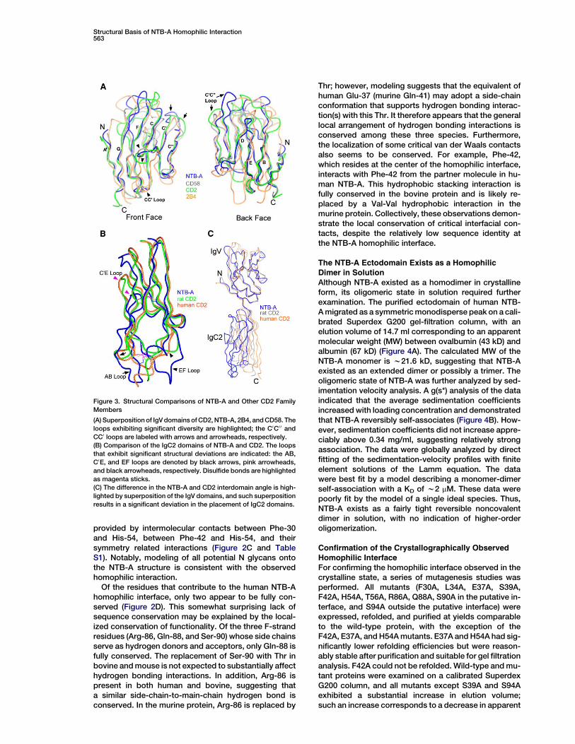
The IgV domains of NTB-A and the related CD2, CD58,and 2B4 receptors adopt a similar b-strand frameworkdespite a low sequence identity (Figure 3A). NTB-Aand CD2 (Wang et al., 1999) exhibit a root mean squaredeviation (RMSD) of w1.5 A based on 86 equivalent Caatoms, confirming their structural similarity. Similarly,NTB-A and 2B4 exhibit an RMSD of w2.2 A (86 Ca)(Ames et al., 2005), and NTB-A and CD58 exhibit anRMSD of w1.7 A (87 Ca) (Wang et al., 1999). Whereasthe overall disposition of the b strands of these IgVdomains is highly similar, the loops exhibit considerablediversity in both length and orientation. Specifically,NTB-A and 2B4 have longer C0C0 0 and CC0 loops, respec-tively, whereas CD58 possesses a shorter FG loop. TheCC0 loop of 2B4 is almost coplanar with the frontb strands, resulting in a relatively flat front face. In con-trast, the CC0 loop in CD2, NTB-A, and CD58 curvesaway from the back sheets, resulting in more concavefront surfaces. These IgV domains also differ substan-tially in the electrostatic properties of their front sur-faces. CD58 and CD2 have a large number of acidicand basic residues on their front sheets, respectively,whereas both NTB-A and 2B4 have predominantlyneutral, albeit polar, front surfaces.
The IgC2 DomainPrior to the present work, rat and human CD2 were theonly CD2 family members for which structures of boththe IgV and IgC2 domains were available (Bodianet al., 1994; Jones et al., 1992). The overall b-strand to-pology of the IgC2 domains is conserved betweenNTB-A and CD2 (RMSD of w2.2–2.6 A), with the frontsheet formed by the G, F, C, and C0 strands and theback sheet formed by the A, B, and E strands (Figure 3B).The most notable differences are that the NTB-A IgC2domain has longer AB and EF loops as well as a shorterC0 strand and C0E loop compared to the CD2 IgC2 do-mains. Notably, in addition to the presence of thehallmark disulfide bond connecting the B and F strands(Cys-131–Cys-173 in NTB-A), the IgC2 domains of all
CD2-family receptors contain a second noncanonicaldisulfide bond. In NTB-A, this additional disulfide bond(Cys-125–Cys-192) tethers the beginning of the B strandand the C terminus of the IgC2 domain, whereas in CD2,it links the end of the A strand to the C terminus of theIgC2 domain.
The IgV-IgC2 Interface
The overall rod-like architecture of the NTB-A monomeris the consequence of specific interactions between theIgV and IgC2 domains (Figure 1B). The G strand from theIgV domain ends at Leu-105 and is followed by a five-residue tether (106RQLRN110) that connects the twodomains. Comparison of the four NTB-A moleculesshowed that the interdomain angles formed by the IgVand IgC2 domains range from w120�–134�. This struc-tural plasticity is reminiscent of that present in rat CD2,where the interdomain angles of two independent mole-cules differ by 7� (Jones et al., 1992). The human and ratCD2 structures exhibit interdomain angles of w160� andw140�–147�, respectively, which are larger than that ofhuman NTB-A. This difference in interdomain angle isbest appreciated by superimposition of the IgV domainsof NTB-A and CD2 and observation of the dramaticdeviation in the placement of the IgC2 domains(Figure 3C). The IgV-IgC2 domain interface of NTB-A isstabilized by w589–671 A2 of total buried surface area,comparable to the 585–627 A2 of buried surface area inrat and human CD2. Four residues from the IgV domain(Asn-9, Ile-11, Leu-12, and Leu-105), two residues fromthe tether region (Arg-106 and Gln-107) and three resi-dues from the IgC2 domain (Asn-177, Ala-178, andVal-179) account for w80% of the total buried surfacearea at the IgV-IgC2 interface in NTB-A (Table S2). Inaddition to the considerable van der Waals contactsinvolving these residues, a hydrogen bond formed bythe main-chain oxygen of Arg-106 and Asn-177 ND2
Figure 1. Organization of the NTB-A Monomer
(A) The NTB-A ectodomain consists of membrane-distal IgV and
membrane-proximal IgC2 domains. The b strands are labeled and
the potential N-glycosylation sites are highlighted.
(B) The interdomain interface is stabilized by a number of van der
Waals interactions and an invariant hydrogen bond involving
Asn-177 ND2 and the main-chain oxygen of Arg-106 (black dash
line). Asn-177 OD1 also forms potential hydrogen bonds with
main-chain nitrogen atoms in the FG loop.

Immunity562
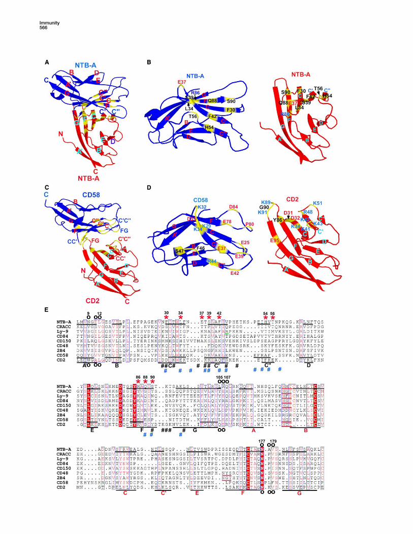
Figure 2. Organization of the NTB-A Homophilic Dimer
(A) The NTB-A dimer is composed of two monomers, shown as blue and red, which interact in a nearly orthogonal fashion and result in an end-
to-end distance of w100 A.
(B) Expanded view along the local two-fold symmetry axis of the interface, showing the orthogonal association of the front b sheets with
predominant contributions from the C, C0, C0 0, and F strands of each monomer.
(C) Detailed view of the NTB-A homophilic interface. In the left panel, residues forming hydrogen bonds, as well as Phe-42 that marks the
approximate center, are shown at same orientation as (B); hydrogen bonds are represented by dashed lines. In the right panel, the dimer is
rotated w180� along the indicated axis so that the hydrophobic interactions involving aromatic residues at the interface are best illustrated.
(D) Alignment of NTB-A ectodomain sequences. The conserved residues are shaded red, and residues with similar properties are labeled red.
Residues involved in homophilic and interdomain interactions are denoted ‘‘*’’ and ‘‘B’’, respectively. The b strands are denoted as underlined
segments in the human sequence and labeled with black (IgV) and red (IgC2) letters.
connects the tether region and FG loop of the IgC2domain (Figure 1B and Table S2).
The NTB-A Homophilic Interface
The NTB-A homophilic interface is formed by the nearlyorthogonal association of front b strands from the twoengaging IgV domains and buries a total surface areaof w1583 A2. As expected, the NTB-A homophilic inter-face displays rough two-fold symmetry (Figures 2B and2C). Twelve residues (Glu-37, Ser-39, Thr-56, Arg-86,Gln-88, Ser-90, and their symmetry mates) are involved
in the formation of eleven potential hydrogen bonds(Table S1); the side chains of Arg-86, Gln-88, and Ser-90, contributed from the F strand of one NTB-A mole-cule, make potential hydrogen bonds with main-chainatoms of Glu-37, Ser-39/Thr-56 and Thr-56 from thepartner molecule, respectively (Figure 2C). van derWaals contacts also make important contributions,with Leu-34 and Phe-42 near the center of the homo-philic interface making hydrophobic interactions withLeu-34 and Phe-42 from the opposing molecule, respec-tively. Four additional hydrophobic interactions are
Structural Basis of NTB-A Homophilic Interaction563
provided by intermolecular contacts between Phe-30and His-54, between Phe-42 and His-54, and theirsymmetry related interactions (Figure 2C and TableS1). Notably, modeling of all potential N glycans ontothe NTB-A structure is consistent with the observedhomophilic interaction.
Of the residues that contribute to the human NTB-Ahomophilic interface, only two appear to be fully con-served (Figure 2D). This somewhat surprising lack ofsequence conservation may be explained by the local-ized conservation of functionality. Of the three F-strandresidues (Arg-86, Gln-88, and Ser-90) whose side chainsserve as hydrogen donors and acceptors, only Gln-88 isfully conserved. The replacement of Ser-90 with Thr inbovine and mouse is not expected to substantially affecthydrogen bonding interactions. In addition, Arg-86 ispresent in both human and bovine, suggesting thata similar side-chain-to-main-chain hydrogen bond isconserved. In the murine protein, Arg-86 is replaced by
Figure 3. Structural Comparisons of NTB-A and Other CD2 Family
Members
(A) Superposition of IgV domains of CD2, NTB-A, 2B4, and CD58. The
loops exhibiting significant diversity are highlighted; the C0C0 0 and
CC0 loops are labeled with arrows and arrowheads, respectively.
(B) Comparison of the IgC2 domains of NTB-A and CD2. The loops
that exhibit significant structural deviations are indicated: the AB,
C0E, and EF loops are denoted by black arrows, pink arrowheads,
and black arrowheads, respectively. Disulfide bonds are highlighted
as magenta sticks.
(C) The difference in the NTB-A and CD2 interdomain angle is high-
lighted by superposition of the IgV domains, and such superposition
results in a significant deviation in the placement of IgC2 domains.
Thr; however, modeling suggests that the equivalent ofhuman Glu-37 (murine Gln-41) may adopt a side-chainconformation that supports hydrogen bonding interac-tion(s) with this Thr. It therefore appears that the generallocal arrangement of hydrogen bonding interactions isconserved among these three species. Furthermore,the localization of some critical van der Waals contactsalso seems to be conserved. For example, Phe-42,which resides at the center of the homophilic interface,interacts with Phe-42 from the partner molecule in hu-man NTB-A. This hydrophobic stacking interaction isfully conserved in the bovine protein and is likely re-placed by a Val-Val hydrophobic interaction in themurine protein. Collectively, these observations demon-strate the local conservation of critical interfacial con-tacts, despite the relatively low sequence identity atthe NTB-A homophilic interface.
The NTB-A Ectodomain Exists as a HomophilicDimer in Solution
Although NTB-A existed as a homodimer in crystallineform, its oligomeric state in solution required furtherexamination. The purified ectodomain of human NTB-A migrated as a symmetric monodisperse peak on a cali-brated Superdex G200 gel-filtration column, with anelution volume of 14.7 ml corresponding to an apparentmolecular weight (MW) between ovalbumin (43 kD) andalbumin (67 kD) (Figure 4A). The calculated MW of theNTB-A monomer is w21.6 kD, suggesting that NTB-Aexisted as an extended dimer or possibly a trimer. Theoligomeric state of NTB-A was further analyzed by sed-imentation velocity analysis. A g(s*) analysis of the dataindicated that the average sedimentation coefficientsincreased with loading concentration and demonstratedthat NTB-A reversibly self-associates (Figure 4B). How-ever, sedimentation coefficients did not increase appre-ciably above 0.34 mg/ml, suggesting relatively strongassociation. The data were globally analyzed by directfitting of the sedimentation-velocity profiles with finiteelement solutions of the Lamm equation. The datawere best fit by a model describing a monomer-dimerself-association with a KD of w2 mM. These data werepoorly fit by the model of a single ideal species. Thus,NTB-A exists as a fairly tight reversible noncovalentdimer in solution, with no indication of higher-orderoligomerization.
Confirmation of the Crystallographically ObservedHomophilic Interface
For confirming the homophilic interface observed in thecrystalline state, a series of mutagenesis studies wasperformed. All mutants (F30A, L34A, E37A, S39A,F42A, H54A, T56A, R86A, Q88A, S90A in the putative in-terface, and S94A outside the putative interface) wereexpressed, refolded, and purified at yields comparableto the wild-type protein, with the exception of theF42A, E37A, and H54A mutants. E37A and H54A had sig-nificantly lower refolding efficiencies but were reason-ably stable after purification and suitable for gel filtrationanalysis. F42A could not be refolded. Wild-type and mu-tant proteins were examined on a calibrated SuperdexG200 column, and all mutants except S39A and S94Aexhibited a substantial increase in elution volume;such an increase corresponds to a decrease in apparent

Immunity564
MW compared to the wild-type protein (Figure S1). Asmain-chain oxygen and nitrogen atoms of Ser-39 formhydrogen bonds with Gln-88 NE2 and OE1, respectively,alteration of this side chain was not expected to disruptthese hydrogen bonds. Although the side chain of Ser-39 makes van der Waals contacts with Gln-88, the effectof the S39A mutation on van der Waals interactions wasalso predicted to be small because Ala and Ser havegrossly similar steric properties. It was thus not surpris-ing that the S39A mutant exhibited chromatographicbehavior similar to the wild-type protein (Figure S1C).E37A and T56A were also predicted to minimally affecthydrogen-bonding interactions because only main-chainatoms of these residues are involved. However, Glu-37and Thr-56 are strongly buried at the homophilic inter-
Figure 4. Oligomeric States of the Wild-Type and S90A NTB-A
Ectodomains
(A) Elution profile of the human NTB-A ectodomain from Superdex
G200. The single monodisperse peak at 14.7 ml corresponds to
a MW between 43 and 67 kD.
(B) g(s*) analysis of the wild-type NTB-A by sedimentation velocity.
Protein concentration ranges from 0.14–1.38 mg/ml.
(C) g(s*) analysis of S90A by sedimentation velocity. Protein concen-
tration ranges from 0.17– 1.48 mg/ml.
face, making extensive van der Waals contacts withLeu-34/Arg-86 and Phe-30/Phe-42/Gln-88 from the op-posing molecule, respectively. It is plausible that substi-tutions of Glu-37 and Thr-56 to Ala would adverselyaffect the homophilic interface because of the reducedvan der Waals contacts, consistent with the experimen-tal results (Figure S1A).
Hydrogen bonds involving Arg-86, Gln-88, and Ser-90appear to make significant contributions to the stabilityof the homophilic interface because the R86A, Q88A,and S90A mutants each exhibited decreased apparentMW as shown by gel filtration (Figure S1B). Phe-30,Leu-34, and His-54 also make significant contributionsto the homophilic interface, with more than half of theirtotal accessible area being buried (Table S1). Replace-ment of these bulky side chains with Ala likely removedcritical van der Waals interactions and resulted in desta-bilization of the NTB-A dimer in solution (Figures S1Aand S1B). As expected, the S94A mutation, which islocated outside the binding interface, behaved similarlyto the wild-type protein (Figure S1C).
For further confirmation of the crystallographicallyobserved interface, sedimentation-velocity analysiswas performed with purified S90A mutant protein. Asfor the wild-type protein, data from the mutant proteinwere best fit by a model of a monomer-dimer self-asso-ciation. However, consistent with its chromatographicbehavior, the sedimentation coefficients for S90A con-tinued to increase up to 1.48 mg/ml, suggestinga weak association. The KD for the S90A mutant deter-mined by global analysis is 32 mM compared to 2 mMfor the wild-type protein. Taken together, the mutagen-esis experiments confirmed the contributions of severalspecific residues and supported the crystallographicallyobserved homophilic interface.
Comparison of the NTB-A Homophilic and CD2-CD58
Heterophilic DimersThe NTB-A homophilic and CD2-CD58 heterophilic di-mers show overall similarities, with their interfacesformed by the front sheets of the interacting IgV do-mains. However, there are significant differences, whichare best appreciated by superimposition of one NTB-Amolecule and CD58 and observation of the deviation inthe placement of the second NTB-A molecule and CD2(Figures 5A and 5C). The NTB-A homophilic interface isformed by the nearly orthogonal association of frontb strands, whereas CD2 and CD58 contact each otherat a slightly larger angle and exhibit a translation withrespect to one another, as compared to the two-foldsymmetric NTB-A dimer. In the NTB-A dimer, the C0 0
strands and a single residue in each C0 0D loop are buriedat the homphilic interface. In contrast, in the CD2-CD58heterodimer, only the analogous strand and loop fromCD58 contribute to the heterophilic interface. As a furtherresult of differences in relative subunit orientations, theCD2-CD58 heterophilic interface involves a considerablenumber of residues contributed from the CC0, C0C0 0, andFG loops of both CD2 and CD58, whereas the NTB-Ahomophilic interface is formed predominantly by strand-to-strand contacts involving the C, C0, C0 0, and F strands(Figures 5B and 5D).
The NTB-A dimer is stabilized by a slightly larger totalburied surface area than the CD2-CD58 dimer (i.e., of

Structural Basis of NTB-A Homophilic Interaction565
1583 A2 versus 1422 A2). The NTB-A dimer also exhibitsbetter surface complementarity (sc) compared to theCD2-CD58 dimer as shown by a larger sc value for theNTB-A dimer (0.68) than the CD2-CD58 heterodimer(0.56). In addition, the physicochemical compositionsof two interfaces differ. The NTB-A interface is predom-inantly stabilized by noncharged hydrogen bonds andvan der Waals contacts and thus contrasts with thepreponderance of ionic interactions present in theCD2-CD58 interface (Wang et al., 1999). Despite thesesignificant differences, the NTB-A and CD2-CD58 di-mers are characterized by roughly similar KDs of w2 mMand 10 mM (van der Merwe et al., 1994), respectively.
Discussion
It has been suggested that the CD2 family of receptorsarose via successive duplication events. In combinationwith previous studies, the current NTB-A structure high-lights a number of features conserved within the CD2family and provides structural evidence supporting thegene-duplication hypothesis. All CD2-family receptorslack the characteristic disulfide bond that tethers the Band F strands in their IgV domains. In addition, classicalIgV domains start with the A strand, followed by the A0
strand, and they hydrogen bond to the B and G strands,respectively. Direct structural analysis demonstratesthat NTB-A, CD2, CD58, and 2B4 all lack the A strand.Moreover, in the IgC2 domains, in addition to the hall-mark intersheet disulfide bond between the B and Fstrands, a second disulfide bond that bridges the endof the A strand (CD2 and CD58) or the beginning of theB strand (CD48 and all SLAM-family receptors) to theend of the G strand is present. All CD2-family receptorsharbor a potential N-glycosylation site in the AB loop ofthe IgC2 domain, except CD48 and 2B4 that beara potential N-glycosylation site at the nearby EF loop.Finally, the CD2-CD58 heterophilic and NTB-A homo-philic dimers exhibit gross overall structural similarity,with their interfaces formed by the front sheets of theIgV domains, suggesting that all CD2 family membersshare a common organization that is relevant to function(see below).
Sequence alignment of the homophilic receptors inthe SLAM family indicates that the covalent structuresof their IgV domains are very similar (i.e., there are fewinsertions/deletions). Therefore, the current structureserves as a possible template to model these homo-philic interactions and provides insight regarding thestructural determinants responsible for specificity.
In human NTB-A, Arg-86, Gln-88 and Ser-90, which arecontributed from the F strand, all utilize side-chain func-tionalities to participate in potential hydrogen bonds inwhich main-chain atoms serve as a donor or acceptor.The recognition of invariant main-chain atoms makesthese residues particularly amenable to substitution,and the equivalent residues in CD84, Ly-9, and CRACCare all capable of participating in hydrogen-bond inter-actions through their side chains. The sole exception isGly-85 in CRACC; this is equivalent to Gln-88 in humanNTB-A. Accordingly, we postulate that these residuesin the F strand are of crucial importance for homophilicinteractions in CD84, Ly-9, and CRACC as well. Further-more, the Phe-42–Phe-42 hydrophobic stacking interac-
tion, which appears to mark the center of the homophilicinterface in NTB-A, is conserved in Ly-9 and is replacedby the highly conservative Tyr-Tyr pair in CD84.
SLAM is distantly related to NTB-A, CRACC, CD84,and Ly-9, as suggested by w20% sequence homologybetween SLAM and each of these homophilic receptors.Notably, the SLAM IgV domain lacks the intersheet hy-drogen bond formed by the conserved Trp33 NE2 inthe middle of the C strand and the main-chain carbonylof a residue preceding the E strand; this bond is presentin almost all other IgV domains. Moreover, the KD for theSLAM homophilic interaction is w200 mM (Mavaddatet al., 2000), at least two orders of magnitude weakerthan other homophilic SLAM family members examinedto date. In light of this modest KD, it is tempting to spec-ulate that additional binding partner(s) for SLAM remainto be identified. Both sequence divergence and itsunique biochemical properties set SLAM apart as a dis-tinct homophilic receptor in the SLAM family.
Interestingly, a recent study (Romero et al., 2005) indi-cated that in Ly-9 the R44A mutation (the equivalent ofNTB-A Val-44), which is predicted to reside near the cen-ter of the homophilic interface, resulted in an enhancedhomophilic association. This effect may be due to theelimination of the unfavorable repulsive electrostaticsresulting from the close proximity of these two Argside chains in the Ly-9 homophilic interface. This obser-vation highlights the critical concept that receptor-ligand interactions do not evolve to produce the highestpossible affinity but instead evolve to select the affinitythat allows for the specificity, kinetics, and associatedsignaling properties that are biologically optimal. Thus,the specific differences in the SLAM-family-receptorinterfaces are likely to serve a number of importantbiological functions, including the realization of biologi-cally relevant KD values that span at least three orders ofmagnitude. Specifically, the KDs for NTB-A and SLAMare w2 mM and 200 mM, respectively, whereas the disso-ciation of CD84 was undetectable by sedimentation-velocity analysis (Q.Y., unpublished data), consistentwith a KD in the nM range. This observation suggeststhat, analogous to the dwell time associated with TCR-MHC interactions (Kalergis et al., 2001), affinity mightbe an important determinant of the distinct functions as-sociated with the different SLAM-family homophilic re-ceptors. Furthermore, the divergence of interfacial resi-dues provides unique determinants that promote thespecificity of each particular homophilic interactionand interfere with inappropriate heterodimerizationamong the family members.
The NTB-A mutagenesis studies highlight some po-tentially important general principles that are relevantto homophilic interactions within the SLAM family. TheS90A mutation in the NTB-A homophilic interface causeda w15 fold increase in KD (lower affinity) compared to thewild-type protein and thus resulted in a significantmonomer population in solution. In addition, other singlemutations (F30A, L34A, E37A, H54A, T56A, R86A, andQ88A) all dramatically affected the homophilic associa-tion and resulted in a predominantly monomer popula-tion as judged by gel filtration. Taken together, thesedata suggest that, despite the large homophilic inter-face, the basis of specificity of homophilic interactionsin the SLAM family may be subtle because the change
Immunity566

Structural Basis of NTB-A Homophilic Interaction567
Figure 6. Functional Implications of the NTB-A Structure
Homophilic engagement of NTB-A may trigger recruitment of the NTB-A to the immunological synapse. The linear dimensions of the NTB-A
dimer are similar to those of other molecular signaling pairs involved in T cell regulation, consistent with a sorting mechanism based on molecular
extent. In T cells, the local enrichment of NTB-A and its associated signaling components, including SAP and FynT, initiates the downstream
signaling events that culminate in modulation of IFN-g secretion and stimulation of Th2 responses. In APCs, EAT-2-coupled signaling cascades
are similarly activated upon NTB-A homophilic engagement, although all of the analogous signaling molecules remain to be identified.
of only a few (or single) residues may significantly alterthe monomer-dimer equilibrium. This apparent sensitiv-ity may be related to the two-fold symmetric organizationof the homphilic dimer because a single mutation in theprimary sequence typically results in the disruption oftwo sets of contacts at the binding interface. This sensi-tivity might also represent an evolutionary mechanismfor the efficient generation of new functional receptorswith novel recognition properties.
A number of intrinsic structural features are relevant tothe physiological function of the CD2 family. The experi-mentally determined interfaces suggest that, with the ex-ception of Ly-9, all members of the CD2 family will exhibita similar overall organization with maximal linear dimen-sions of w100–140A (Figure 6). It has been speculatedthat the unique compartmentalization of moleculesobserved at the immunological synapse is driven bya sorting mechanism based on molecular dimension.Notably, the proposed CD2-family-receptor dimensionsare consistent with those of other pairs of signalingmolecules, including the TCR-pMHC and CTLA-4-B7
complexes, that localize at the central zone of the immu-nological synapse (van der Merwe et al., 2000).
The common molecular dimensions may be of partic-ular relevance to the SLAM-family receptors because itaffords the opportunity for all homophilic- and hetero-philic-receptor pairs to colocalize with the same set ofsignaling molecules. These molecules are likely to ex-hibit overlapping but unique functions because theyhave distinct cytoplasmic domains with different signal-ing. The ability of the family members to colocalize witha common set of signaling partners could allow for thesubtle modulation of signaling mechanisms under a vari-ety of stimuli in different cell types. Consistent with thisnotion, several CD2 family members such as CD2 (vander Merwe et al., 2000), CD48 (Moran and Miceli,1998), and also the SAP adaptor protein (Cannonset al., 2004), are recruited to the immunological synapseformed between T cells and antigen-presenting cells(APCs). Additionally, 2B4 and SAP are recruited to theimmunological synapse formed at the NK-target cell in-terface (Roda-Navarro et al., 2004).
Figure 5. Comparsion of Homophilic and Heterophilic Interfaces
(A and C) The overall organization of NTB-A homophilic and CD2-CD58 heterophilic dimers. One NTB-A molecule (blue) and CD58 (blue) are dis-
played in the same orientation, highlighting the detailed differences in overall organization of the two dimers. Residues contributing to the two
interfaces are highlighted yellow.
(B) Detailed view of the NTB-A homophilic interface. The blue monomer is rotated w180� about a horizontal axis relative to (A) so that the residues
that contribute to the binding surface are exposed. The labels of individual residues are color coded blue, red, and black to represent basic,
acidic, and neutral residues, respectively.
(D) Residues contributing to the CD2-CD58 interface displayed as in (C). The significant contribution of loop residues and charged residues, rel-
ative to NTB-A, is demonstrated.
(E) Alignment of the CD2-family ectodomain sequences. The strictly conserved residues are shaded red, and residues with similar properties are
labeled red. Residues critical for homophilic interactions in NTB-A are denoted with ‘‘*’’, residues involved in CD2-CD58 heterophilic interaction
are denoted with black ‘‘#’’ (CD2) and blue ‘‘#’’ (CD58), and residues contributing to the interdomain interface in NTB-A and CD2 are denoted with
‘‘B’’. Ly-9 Arg-44 is underlined in green. The b strands identified from existing structures are underlined in black, and labeled with black (IgV) or
red (IgC2) capital letters. Potential N-glycosylation sites in the AB and EF loops in the IgC2 domains are underlined red.

Immunity568
All CD2 family members possess a potential N-glyco-sylation site at the base of their IgC2 domains and prox-imal to the plasma membrane. As previously suggestedfor CD2 and ICAM (Wang and Springer, 1998), the pres-ence of such N glycans may provide important stericconstraints that bias the orientation of the rod-likeNTB-A molecule with respect to the membrane andcould be an important determinant for the proper pre-sentation of the ligand-binding surface. Furthermore,both NTB-A and CD2 exhibit some degree of intrinsicflexibility in the tether region joining the IgV and IgC2domains, and this region may be of general importancefor receptor-ligand interactions in a physiological con-text (Davis et al., 2003). Unlike the protein-protein inter-actions observed in solution, the in vivo interactionsbetween membrane-anchored receptors and ligandsare subjected to forces that oppose binding, includingcell migration and fluctuations in inter-membrane dis-tance. The plasticity of the tether region could allowthese membrane-anchored cell-surface proteins toadapt to the dynamic environment without rupturingthe receptor-ligand interface. In addition, the IgV-IgC2interface is essential for maintaining the overall architec-ture responsible for presentation of the ligand-bindingsurface. The mechanisms that control the finely bal-anced dynamics at the domain interface are likely tobe shared through the entire CD2 family. Notably,comparison of the NTB-A structure with the full-lengthCD2 structures revealed that the same set of equivalentresidues are involved in the interdomain interactions,including Asn-177 in the FG loop, which forms thesame side-chain-to-main-chain hydrogen bonds inthese structures. Because Asn-177 is invariant, thesecontacts are likely to be functionally relevant to the entirefamily; in fact, these contacts are conserved in a largenumber of IgC2 domains and are responsible for main-taining the overall compact state of the domain. More-over, a noncanonical disulfide bond is present atthe base of IgC2 domains of all CD2-family receptors.This modification serves to tether the two b sheets. Ithas also been previously suggested to stabilize this do-main against forces experienced in vivo (Wang et al.,1999).
Crystallographic analysis supports a model in whichthe NTB-A ectodomain is a homophilic dimer. Solutionstudies also indicated that the formation of the homo-philic dimer did not lead to any higher oligomerizationstate, suggesting that the ligand-induced oligomeriza-tion proposed for the tumor necrosis factor receptor(TNFR) and epidermal growth factor receptor (EGFR)families is unlikely to be part of the signaling mechanismutilized by NTB-A. Furthermore, the compact nature ofthe IgSF domains does not readily appear to lend itselfto a structural reorganization that might facilitate signal-ing. Instead we favor a model in which the homophilicengagement triggers the redistribution of freely diffusingNTB-A molecules to the immunological synapse, result-ing in the local enrichment of NTB-A and its associatedsignaling components, including the adaptor SAP. Thisenrichment results in a series of localized and concertedmolecular activation events (e.g., phosphorylation),which ultimately exceed an existing threshold and resultin modulation of IFN-g secretion and enhancement ofTh2 immune responses.
Experimental Procedures
Cloning, Expression, and Purification of Human NTB-A
Ectodomain
The human NTB-A ectodomain (24-215 plus the initiator methionine)
and mutants (F30A, E37A, S39A, F42A, H54A, T56A, R86A, Q88A,
S90A, and S94A) were expressed from pET-3a in Rosseta (DE3)
pLysS E. coli strain. Selenomethionine (SeMet)-substituted protein
was produced in the methionine auxotroph B834 (DE3) E. coli strain.
All proteins were refolded as described for B7-2 (Zhang et al., 2002).
Proteins were purified by gel filtration and anion exchange chroma-
tography.
Sedimentation Velocity Analysis
Wild-type and S90A mutant NTB-A proteins were exchanged into
a buffer containing 20 mM Tris (pH 7.45) and 50 mM NaCl. Sedimen-
tation velocity analysis was performed at protein concentrations of
1.38, 1.0, 0.34, and 0.14 mg/ml for the wild-type protein and 1.48,
0.5, and 0.17 mg/ml for the S90A protein in a Beckman coulter
XL-I. Data were collected at 20�C and 55,000 rpm, and interference
scans were acquired at 1 min intervals for 5 or 7 hr. Data were first
analyzed with the program DcDt+ (Philo, 2000). The KDs of the
homophilic interactions were obtained by global fitting of all the
data sets collected at different concentrations with sedanal (Stafford
and Sherwood, 2004) or sedphat (Schuck, 2003).
Structure Determination
NTB-A (4 mg/ml in 10 mM Tris [pH 8.5]) was crystallized by sitting
drop vapor diffusion with 2.5 ml of protein and 2.5 ml of precipitant
composed of 0.42–0.5 M CaCl2, 100 mM HEPES buffer (pH 7.4),
1.0 mM Pt(NH3)2Cl2, and 4% PEG400 at 4�C. SeMet-substituted
NTB-A was crystallized by microseeding with native crystals. Prior
to flash-cooling (100 K), crystals were cryoprotected in mother liquor
containing 25% PEG400 supplemented with 15% MPD. Native
(3.0 A) and MAD (3.3 A) data were collected at beam line X29 at
the National Synchrotron Light Source (NSLS) in Brookhaven Na-
tional Laboratory. Native and SeMet-substituted crystals exhibited
diffraction consistent with the space group C2; data were integrated
and scaled with HKL2000 (Otwinowski and Minor, 1997).
The heavy atom substructure was solved with SHELXD (Schneider
and Sheldrick, 2002) with peak data in the 40–3.7 A resolution range;
phase determination and density modification with SOLVE (Terwil-
liger and Berendzen, 1999) and RESOLVE (Terwilliger, 2000), respec-
tively, were then performed. The main chain was manually built into
the electron density with programs O (Jones et al., 1991) and COOT
(Emsley and Cowtan, 2004) and then rigid body refinement and
simulated annealing with CNS were performed with native data
(Brunger et al., 1998). ARP/wARP was used so that the map together
with restraints from experimental phases could be further improved.
The model was further developed by alternate cycles of manual
revision and refinement with refmac5 (Murshudov et al., 1997). TLS
refinement was used in the final stages, and omit maps were calcu-
lated to check the accuracy of the model. The four molecules in the
asymmetric unit are denoted as A, B, C, and D. Electron density for
all the four molecules was continuous except for Gln-118–Phe-120,
Gln-118–Gln-121 in the AB loop of IgC2 domains of A and C mole-
cules, respectively. The IgV domain of the C molecule exhibited rel-
atively poor electron density throughout the entire refinement; this
may be explained by the relatively high solvent exposure of its
back face, although the electron density for residues in the homo-
philic interface was unambiguous. Residues in the generously al-
lowed and disallowed regions in the Ramachandran plot account
for 1.9% and 0.6% of the total residues, respectively; these residues
are predominately contributed from connecting loops, mostly from
C molecule.
Analysis of superpositions were performed with the CCP4 suite
(Mitchell et al., 1990; Potterton et al., 2002; Potterton et al., 2004).
Surface complementarities, accessible surface area, and atomic
contacts were calculated with the sc (Lawrence and Colman,
1993), areaimol (Lee and Richards, 1971), and contact (Skarzynski
T. and Leslie A.) programs in CCP4 Suite (CCP4, 1994), respectively.
Structure-based sequence alignment was rendered with the pro-
gram ESPript (Gouet et al., 1999). Ribbon diagrams were generated
with Pymol (DeLano W.L., http://pymol.sourceforge.net/). The

Structural Basis of NTB-A Homophilic Interaction569
modeling of N glycans was performed with GlyProt (Bohne-Lang
and von der Lieth, 2005).
Supplemental Data
Supplemental Data include one figure and two tables and can be
found with this article online at http://www.immunity.com/cgi/
content/full/25/4/559/DC1/.
Acknowledgments
We gratefully acknowledge the staff of the X9A and X29 beam lines
at the National Synchrotron Light Source and Teresa P. DiLorenzo
for critical reading of the manuscript. This work was supported by
National Institute of Health Grants (to S.G.N. and S.C.A.; AI07289).
Received: May 3, 2006
Revised: June 20, 2006
Accepted: June 27, 2006
Published online: October 12, 2006
References
Ames, J.B., Vyas, V., Lusin, J.D., and Mariuzza, R. (2005). NMR struc-
ture of the natural killer cell receptor 2B4 (CD244): Implications for
ligand recognition. Biochemistry 44, 6416–6423.
Bloch-Queyrat, C., Fondaneche, M.C., Chen, R., Yin, L., Relouzat, F.,
Veillette, A., Fischer, A., and Latour, S. (2005). Regulation of natural
cytotoxicity by the adaptor SAP and the Src-related kinase Fyn.
J. Exp. Med. 202, 181–192.
Bodian, D.L., Jones, E.Y., Harlos, K., Stuart, D.I., and Davis, S.J.
(1994). Crystal structure of the extracellular region of the human
cell adhesion molecule CD2 at 2.5 A resolution. Structure 2, 755–766.
Bohne-Lang, A., and von der Lieth, C.W. (2005). GlyProt: In silico
glycosylation of proteins. Nucleic Acids Res. 33, W214–W219.
Bottino, C., Falco, M., Parolini, S., Marcenaro, E., Augugliaro, R., Si-
vori, S., Landi, E., Biassoni, R., Notarangelo, L.D., Moretta, L., and
Moretta, A. (2001). NTB-A [correction of GNTB-A], a novel SH2D1A-
associated surface molecule contributing to the inability of natural
killer cells to kill Epstein-Barr virus-infected B cells in X-linked
lymphoproliferative disease. J. Exp. Med. 194, 235–246.
Brunger, A.T., Adams, P.D., Clore, G.M., DeLano, W.L., Gros, P.,
Grosse-Kunstleve, R.W., Jiang, J.S., Kuszewski, J., Nilges, M.,
Pannu, N.S., et al. (1998). Crystallography & NMR system: A new
software suite for macromolecular structure determination. Acta
Crystallogr. D Biol. Crystallogr. 54, 905–921.
Cannons, J.L., Yu, L.J., Hill, B., Mijares, L.A., Dombroski, D., Nichols,
K.E., Antonellis, A., Koretzky, G.A., Gardner, K., and Schwartzberg,
P.L. (2004). SAP regulates T(H)2 differentiation and PKC-theta-medi-
ated activation of NF-kappaB1. Immunity 21, 693–706.
CCP4 (Collaborative Computational Project, Number 4) (1994). The
CCP4 suite: Programs for protein crystallography. Acta Crystallogr.
D Biol. Crystallogr. 50, 760–763.
Czar, M.J., Kersh, E.N., Mijares, L.A., Lanier, G., Lewis, J., Yap, G.,
Chen, A., Sher, A., Duckett, C.S., Ahmed, R., and Schwartzberg,
P.L. (2001). Altered lymphocyte responses and cytokine production
in mice deficient in the X-linked lymphoproliferative disease gene
SH2D1A/DSHP/SAP. Proc. Natl. Acad. Sci. USA 98, 7449–7454.
Davis, S.J., Ikemizu, S., Evans, E.J., Fugger, L., Bakker, T.R., and van
der Merwe, P.A. (2003). The nature of molecular recognition by T
cells. Nat. Immunol. 4, 217–224.
Emsley, P., and Cowtan, K. (2004). Coot: Model-building tools for
molecular graphics. Acta Crystallogr. D Biol. Crystallogr. 60, 2126–
2132.
Engel, P., Eck, M.J., and Terhorst, C. (2003). The SAP and SLAM
families in immune responses and X-linked lymphoproliferative
disease. Nat. Rev. Immunol. 3, 813–821.
Gouet, P., Courcelle, E., Stuart, D.I., and Metoz, F. (1999). ESPript:
Analysis of multiple sequence alignments in PostScript. Bioinfor-
matics 15, 305–308.
Howie, D., Laroux, F.S., Morra, M., Satoskar, A.R., Rosas, L.E.,
Faubion, W.A., Julien, A., Rietdijk, S., Coyle, A.J., Fraser, C., and
Terhorst, C. (2005). Cutting edge: The SLAM family receptor Ly108
controls T cell and neutrophil functions. J. Immunol. 174, 5931–5935.
Ikemizu, S., Sparks, L.M., van der Merwe, P.A., Harlos, K., Stuart,
D.I., Jones, E.Y., and Davis, S.J. (1999). Crystal structure of the
CD2-binding domain of CD58 (lymphocyte function-associated anti-
gen 3) at 1.8-A resolution. Proc. Natl. Acad. Sci. USA 96, 4289–4294.
Jones, E.Y., Davis, S.J., Williams, A.F., Harlos, K., and Stuart, D.I.
(1992). Crystal structure at 2.8 A resolution of a soluble form of the
cell adhesion molecule CD2. Nature 360, 232–239.
Jones, T.A., Zou, J.Y., Cowan, S.W., and Kjeldgaard. (1991).
Improved methods for building protein models in electron density
maps and the location of errors in these models. Acta Crystallogr.
A 47, 110–119.
Kalergis, A.M., Boucheron, N., Doucey, M.-A., Palmieri, E., Goyarts,
E.C., Vegh, Z., Luescher, I.F., and Nathenson, S.G. (2001). Efficient T
cell activation requires an optimal dwell-time of interaction between
the TCR and the pMHC complex. Nat. Immunol. 2, 229–234.
Kumar, K.R., Li, L., Yan, M., Bhaskarabhatla, M., Mobley, A.B.,
Nguyen, C., Mooney, J.M., Schatzle, J.D., Wakeland, E.K., and
Mohan, C. (2006). Regulation of B cell tolerance by the lupus suscep-
tibility gene Ly108. Science 312, 1665–1669.
Latour, S., Gish, G., Helgason, C.D., Humphries, R.K., Pawson, T.,
and Veillette, A. (2001). Regulation of SLAM-mediated signal trans-
duction by SAP, the X-linked lymphoproliferative gene product.
Nat. Immunol. 2, 681–690.
Latour, S., and Veillette, A. (2004). The SAP family of adaptors in
immune regulation. Semin. Immunol. 16, 409–419.
Lawrence, M.C., and Colman, P.M. (1993). Shape complementarity
at protein/protein interfaces. J. Mol. Biol. 234, 946–950.
Lee, B., and Richards, F.M. (1971). The interpretation of protein struc-
tures: Estimation of static accessibility. J. Mol. Biol. 55, 379–400.
Mavaddat, N., Mason, D.W., Atkinson, P.D., Evans, E.J., Gilbert,
R.J., Stuart, D.I., Fennelly, J.A., Barclay, A.N., Davis, S.J., and
Brown, M.H. (2000). Signaling lymphocytic activation molecule
(CDw150) is homophilic but self-associates with very low affinity.
J. Biol. Chem. 275, 28100–28109.
Mitchell, E.M., Artymiuk, P.J., Rice, D.W., and Willett, P. (1990). Use
of techniques derived from graph theory to compare secondary
structure motifs in proteins. J. Mol. Biol. 212, 151–166.
Moran, M., and Miceli, M.C. (1998). Engagement of GPI-linked CD48
contributes to TCR signals and cytoskeletal reorganization: A role
for lipid rafts in T cell activation. Immunity 9, 787–796.
Murshudov, G.N., Vagin, A.A., and Dodson, E.J. (1997). Refinement
of macromolecular structures by the maximum-likelihood method.
Acta Crystallogr. D Biol. Crystallogr. 53, 240–255.
Otwinowski, Z., and Minor, W. (1997). Processing of X-ray diffraction
data collected in oscillation mode. Methods Enzymol. 276, 307–326.
Philo, J.S. (2000). A method for directly fitting the time derivative of
sedimentation velocity data and an alternative algorithm for calculat-
ing sedimentation coefficient distribution functions. Anal. Biochem.
279, 151–163.
Potterton, E., McNicholas, S., Krissinel, E., Cowtan, K., and Noble,
M. (2002). The CCP4 molecular-graphics project. Acta Crystallogr.
D Biol. Crystallogr. 58, 1955–1957.
Potterton, L., McNicholas, S., Krissinel, E., Gruber, J., Cowtan, K.,
Emsley, P., Murshudov, G.N., Cohen, S., Perrakis, A., and Noble,
M. (2004). Developments in the CCP4 molecular-graphics project.
Acta Crystallogr. D Biol. Crystallogr. 60, 2288–2294.
Roda-Navarro, P., Mittelbrunn, M., Ortega, M., Howie, D., Terhorst,
C., Sanchez-Madrid, F., and Fernandez-Ruiz, E. (2004). Dynamic
redistribution of the activating 2B4/SAP complex at the cytotoxic
NK cell immune synapse. J. Immunol. 173, 3640–3646.
Romero, X., Zapater, N., Calvo, M., Kalko, S.G., de la Fuente, M.A.,
Tovar, V., Ockeloen, C., Pizcueta, P., and Engel, P. (2005). CD229
(Ly9) lymphocyte cell surface receptor interacts homophilically
through its N-terminal domain and relocalizes to the immunological
synapse. J. Immunol. 174, 7033–7042.
Roncagalli, R., Taylor, J.E., Zhang, S., Shi, X., Chen, R., Cruz-Munoz,
M.E., Yin, L., Latour, S., and Veillette, A. (2005). Negative regulation

Immunity570
of natural killer cell function by EAT-2, a SAP-related adaptor. Nat.
Immunol. 6, 1002–1010.
Schneider, T.R., and Sheldrick, G.M. (2002). Substructure solution
with SHELXD. Acta Crystallogr. D Biol. Crystallogr. 58, 1772–1779.
Schuck, P. (2003). On the analysis of protein self-association by sed-
imentation velocity analytical ultracentrifugation. Anal. Biochem.
320, 104–124.
Stafford, W.F., and Sherwood, P.J. (2004). Analysis of heterologous
interacting systems by sedimentation velocity: Curve fitting algo-
rithms for estimation of sedimentation coefficients, equilibrium
and kinetic constants. Biophys. Chem. 108, 231–243.
Terwilliger, T.C. (2000). Maximum-likelihood density modification.
Acta Crystallogr. D Biol. Crystallogr. 56, 965–972.
Terwilliger, T.C., and Berendzen, J. (1999). Automated MAD and MIR
structure solution. Acta Crystallogr. D Biol. Crystallogr. 55, 849–861.
Valdez, P.A., Wang, H., Seshasayee, D., van Lookeren Campagne,
M., Gurney, A., Lee, W.P., and Grewal, I.S. (2004). NTB-A, a new
activating receptor in T cells that regulates autoimmune disease.
J. Biol. Chem. 279, 18662–18669.
van der Merwe, P.A., Barclay, A.N., Mason, D.W., Davies, E.A., Mor-
gan, B.P., Tone, M., Krishnam, A.K., Ianelli, C., and Davis, S.J. (1994).
Human cell-adhesion molecule CD2 binds CD58 (LFA-3) with a very
low affinity and an extremely fast dissociation rate but does not bind
CD48 or CD59. Biochemistry 33, 10149–10160.
van der Merwe, P.A., Davis, S.J., Shaw, A.S., and Dustin, M.L. (2000).
Cytoskeletal polarization and redistribution of cell-surface mole-
cules during T cell antigen recognition. Semin. Immunol. 12, 5–21.
Veillette, A. (2006). Immune regulation by SLAM family receptors and
SAP-related adaptors. Nat. Rev. Immunol. 6, 56–66.
Veillette, A., Cruz-Munoz, M.E., and Zhong, M.C. (2006). SLAM family
receptors and SAP-related adaptors: Matters arising. Trends Immu-
nol. 27, 228–234.
Wandstrat, A.E., Nguyen, C., Limaye, N., Chan, A.Y., Subramanian,
S., Tian, X.H., Yim, Y.S., Pertsemlidis, A., Garner, H.R., Jr., Morel,
L., and Wakeland, E.K. (2004). Association of extensive polymor-
phisms in the SLAM/CD2 gene cluster with murine lupus. Immunity
21, 769–780.
Wang, J., and Springer, T.A. (1998). Structural specializations of
immunoglobulin superfamily members for adhesion to integrins
and viruses. Immunol. Rev. 163, 197–215.
Wang, J.H., Smolyar, A., Tan, K., Liu, J.H., Kim, M., Sun, Z.Y., Wagner,
G., and Reinherz, E.L. (1999). Structure of a heterophilic adhesion
complex between the human CD2 and CD58 (LFA-3) counterrecep-
tors. Cell 97, 791–803.
Wang, N., Satoskar, A., Faubion, W., Howie, D., Okamoto, S., Feske,
S., Gullo, C., Clarke, K., Sosa, M.R., Sharpe, A.H., and Terhorst, C.
(2004). The cell surface receptor SLAM controls T cell and macro-
phage functions. J. Exp. Med. 199, 1255–1264.
Wu, C., Nguyen, K.B., Pien, G.C., Wang, N., Gullo, C., Howie, D.,
Sosa, M.R., Edwards, M.J., Borrow, P., Satoskar, A.R., et al.
(2001). SAP controls T cell responses to virus and terminal differen-
tiation of TH2 cells. Nat. Immunol. 2, 410–414.
Zhang, X., Schwartz, J.C., Almo, S.C., and Nathenson, S.G. (2002).
Expression, refolding, purification, molecular characterization, crys-
tallization, and preliminary X-ray analysis of the receptor binding
domain of human B7-2. Protein Expr. Purif. 25, 105–113.
Accession Numbers
The atomic coordinates and reflections have been deposited in the
Protein Data Bank under accession code 2IF7.