indications and limitations of cardiac imaging tests · indications and limitations of cardiac...
TRANSCRIPT

Indications and Limitations of
Cardiac Imaging Tests
April 1, 2011
Joe M. Moody, Jr, MD
UTHSCSA and STVHCS
Cardiology is a very visual specialty,
despite the importance of auscultation
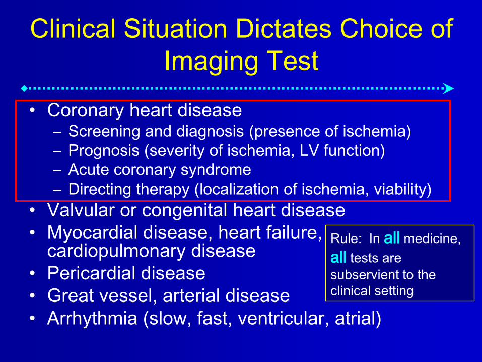
Clinical Situation Dictates Choice of
Imaging Test
• Coronary heart disease– Screening and diagnosis (presence of ischemia)
– Prognosis (severity of ischemia, LV function)
– Acute coronary syndrome
– Directing therapy (localization of ischemia, viability)
• Valvular or congenital heart disease
• Myocardial disease, heart failure, cardiopulmonary disease
• Pericardial disease
• Great vessel, arterial disease
• Arrhythmia (slow, fast, ventricular, atrial)
Rule: In all medicine,
all tests are
subservient to the
clinical setting

Local Expertise Also Affects
Choice of Imaging Test
• Availability of technical support
• Expertise and experience of technical
personnel
• Availability of equipment
• Expertise and experience of interpreter
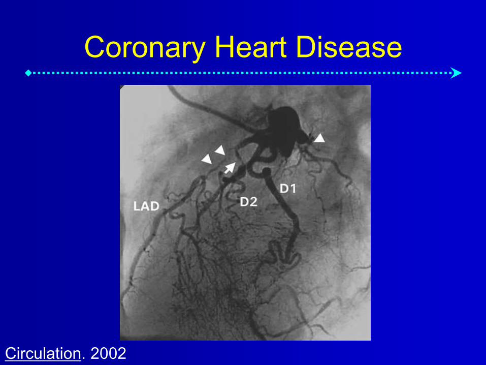
Coronary Heart Disease
Circulation. 2002

Physiology: Ischemic Cascade
1. Supply/demand imbalance
2. Diastolic dysfunction (4 sec)
3. Systolic dysfunction (6 sec)
4. Elevated LV filling pressure
5. ECG changes (20 sec)
6. Angina (25 sec)
From Sigwart U, et al, Silent Myocardial Ischemia 1984
and Armstrong WF, Prog Cardiov Dis 1997;39:499-522

Time Course of Ischemia
• Experimental Ischemia– Threshold of 10-20% reduction in blood flow impairs
wall thickening and 80% reduction results in akinesis
– Decreased wall thickening extends beyond reduced flow (“tethering”)
• Clinical Ischemia (complete balloon occlusion)– Regional endocardial dysfunction in 19 sec
– ECG change in 30 sec
– Chest pain 39 sec
• Clinical Ischemia (stress in region of coronary stenosis)– Wall motion abnormality in 30 sec
– ECG change in 90 sec
Otto CM. 2nd Ed. 2002. p. 275, 307

Cardiac Imaging Tests in Coronary
Disease
• Invasive: Cardiac catheterization(ventriculography, coronary angiography)
• Noninvasive:– Electric* “imaging” (ST segment – ischemia, arrhythmia)
– Nuclear* (myocardial perfusion, pump function, viability)
– Echocardiography* (wall motion, pump function, valve function)
– Chest X-Ray*
– CT Scan (coronary anatomy, coronary calcification)
– MRI (coronary anatomy, ventricular function, myocardial perfusion, viability, valve function)
* = common, high volume use

Imaging is Used When Exercise
ECG is not Adequate
• Patients unable to exercise (intermediate or high clinical likelihood of CAD based on age and symptoms)
• ECG not interpretable (LVH, WPW, Paced QRS, LBBB, digoxin therapy, >1mm ST depression)
• Clinical situation demands more information (angiographically intermediate lesions, intermediate Duke treadmill score, changing clinical situation with prior imaging study)

Effect of Symptoms on Risk of CAD
Hurst, 1998, Ch. 45
Intermediate risk is 10-90%

History of Noninvasive Testing
Master’s 2 step
Motorized treadmill ECG
Myocardial perfusion imaging
Stress echocardiography
Coronary calcification
Magnetic resonance imaging
Time
Search for
optimized
sensitivity and
specificity
Balance of cost and
benefit and risk
Choice depends on particular
clinical question
*
*
*

Types of Stress
• Exercise is most desirable, treadmill in USA– Exercise tolerance has prognostic value, and
clinical appearance and HR and BP response information are useful
• In patients who can’t exercise:– Vasodilator for perfusion testing (dipyridamole,
adenosine, regadenoson=Lexiscan)
– Adrenergic for perfusion or wall motion (dobutamine)
• In other special circumstances– LBBB, Paced QRS (prefer vasodilator)

Simplified Summary of
Noninvasive Assessment in CAD
Stress Assessment
ECG
Perfusion
(nuclear)
Function
(echo)
(MRI)
Exercise
Vasodilator
Inotropic/
adrenergic
Exercise ECG
Exercise
perfusion scan
Vasodilator
perfusion scan
Exercise echo
Dobutamine
stress echo

Exercise ECG in CAD
• Usefulness:
– Widely available
– Optimal type of
stress
– Least expensive
procedure
– Wealth of prognostic
information
• Limitations:
– Less sensitive than
most imaging tests
– False positive results
– Many patients
cannot exercise
– Many patients have
uninterpretable ECG

Exercise Nuclear Myocardial
Perfusion Imaging (MPI) in CAD
• Usefulness– Widely available
– Provides prognostic information from exercise
– Useful in abnormal ECG with ST-T wave abnormalities
– Very sensitive
– Wealth of prognostic information
• Limitations– Expensive
– False positive results
– Many patients cannot exercise

Exercise Echocardiography in
CAD
• Usefulness
– Provides prognostic
information from
exercise
– Useful in abnormal
ECG with ST-T wave
abnormalities
– Very specific
– Adequate prognostic
information
• Limitations
– Technically demanding,
so less widely available
– Expensive
– Difficult to interpret in
presence of resting wall
motion abnormalities
– Many patients cannot
exercise
– May be less sensitive
than MPI
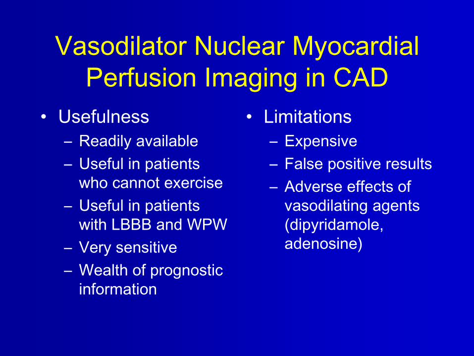
Vasodilator Nuclear Myocardial
Perfusion Imaging in CAD
• Usefulness
– Readily available
– Useful in patients
who cannot exercise
– Useful in patients
with LBBB and WPW
– Very sensitive
– Wealth of prognostic
information
• Limitations
– Expensive
– False positive results
– Adverse effects of
vasodilating agents
(dipyridamole,
adenosine)

Vasodilator Echocardiography in
CAD
• Infrequently used in USA, reported
useful in Europe

Inotropic/Adrenergic Stress
Echocardiography (Dobutamine)
• Usefulness
– Useful in patients
who cannot exercise
and who have
contraindication to
vasodilator
– Very specific, used
for positive MPI
– Adequate prognostic
information
• Limitations
– Expensive
– Difficult to interpret in
presence of resting
wall motion
abnormalities
– May be less
sensitive than MPI
– May be less safe
than MPI

Summary: Imaging Tests and
Usefulness In CAD
Feature Ex
ECG
Ex Nuc Ex
Echo
Vaso
Nuc
Ino Echo
Expense ++ ++++ +++ ++++ +++
Availability ++++ +++ ++ +++ +++
Useful in
LBBB- - - +++ -
Unable to
exercise- - - +++ +++
Prognost info +++ ++++ +++ ++++ +++
Use in ST-T - ++++ ++++ ++++ ++++
Specificity + ++ ++++ ++ ++++
Sensitivity ++ ++++ +++ ++++ +++
Wheezing - - - - ++++

Summary: Imaging Tests and Usefulness
by Clinical Situation (Thin Ice)
Setting ECG Echo Nuc CXR CT
scan
MRI Cath
Ischemia
diagnosis++++ +++S ++++ - + ++ ++
Ischemia
severity++++ +++S ++++ - - ++ ++
MI
diagnosis++++ +++ +++ - - - +++
MI
prognosis++ +++ +++ + - - ++++
Viability + ++++S ++++ - - +++ +++
Valve/cong
. disease+ ++++ + + - +++ +++
Arrhythmia ++++ + + - + + +
CHF +++ ++++ ++ ++ - ++ ++++
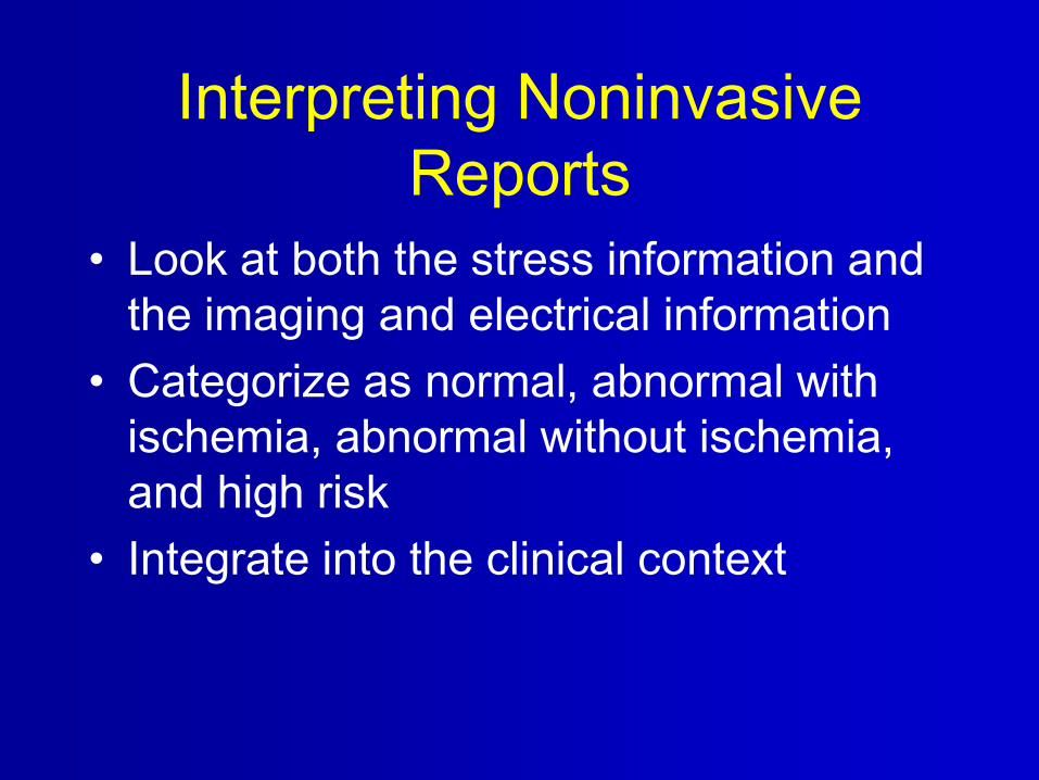
Interpreting Noninvasive
Reports
• Look at both the stress information and
the imaging and electrical information
• Categorize as normal, abnormal with
ischemia, abnormal without ischemia,
and high risk
• Integrate into the clinical context

Choosing the Best Test• Patient can exercise
– and ECG is normal: Exercise ECG
– and ECG is not interpretable: exercise perfusion study or
exercise echocardiogram
– and LBBB or LVH: vasodilator perfusion study
• Patient cannot exercise: vasodilator perfusion study or
dobutamine stress echocardiogram
• Patient with asthma and wheezing: no vasodilator -
dobutamine stress echo or perfusion study
• Patient with poor echo windows (COPD): may need
echo contrast agent, or change to perfusion study
• Patient over 350 pounds: difficult - perfusion will be
planar, echo also will likely be poor (also, can’t do
cardiac catheterization!)

Stress ECG Testing
≥1 mm horizontal ST
segment depression

Nuclear Perfusion Imaging

Heart Tests – Coronary Disease - 2
• Coronary Artery Calcium (CAC) score
– Asymptomatic and intermediate risk of CAD (10-20% 10-yr risk)
– Symptomatic and low risk of CAD (low score has high neg predict value)
• CT Coronary Angiography (Iodine +)
– Anomalous coronary (also cardiac MR)
– Acute chest pain, intermediate risk, neg Trop
– Maybe Sx + (intermed prob – abn ECG –can’t exercise – prior equivocal test)
• PET Scan, alternative to Nuc

Coronary Artery Calcium

CT Coronary Angiography
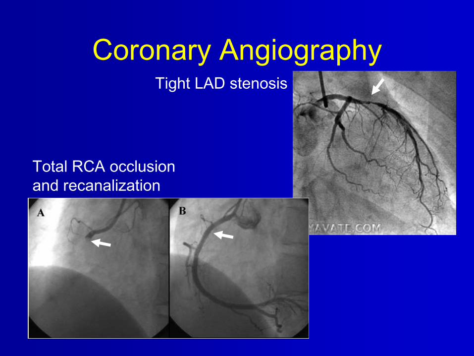
Coronary AngiographyTight LAD stenosis
Total RCA occlusion
and recanalization

Cardiac PET Scan

Heart Tests – Coronary Disease
– Radiation Exposure
Procedure # CXRs
Stress echocardiography 0
Cardiac MR Angiography 0
Coronary artery Calcium score 20-40
Coronary angiography (diagnostic) 200-500
Nuclear perfusion imaging 100-500
PET perfusion imaging 100-400
CT coronary angiography 700-2100

Definition of Angina
ACC/AHA Clinical Practice Guideline, Chronic Stable Angina, 2002.

Diagnosis: Pretest Probability of
Obstructive Disease at Catheterization
ACC/AHA Guideline Exercise Testing 2002, p. 7.
Diagnostic testing is appropriate - intermediate pretest probability

Stable CAD: Diagnostic Tests
• ECG normal and able to exercise = ETT
• ECG abnl and able to exercise = ETT
with imaging (nuc-perfusion or echo-wall
motion)
– Exception: LBBB, use vasodilator
• Unable to exercise = vasodilator or
dobutamine stress

Contraindications to Exercise Testing
• MI or UA <48 h
• Uncontrolled ventricular arrhythmia
• Symptomatic severe AS
• HCM (?)
• Decompensated HF
• Acute pulmonary embolism
• Acute aortic dissection
• Acute pericarditis
ACC/AHA Guideline Exercise Testing 2002, p. 5.

Stable CAD: Low Risk Test Results
• ECG result: Low risk Duke treadmill score (≥5)
– Number of minutes of Bruce protocol
– Minus 5 times number of mm ST depression
– Minus 4 times angina score (0=none, 1=some, 2=limiting)
• Nuclear result: normal, or small perfusion defect at rest or stress
• Stress Echo result: Normal wall motion or no change in limited resting wall motion abnormalities with stress
ACC/AHA Guideline, Stable Angina, 2002

Stable CAD: Strongly Positive (High
Risk) Test Results
• Markedly positive result = coronary angio
• ECG result: Significant ST depression at
low workload, ST elevation, low BP (Duke
treadmill score ≤-11)
• Nuclear result: TID, lung uptake,
multizone ischemia, EF<35%
• Stress Echo result: Fall in EF with stress,
multizone hypokinesis, EF<35%
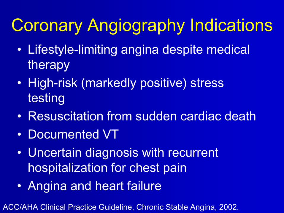
Coronary Angiography Indications
• Lifestyle-limiting angina despite medical
therapy
• High-risk (markedly positive) stress
testing
• Resuscitation from sudden cardiac death
• Documented VT
• Uncertain diagnosis with recurrent
hospitalization for chest pain
• Angina and heart failure
ACC/AHA Clinical Practice Guideline, Chronic Stable Angina, 2002.

Stress Myocardial Perfusion
Imaging
Courtesy of Dr. Janet Hays, UTHSCSANormal

NormalCourtesy of Dr. Janet Hays, UTHSCSA

NormalCourtesy of Dr. Janet Hays, UTHSCSA

NormalCourtesy of Dr. Janet Hays, UTHSCSA

AbnormalCourtesy of Dr. Janet Hays, UTHSCSA

AbnormalCourtesy of Dr. Janet Hays, UTHSCSA

AbnormalCourtesy of Dr. Janet Hays, UTHSCSA

Abnormal
Myocardial
Perfusion
Image
From Industry Advertisement for
Technicium Sestamibi (Cardiolite)
Inferior fixed defect
Small-moderate size,
moderate severity
reversible in apical septum

From South Texas Veterans’ Health Care SystemHigh grade LCX and RCA

Thank You!

What is the Appropriate Test?
• Age: 67
• Gender: M
• Htn: +
• DM: +
• HLP: -
• Tob: +
• ECG: Normal
• Hx: Employed postal
worker with no prior
history of heart
disease, with sharp
left-sided pain
lasting 2 seconds at
rest
• Answer:

What is the Appropriate Test?
• Age: 67
• Gender: M
• Htn: +
• DM: +
• HLP: -
• Tob: +
• ECG: Normal
• Hx: Employed postal
worker with no prior
history of heart
disease, with sharp
left-sided pain
lasting 2 seconds at
rest
• Answer: Treadmill exercise ECG

What is the Appropriate Test?
• Age: 68
• Gender: M
• Htn: +
• DM: -
• HLP: +
• Tob: +
• ECG: T flattening
• Hx: thin patient with
significant COPD on
home O2, wheezing,
with increasing
dyspnea and mild
chest tightness
• Answer:

What is the Appropriate Test?
• Age: 68
• Gender: M
• Htn: +
• DM: -
• HLP: +
• Tob: +
• ECG: T flattening
• Hx: thin patient with
significant COPD on
home O2, wheezing,
with increasing
dyspnea and mild
chest tightness
• Answer: Adrenergic stress echo (Vasodilator- no)

What is the Appropriate Test?
• Age: 35
• Gender: F
• Htn: +
• DM: -
• HLP: -
• Tob: -
• ECG: minor T
wave flattening
• Hx: asymptomatic
secretary wishes
permission to join an
exercise program
• Answer:

What is the Appropriate Test?
• Age: 35
• Gender: F
• Htn: +
• DM: -
• HLP: -
• Tob: -
• ECG: minor T
wave flattening
• Hx: asymptomatic
secretary wishes
permission to join an
exercise program
• Answer: No test, reassure, treat hypertension

What is the Appropriate Test?
• Age: 72
• Gender: M
• Htn: +
• DM: -
• HLP: +
• Tob: 0 for 9 yr
• ECG: LBBB
• Hx: patient 9 years
post CABG with
return of his former
chest pain,
occurring about
twice a week
• Answer:

What is the Appropriate Test?
• Age: 72
• Gender: M
• Htn: +
• DM: -
• HLP: +
• Tob: 0* 9 yr
• ECG: LBBB
• Hx: patient 9 years
post CABG with
return of his former
chest pain,
occurring about
twice a week
• Answer: Vasodilator thallium (dobutamine not good)

What is the Appropriate Test?
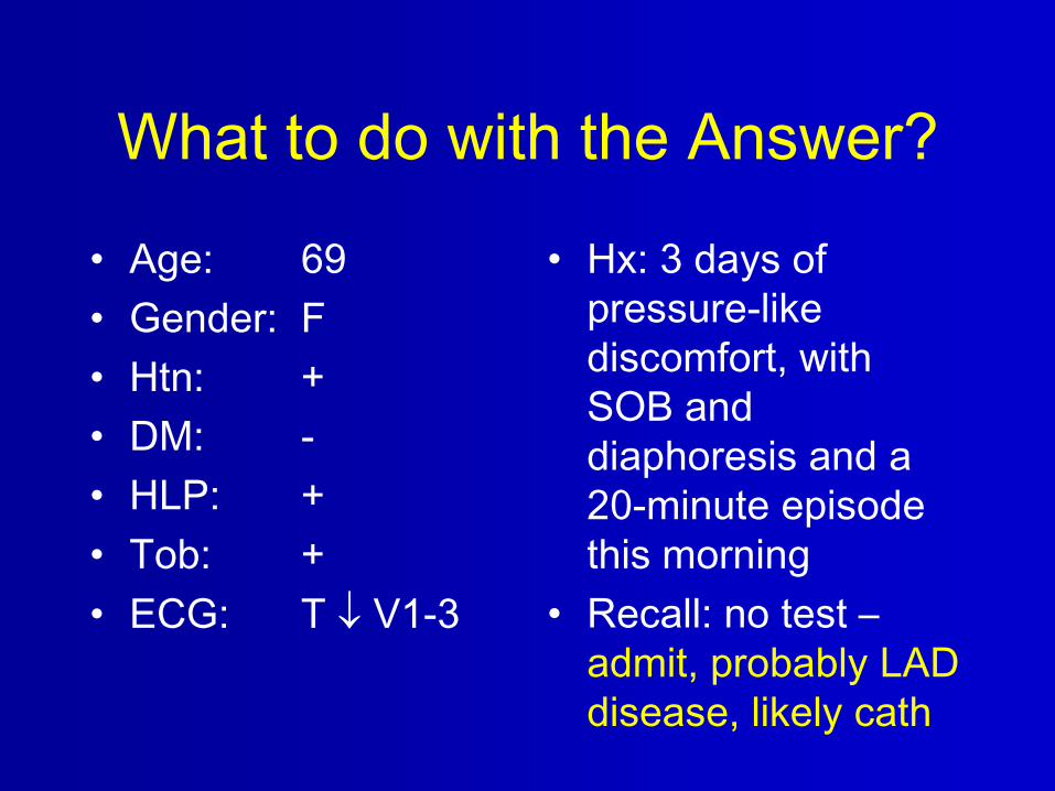
• Age: 69
• Gender: F
• Htn: +
• DM: -
• HLP: +
• Tob: +
• ECG: T V1-3
• Hx: 3 days of
pressure-like
discomfort, with
SOB and
diaphoresis and a
20-minute episode
this morning
• Answer:

What is the Appropriate Test?
• Age: 69
• Gender: F
• Htn: +
• DM: -
• HLP: +
• Tob: +
• ECG: T V1-3
• Hx: 3 days of
pressure-like
discomfort, with
SOB and
diaphoresis and a
20-minute episode
this morning
• Answer: No test, too sick, Wellens’, admit

What is the Appropriate Test?
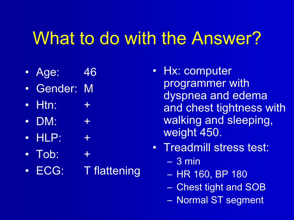
• Age: 46
• Gender: M
• Htn: +
• DM: +
• HLP: +
• Tob: +
• ECG: T flattening
• Hx: computer
programmer with
dyspnea and edema
and chest tightness
with walking and
sleeping, weight
450.
• Answer:

What is the Appropriate Test?
• Age: 46
• Gender: M
• Htn: +
• DM: +
• HLP: +
• Tob: +
• ECG: T flattening
• Hx: computer
programmer with
dyspnea and edema
and chest tightness
with walking and
sleeping, weight
450.
• Answer: No good answer.
Vasodilator sestamibi?
Dobutamine echo?
Exercise stress test?
Medical therapy?

What is the Appropriate Test?
• Age: 71
• Gender: F
• Htn: +
• DM: -
• HLP: -
• Tob: +
• ECG: normal
• Hx: patient admitted
with chest pain and
ST depression and
mild elevation of
Tro-I, also weight
loss, pulmonary
nodules and cervical
adenopathy, 5 years
post mastectomy
• Answer:

What is the Appropriate Test?
• Age: 71
• Gender: F
• Htn: +
• DM: -
• HLP: -
• Tob: +
• ECG: normal
• Hx: patient admitted
with chest pain and
ST depression and
mild elevation of
Tro-I, also weight
loss, pulmonary
nodules and cervical
adenopathy, 5 years
post mastectomy
• Answer: Consult Oncology first

What is the Appropriate Test?
• Age: 42
• Gender: F
• Htn: +
• DM: +
• HLP: +
• Tob: -
• ECG: PPRWP
• Hx: patient with
amputation left great
toe for vascular
disease, no chest
pain, mild dyspnea
when upset
• Answer:

What is the Appropriate Test?
• Age: 42
• Gender: F
• Htn: +
• DM: +
• HLP: +
• Tob: -
• ECG: PPRWP
• Hx: patient with
amputation left great
toe for vascular
disease, no chest
pain, mild dyspnea
when upset
• Answer: Vasodilator thallium (DSE OK)

What is the Appropriate Test?
• Age: 72
• Gender: F
• Htn: +
• DM: -
• HLP: -
• Tob: -
• ECG: A Fib, LVH
• Hx: patient with
chronic shortness of
breath, slightly
worsening
• Answer:

What is the Appropriate Test?
• Age: 72
• Gender: F
• Htn: +
• DM: -
• HLP: -
• Tob: -
• ECG: A Fib, LVH
• Hx: patient with
chronic shortness of
breath, slightly
worsening
• Answer: Vasodilator thallium (DSE NOT OK)

What is the Appropriate Test?
• Age: 45
• Gender: M
• Htn: +
• DM: -
• HLP: +
• Tob: - * 2 yr
• ECG: normal
• Hx: patient with no
symptoms, with
abnormal cine CT
result, worried
• Answer:

What is the Appropriate Test?
• Age: 45
• Gender: M
• Htn: +
• DM: -
• HLP: +
• Tob: none for 2
yr
• ECG: normal
• Hx: patient with no
symptoms, with
abnormal cine CT
result, worried
• Answer: Exercise ECG, but difficult decision

What is the Appropriate Test?
• Age: 59
• Gender: M
• Htn: +
• DM: -
• HLP: +
• Tob: - * 2 yr
• ECG: Ant MI
• Hx: patient with
refractory chest pain
despite optimal
medication, S/P
CABG 1994, cath
1998 2 grafts down,
native disease
progression
• Answer:

What is the Appropriate Test?
• Age: 59
• Gender: M
• Htn: +
• DM: -
• HLP: +
• Tob: - * 2 yr
• ECG: Ant MI
• Hx: patient with
refractory chest pain
despite optimal
medication, S/P
CABG 1994, cath
1998 2 grafts down,
native disease
progression
• Answer: Review cath; if candidate, thallium

What is the Appropriate Test?
• Age: 63
• Gender: M
• Htn: +
• DM: +
• HLP: +
• Tob: - * 2 yr
• ECG: LVH, A Fib
• Hx: patient with no
chest pain and no
prior cardiac history
admitted with new
onset CHF
• Answer:

What is the Appropriate Test?
• Age: 63
• Gender: M
• Htn: +
• DM: +
• HLP: +
• Tob: - * 2 yr
• ECG: LVH, A Fib
• Hx: patient with no
chest pain and no
prior cardiac history
admitted with new
onset CHF
• Answer: Either Cath first, or viability first

What to do with the Answer?
• Age: 67
• Gender: M
• Htn: +
• DM: +
• HLP: -
• Tob: +
• ECG: Normal
• Hx: Employed postal
worker with no prior
history of heart
disease, with sharp
left-sided pain
lasting 2 seconds at
rest
• Treadmill ECG
– Normal/normal
– Abnormal

What to do with the Answer?
• Age: 67
• Gender: M
• Htn: +
• DM: +
• HLP: -
• Tob: +
• ECG: Normal
• Hx: Employed postal worker with no prior history of heart disease, with sharp left-sided pain lasting 2 seconds at rest
• Treadmill ECG
– Normal/normal reassure
– Abnormal imaging study or cath

What to do with the Answer?
• Age: 68
• Gender: M
• Htn: +
• DM: -
• HLP: +
• Tob: +
• ECG: T flattening
• Hx: thin patient with
significant COPD on
home O2, wheezing,
with increasing
dyspnea and mild
chest tightness
• Stress echo
– Normal
– Abnormal

What to do with the Answer?
• Age: 68
• Gender: M
• Htn: +
• DM: -
• HLP: +
• Tob: +
• ECG: T flattening
• Hx: thin patient with significant COPD on home O2, wheezing, with increasing dyspnea and mild chest tightness
• Stress echo
– Normal reassure
– Abnormal medical management or cath

What to do with the Answer?
• Age: 35
• Gender: F
• Htn: +
• DM: -
• HLP: -
• Tob: -
• ECG: minor T
wave flattening
• Hx: asymptomatic
secretary wishes
permission to join an
exercise program
• Recall, no test
needed
• Inappropriate test
leads to
– More inappropriate
tests

What to do with the Answer?
• Age: 72
• Gender: M
• Htn: +
• DM: -
• HLP: +
• Tob: 0* 9 yr
• ECG: LBBB
• Hx: patient 9 years
post CABG with
return of his former
chest pain,
occurring about
twice a week
• Vasodilator thallium
– Normal
– Abnormal
– High-risk

What to do with the Answer?
• Age: 72
• Gender: M
• Htn: +
• DM: -
• HLP: +
• Tob: 0* 9 yr
• ECG: LBBB
• Hx: patient 9 years post CABG with return of his former chest pain, occurring about twice a week
• Vasodilator thallium
– Normal reassure, medical therapy
– Abnormal medical therapy
– High-risk cath
What to do with the Answer?
• Age: 69
• Gender: F
• Htn: +
• DM: -
• HLP: +
• Tob: +
• ECG: T V1-3
• Hx: 3 days of
pressure-like
discomfort, with
SOB and
diaphoresis and a
20-minute episode
this morning
• Recall: no test –
admit, probably LAD
disease, likely cath
What to do with the Answer?
• Age: 46
• Gender: M
• Htn: +
• DM: +
• HLP: +
• Tob: +
• ECG: T flattening
• Hx: computer programmer with dyspnea and edema and chest tightness with walking and sleeping, weight 450.
• Treadmill stress test:– 3 min
– HR 160, BP 180
– Chest tight and SOB
– Normal ST segment

What to do with the Answer?
• Age: 46
• Gender: M
• Htn: +
• DM: +
• HLP: +
• Tob: +
• ECG: T flattening
• Hx: computer programmer with dyspnea and edema and chest tightness with walking and sleeping, weight 450.
• Treadmill stress test:– 3 min
– HR 160, BP 180
– Chest tight and SOB
– Normal ST segmentAnswer: medical therapy, no
real change in management

What to do with the Answer?
• Age: 42
• Gender: F
• Htn: +
• DM: +
• HLP: +
• Tob: -
• ECG: PPRWP
• Hx: patient with amputation left great toe for vascular disease, no chest pain, mild dyspnea when upset
• Pthall –
– Normal
– Abnormal
– High risk

What to do with the Answer?
• Age: 42
• Gender: F
• Htn: +
• DM: +
• HLP: +
• Tob: -
• ECG: PPRWP
• Hx: patient with
amputation left great toe
for vascular disease, no
chest pain, mild dyspnea
when upset
• Pthall –
– Normal reassure
– Abnormal cath or
medical therapy
– High risk cath

Last Slide: Hurray!
• Recall the
physiology
• Means of assessing
ischemia
– Electrical
– Perfusion
– Function (wall motion)
• Means of producing
stress
– Exercise
– Adrenergic agent
– Vasodilator
• Integrate into clinical
context