innate chap
DESCRIPTION
innateTRANSCRIPT
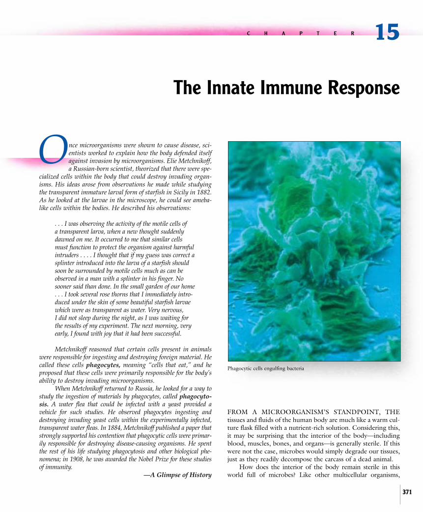
Once microorganisms were shown to cause disease, sci-entists worked to explain how the body defended itselfagainst invasion by microorganisms. Elie Metchnikoff,a Russian-born scientist, theorized that there were spe-
cialized cells within the body that could destroy invading organ-isms. His ideas arose from observations he made while studyingthe transparent immature larval form of starfish in Sicily in 1882.As he looked at the larvae in the microscope, he could see ameba-like cells within the bodies. He described his observations:
. . . I was observing the activity of the motile cells of a transparent larva, when a new thought suddenlydawned on me. It occurred to me that similar cellsmust function to protect the organism against harmfulintruders . . . . I thought that if my guess was correct asplinter introduced into the larva of a starfish shouldsoon be surrounded by motile cells much as can beobserved in a man with a splinter in his finger. Nosooner said than done. In the small garden of our home. . . I took several rose thorns that I immediately intro-duced under the skin of some beautiful starfish larvaewhich were as transparent as water. Very nervous, I did not sleep during the night, as I was waiting forthe results of my experiment. The next morning, veryearly, I found with joy that it had been successful.
Metchnikoff reasoned that certain cells present in animalswere responsible for ingesting and destroying foreign material. Hecalled these cells phagocytes, meaning “cells that eat,” and heproposed that these cells were primarily responsible for the body’sability to destroy invading microorganisms.
When Metchnikoff returned to Russia, he looked for a way tostudy the ingestion of materials by phagocytes, called phagocyto-sis. A water flea that could be infected with a yeast provided a vehicle for such studies. He observed phagocytes ingesting anddestroying invading yeast cells within the experimentally infected,transparent water fleas. In 1884, Metchnikoff published a paper thatstrongly supported his contention that phagocytic cells were primar-ily responsible for destroying disease-causing organisms. He spentthe rest of his life studying phagocytosis and other biological phe-nomena; in 1908, he was awarded the Nobel Prize for these studiesof immunity.
—A Glimpse of History
FROM A MICROORGANISM’S STANDPOINT, THE tissues and fluids of the human body are much like a warm cul-ture flask filled with a nutrient-rich solution. Considering this,it may be surprising that the interior of the body—includingblood, muscles, bones, and organs—is generally sterile. If thiswere not the case, microbes would simply degrade our tissues,just as they readily decompose the carcass of a dead animal.
How does the interior of the body remain sterile in thisworld full of microbes? Like other multicellular organisms,
15C H A P T E R
The Innate Immune Response
371
Phagocytic cells engulfing bacteria

humans have evolved several mechanisms of defense. First, weare covered with skin and mucous membranes that prevententry of most foreign material, including microbes, into thebody. Ready in case the barriers are breached are sensor systemsthat detect molecules associated with danger; for example, com-pounds that are unique to bacteria or are typically released onlywhen tissues are damaged. These sensors can direct and assistother host defenses, facilitating the destruction of the foreignmaterial. Also lying in wait are host cells that specialize in ingest-ing and digesting foreign material; if needed, additional rein-forcements can be recruited to the site of breach. The protec-tion provided by these systems is termed innate immunity,reflecting that we are born with it. Innate immunity differs fromadaptive immunity, which will be described shortly, in that allinvaders are dealt with using a limited set of weapons. Althoughthe number of copies of the various weapons can be modulatedin response to an invader, their mechanisms cannot be modifiedto enhance the reaction.
The components of innate immunity have been callednon-specific defenses, but recent discoveries have shown thatmost of these components are far from unfocused; instead,they rely on the recognition of certain molecular patternsassociated with invading microbes or tissue damage, a featurereferred to as pattern recognition. Molecular patterns asso-ciated with pathogens include various compounds unique tobacterial cell walls, such as lipopolysaccharide, lipoteichoicacid, and peptidoglycan, and other molecules. Those associat-ed with damage include various proteins that are normallyintracellular and are now outside cells, and substances pro-duced during tissue necrosis and damage. ■ lipopolysaccharide, p. 59■ lipoteichoic acid, p. 59 ■ peptidoglycan, p. 58
In addition to innate immunity, vertebrates have evolved amore specialized response, termed the adaptive immuneresponse; this develops throughout life and substantiallyincreases the ability of the host to defend itself. Each time thebody is exposed to foreign material, the adaptive defense systemfirst “learns” and then “remembers” the most effective responseto that specific material; it then reacts accordingly if the materi-al is encountered again. The foreign material to which theimmune system responds is called an antigen. On first exposureto an invading microbe or other antigen, the response developsrelatively slowly, during which time the microbe may causedamage if the innate defenses are unable to contain it.Successive exposures, however, lead to a swift and greater repeatresponse, generally eliminating the invader before it causes obvi-ous harm.
There are two general mechanisms used by the adaptiveimmune response to eliminate an invader. If the antigen is with-in one of the body’s own cells, which are referred to as either ahost cell or a “self’’ cell, then the cell may be sacrificed as ameans of destroying the invader. If the antigen is extracellular,then the body responds by making antibodies. These glycopro-tein molecules have two functional regions; one region bindsspecifically to the antigen and the other functions as a “red flag,”directing other host defenses to remove or destroy the antigen.
The study of the many mechanisms the body uses todefend itself against invading microbes is called immunology.
372 Chapter 15 The Innate Immune Response
It encompasses not only the study of protection against infec-tious agents, but also cancers and the acceptance or rejectionof transplanted cells and organs. Immunologists also study theeffects of the immune response that can damage the body,such as autoimmunity, which occurs when the immuneresponse is inappropriately directed against the cells of one’sown body, and hypersensitivity, or allergic reactions.
To simplify the description of a network as complex and intri-cate as the immune system, it is helpful to consider it as a series ofindividual parts. This chapter, for example, will focus almostexclusively on innate immunity. Bear in mind, however, thatalthough the various parts are discussed separately, in the bodytheir actions are intimately connected and coordinated. In fact, asyou will see in chapter 16, certain components of the innatedefenses are instrumental in educating the adaptive defenses,helping them to distinguish antigens that represent danger.
15.1 Overview of the InnateDefenses
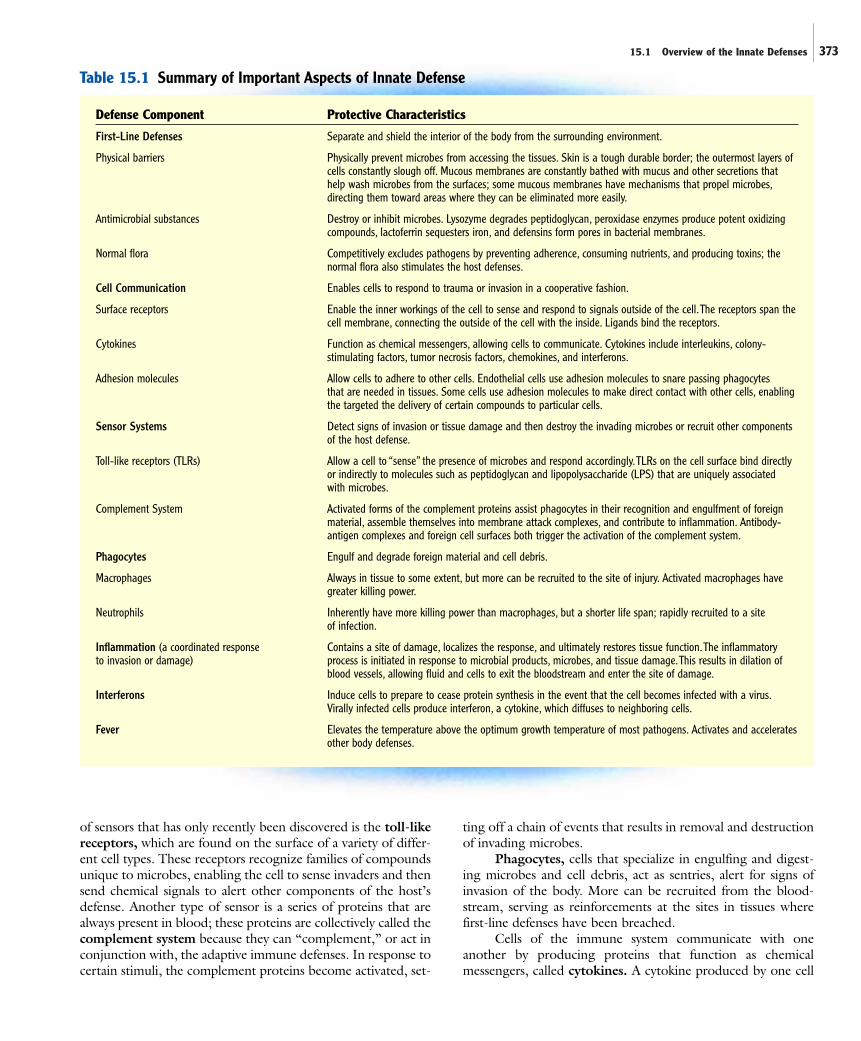
First-line defenses are the barriers that separate and shield theinterior of the body from the surrounding environment; theyare the initial obstacles that microorganisms must overcome toinvade the tissues. The anatomical barriers, which include theskin and mucous membranes, not only provide physical separa-tion, but they are often bathed in secretions containing sub-stances that have antimicrobial properties (figure 15.1).Characteristics of the components of innate immunity, includ-ing the first-line defenses, are summarized in table 15.1.
Sensor systems within the body recognize when the first-line barriers have been breached and then relay that informationto other components of the host defenses. An important group
MouthEye
Anus Skin
Urogenital tract
Alimentary tract
Respiratory tract
Skin
Mucous membranes
Figure 15.1 Anatomical Barriers These barriers separate the interior of thebody from the surrounding environment; they are the initial obstacles microorganismsmust overcome to invade tissues.The skin is shown in purple.
15.1 Overview of the Innate Defenses 373
Defense Component Protective Characteristics
First-Line Defenses Separate and shield the interior of the body from the surrounding environment.
Physical barriers Physically prevent microbes from accessing the tissues. Skin is a tough durable border; the outermost layers ofcells constantly slough off. Mucous membranes are constantly bathed with mucus and other secretions thathelp wash microbes from the surfaces; some mucous membranes have mechanisms that propel microbes,directing them toward areas where they can be eliminated more easily.
Antimicrobial substances Destroy or inhibit microbes. Lysozyme degrades peptidoglycan, peroxidase enzymes produce potent oxidizingcompounds, lactoferrin sequesters iron, and defensins form pores in bacterial membranes.
Normal flora Competitively excludes pathogens by preventing adherence, consuming nutrients, and producing toxins; thenormal flora also stimulates the host defenses.
Cell Communication Enables cells to respond to trauma or invasion in a cooperative fashion.
Surface receptors Enable the inner workings of the cell to sense and respond to signals outside of the cell.The receptors span thecell membrane, connecting the outside of the cell with the inside. Ligands bind the receptors.
Cytokines Function as chemical messengers, allowing cells to communicate. Cytokines include interleukins, colony-stimulating factors, tumor necrosis factors, chemokines, and interferons.
Adhesion molecules Allow cells to adhere to other cells. Endothelial cells use adhesion molecules to snare passing phagocytes that are needed in tissues. Some cells use adhesion molecules to make direct contact with other cells, enablingthe targeted the delivery of certain compounds to particular cells.
Sensor Systems Detect signs of invasion or tissue damage and then destroy the invading microbes or recruit other componentsof the host defense.
Toll-like receptors (TLRs) Allow a cell to “sense” the presence of microbes and respond accordingly.TLRs on the cell surface bind directlyor indirectly to molecules such as peptidoglycan and lipopolysaccharide (LPS) that are uniquely associated with microbes.
Complement System Activated forms of the complement proteins assist phagocytes in their recognition and engulfment of foreignmaterial, assemble themselves into membrane attack complexes, and contribute to inflammation. Antibody-antigen complexes and foreign cell surfaces both trigger the activation of the complement system.
Phagocytes Engulf and degrade foreign material and cell debris.
Macrophages Always in tissue to some extent, but more can be recruited to the site of injury. Activated macrophages havegreater killing power.
Neutrophils Inherently have more killing power than macrophages, but a shorter life span; rapidly recruited to a site of infection.
Inflammation (a coordinated response Contains a site of damage, localizes the response, and ultimately restores tissue function.The inflammatoryto invasion or damage) process is initiated in response to microbial products, microbes, and tissue damage.This results in dilation of
blood vessels, allowing fluid and cells to exit the bloodstream and enter the site of damage.
Interferons Induce cells to prepare to cease protein synthesis in the event that the cell becomes infected with a virus.Virally infected cells produce interferon, a cytokine, which diffuses to neighboring cells.
Fever Elevates the temperature above the optimum growth temperature of most pathogens. Activates and acceleratesother body defenses.
Table 15.1 Summary of Important Aspects of Innate Defense
of sensors that has only recently been discovered is the toll-likereceptors, which are found on the surface of a variety of differ-ent cell types. These receptors recognize families of compoundsunique to microbes, enabling the cell to sense invaders and thensend chemical signals to alert other components of the host’sdefense. Another type of sensor is a series of proteins that arealways present in blood; these proteins are collectively called thecomplement system because they can “complement,” or act inconjunction with, the adaptive immune defenses. In response tocertain stimuli, the complement proteins become activated, set-
ting off a chain of events that results in removal and destructionof invading microbes.
Phagocytes, cells that specialize in engulfing and digest-ing microbes and cell debris, act as sentries, alert for signs ofinvasion of the body. More can be recruited from the blood-stream, serving as reinforcements at the sites in tissues wherefirst-line defenses have been breached.
Cells of the immune system communicate with oneanother by producing proteins that function as chemical messengers, called cytokines. A cytokine produced by one cell
374 Chapter 15 The Innate Immune Response
diffuses to another and binds to the appropriate cytokine recep-tor of that cell. When a cytokine binds a receptor, the receptortransmits a signal to the interior of the cell, inducing certainchanges in the activities of the cell. Some types of cytokinesendow cells with enhanced powers; others prompt cells tomigrate to specific locations within the body.
When invading microorganisms or tissue damage isdetected, inflammation ensues; this is a coordinated responseinvolving many aspects of the innate defenses. During inflam-mation, the cells that line local blood vessels near the area ofinvasion or damage undergo changes that allow antibodies,complement proteins, and coagulation proteins in plasma, thefluid portion of the blood, to leak into tissues. Other changesallow phagocytic cells in the bloodstream to adhere to the ves-sels and then squeeze between cells, exiting the bloodstream.Phagocytic cells then migrate to the area of infection or damagewhere they ingest and destroy foreign material. Some types ofphagocytes play a dual role, destroying invaders while also com-municating with cells of the adaptive immune system, enlistingtheir far more powerful effects.
The body also has physiological defense mechanisms, suchas the increase in internal body temperature called fever, whichacts in several ways to discourage infection.
M I C R O C H E C K 1 5 . 1
First-line defenses are the initial obstacles that microbesmust overcome to invade the tissues. Within the bodyare sensor systems such as toll-like receptors and thecomplement system that recognize when the barriershave been breached. Phagocytic cells engulf foreignmaterial; they can communicate with other cells viacytokines. Inflammation is a coordinated response toinvasion or tissue damage.
■ How do cytokines function?■ Describe the dual roles played by some types of
phagocytes.■ What types of molecules that are unique to microbes
might toll-like receptors recognize?
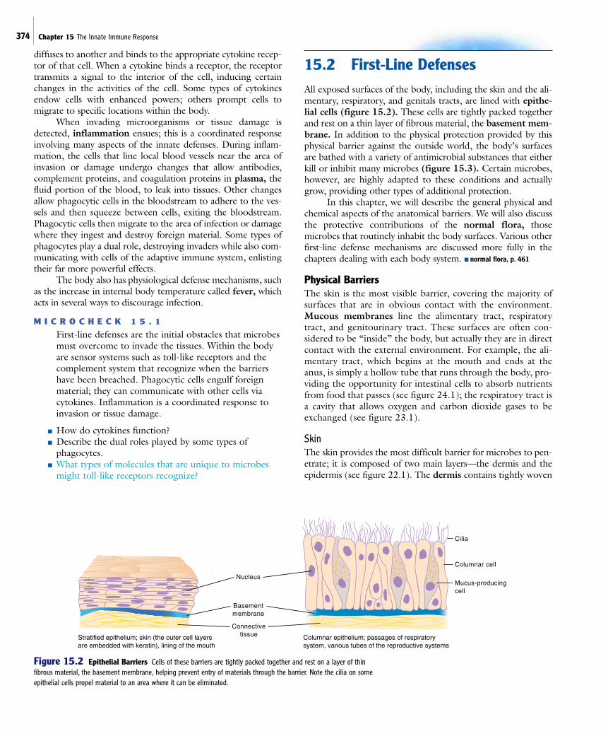
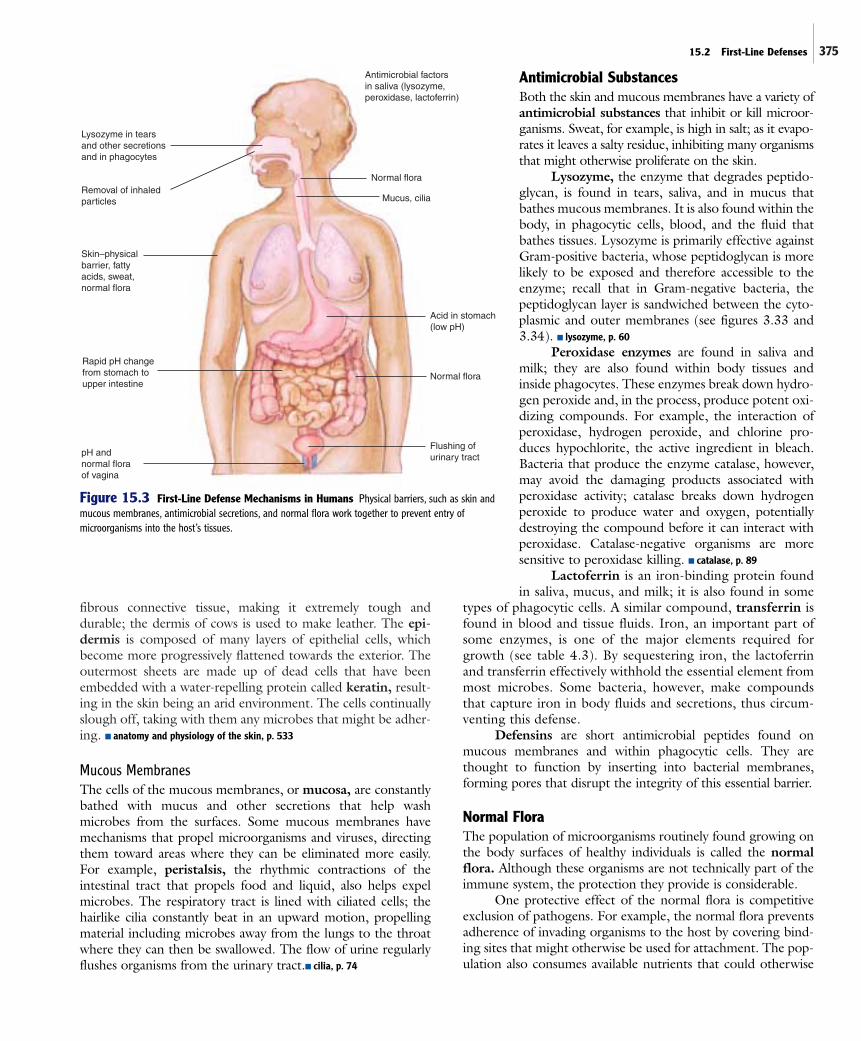
15.2 First-Line DefensesAll exposed surfaces of the body, including the skin and the ali-mentary, respiratory, and genitals tracts, are lined with epithe-lial cells (figure 15.2). These cells are tightly packed togetherand rest on a thin layer of fibrous material, the basement mem-brane. In addition to the physical protection provided by thisphysical barrier against the outside world, the body’s surfacesare bathed with a variety of antimicrobial substances that eitherkill or inhibit many microbes (figure 15.3). Certain microbes,however, are highly adapted to these conditions and actuallygrow, providing other types of additional protection.
In this chapter, we will describe the general physical andchemical aspects of the anatomical barriers. We will also discussthe protective contributions of the normal flora, thosemicrobes that routinely inhabit the body surfaces. Various otherfirst-line defense mechanisms are discussed more fully in thechapters dealing with each body system. ■ normal flora, p. 461
Physical BarriersThe skin is the most visible barrier, covering the majority ofsurfaces that are in obvious contact with the environment.Mucous membranes line the alimentary tract, respiratorytract, and genitourinary tract. These surfaces are often con-sidered to be “inside” the body, but actually they are in directcontact with the external environment. For example, the ali-mentary tract, which begins at the mouth and ends at theanus, is simply a hollow tube that runs through the body, pro-viding the opportunity for intestinal cells to absorb nutrientsfrom food that passes (see figure 24.1); the respiratory tract isa cavity that allows oxygen and carbon dioxide gases to beexchanged (see figure 23.1).
SkinThe skin provides the most difficult barrier for microbes to pen-etrate; it is composed of two main layers—the dermis and theepidermis (see figure 22.1). The dermis contains tightly woven
Nucleus
Basementmembrane
Connectivetissue
Cilia
Columnar cell
Mucus-producing cell
Columnar epithelium; passages of respiratory system, various tubes of the reproductive systems
Stratified epithelium; skin (the outer cell layers are embedded with keratin), lining of the mouth
Figure 15.2 Epithelial Barriers Cells of these barriers are tightly packed together and rest on a layer of thinfibrous material, the basement membrane, helping prevent entry of materials through the barrier. Note the cilia on someepithelial cells propel material to an area where it can be eliminated.
375
fibrous connective tissue, making it extremely tough anddurable; the dermis of cows is used to make leather. The epi-dermis is composed of many layers of epithelial cells, whichbecome more progressively flattened towards the exterior. Theoutermost sheets are made up of dead cells that have beenembedded with a water-repelling protein called keratin, result-ing in the skin being an arid environment. The cells continuallyslough off, taking with them any microbes that might be adher-ing. ■ anatomy and physiology of the skin, p. 533
Mucous MembranesThe cells of the mucous membranes, or mucosa, are constantlybathed with mucus and other secretions that help washmicrobes from the surfaces. Some mucous membranes havemechanisms that propel microorganisms and viruses, directingthem toward areas where they can be eliminated more easily.For example, peristalsis, the rhythmic contractions of theintestinal tract that propels food and liquid, also helps expelmicrobes. The respiratory tract is lined with ciliated cells; thehairlike cilia constantly beat in an upward motion, propellingmaterial including microbes away from the lungs to the throatwhere they can then be swallowed. The flow of urine regularlyflushes organisms from the urinary tract.■ cilia, p. 74
Antimicrobial SubstancesBoth the skin and mucous membranes have a variety ofantimicrobial substances that inhibit or kill microor-ganisms. Sweat, for example, is high in salt; as it evapo-rates it leaves a salty residue, inhibiting many organismsthat might otherwise proliferate on the skin.
Lysozyme, the enzyme that degrades peptido-glycan, is found in tears, saliva, and in mucus thatbathes mucous membranes. It is also found within thebody, in phagocytic cells, blood, and the fluid thatbathes tissues. Lysozyme is primarily effective againstGram-positive bacteria, whose peptidoglycan is morelikely to be exposed and therefore accessible to theenzyme; recall that in Gram-negative bacteria, thepeptidoglycan layer is sandwiched between the cyto-plasmic and outer membranes (see figures 3.33 and3.34). ■ lysozyme, p. 60
Peroxidase enzymes are found in saliva andmilk; they are also found within body tissues andinside phagocytes. These enzymes break down hydro-gen peroxide and, in the process, produce potent oxi-dizing compounds. For example, the interaction ofperoxidase, hydrogen peroxide, and chlorine pro-duces hypochlorite, the active ingredient in bleach.Bacteria that produce the enzyme catalase, however,may avoid the damaging products associated withperoxidase activity; catalase breaks down hydrogenperoxide to produce water and oxygen, potentiallydestroying the compound before it can interact withperoxidase. Catalase-negative organisms are moresensitive to peroxidase killing. ■ catalase, p. 89
Lactoferrin is an iron-binding protein foundin saliva, mucus, and milk; it is also found in some
types of phagocytic cells. A similar compound, transferrin isfound in blood and tissue fluids. Iron, an important part ofsome enzymes, is one of the major elements required forgrowth (see table 4.3). By sequestering iron, the lactoferrinand transferrin effectively withhold the essential element frommost microbes. Some bacteria, however, make compoundsthat capture iron in body fluids and secretions, thus circum-venting this defense.
Defensins are short antimicrobial peptides found onmucous membranes and within phagocytic cells. They arethought to function by inserting into bacterial membranes,forming pores that disrupt the integrity of this essential barrier.
Normal FloraThe population of microorganisms routinely found growing onthe body surfaces of healthy individuals is called the normalflora. Although these organisms are not technically part of theimmune system, the protection they provide is considerable.
One protective effect of the normal flora is competitiveexclusion of pathogens. For example, the normal flora preventsadherence of invading organisms to the host by covering bind-ing sites that might otherwise be used for attachment. The pop-ulation also consumes available nutrients that could otherwise
Lysozyme in tearsand other secretionsand in phagocytes
Removal of inhaledparticles
Skin–physicalbarrier, fattyacids, sweat,normal flora
Rapid pH changefrom stomach toupper intestine
pH and normal floraof vagina
Antimicrobial factorsin saliva (lysozyme,peroxidase, lactoferrin)
Normal flora
Normal flora
Mucus, cilia
Acid in stomach(low pH)
Flushing ofurinary tract
Figure 15.3 First-Line Defense Mechanisms in Humans Physical barriers, such as skin andmucous membranes, antimicrobial secretions, and normal flora work together to prevent entry ofmicroorganisms into the host’s tissues.
15.2 First-Line Defenses
376 Chapter 15 The Innate Immune Response
be used by less desirable organisms. Members of the normalflora also produce compounds that are toxic to other bacteria.For example, in the hair follicles of the skin, Propionibacteriumspecies degrade the lipids found in body secretions, releasing fattyacids that inhibit the growth of many potential disease-producers.In the gastrointestinal tract, some strains of E. coli synthesize colicins, proteins that are toxic to some strains of bacteria.Lactobacillus species growing in the vagina produce lactic acid asa fermentation end product, resulting in an acidic pH thatinhibits the growth of many potential disease-causing organisms.Disruption of the normal flora, which occurs when antibiotics areused, can predispose a person to various infections. Examplesinclude antibiotic-associated colitis, caused by the growth oftoxin-producing strains of Clostridium difficile in the intestine,and vulvovaginitis, caused by excessive growth of Candida albi-cans in the vagina. ■ antibiotic-associated colitis, p. 601 ■ vulvovaginitis, p. 640
The normal flora also stimulates the host defenses, effec-tively providing a moderate amount of “exercise’’ to the system,thereby enhancing its function. Other aspects of the normalflora will be discussed in chapter 19.
M I C R O C H E C K 1 5 . 2
Physical barriers that prevent entry of microorganismsinto the body include the skin and mucous membranes.Various antimicrobial substances, including lysozyme,peroxidase enzymes, lactoferrin, and defensins arefound on the body surfaces. The normal flora plays aprotective role by excluding certain other microbes.
■ What is peristalsis?■ What is the role of lactoferrin?■ How would damage to the ciliated cells of the respira-
tory tract predispose a person to infection?
15.3 The Cells of the ImmuneSystem
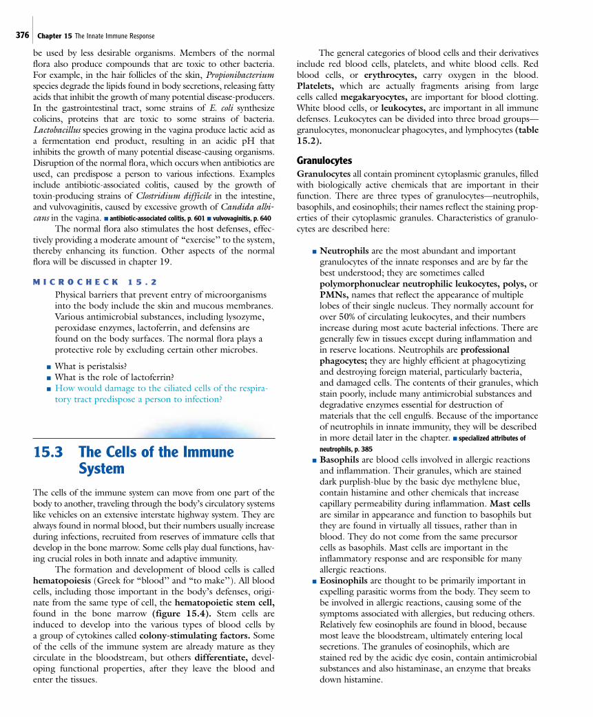
The cells of the immune system can move from one part of thebody to another, traveling through the body’s circulatory systemslike vehicles on an extensive interstate highway system. They arealways found in normal blood, but their numbers usually increaseduring infections, recruited from reserves of immature cells thatdevelop in the bone marrow. Some cells play dual functions, hav-ing crucial roles in both innate and adaptive immunity.
The formation and development of blood cells is calledhematopoiesis (Greek for “blood’’ and “to make’’). All bloodcells, including those important in the body’s defenses, origi-nate from the same type of cell, the hematopoietic stem cell,found in the bone marrow (figure 15.4). Stem cells areinduced to develop into the various types of blood cells by a group of cytokines called colony-stimulating factors. Someof the cells of the immune system are already mature as they circulate in the bloodstream, but others differentiate, devel-oping functional properties, after they leave the blood andenter the tissues.
The general categories of blood cells and their derivativesinclude red blood cells, platelets, and white blood cells. Redblood cells, or erythrocytes, carry oxygen in the blood.Platelets, which are actually fragments arising from large cells called megakaryocytes, are important for blood clotting.White blood cells, or leukocytes, are important in all immunedefenses. Leukocytes can be divided into three broad groups—granulocytes, mononuclear phagocytes, and lymphocytes (table15.2).
GranulocytesGranulocytes all contain prominent cytoplasmic granules, filledwith biologically active chemicals that are important in theirfunction. There are three types of granulocytes—neutrophils,basophils, and eosinophils; their names reflect the staining prop-erties of their cytoplasmic granules. Characteristics of granulo-cytes are described here:
■ Neutrophils are the most abundant and importantgranulocytes of the innate responses and are by far thebest understood; they are sometimes calledpolymorphonuclear neutrophilic leukocytes, polys, orPMNs, names that reflect the appearance of multiplelobes of their single nucleus. They normally account forover 50% of circulating leukocytes, and their numbersincrease during most acute bacterial infections. There aregenerally few in tissues except during inflammation andin reserve locations. Neutrophils are professionalphagocytes; they are highly efficient at phagocytizingand destroying foreign material, particularly bacteria,and damaged cells. The contents of their granules, whichstain poorly, include many antimicrobial substances anddegradative enzymes essential for destruction ofmaterials that the cell engulfs. Because of the importanceof neutrophils in innate immunity, they will be describedin more detail later in the chapter. ■ specialized attributes ofneutrophils, p. 385
■ Basophils are blood cells involved in allergic reactionsand inflammation. Their granules, which are staineddark purplish-blue by the basic dye methylene blue,contain histamine and other chemicals that increasecapillary permeability during inflammation. Mast cellsare similar in appearance and function to basophils butthey are found in virtually all tissues, rather than inblood. They do not come from the same precursor cells as basophils. Mast cells are important in theinflammatory response and are responsible for manyallergic reactions.
■ Eosinophils are thought to be primarily important inexpelling parasitic worms from the body. They seem tobe involved in allergic reactions, causing some of thesymptoms associated with allergies, but reducing others.Relatively few eosinophils are found in blood, becausemost leave the bloodstream, ultimately entering localsecretions. The granules of eosinophils, which arestained red by the acidic dye eosin, contain antimicrobialsubstances and also histaminase, an enzyme that breaksdown histamine.
15.3 The Cells of the Immune System 377
Natural killer (NK) cells
Macrophage
Mononuclear phagocytes
Dendritic cell
Plasma cell
MyeloblastMegakaryoblast
Megakaryocyte
Erythroblast
Red blood cells (erythrocytes)
White blood cells (leukocytes)
Platelets(thrombocytes)
BasophilEosinophil NeutrophilMonocyte
Granulocytes
Lymphocytes
T lymphocyteprocessed inthymus
B lymphocyteprocessed inbone marrow
Monoblast Lymphoblasts
Hematopoieticstem cell(in bone marrow)
Lymphoidstem cell
Stem cellfor all exceptlymphocytes
(in bone marrow)
Colony-stimulating factors
Figure 15.4 Blood and Lymphoid Cells All these types of cells are derived from precursor stem cells found in thebone marrow. Some of the steps not yet clearly defined are indicated by dotted arrows. Multiple steps occur between the stemcell and the final cells produced.The role of these cells in the immune response will be explained in this chapter and chapter 16.
Mononuclear PhagocytesTwo functional types of mononuclear phagocytes, macro-phages and dendritic cells, arise from the same precursor, themonocyte. Characteristics of mononuclear phagocytes aredescribed here:
■ Monocytes circulate in blood after leaving the bonemarrow. When they migrate into tissues they developinto either macrophages or dendritic cells.
■ Macrophages, like neutrophils, are professionalphagocytes. Although macrophages are present in
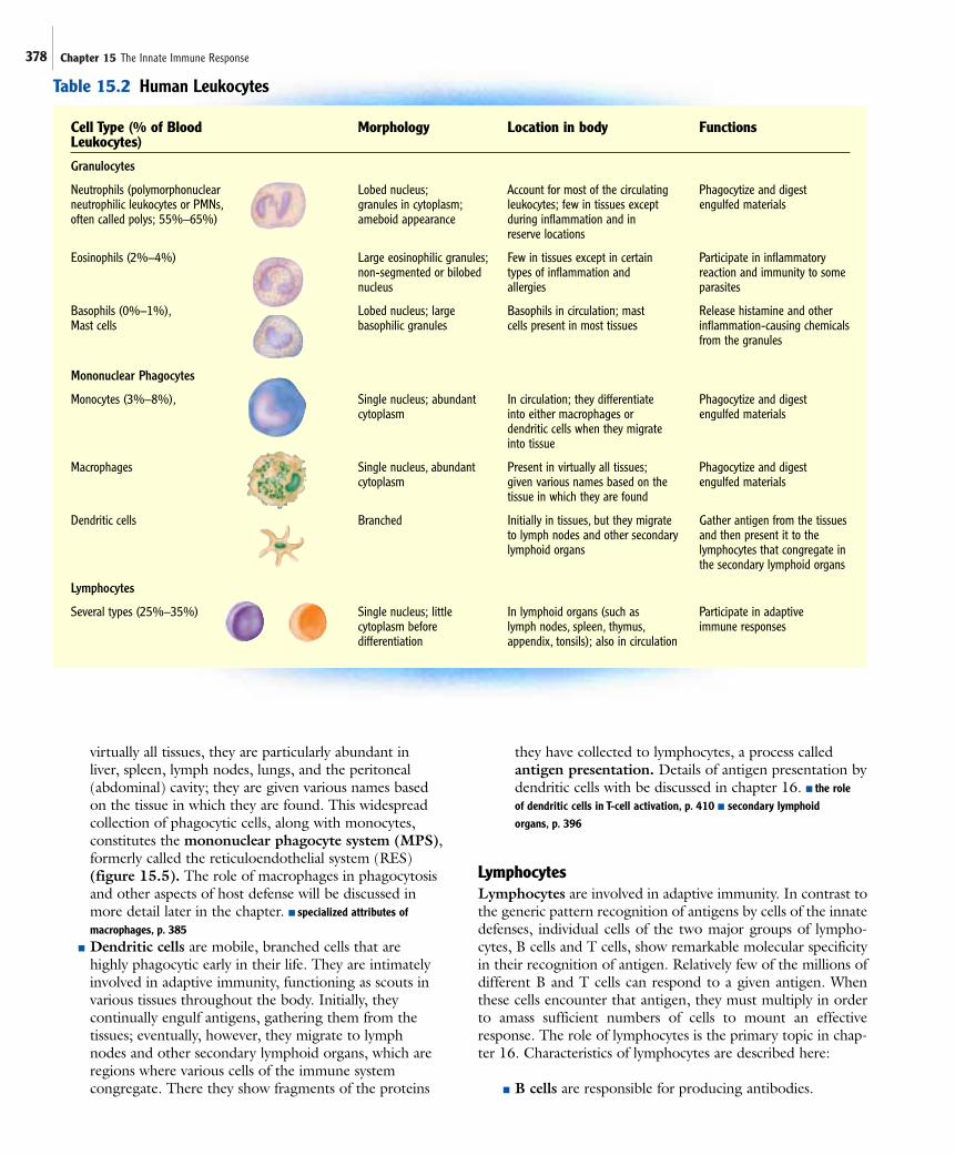
378 Chapter 15 The Innate Immune Response
Cell Type (% of Blood Morphology Location in body FunctionsLeukocytes)
Granulocytes
Neutrophils (polymorphonuclear Lobed nucleus; Account for most of the circulating Phagocytize and digestneutrophilic leukocytes or PMNs, granules in cytoplasm; leukocytes; few in tissues except engulfed materialsoften called polys; 55%–65%) ameboid appearance during inflammation and in
reserve locations
Eosinophils (2%–4%) Large eosinophilic granules; Few in tissues except in certain Participate in inflammatorynon-segmented or bilobed types of inflammation and reaction and immunity to somenucleus allergies parasites
Basophils (0%–1%), Lobed nucleus; large Basophils in circulation; mast Release histamine and otherMast cells basophilic granules cells present in most tissues inflammation-causing chemicals
from the granules
Mononuclear Phagocytes
Monocytes (3%–8%), Single nucleus; abundant In circulation; they differentiate Phagocytize and digest cytoplasm into either macrophages or engulfed materials
dendritic cells when they migrate into tissue
Macrophages Single nucleus, abundant Present in virtually all tissues; Phagocytize and digest cytoplasm given various names based on the engulfed materials
tissue in which they are found
Dendritic cells Branched Initially in tissues, but they migrate Gather antigen from the tissues to lymph nodes and other secondary and then present it to thelymphoid organs lymphocytes that congregate in
the secondary lymphoid organs
Lymphocytes
Several types (25%–35%) Single nucleus; little In lymphoid organs (such as Participate in adaptivecytoplasm before lymph nodes, spleen, thymus, immune responsesdifferentiation appendix, tonsils); also in circulation
Table 15.2 Human Leukocytes
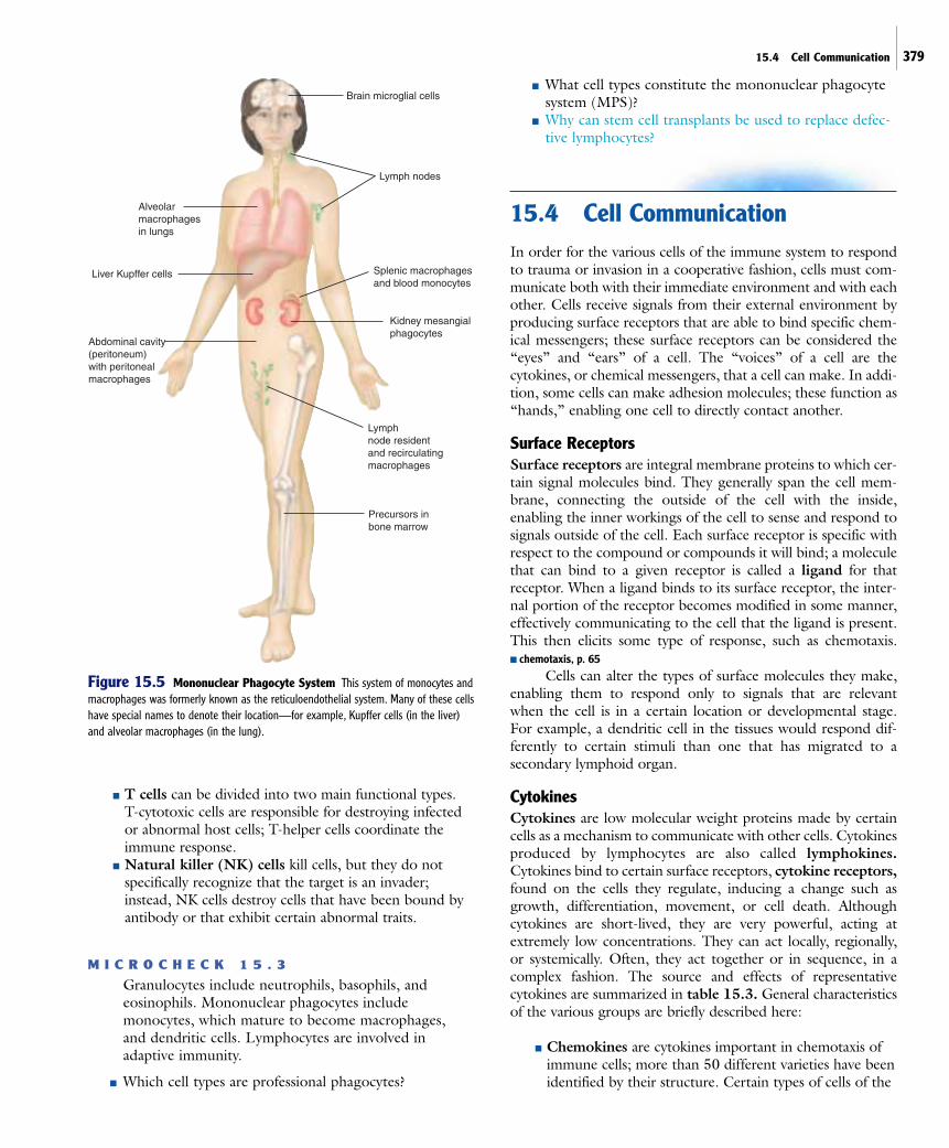
virtually all tissues, they are particularly abundant inliver, spleen, lymph nodes, lungs, and the peritoneal(abdominal) cavity; they are given various names basedon the tissue in which they are found. This widespreadcollection of phagocytic cells, along with monocytes,constitutes the mononuclear phagocyte system (MPS),formerly called the reticuloendothelial system (RES)(figure 15.5). The role of macrophages in phagocytosisand other aspects of host defense will be discussed inmore detail later in the chapter. ■ specialized attributes ofmacrophages, p. 385
■ Dendritic cells are mobile, branched cells that arehighly phagocytic early in their life. They are intimatelyinvolved in adaptive immunity, functioning as scouts invarious tissues throughout the body. Initially, theycontinually engulf antigens, gathering them from thetissues; eventually, however, they migrate to lymphnodes and other secondary lymphoid organs, which areregions where various cells of the immune systemcongregate. There they show fragments of the proteins
they have collected to lymphocytes, a process calledantigen presentation. Details of antigen presentation bydendritic cells with be discussed in chapter 16. ■ the role of dendritic cells in T-cell activation, p. 410 ■ secondary lymphoid organs, p. 396
LymphocytesLymphocytes are involved in adaptive immunity. In contrast tothe generic pattern recognition of antigens by cells of the innatedefenses, individual cells of the two major groups of lympho-cytes, B cells and T cells, show remarkable molecular specificityin their recognition of antigen. Relatively few of the millions ofdifferent B and T cells can respond to a given antigen. Whenthese cells encounter that antigen, they must multiply in orderto amass sufficient numbers of cells to mount an effectiveresponse. The role of lymphocytes is the primary topic in chap-ter 16. Characteristics of lymphocytes are described here:
■ B cells are responsible for producing antibodies.
15.4 Cell Communication 379
Precursors inbone marrow
Lymphnode residentand recirculatingmacrophages
Kidney mesangialphagocytes
Splenic macrophagesand blood monocytes
Lymph nodes
Brain microglial cells
Alveolarmacrophagesin lungs
Liver Kupffer cells
Abdominal cavity(peritoneum) with peritonealmacrophages
Figure 15.5 Mononuclear Phagocyte System This system of monocytes andmacrophages was formerly known as the reticuloendothelial system. Many of these cellshave special names to denote their location—for example, Kupffer cells (in the liver)and alveolar macrophages (in the lung).
■ T cells can be divided into two main functional types. T-cytotoxic cells are responsible for destroying infectedor abnormal host cells; T-helper cells coordinate theimmune response.
■ Natural killer (NK) cells kill cells, but they do notspecifically recognize that the target is an invader;instead, NK cells destroy cells that have been bound byantibody or that exhibit certain abnormal traits.
M I C R O C H E C K 1 5 . 3
Granulocytes include neutrophils, basophils, andeosinophils. Mononuclear phagocytes includemonocytes, which mature to become macrophages, and dendritic cells. Lymphocytes are involved inadaptive immunity.
■ Which cell types are professional phagocytes?
■ What cell types constitute the mononuclear phagocytesystem (MPS)?
■ Why can stem cell transplants be used to replace defec-tive lymphocytes?
15.4 Cell CommunicationIn order for the various cells of the immune system to respondto trauma or invasion in a cooperative fashion, cells must com-municate both with their immediate environment and with eachother. Cells receive signals from their external environment byproducing surface receptors that are able to bind specific chem-ical messengers; these surface receptors can be considered the“eyes” and “ears” of a cell. The “voices” of a cell are thecytokines, or chemical messengers, that a cell can make. In addi-tion, some cells can make adhesion molecules; these function as“hands,” enabling one cell to directly contact another.
Surface ReceptorsSurface receptors are integral membrane proteins to which cer-tain signal molecules bind. They generally span the cell mem-brane, connecting the outside of the cell with the inside,enabling the inner workings of the cell to sense and respond tosignals outside of the cell. Each surface receptor is specific withrespect to the compound or compounds it will bind; a moleculethat can bind to a given receptor is called a ligand for thatreceptor. When a ligand binds to its surface receptor, the inter-nal portion of the receptor becomes modified in some manner,effectively communicating to the cell that the ligand is present.This then elicits some type of response, such as chemotaxis. ■ chemotaxis, p. 65
Cells can alter the types of surface molecules they make,enabling them to respond only to signals that are relevant when the cell is in a certain location or developmental stage. For example, a dendritic cell in the tissues would respond dif-ferently to certain stimuli than one that has migrated to a secondary lymphoid organ.
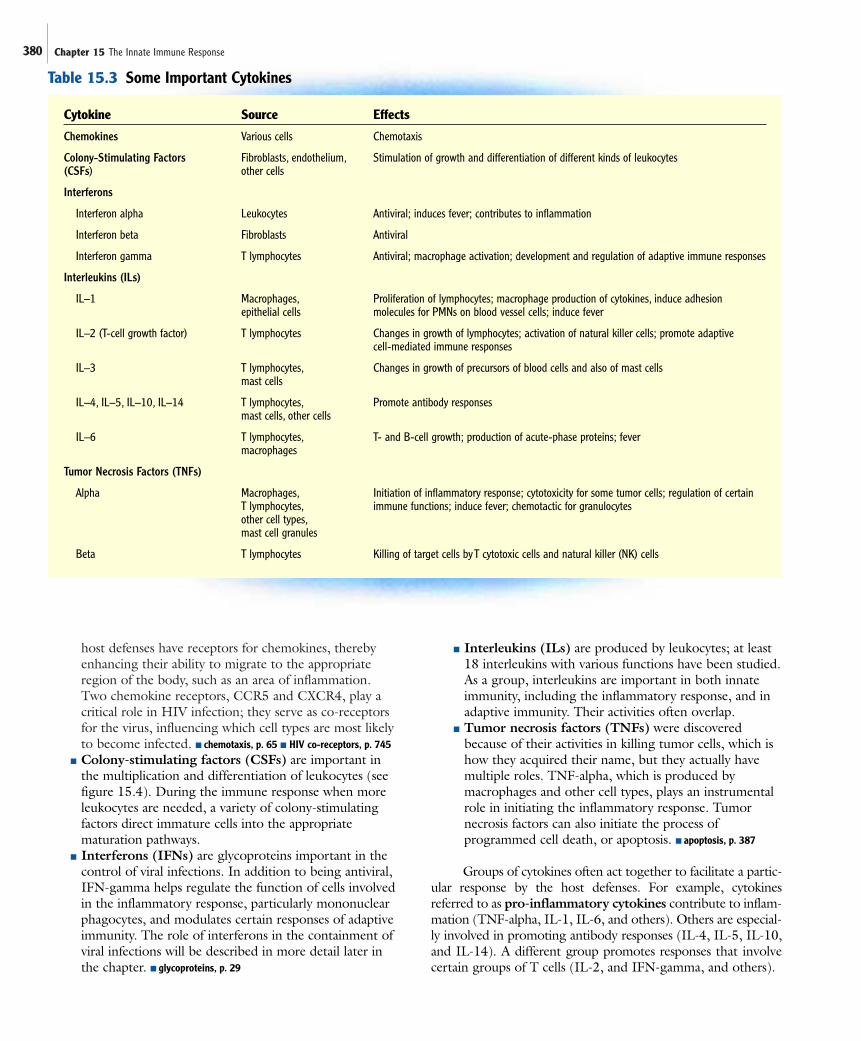
CytokinesCytokines are low molecular weight proteins made by certaincells as a mechanism to communicate with other cells. Cytokinesproduced by lymphocytes are also called lymphokines.Cytokines bind to certain surface receptors, cytokine receptors,found on the cells they regulate, inducing a change such asgrowth, differentiation, movement, or cell death. Althoughcytokines are short-lived, they are very powerful, acting atextremely low concentrations. They can act locally, regionally,or systemically. Often, they act together or in sequence, in acomplex fashion. The source and effects of representativecytokines are summarized in table 15.3. General characteristicsof the various groups are briefly described here:
■ Chemokines are cytokines important in chemotaxis ofimmune cells; more than 50 different varieties have beenidentified by their structure. Certain types of cells of the
380 Chapter 15 The Innate Immune Response
host defenses have receptors for chemokines, therebyenhancing their ability to migrate to the appropriateregion of the body, such as an area of inflammation.Two chemokine receptors, CCR5 and CXCR4, play acritical role in HIV infection; they serve as co-receptorsfor the virus, influencing which cell types are most likelyto become infected. ■ chemotaxis, p. 65 ■ HIV co-receptors, p. 745
■ Colony-stimulating factors (CSFs) are important inthe multiplication and differentiation of leukocytes (seefigure 15.4). During the immune response when moreleukocytes are needed, a variety of colony-stimulatingfactors direct immature cells into the appropriatematuration pathways.
■ Interferons (IFNs) are glycoproteins important in thecontrol of viral infections. In addition to being antiviral,IFN-gamma helps regulate the function of cells involvedin the inflammatory response, particularly mononuclearphagocytes, and modulates certain responses of adaptiveimmunity. The role of interferons in the containment ofviral infections will be described in more detail later inthe chapter. ■ glycoproteins, p. 29
■ Interleukins (ILs) are produced by leukocytes; at least18 interleukins with various functions have been studied.As a group, interleukins are important in both innateimmunity, including the inflammatory response, and inadaptive immunity. Their activities often overlap.
■ Tumor necrosis factors (TNFs) were discoveredbecause of their activities in killing tumor cells, which ishow they acquired their name, but they actually havemultiple roles. TNF-alpha, which is produced bymacrophages and other cell types, plays an instrumentalrole in initiating the inflammatory response. Tumornecrosis factors can also initiate the process ofprogrammed cell death, or apoptosis. ■ apoptosis, p. 387
Groups of cytokines often act together to facilitate a partic-ular response by the host defenses. For example, cytokinesreferred to as pro-inflammatory cytokines contribute to inflam-mation (TNF-alpha, IL-1, IL-6, and others). Others are especial-ly involved in promoting antibody responses (IL-4, IL-5, IL-10,and IL-14). A different group promotes responses that involvecertain groups of T cells (IL-2, and IFN-gamma, and others).
Cytokine Source Effects
Chemokines Various cells Chemotaxis
Colony-Stimulating Factors Fibroblasts, endothelium, Stimulation of growth and differentiation of different kinds of leukocytes(CSFs) other cells
Interferons
Interferon alpha Leukocytes Antiviral; induces fever; contributes to inflammation
Interferon beta Fibroblasts Antiviral
Interferon gamma T lymphocytes Antiviral; macrophage activation; development and regulation of adaptive immune responses
Interleukins (ILs)
IL–1 Macrophages, Proliferation of lymphocytes; macrophage production of cytokines, induce adhesion epithelial cells molecules for PMNs on blood vessel cells; induce fever
IL–2 (T-cell growth factor) T lymphocytes Changes in growth of lymphocytes; activation of natural killer cells; promote adaptive cell-mediated immune responses
IL–3 T lymphocytes, Changes in growth of precursors of blood cells and also of mast cellsmast cells
IL–4, IL–5, IL–10, IL–14 T lymphocytes, Promote antibody responsesmast cells, other cells
IL–6 T lymphocytes, T- and B-cell growth; production of acute-phase proteins; fevermacrophages
Tumor Necrosis Factors (TNFs)
Alpha Macrophages, Initiation of inflammatory response; cytotoxicity for some tumor cells; regulation of certain T lymphocytes, immune functions; induce fever; chemotactic for granulocytesother cell types,mast cell granules
Beta T lymphocytes Killing of target cells by T cytotoxic cells and natural killer (NK) cells
Table 15.3 Some Important Cytokines
15.5 Sensor Systems 381
Adhesion MoleculesAdhesion molecules on the surface of cells allow those cells toadhere to other cells. Some cells use adhesion molecules to“grab” other cells as they pass by. For example, when phago-cytic cells in the blood are needed in tissues, the endothelialcells, which are the cells that line the blood vessels, synthesizeadhesion molecules, snaring passing phagocytic cells. This slowsdown the rapidly moving phagocytic cells, and provides themwith the opportunity to exit the bloodstream. Other types ofadhesion molecules allow cells to make direct contact with oneanother, thereby enabling cells to target the delivery ofcytokines or other compounds to a particular cell.
M I C R O C H E C K 1 5 . 4
Surface receptors allow a cell to detect molecules thatare present outside of that cell. Cytokines provide cellswith a mechanism of communication. Adhesionmolecules allow a cell to adhere to other cells.
■ What is a ligand?■ What is the function of colony-stimulating factors?■ How could colony-stimulating factors be used as
a therapy?
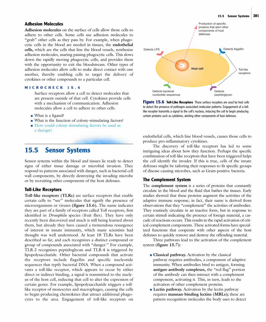
15.5 Sensor SystemsSensor systems within the blood and tissues lie ready to detectsigns of either tissue damage or microbial invasion. Theyrespond to patterns associated with danger, such as bacterial cellwall components, by directly destroying the invading microbeor by recruiting other components of the host defenses.
Toll-Like ReceptorsToll-like receptors (TLRs) are surface receptors that enablecertain cells to “see’’ molecules that signify the presence ofmicroorganisms or viruses (figure 15.6). The name indicatesthey are part of a family of receptors called Toll receptors, firstidentified in Drosophila species (fruit flies). They have onlyrecently been discovered and much is still being learned aboutthem, but already they have caused a tremendous resurgenceof interest in innate immunity, which many scientists hadthought was well understood. At least 10 TLRs have beendescribed so far, and each recognizes a distinct compound orgroup of compounds associated with “danger.’’ For example,TLR-2 recognizes peptidoglycan and TLR-4 is triggered bylipopolysaccharide. Other bacterial compounds that activatethe receptors include flagellin and specific nucleotidesequences that typify bacterial DNA. When a compound acti-vates a toll-like receptor, which appears to occur by eitherdirect or indirect binding, a signal is transmitted to the nucle-us of the host cell, inducing that cell to alter the expression ofcertain genes. For example, lipopolysaccharide triggers a toll-like receptor of monocytes and macrophages, causing the cellsto begin producing chemokines that attract additional phago-cytes to the area. Engagement of toll-like receptors on
endothelial cells, which line blood vessels, causes those cells toproduce pro-inflammatory cytokines.
The discovery of toll-like receptors has led to someintriguing ideas about how they function. Perhaps the specificcombination of toll-like receptors that have been triggered helpsthe cell identify the invader. If this is true, cells of the innatedefenses might be tailoring their responses to fit specific groupsof disease-causing microbes, such as Gram-positive bacteria.
The Complement SystemThe complement system is a series of proteins that constantlycirculate in the blood and the fluid that bathes the tissues. Earlystudies showed that these proteins augment the activities of theadaptive immune response; in fact, their name is derived fromobservations that they “complement” the activities of antibodies.They routinely circulate in an inactive form, but in response tocertain stimuli indicating the presence of foreign material, a cas-cade of reactions occurs. This results in the rapid activation of crit-ical complement components. These activated forms have special-ized functions that cooperate with other aspects of the hostdefenses to quickly remove and destroy the offending material.
Three pathways lead to the activation of the complementsystem (figure 15.7):
■ Classical pathway. Activation by the classical pathway requires antibodies, a component of adaptiveimmunity. When antibodies bind to antigen, formingantigen-antibody complexes, the “red flag” portion of the antibody can then interact with a complementcomponent, activating it. This, in turn, leads to theactivation of other complement proteins.
■ Lectin pathway. Activation by the lectin pathwayrequires mannan-binding lectins (MBLs); these arepattern-recognition molecules the body uses to detect
Host cell
Production of specificproteins that alert othercomponents of host defenses
Detects flagellin
Detects peptidoglycan
Detects LPS
Detects bacterialnucleotide sequences
Toll-like receptors
Figure 15.6 Toll-Like Receptors These surface receptors are used by host cellsto detect the presence of pathogen-associated molecular patterns. Engagement of a toll-like receptor transmits a signal to the cell’s nucleus, inducing the cell to begin producingcertain proteins such as cytokines, alerting other components of host defenses.

382 Chapter 15 The Innate Immune Response
mannan, a polymer of mannose typically found onmicrobial but not mammalian cells. When MBL binds toa surface, it can then interact with the complementcomponent involved in initiating the classical pathway. ■ mannose, p. 384
■ Alternative pathway. The alternative pathway is quiteunlike the other pathways in how it is initiated; nearlyany cell surface automatically triggers the pathwayunless regulatory proteins specifically halt the process. This occurs because one of the complement proteins,C3b, readily binds cell surfaces. Unless regulatoryproteins quickly inactivate C3b, a stabilizing proteinwill bind to it, allowing a subsequent cascade ofreactions to occur. Host cell membranes containmolecules that bind those regulatory proteins,
facilitating the inactivation of C3b before thealternative pathway is triggered. Those regulatoryproteins are generally not associated with microbialsurfaces, however, leading to complement activation by the alternative pathway. As we will discuss in chapter 19, some disease-causing bacteria havedeveloped mechanisms to thwart complementactivation by this pathway.
The nature of the complement system allows an exceed-ingly rapid and powerful response. Its activation occurs by a cas-cade of reactions; once a specific protein becomes activated, itfunctions as an enzyme, cleaving and therefore activating mil-lions of molecules of the next protein in the cascade. In turn,each of those molecules activates multiple molecules of the next
Classical pathway initiated by:
Antigen-antibody complexes
InflammationC3 and C5a induce changes that contribute
to local vascular permeability and attractphagocytes.
Lectin pathwayinitiated by:
Binding of mannan-binding lectins to cell surfaces
Alternative pathwayinitiated by:
Binding of C3b to cell surfaces(regulatory proteins protect host cell surfaces)
C3
C3a C3b
C5
C5aC5b
C6 C7C8 C9
OpsonizationC3b binds to microbial cells, functioning as an opsonin.
C3b
Lysis of foreign cellsFormation of a membrane attack complex,
which creates pores in cell membranes,disrupting the integrity of the cell.
+
Figure 15.7 Complement System Activation of the complement system leads to inflammation, lysis of foreigncells, and opsonization.The three mechanisms that trigger the cascade include the classical pathway, the lectin pathway,and the alternative pathway. Not all of the steps in these pathways are shown.
15.5 Sensor Systems 383
protein in the cascade, and so on. Generally, activation involvessplitting the protein into two parts, each of which then carriesout a specific function. Stringent mechanisms operate to controlthe complement system at various points.
The major complement components have each beengiven a number along with the letter C, for complement. Thenine major components, C1 through C9, were numbered inthe order in which they were discovered and not the order inwhich they react. When one of these components is split intotwo molecules, a lowercase letter is added to the name. Forexample, the activation of C3 splits it into C3a and C3b. Notethat C3 spontaneously splits into C3a and C3b even when thecomplement system has not been activated, but does so at avery low rate; this spontaneous hydrolysis allows enough C3bto be present to potentially trigger the alternative pathway ofcomplement activation. ■ hydrolysis, p. 25
Activation of the complement system eventually leads tothree major protective outcomes:
■ Inflammation. The complement components C3a andC5a induce changes in endothelial cells, which line theblood vessels, and in mast cells. These effects contributeto the vascular permeability associated with inflammation.C5a is a potent chemoattractant, drawing phagocytesinto the area where complement was activated.
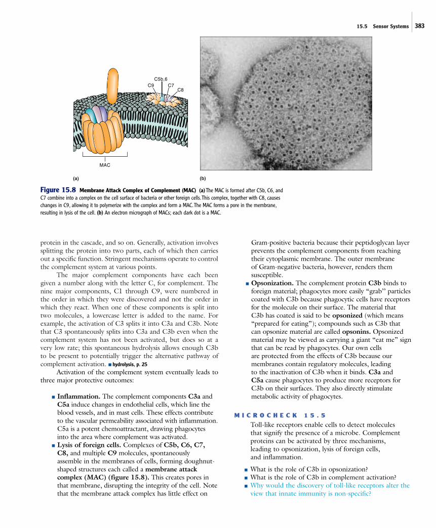
■ Lysis of foreign cells. Complexes of C5b, C6, C7, C8, and multiple C9 molecules, spontaneously assemble in the membranes of cells, forming doughnut-shaped structures each called a membrane attackcomplex (MAC) (figure 15.8). This creates pores inthat membrane, disrupting the integrity of the cell. Notethat the membrane attack complex has little effect on
Gram-positive bacteria because their peptidoglycan layerprevents the complement components from reachingtheir cytoplasmic membrane. The outer membrane of Gram-negative bacteria, however, renders themsusceptible.
■ Opsonization. The complement protein C3b binds toforeign material; phagocytes more easily “grab’’ particlescoated with C3b because phagocytic cells have receptorsfor the molecule on their surface. The material that C3b has coated is said to be opsonized (which means“prepared for eating”); compounds such as C3b that can opsonize material are called opsonins. Opsonizedmaterial may be viewed as carrying a giant “eat me” signthat can be read by phagocytes. Our own cells are protected from the effects of C3b because ourmembranes contain regulatory molecules, leading to the inactivation of C3b when it binds. C3a and C5a cause phagocytes to produce more receptors forC3b on their surfaces. They also directly stimulatemetabolic activity of phagocytes.
M I C R O C H E C K 1 5 . 5
Toll-like receptors enable cells to detect moleculesthat signify the presence of a microbe. Complementproteins can be activated by three mechanisms,leading to opsonization, lysis of foreign cells,and inflammation.
■ What is the role of C3b in opsonization?■ What is the role of C3b in complement activation?■ Why would the discovery of toll-like receptors alter the
view that innate immunity is non-specific?
(b)(a)
MAC
C9C5b,6
C7C8
Figure 15.8 Membrane Attack Complex of Complement (MAC) (a)The MAC is formed after C5b, C6, andC7 combine into a complex on the cell surface of bacteria or other foreign cells.This complex, together with C8, causeschanges in C9, allowing it to polymerize with the complex and form a MAC.The MAC forms a pore in the membrane,resulting in lysis of the cell. (b) An electron micrograph of MACs; each dark dot is a MAC.

384 Chapter 15 The Innate Immune Response
15.6 PhagocytosisPhagocytes are cells that routinely engulf and digest material,including invading organisms. Yet, with the multitude of differ-ent particles in the body, how do those cells determine whichones to engulf? The answer lies in the various pattern recogni-tion receptors that stud the phagocyte surface, binding to certain molecular configurations often found on cell debris and foreign material. Binding of a substance to certain patternrecognition receptors induces the phagocytic cell to engulf thatmaterial. A receptor called the scavenger receptor, for example,facilitates the engulfment of various materials that have chargedmolecules on their surface.
In routine situations, such as when organisms are introducedthrough a minor skin wound, macrophages that reside in the tis-sues readily destroy the relatively few bacteria that have entered. If the invading microbes are not rapidly cleared, however,macrophages can produce cytokines to recruit additional phago-cytes, particularly neutrophils, for extra help. It is the toll-like recep-tors on macrophages that enable them to sense that the material is microbial in origin, and must therefore be eliminated quickly.
The Process of PhagocytosisPhagocytosis involves a series of complex steps by which phago-cytes engulf and kill invading microorganisms (figure 15.9). The
steps are particularly relevant medically, because most disease-causing organisms have evolved the ability to evade one or moreof the steps. Chapter 19 will describe some of the mechanismsmicroorganisms have developed to circumvent this importantaspect of innate immunity. The steps of phagocytosis include:
■ Chemotaxis. The phagocytic cells are recruited to thesite of infection or tissue damage by certain chemicalstimuli that act as chemoattractants. Compounds thatpromote chemotaxis of phagocytes include products ofmicroorganisms, phospholipids released by injuredmammalian cells, and the complement component C5a.
■ Recognition and attachment. Phagocytic cells use various receptors to bind invading microbes either directly or indirectly. Direct binding occursthrough receptors that bind patterns associated withcompounds found on microbes. For example, one type of receptor on phagocytic cells binds mannose, atype of sugar found on the surface of some types ofbacteria and yeasts. Indirect binding occurs when aparticle has first been opsonized, dramatically enhancingthe phagocytes’ ability to attach and subsequently engulf the material. Opsonins include the complementcomponent C3b and certain classes of antibodymolecules; phagocytes have receptors for specific parts of these molecules.
(b)
(c)
(d)
(e)(f)
(g)
Phagocyte
Lysosomes
Phagosome
Phagolysosome
(b) C3b coatsorganisms andattaches to C3breceptors onphagocyte
(c) Organism is engulfed into
a phagosome
(d) Phagosome fuses with lysosome to produce phagolysosome
(e) Organism iskilled within thephagolysosome
(f) Digestion andbreakdown oforganism withinphagolysosome
(g) Contents ofphagolysosomeeliminated byexocytosis
C5a
C3b
Microbe
(a) Chemoattractants,such as C5a, attractphagocyte toorganisms to be ingested
C3b
C3b receptorson phagocyte
Multiple contacts aremade by C3b on themicrobe with C3b receptors on phagocyte
Figure 15.9 Phagocytosis and Intracellular Destruction of PhagocytizedMaterial This diagram shows a microbe that has been opsonized by the complement proteinC3b; certain classes of antibodies can also function as opsonins.
15.7 Inflammation—A Coordinated Response to Invasion or Damage 385
■ Engulfment. The phagocytic cell engulfs the invader,forming a membrane-bound vacuole called aphagosome. This process involves rearrangement of thephagocyte’s cytoskeleton, forming armlike extensionscalled pseudopods that surround the material beingengulfed. Engulfment itself does not destroy themicrobe. ■ cytoskeleton, p. 73 ■ pseudopod, p. 73
■ Fusion of the phagosome with the lysosome.Within the phagocyte, the phagosome is transportedalong the cytoskeleton to a point where it can fusewith a lysosome, a membrane-bound body filled withvarious digestive enzymes, including lysozyme andproteases. The fusion results in the formation of aphagolysosome. In neutrophils, the membrane-boundbodies are referred to as granules.
■ Destruction and digestion. Within the fusion product,oxygen consumption increases enormously as sugars areoxidized via the TCA cycle, with the production ofhighly toxic oxygen products such as superoxide,hydrogen peroxide, singlet oxygen and hydroxyl radicals. As the available oxygen in the phagolysosome is consumed, the metabolic pathway switches tofermentation with the production of lactic acid, loweringthe pH. Various enzymes degrade the peptidoglycan ofthe bacterial cell walls, and other components of the cell.■ TCA cycle, pp. 137, 144 ■ superoxide, p. 89 ■ hydrogen peroxide, p. 89
■ Exocytosis. Following the digestion of themicroorganisms, the membrane-bound vesiclecontaining the digested material fuses with the plasmamembrane. This expels the material to the externalenvironment. ■ exocytosis, p. 72
Specialized Attributes of MacrophagesMacrophages can be viewed as the scavengers and sentries—routinely phagocytizing dead cells and debris, but always on thelookout, ready to destroy invaders, and able to call in reinforce-ments when needed. They are always present in tissues to atleast some extent, where they either slowly wander or remainstationary. Macrophages that remain fixed in tissues are oftenreferred to by different names according to the type of tissue inwhich they reside (see figure 15.5). These phagocytic cells playan essential role in every major tissue in the body.
Macrophages live for weeks to months, and maintain theirkilling power by continually regenerating their lysosomes. Asmacrophages die they are continually replaced by circulatingmonocytes that leave the blood and migrate to the tissues; recallthat monocytes can differentiate into macrophages. Migrationof monocytes is enhanced by certain stimuli associated withinvasion and tissue damage.
Macrophages have several important characteristics thatenable them to accomplish their diverse tasks. Various toll-likereceptors enable them to sense material that signifies danger.When these receptors are triggered, the macrophage producespro-inflammatory cytokines to alert and stimulate various othercells of the immune system. A macrophage can increase its other-wise limited killing power with the assistance of a subset of T-helper cells to become an activated macrophage. This is anexample of the cooperation between the innate and adaptive
host defenses. Activation of macrophages induces the produc-tion of nitric oxide (NO) and oxygen radicals, which moreeffectively destroy microbes. These products also damage tissueswhen they are released, a reason why it would be detrimental formacrophages to continually maintain an activated state. Detailsof the activation process, including the roles of T-helper cells,will be discussed in chapter 16. ■ macrophage activation, p. 409
If activated macrophages fail to destroy microbes andchronic infection ensues, large numbers of macrophages can fusetogether to form giant cells. Macrophages, giant cells, and T-helper cells form concentrated groups of cells called granulo-mas that wall off and retain organisms or other material that can-not be destroyed by the cells; again, this is an example of thecooperation between defense systems. This prevents the microbesfrom escaping to infect other cells (see figure 23.18). Granulomasare part of the disease process in tuberculosis, histoplasmosis, andother illnesses. ■ tuberculosis, p. 580 ■ histoplasmosis, p. 592
Specialized Attributes of NeutrophilsNeutrophils can be viewed as the rapid response team—quick tomove into an area of trouble so that the offending invaders canbe removed. They play a critical role during the early stages ofinflammation, being the first cell type recruited from the blood-stream to the site of damage. They inherently have more killingpower than macrophages, including those that have been acti-vated. The cost for their effectiveness, however, is a relativelyshort life span of only 1 to 2 days in the tissues; once they haveexpended their granules, they die. Many more are in reserve,however, for it is estimated that for every neutrophil in the cir-culatory system, 100 more are waiting in the bone marrow,ready to be mobilized when needed.
M I C R O C H E C K 1 5 . 6
The process of phagocytosis includes chemotaxis,recognition and attachment, engulfment, fusion of the phagosome with the lysosome, destruction anddigestion of the ingested material, and exocytosis.Macrophages are long-lived phagocytic cells that arealways present in tissues; they can be activated toenhance their killing power. Neutrophils are highlyactive, short-lived phagocytic cells that must berecruited to the site of damage.
■ How does a phagolysosome differ from a phagosome?■ What is a granuloma?■ What could a microorganism do to avoid engulfment?
15.7 Inflammation—A CoordinatedResponse to Invasion or Damage
When tissues have been damaged, such as when an object pen-etrates the skin or when microbes produce toxic compounds, acoordinated response called the inflammatory response, orinflammation occurs. Everyone has experienced the signs of
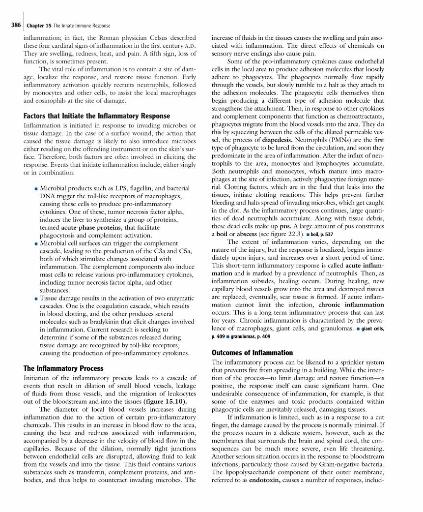
386 Chapter 15 The Innate Immune Response
inflammation; in fact, the Roman physician Celsus describedthese four cardinal signs of inflammation in the first century A.D.They are swelling, redness, heat, and pain. A fifth sign, loss offunction, is sometimes present.
The vital role of inflammation is to contain a site of dam-age, localize the response, and restore tissue function. Earlyinflammatory activation quickly recruits neutrophils, followedby monocytes and other cells, to assist the local macrophagesand eosinophils at the site of damage.
Factors that Initiate the Inflammatory ResponseInflammation is initiated in response to invading microbes ortissue damage. In the case of a surface wound, the action thatcaused the tissue damage is likely to also introduce microbeseither residing on the offending instrument or on the skin’s sur-face. Therefore, both factors are often involved in eliciting theresponse. Events that initiate inflammation include, either singlyor in combination:
■ Microbial products such as LPS, flagellin, and bacterialDNA trigger the toll-like receptors of macrophages,causing these cells to produce pro-inflammatorycytokines. One of these, tumor necrosis factor alpha,induces the liver to synthesize a group of proteins,termed acute-phase proteins, that facilitatephagocytosis and complement activation.
■ Microbial cell surfaces can trigger the complementcascade, leading to the production of the C3a and C5a, both of which stimulate changes associated withinflammation. The complement components also inducemast cells to release various pro-inflammatory cytokines,including tumor necrosis factor alpha, and othersubstances.
■ Tissue damage results in the activation of two enzymaticcascades. One is the coagulation cascade, which resultsin blood clotting, and the other produces severalmolecules such as bradykinin that elicit changes involvedin inflammation. Current research is seeking todetermine if some of the substances released duringtissue damage are recognized by toll-like receptors,causing the production of pro-inflammatory cytokines.
The Inflammatory ProcessInitiation of the inflammatory process leads to a cascade ofevents that result in dilation of small blood vessels, leakage of fluids from those vessels, and the migration of leukocytes out of the bloodstream and into the tissues (figure 15.10).
The diameter of local blood vessels increases duringinflammation due to the action of certain pro-inflammatorychemicals. This results in an increase in blood flow to the area,causing the heat and redness associated with inflammation,accompanied by a decrease in the velocity of blood flow in thecapillaries. Because of the dilation, normally tight junctionsbetween endothelial cells are disrupted, allowing fluid to leakfrom the vessels and into the tissue. This fluid contains varioussubstances such as transferrin, complement proteins, and anti-bodies, and thus helps to counteract invading microbes. The
increase of fluids in the tissues causes the swelling and pain asso-ciated with inflammation. The direct effects of chemicals onsensory nerve endings also cause pain.
Some of the pro-inflammatory cytokines cause endothelialcells in the local area to produce adhesion molecules that looselyadhere to phagocytes. The phagocytes normally flow rapidlythrough the vessels, but slowly tumble to a halt as they attach tothe adhesion molecules. The phagocytic cells themselves thenbegin producing a different type of adhesion molecule thatstrengthens the attachment. Then, in response to other cytokinesand complement components that function as chemoattractants,phagocytes migrate from the blood vessels into the area. They dothis by squeezing between the cells of the dilated permeable ves-sel, the process of diapedesis. Neutrophils (PMNs) are the firsttype of phagocyte to be lured from the circulation, and soon theypredominate in the area of inflammation. After the influx of neu-trophils to the area, monocytes and lymphocytes accumulate.Both neutrophils and monocytes, which mature into macro-phages at the site of infection, actively phagocytize foreign mate-rial. Clotting factors, which are in the fluid that leaks into the tissues, initiate clotting reactions. This helps prevent furtherbleeding and halts spread of invading microbes, which get caughtin the clot. As the inflammatory process continues, large quanti-ties of dead neutrophils accumulate. Along with tissue debris,these dead cells make up pus. A large amount of pus constitutesa boil or abscess (see figure 22.3). ■ boil, p. 537
The extent of inflammation varies, depending on thenature of the injury, but the response is localized, begins imme-diately upon injury, and increases over a short period of time.This short-term inflammatory response is called acute inflam-mation and is marked by a prevalence of neutrophils. Then, asinflammation subsides, healing occurs. During healing, newcapillary blood vessels grow into the area and destroyed tissuesare replaced; eventually, scar tissue is formed. If acute inflam-mation cannot limit the infection, chronic inflammationoccurs. This is a long-term inflammatory process that can lastfor years. Chronic inflammation is characterized by the preva-lence of macrophages, giant cells, and granulomas. ■ giant cells,p. 409 ■ granulomas, p. 409
Outcomes of InflammationThe inflammatory process can be likened to a sprinkler systemthat prevents fire from spreading in a building. While the inten-tion of the process—to limit damage and restore function—ispositive, the response itself can cause significant harm. Oneundesirable consequence of inflammation, for example, is thatsome of the enzymes and toxic products contained withinphagocytic cells are inevitably released, damaging tissues.
If inflammation is limited, such as in a response to a cutfinger, the damage caused by the process is normally minimal. Ifthe process occurs in a delicate system, however, such as themembranes that surrounds the brain and spinal cord, the con-sequences can be much more severe, even life threatening.Another serious situation occurs in the response to bloodstreaminfections, particularly those caused by Gram-negative bacteria.The lipopolysaccharide component of their outer membrane,referred to as endotoxin, causes a number of responses, includ-
15.7 Inflammation—A Coordinated Response to Invasion or Damage 387
(a) Normal blood flow in the tissues as injury occurs. (b) Substances released cause dilation of small blood vessels and increased blood flow in the immediate area.
(c) Phagocytes attach to the endothelial cells and then squeeze between the cells into surrounding tissue.
(d) The attraction of phagocytes causes them to move to the site of damage and inflammation. Collections of dead phagocytes and tissue debris make up the pus often found at sites of an active inflammatory response.
Skinsurface
Arteriole
Venule
Capillaries
Blood vesseldilation
• Microbial products• Microbes• Tissue damage
Bacteria
Site of tissuedamage andinflammation
Diapedesis
Pus formation
Loose adherence; cells tumble to a halt
Tighter adherence Diapedesis
Figure 15.10 The Inflammatory Process This coordinated response to microbial invasion or tissue damagebrings phagocytes and other leukocytes to the site.The role of inflammation is to contain a site of damage, localize the response, and restore tissue function.
ing the release of pro-inflammatory cytokines by monocytes,activation of the complement cascade and activation of the clot-ting cascade. The net result is a rapid loss in blood pressure,leading to shock, extensive tissue damage, and widespread for-mation of clots that plug the capillaries, cutting off the bloodsupply; this manifestation of a bloodstream infection (sepsis) iscalled septic shock (see figure 28.3). The cell wall components
of Gram-positive bacteria can also elicit septic shock. ■ meninges,p. 664 ■ endotoxin, p. 59 ■ Gram-negative sepsis, p. 719
Apoptosis—Controlled Cell Death that Circumvents the Inflammatory ProcessThe inflammatory response represents a potential problem for thehost; that is, how to distinguish cell death caused by abnormal
PERSPECTIVE 15.1 For Schistosoma, the Inflammatory Response Delivers
Just as our immune system has evolved to protect us fromnew and different invasions, it is not surprising that in theopportunistic and adaptable world of microbes, some wouldfind ways to use our defenses to their advantage.Theparasitic flatworms that cause schistosomiasis do not shyfrom the immune response when it comes to procreation;instead they appear to use it to deliver their ova to anenvironment where they might hatch. Adult females ofSchistosoma species, which live in the bloodstream ofinfected hosts, lay their ova in veins near the intestine orbladder; they seem to rely on a robust inflammatory responseto expel the ova, completing one portion of a complex lifecycle.The ova released in feces or urine can hatch to form alarval form called a miracidium if untreated sewage reacheswater.The miracidium then infects a specific freshwater snailhost and undergoes asexual multiplication.The infected snailthen releases large numbers of another larval form, cercariae,which swim about in search of a human host.
The parasite is acquired when a person wades orswims in contaminated water.The cercariae penetrate theskin by burrowing through it with the aid of digestiveenzymes; schistosomes are rare among pathogens becausethey can actually penetrate intact skin.The larvae thenproceed to enter the circulatory system where they can livefor over a quarter of a century. Schisotosoma species haveseparate sexes and, remarkably, the male and femaleworms locate one another in the bloodstream.The male’sbody has a deep longitudinal groove in which he clasps hisfemale partner to live in copulatory embrace (shisto-somameans “split-body,” referring to the long slit).The adultworms effectively mask themselves from the immunesystem by adsorbing various blood proteins; this providesthem with a primitive stealth “cloaking device.”
Depending on the species, the female worm migratesto the veins of either the intestine or bladder to layhundreds of ova per day.The body responds vigorously to
the highly antigenic eggs, ejecting them in manner thatappears similar to what is experienced as a sliver in theskin works its way to the surface. Over half of the ova arenot expelled, however, and many of these are instead sweptaway by the bloodstream to the liver.The inflammatoryprocess and granuloma formation there gradually destroysliver cells, replacing the cells with scar tissue. Malfunction of the liver results in malnutrition and a buildup of pressurein the esophagus. Fluid accumulates in the abdominalcavity and hemorrhage occurs if the engorged esophagealveins rupture.
Despite their complex life cycle, Schistosoma speciesare highly successful. Not only are they adept at avoidingcertain immune responses that would otherwise lead totheir destruction, they have learned to exploit inflammationfor their own dissemination. Over 200 million peopleworldwide are infected with these parasites, resulting in thedeath of over 500,000 people each year.
388 Chapter 15 The Innate Immune Response
events, such as injury, from that caused by normal events such astissue remodeling that render certain cells unnecessary or poten-tially harmful. The former merits an inflammatory responsewhereas the latter does not and, in fact, would be unnecessarilydestructive to normal tissue. Apoptosis (Greek, apo for “falling”;ptosis for “off”), or programmed cell death, is a process thatdestroys self-cells without eliciting inflammation. During apopto-sis, the dying cells undergo certain changes. For example, theshape of the cell changes, enzymes cut the DNA, and portions ofthe cell bud off, effectively shrinking the cell. Some changesappear to signal to macrophages that the remains of the cell areto be engulfed without the commotion associated with inflam-mation. For example, some parts of the membrane invert, expos-ing molecules that are generally restricted to the inner leaflet.
The mechanisms and events connected to apoptosis arecurrently the focus of a great deal of research. It is now recog-nized that the process is used to eliminate a wide range of cellsfrom the body, including virally infected cells, as well as thoselymphocytes whose function is rendered obsolete by the suc-cessful elimination of an antigen.
M I C R O C H E C K 1 5 . 7
Inflammation is a cascade of events initiated in responseto invading microbes or tissue damage. The outcome isdilation of small blood vessels, leakage of fluids fromthose vessels, and migration of leukocytes out of thebloodstream and into the tissue. Inflammation can helpcontain an infection, but the response itself can causedamage. Apoptosis provides a mechanism for thedestruction of self-cells without initiating inflammation.
■ Describe three general events that can initiateinflammation.
■ Describe the changes that characterize cells that areundergoing apoptosis.
■ How could infection of the fallopian tubes lead tosterility and ectopic pregnancy?
15.8 InterferonsInterferons are a group of glycoproteins important in the con-trol of viral infections as well as other immune responses. Oneof their most important functions is to prepare cells in the vicin-ity of a virally infected cell to cease protein synthesis in the eventthey become infected with a virus themselves. This preventsviral replication within those neighboring cells, limiting thespread of the virus.
Cells use the presence of double-stranded RNA to indi-cate they have been infected with a virus. Eukaryotic cells typ-ically do not contain double-stranded RNA because only onestrand of DNA in a gene is used as a template for RNA syn-thesis. Replication of RNA viruses other than retroviruses,however, routinely generates double-stranded RNA. EvenDNA viruses often give rise to double-stranded RNA as a con-sequence of their efficient use of their relatively small genomes;in some regions, both strands of DNA are transcribed intomRNA.
Double-stranded RNA in an animal cell induces the syn-thesis and subsequent secretion of interferon (figure 15.11).The interferon molecules then attach to a specific receptor onboth the infected cell and neighboring cells, causing them toactivate genes encoding enzymes capable of directing the degra-dation of mRNA and inhibition of protein synthesis. The actionof these enzymes requires the presence of double-strandedRNA, preventing viral replication in infected cells withoutimpacting uninfected cells. This process essentially sacrifices aninfected host cell in order to prevent viral spread. Interferonsprovide some protection against most types of viruses.
Three types of interferon are known. One type, interferonalpha, is a family of closely related proteins produced by variouswhite blood cells. In addition to its antiviral activity, it also con-tributes to fever production. A second type, interferon beta, ismade by fibroblasts, which are cells of fibrous supporting tissue.

15.9 Fever 389
Both alpha and beta interferons are made by many cell typeswhen infected with viruses. A third type, inteferon gamma, ismade by lymphocytes; unlike the other interferons, its synthesis isnot directly related to viral infection. In addition to being antivi-ral, interferon gamma is very important in enhancing the killingpower of macrophages, and also functions in the developmentand regulation of the adaptive immune response.
Interferons are quite species specific with regard to host,which initially prevented their widespread therapeutic use; interferon from other animals is not effective in humans.Microorganisms, however, have now been genetically engi-neered to produce human interferons. Interferon alpha has beenapproved in the United States for treatment of Kaposi’s sarcomain AIDS patients, chronic hepatitis B and hepatitis C infections,and several other diseases. Interferon beta is used to slow theprogression of multiple sclerosis (MS), but the mechanisms ofits beneficial effects are unclear. ■ genetic engineering, pp. 220, 230
M I C R O C H E C K 1 5 . 8
Interferons induce cells in the vicinity of a virally infected cell to prepare to cease protein synthesis shouldthey become infected with virus. Double-stranded RNAfunctions as the signal to a cell that it is infected with a virus.
■ What activities does interferon gamma have thatinterferons alpha and beta do not?
■ What is the source of interferon that is usedtherapeutically?
■ Considering that retroviruses have an RNA genome,why would their replication not generate double-stranded RNA?
15.9 FeverFever is one of the strongest indications of infectious disease,especially those of bacterial origin, although significant infec-tions can occur without it. There is abundant evidence that feveris an important host defense mechanism in a number of verte-brates, including humans. Within the human body, the temper-ature is normally kept within a narrow range, around 37°C, bya temperature-regulation center in the hypothalamus of thebrain. The hypothalamus controls temperature by regulatingblood flow to the skin, and the amount of sweating and respi-ration. In this way, the heat produced by metabolism is con-served or lost in order to maintain a fairly constant temperature.During an infection, the regulating center continues to functionbut the body’s thermostat is “set” at higher levels. An oral tem-perature above 37.8°C is regarded as fever.
A higher temperature setting occurs as a result of certainpro-inflammatory cytokines released by macrophages whentheir toll-like receptors detect microbial products. Thecytokines are carried in the bloodstream to the hypothalamus,where they act as messages that microorganisms have invadedthe body. These cytokines and other fever-inducing substancesare pyrogens. Fever-inducing cytokines are called endoge-neous pyrogens, indicating that the body makes them, where-as microbial products, such as bacterial endotoxins, are calledexogenous pyrogens, indicating that they are introduced fromexternal sources. The temperature-regulating center respondsto pyrogens by raising body temperature. The resulting feverinhibits the growth of many pathogens by at least two mecha-nisms: (1) elevating the temperature above the optimum
Virus infects cell
Virus (RNA or DNA)
Virus infects cell
Virus replicates
Nucleus
InterferonInterferon attaches toreceptors on cell surface
Double-stranded RNAfrom viral replicationinitiates production of interferon
Double-strandedRNA from virus
mRNA
Genes for mRNA-degradingenzymes are activated inresponse to signal from interferon
Cell producesvirus and dies
Cell dies, but novirus is produced
EnzymesWhen viral double-stranded RNAis present, enzymes actively degrademRNA; protein synthesis stops;cell dies and virus cannot replicate
Figure 15.11 Mechanism of the Antiviral Activity of Interferons Interferons are small glycoproteins madeby virus-infected cells that act on nearby cells, causing them to produce antiviral proteins.These proteins are enzymesthat inhibit virus replication in various ways.

390 Chapter 15 The Innate Immune Response
growth temperature of the pathogen, and (2) activating andspeeding up a number of other body defenses.
The adverse effects of fever on pathogens correlates inpart with their ideal growth temperature. Bacteria that growbest at 37°C are less likely to cause disease in people with fever.The growth rate of bacteria often declines sharply as the tem-perature rises above their optimum growth temperature. Aslower growth rate allows more time for other defenses todestroy the invaders. ■ temperature requirements, p. 86
A moderate rise in temperature increases the rate of enzy-matic reactions. It is thus not surprising that fever has beenshown to enhance the inflammatory response, phagocytickilling by leukocytes, the multiplication of lymphocytes, therelease of substances that attract neutrophils, and the produc-tion of interferons and antibodies. Release of leukocytes into theblood from the bone marrow is also enhanced. For all these rea-sons, it is wise to consult a physician before taking drugs toreduce the fever of infectious disease.
M I C R O C H E C K 1 5 . 9
Fever results when macrophages releasepro-inflammatory cytokines; this occurs whenthe toll-like receptors on the macrophagesare engaged by microbial products.
■ What is an endogenous pyrogen? What is an exogenous pyrogen?
■ How does fever inhibit the growth of pathogens?■ Syphilis was once treated by intentionally infecting the
patient with the parasite that causes malaria, a diseasecharacterized by repeated bouts of fever, shaking, andchills. Why would this treatment cure syphilis?
F U T U R E C H A L L E N G E S
Polishing the Magic Bullet
Very early in the twentieth century, Paul Ehrlich lookedfor a “magic bullet,” a drug that could affect onlyinvading microorganisms and not host cells, one thatcould be delivered directly to the area of infection or
inflammation. In spite of extensive investigations, Ehrlich did notfind a magic bullet, but others carried on the search. Through theyears many attempts have been made to attach drugs to variousmolecules, such as specific antibodies, that could home to the areawhere the drugs were needed.
Currently, a promising approach to finding the elusivemagic bullet is based on ongoing investigations of the mechanismsof inflammation. Early during the inflammatory response, a fami-ly of adhesion molecules called selectins is expressed on cells ofsmall blood vessels in the area of injury. Selectins bind to carbohy-drate molecules on circulating leukocytes. As the leukocytes rollrapidly through the blood vessels, selectin molecules bind them,stopping them in their tracks. The leukocytes are held captive in theregion of inflammation. Other molecules in that area attract the cap-tured white blood cells across the vessel wall and into the injured tis-sue. Selectin activity is a marker for tissue damage and, therefore, alogical target for delivery of drugs into areas of inflammationresulting from infections. The drugs could be antimicrobials to actagainst invading microorganisms, or anti-inflammatory agents tolessen the symptoms caused by inflammation.
In some diseases, such as arthritis and other autoimmunediseases, the tissue damage caused by inflammation is the majorfactor in the disease process. In addition to delivering anti-inflammatory drugs to the area, blocking the actions of selectinsby a selectin inhibitor might prevent the development of inflam-matory damage.
Scientists working in this area are studying the exact struc-ture of the various selectin molecules and their characteristics.One selectin is stored in cells of the blood vessel and is expressedon the cell surface within minutes of tissue injury. Another is synthesized after injury and expressed on cell surfaces afterabout 4 hours. The structure of leukocyte surface molecules that will bind to selectins has also been determined. It should be possible to attach antimicrobial or anti-inflammatory drugs tothe small portions of binding molecules that actually bind to theselectin. This combination would then be selectively removedfrom the circulation in the areas of inflammation, the only areaswhere selectin is produced. It is estimated that preparations ofselectin-binding antimicrobials or anti-inflammatory drugs couldbe available within several years, making Ehrlich’s long-agodream a reality.
S U M M A R Y
15.1 Overview of the Innate Defenses (Table 15.1)
1. The innate defense system is composed of first-line defenses,sensor systems such as toll-like receptors and complement,and phagocytes. Inflammation is a coordinated responsethat involves many aspects of the innate defenses.
15.2 First-Line Defenses (Figures 15.2, 15.3)
Physical Barriers
1. The skin provides the most difficult barrier for microbes topenetrate; it is composed of two main layers—the dermisand the epidermis.
2. The cells of the mucous membranes are constantly bathed with mucus and other secretions that help wash microbes from the surfaces. Some mucous membranes have mechanisms that propel microbes, directing them toward areas where they can be eliminated more easily.
Antimicrobial Substances
1. Lysozyme, peroxidase enzymes, lactoferrin, and defensins are antimicrobial substances that inhibit or killmicroorganisms.
Summary 391
Normal Flora
1. Members of the normal flora competitively excludepathogens and stimulate the host defenses.
15.3 The Cells of the Immune System (Figure 15.4, Table 15.2)
Granulocytes
1. There are three types of granulocytes—neutrophils,basophils, and eonsinophils.
Mononuclear Phagocytes
1. Monocytes differentiate into either macrophages ordendritic cells. (Figure 15.5)
Lymphocytes
1. Lymphocytes, which include B cells, T cells, and naturalkiller (NK) cells, are involved in adaptive immunity.
15.4 Cell Communication
Surface Receptors
1. Surface receptors bind ligands that are on the outside of thecell, enabling the cell to detect that the ligand is present.
Cytokines (Table 15.3)
1. Cytokines include interleukins (ILs), colony-stimulatingfactors (CSFs), tumor necrosis factors (TNFs),chemokines, and interferons.
Adhesion Molecules
1. Adhesion molecules allow cells to adhere to other cells.
15.5 Sensor Systems
Toll-Like Receptors (Figure 15.6)
1. Toll-like receptors enable cells to detect molecules that signifythe presence of an invading microbe.
The Complement System (Figure 15.7)
1. Complement proteins circulate in the blood and the fluidthat bathes tissues; in response to certain stimuli that indicatethe presence of foreign material, they become activated.
2. The major protective outcomes of complement activationinclude opsonization, lysis of foreign cells, and initiation of inflammation.
15.6 Phagocytosis
The Process of Phagocytosis (Figure 15.9)
1. The steps of phagocytosis include chemotaxis, recognitionand attachment, engulfment, destruction and digestion, andexocytosis.
Specialized Attributes of Macrophages
1. Macrophages are always present in tissues to some extent, butare able to call in reinforcements when needed.
2. A macrophage can increase its killing power, becoming anactivated macrophage.
3. Macrophages, giant cells, and T-helper cells formconcentrated groups called granulomas that wall off andretain organisms or other material that cannot be destroyedby macrophages.
Specialized Attributes of Neutrophils
1. Neutrophils play a critical role during the early stages ofinflammation, being the first cell type recruited from thebloodstream to the site of damage.
15.7 Inflammation—A Coordinated Response to Invasion or Damage (Figure 15.10)
1. Swelling, redness, heat, and pain are the signs ofinflammation, the attempt by the body to contain a site ofdamage, localize the response, and restore tissue function.
Factors that Initiate the Inflammatory Response
1. Inflammation is initiated when pro-inflammatory cytokines orother inflammatory mediators are released as a result of theengagement of toll-like receptors or activation of complementby invading microbes, or when tissue damage occurs.
The Inflammatory Process
1. The inflammatory process leads to a cascade of events thatresult in dilation of small blood vessels, leakage of fluids fromthose vessels, and the migration of leukocytes out of thebloodstream and into the tissues.
2. Acute inflammation is marked by a preponderance ofneutrophils; chronic inflammation is characterized by theprevalence of macrophages, giant cells, and granulomas.
Outcomes of Inflammation
1. Inflammation can contain an infection, but the process itselfcan cause damage; a systemic response can be life threatening.
Apoptosis—Controlled Cell Death that Circumvents the Inflammatory Process
1. Apoptosis is a mechanism of eliminating self-cells withoutevoking an inflammatory response.
15.8 Interferons (Figure 15.11)
1. One of the roles of interferons is to induce cells in the vicinity of a virally infected cell to prepare to cease protein synthesis in the event they become infected with a virus; double-stranded RNA signifies to the cell that it has been infected.
15.9 Fever
1. Fever occurs as a result of certain pro-inflammatory cytokinesreleased by macrophages when their toll-like receptors bindmicrobial products.
2. Fever inhibits the growth of many pathogens and increasesthe rate of various body defenses.
392 Chapter 15 The Innate Immune Response
R E V I E W Q U E S T I O N S
Short Answer
1. Why is iron metabolism important in body defenses?
2. How do phagocytes get into tissues during an inflammatoryresponse?
3. Describe how the skin protects against infection.
4. What are the benefits of saliva in protection against infection?What factors found in saliva aid in protection?
5. Name two categories of cytokines and give their effects.
6. Contrast the classical and alternative pathways of complementactivation.
7. How does the activation of a few molecules in early stages of the complement cascade result in the cleavage of millions of molecules of later ones?
8. How do complement proteins cause foreign cell lysis?
9. Describe three mechanisms of triggering inflammation.
10. Describe the purpose of apoptosis.
Multiple Choice
1. Lysozyme does which of the following:A. disrupts cell membranesB. hydrolyzes peptidoglycanC. waterproofs skinD. propels gastrointestinal contentsE. propels the cilia of the respiratory tract
2. The hematopoietic stem cells in the bone marrow can becomewhich of the following cell types?1. red blood cell 2. T cell 3. B cell4. monocyte 5. macrophageA. 2, 3 B. 2, 4 C. 2, 3, 4, 5 D. 1, 4, 5 E. 1, 2, 3, 4, 5
3. All of the following refer to the same type of cell exceptA. macrophage B. neutrophilC. poly D. PMN
4. Toll-like receptors are triggered by all of the followingcompounds exceptA. peptidoglycan B. glycolysis enzymes C. lipopolysaccharideD. flagellin E. certain nucleotide sequences
5. A pathogen that can avoid the complement component C3bwould directly protect itself fromA. opsonization B. triggering inflammation C. lysisD. inducing interferon E. antibodies
6. Which of the following statements about phagocytosis is false?A. Phagocytes move toward an area of infection by a process
called chemotaxis.B. The vacuole in which bacteria are exposed to degradative
enzymes is called a phagolysosome.C. Phagocytes have receptors that recognize complement pro-
teins bound to bacteria.D. Phagocytes have receptors that recognize antibodies bound
to bacteria.E. Macrophages die after phagocytizing bacteria but neutrophils
regenerate their lysosomes and survive.
7. All of the following cell types are found in a granuloma exceptA. neutrophils B. macrophagesC. giant cells D. T cells
8. All of the following trigger inflammation exceptA. engagement of toll-like receptors.B. activation of complement.C. interferon induction of antiviral protein synthesis.D. tissue damage.
9. Which of the following statements about inflammation is false?A. Vasodilation results in leakage of blood components.B. The process can cause damage to host tissue.C. Neutrophils predominate at the site during the early stages
of acute inflammation.D. Apoptosis induces inflammation.E. The cardinal signs of inflammation are redness, swelling,
heat, and pain.
10. The direct/immediate action of interferon on a cell is toA. interfere with the replication of the virus.B. prevent the virus from entering the cell.C. stimulate synthesis of antiviral proteins.D. stimulate the immune response.E. stop the cell from dividing.
Applications
1. Physicians regularly have to treat recurrent urinary tractinfections in paralyzed paraplegic patients. What explanationwould the physician provide to a patient who asked why the condition keeps coming back in spite of repeated treatment?
2. A cattle farmer sees a sore on the leg of one of his cows. Thefarmer feels the sore and notices that the area just around thesore is warm to the touch. A veterinarian examines the woundand explains that the warmth may be due to inflammation. The farmer wants an explanation of the difference between the localized warmth and fever. What would be the vet’sexplanation to the farmer?
Critical Thinking
1. A student argues that phagocytosis is a wasteful process becauseafter engulfed organisms are digested and destroyed, theremaining material is excreted from the cell (see figure 15.9). A more efficient process would be to release the digestedmaterial inside the cell. This way, the material and enzymes could be reused by the cell. Does the student have a validargument? Why or why not?
2. According to figure 15.11 any cell infected by viruses may die due to the action of interferons. This strategy, however,seems counterproductive. The same result would occur without interferon—any cell infected by a virus might diedirectly from the virus. Is there any apparent benefit from the interferon action?