loss of plakoglobin immunoreactivity in intercalated discs ... · loss of plakoglobin...
TRANSCRIPT

. . . . . . . . . . . . . . . . . . . . . . . . . . . . . . . . . . . . . . . . . . . . . . . . . . . . . . . . . . . . . . . . . . . . . . . . . . . . . . . . . . . . . . . . . . . . . . . . . . . . . . . . . . . . . . . . . . . . . . . . . . . . . . . . . . . . . . . . . . . . . . . . . . . . . . . . . . . . . . . . . . . . . . . . . . . . . . . . . . . . .
. . . . . . . . . . . . . . . . . . . . . . . . . . . . . . . . . . . . . . . . . . . . . . . . . . . . . . . . . . . . . . . . . . . . . . . . . . . . . . . . . . . . . . . . . . . . . . . . . . . . . . . . . . . . . . . . . . . . . . . . . . . . . . . . . . . . . . . . . . . . . . . . . . . . . . . . . . . . . . . . . . . . . . . . . . . . . . . . . . . . .
Loss of plakoglobin immunoreactivity inintercalated discs in arrhythmogenic rightventricular cardiomyopathy: protein mislocalizationversus epitope maskingSebastian Kant1, Claudia A. Krusche1, Anna Gaertner2, Hendrik Milting2,and Rudolf E. Leube1*
1Institute of Molecular and Cellular Anatomy, RWTH Aachen University, Wendlingweg 2, Aachen 52074, Germany; and 2Herz- und Diabeteszentrum NRW,Klinik fur Thorax- und Kardiovaskularchirurgie, Erich und Hanna Klessmann-Institut fur Kardiovaskulare Forschung und Entwicklung, Bad Oeynhausen, Germany
Received 23 December 2014; revised 19 November 2015; accepted 27 November 2015; online publish-ahead-of-print 16 December 2015
Time for primary review: 37 days
Aims To examine the relevance and cause of reduced plakoglobin IF in intercalated discs for arrhythmogenic right ventricularcardiomyopathy (ARVC) and ARVC-like disease in mouse and human.
Methodsand results
Normalized semi-quantitative IF measurements were performed in a standardized format in desmoglein 2-mutant micewith an ARVC-like phenotype (n ¼ 6) and in cardiac biopsies from humans with ARVC and non-ARVC heart disease(n ¼ 10). Reduced plakoglobin staining was detectable in ARVC only with one antibody directed against a defined epi-tope but not with three other antibodies reacting with different epitopes of plakoglobin.
Conclusions Reduced plakoglobin staining in intercalated discs of heart tissue from human ARVC patients and in a murine ARVCmodel is caused by alterations in epitope accessibility and not by protein relocalization.
- - - - - - - - - - - - - - - - - - - - - - - - - - - - - - - - - - - - - - - - - - - - - - - - - - - - - - - - - - - - - - - - - - - - - - - - - - - - - - - - - - - - - - - - - - - - - - - - - - - - - - - - - - - - - - - - - - - - - - - - - - - - - - - - - - - - - - - - - - - - - - - - - - - - - - - - - - -Keywords Wnt signalling † Armadillo repeat † b-catenin † Desmosome † Area composita
1. IntroductionArrhythmogenic right ventricular cardiomyopathy (ARVC) also re-ferred to as arrhythmogenic cardiomyopathy (AC) and arrhythmogenicright ventricular dysplasia (ARVD) is an inherited heart disease with aprevalence of 1:2000–1:10000 (http://www.orpha.net). Since it hasbeen considered to be a disease of the right ventricle, the revisedtask force criteria focus primarily on right ventricular pathology.1 Des-pite these criteria, diagnosis of ARVC has remained difficult, especiallyin light of the growing evidence for biventricular and even predominantleft ventricular involvement.2 This paradigm shift, which is paralleled byfindings in murine ARVC models,3 – 6 has posed additional problems inthe reliable diagnosis of ARVC.
Improvements in genetic testing have shown that mutations ingenes encoding desmosomal proteins are the most common geneticalterations in ARVC, which has been referred to as a disease of thedesmosome.7,8 Most mutations have been identified in the plakophilin2 gene, followed by mutations in the genes encoding desmoglein 2 and
desmoplakin and the fewest mutations in the genes coding for plako-globin, desmocollin 2 and the desmosome-associated intermediatefilament protein desmin.9 – 12 In addition, mutations in the non-desmosomal proteins transforming growth factor b3, ryanodine re-ceptor 2, transmembrane protein 43, phospholamban, lamin, and titinhave been reported with highly variable prevalence in different popu-lations.13,14 Furthermore, the genes in four other ARVC-causing loci(ARVC 3, 4, 6, and 7) have not been identified to date.11 Although thedetection of pathogenic mutations is a major criterion in the diagnosisof ARVC, all known mutations account only for ,65% of ARVCcases.8 – 10
Given the above-mentioned uncertainties in ARVC diagnosis, add-itional diagnostic criteria have been searched for and proposed. Amongthem is the reduction of plakoglobin at the intercalated disc as deter-mined by IF microscopy of sectioned myocardial biopsies.15 Plakoglo-bin localization was examined in this study in biopsies of 11 ARVCpatients, 8 of which had mutations in desmosomal genes. It was com-pared with 14 biopsies of patients with heart disease other than ARVC
* Corresponding author. Tel: +49 2418089107; fax: +49 2418082508, E-mail: [email protected]
Published on behalf of the European Society of Cardiology. All rights reserved. & The Author 2015. For permissions please email: [email protected].
Cardiovascular Research (2016) 109, 260–271doi:10.1093/cvr/cvv270
by guest on October 13, 2016
Dow
nloaded from

and 10 healthy controls. A reduction of plakoglobin localization in in-tercalated discs was detected in all ARVC samples but in none of thecontrols. These findings were corroborated by blinded analyses of an-other sample series.15 Since then reduced plakoglobin localization at in-tercalated discs of ARVC patients was reported repeatedly using eitherIF16 –18 or immunohistochemistry.19– 21 In some studies, however, re-duced plakoglobin immunoreactivity was observed in a considerablenumber of patients with heart diseases other than ARVC.19,22 Evenmore, some authors could not detect a reduction in intercalated disclocalization of plakoglobin in comparison to control groups using eitherIF23 or immunohistochemistry.24 Some of the observed differencesmay be related to technical issues, since quantification of IF and immu-nohistochemical reactions is rather challenging and requires carefulcontrols and standardization.
To resolve the existing discrepancies, we designed a cross-speciesstudy in which variables were reduced as much as possible by adjustingimmunosignals to within the dynamic detection range, by normalizationof recordings using control reactions in the same tissue sections and byrecording entire data sets with identical settings in single sessions. Inthis way, we were able to derive highly reproducible results in a murinemodel of ARVC and in human ARVC patients using different plakoglo-bin antibodies. Our results show that the surmised reduction of plako-globin localization in intercalated discs is most likely caused bydifferential epitope accessibility. The implications of these findings arediscussed with respect to the proposed function of plakoglobin in thepathogenesis of ARVC.25 –27
2. Methods
2.1 Tissue acquisition and IF microscopyMice were housed in the central animal facility of the University Hospital ofRWTH Aachen University. The animals were fed standard rodent lab diet(Sniff) and had free access to food and water. The animal experiments wereapproved by the Landesamt fur Natur, Umwelt und Verbraucherschutz(LANUV) Nordrhein-Westfalen under the reference number 8.87–50.10.37.09.114. This governmental institution is bound to follow the Ger-man law and the guidelines from Directive 2010/63/EU when approving ani-mal test proposals. The characteristics of the used mouse strain have beendescribed previously (supplementary data in Ref. 4). Homozygous mutantmice and wild-type controls were killed at the age of 2 and 12 weeks by cer-vical dislocation. Hearts were dissected, cut in half, and fixed in 4% bufferedformaldehyde overnight. The tissues were then dehydrated using a gradedisopropanol series and were embedded in paraffin according to standardprotocols. It was subsequently cut into 5 mm thick sections, deparaffi-nized, and subsequently rehydrated. For epitope retrieval, sectionswere transferred into 10 mM citrate buffer (pH 6.0) and heated for3 min at maximum pressure in a pressure cooker. A mixture of primaryantibodies diluted in PBS containing 1.5% (w/v) BSA was applied overnight at 48C, and unbound antibodies were removed by two times wash-ing for 10 min in TBST buffer containing 50 mM Tris/HCl (pH 7.5), 0.05%(v/v) Tween 20, and 0.3 M NaCl. Secondary antibodies were diluted1:500 in 1.5% (w/v) BSA/PBS and were applied for 1 h in the dark atroom temperature. Two 10 min TBST washing steps followed and unspe-cific background staining was quenched by 30 min incubation in quench-ing solution [0.1% (w/v) Sudan black B (Merck) dissolved in 70% ethanol].After three times washing with TBST for 10 min, sections were counter-stained with DAPI and mounted in Mowiol 4 – 88-based mountingmedium (Roth).
Human myocardial tissue was obtained from patients with end-stageheart failure and from donor hearts rejected due to technical reasons with-in the heart transplant program at the Herz- und Diabeteszentrum NRW.
End-stage heart failure was caused by dilated cardiomyopathy (DCM) orARVC. Diagnosis of ARVC was based on clinical and genetic parametersthat have been defined by the task force.1 This classification distinguishessix categories concerning global or regional dysfunction and structural al-terations (I), tissue characterization of wall (II), repolarization abnormalities(III), depolarization abnormalities (IV), arrhythmias (V), and family history(VI). Each category comprises several major and minor criteria. Table 1 listsall the major and minor criteria within each category that were fulfilled inthe different patients showing that each fulfils the requirement for definiteARVC diagnosis, i.e. at least two major criteria from different categories. Ofnote, the mutations in patients 1, 2, 3, 6, 7, 8, and 10 have been described aspathogenic in earlier reports.28 – 31 Some are also listed as pathogenic inOMIM (http://www.ncbi.nlm.nih.gov/omim; patients 2, 8 and 10) andsome have been shown to encode either non-functional proteins (patient6),30 severely truncated polypeptides (patient 7), or slightly truncated poly-peptides (patients 1, 2, 3).
Heart tissue was prepared immediately after explantation and cryocon-served at 2808C until usage. Patients gave informed consent to the use oftheir explanted hearts, the human study conformed to the declaration ofHelsinki, and approval was given by an ethics review board. Eight micro-metre thick cryosections were prepared and dried at room temperaturefor 30 min. Afterwards sections were fixed in pre-cooled acetone for10 min at 48C and rehydrated in PBS. A mixture of primary antibodies di-luted in 1.5% (w/v) BSA/PBS was applied over night at 48C, and unboundantibodies were removed by two 10 min washes in PBS. Secondary anti-bodies diluted in PBS containing 1.5% (w/v) BSA were applied for 1 h atroom temperature. Two times washing in PBS followed and sectionswere counterstained with DAPI prior to mounting.
The following primary antibodies were used: polyclonal antibodies fromrabbit against desmocollin 2 (Progen, 610120; 1:1000 for human tissues),histone H3 (Abcam, ab1791; 1:1500 for murine tissues), b-catenin (Sigma,C2206; 1:7000 for human tissues; 1:4000 for murine tissues), N-Cadherin(Abcam, ab12221; 1:3000 for human tissues, 1:1500 for murine tissues), andDsg2 (affinity-purified Dsg2 IC;32 1:3000 for human tissues, 1:1000 for mur-ine tissues), polyclonal antibodies from goat against the carboxyterminus ofplakoglobin (Santa Cruz, C0806; 1:1000 for human tissues, 1:150 for mur-ine tissues), polyclonal antibodies from guinea pig against the aminoterminalpart of plakoglobin (Progen, GP57, 1:500) and desmoplakin (Progen, DP-1;1:5000 for human tissues, 1:2000 for murine tissues) and murine monoclo-nal antibodies against plakophilin 2 [Progen, PP2/86 cell culture supernatant(for epitope mapping, see Ref. 33); 1:500 for human tissues, 1:25 for murinetissues], a circumscribed epitope in the aminoterminal part of plakoglobin[Sigma, clone 15F11 ascites, P8087 (for epitope mapping, see Ref. 34);1:30000 for human tissues, 1:20000 for murine tissues], and a carboxyterm-inal epitope of plakoglobin [Progen, clone PG5.1 (for epitope mapping, seeRef. 35); cell culture supernatant 1:30 for murine tissues; purified antibody1:450 for human tissues]. Secondary antibodies were Alexa488-conjugatedgoat-anti-rabbit and goat-anti-mouse (Invitrogen, A-11070 and A-110029,respectively), Alexa555-conjugated goat-anti-guinea pig (Invitrogen,A-21435; used on human samples), Cy3-conjugated goat-anti-guinea pig(Dianova, 106–166–003; used on murine samples), and Dylight488-conjugated rabbit-anti-goat (KPL, 072–03–13–06). All secondary anti-bodies were diluted 1:500. IF staining was always performed simultaneouslyfor an entire test group using identical antibody mixtures for each section.The IF staining was then recorded in a single session using a confocal laserscanning microscope (LSM 710 DUO, Zeiss). To avoid overexposure, laserintensity and detector sensitivity were adjusted to the dynamic detectorrange using the brightest immunoreaction in wild-type control sections. La-sers were pre-warmed for 1 h prior to the adjustment to further reduce thepossibility of variations in illumination intensity during recording. Subse-quent image recording was done without any further adjustments in a singlerun. The fluorescence of the internal reference staining was used to select asuitable region for unbiased recording of the sought-after fluorescenceintensity.
Plakoglobin and ARVC 261by guest on O
ctober 13, 2016D
ownloaded from

. . . . . . . . . . . . . . . . . . . . . . . . . . . . . . . . . . . . . . . . . . . . . . . . . . . . . . . . . . . . . . . . . . . . . . . . . . . . . . . . . . . . . . . . . . . . . . . . . . . . . . . . . . . . . . . . . . . . . . . . . . . . . . . . . . . . . . . . . . . . . . . . . . . . . . . . . . . . . . . . . . . . . . . . . . . . . . . . . . . . . . . . . . . . . . . . . . . . . . . . . . . . . . . . . . . . . . . . . . . . . . . . . . . . . . . . . . . . . . . . . . . . . .
Table 1 Clinical and genetic characteristics of ARVC patients
Patient GenderAge
Mutation ARVC major criteriaa ARVC minor criteriaa ARVC task forceclassificationb
Additional information
Cohort 1
1 Male62 years † PKP2 heterozygous c.2176C.T28,29 p.Q726X † e-Waves in V1–V3 [IV]
† Identification of a pathogenic mutation in thepatient [VI]
† ARVC confirmed pathologically at autopsy ina first-degree relative [VI]
† .50 to 65% residualmyocytes with fibrousreplacement of the RVc [II]
† inverted T-waves V4–V6[III]
2 major2 minor� definite diagnosis
Right ventricular dilatation(RVEDD ¼ 42 mm);sudden cardiac death dueto suspected ARVC in afirst-degree relative
2 Male65 years † PKP2 heterozygous c.1803delC29 p.D601Efs
† CTNNB1 heterozygous c.1090C.G p.Q364E† ,50% residual myocytes with fibrous replacement
of the RVc [II]† Inverted T-waves in V1–V3 at age .14 years [III]† Identification of a pathogenic mutation [VI]
3 major0 minor� definite diagnosis
Right ventricular dilatation(RVEDD ¼ 65 mm)
3 Female61 years † DSC2 homozygous c.1912_1917delAGAA29 p.Q638Lfs † Right ventricular dilatation (RVEDD ¼ 54 mm) and
dyskinesiad [I]† ,50% residual myocytes with fibrous replacement
of the RVc [II]† Inverted T-waves in V1–V3 at age .14 years [III]† Identification of a pathogenic mutation [VI]
† Sudden cardiac death,35 years in afirst-degree relative dueto suspected ARVC [VI]
4 major0 minor� definite diagnosis
Left ventricular dilatation
4 Male24 years † Unknown † Right ventricular dilatation (RVEDD¼ 52 mm) and
global dyskinesiad [I]† ,50% residual myocytes with fibrous replacement
of the RVc [II]† Inverted T-waves in V1–V3 at age .14 years [III]† e-waves in V1–V3 [IV]
4 major0 minor� definite diagnosis
5 Male51 years † CDH2 heterozygous c.254G.C p.G85A † Right ventricular dilatation (RVEDD ¼ 68 mm) and
dyskinesiad [I]† ,50% residual myocytes with fibrous replacement
of the RVc [II]
2 major0 minor� definite diagnosis
Cohort 2
6 Female16 years † DES heterozygous c.347A.G29,30 p.N116S † Right ventricular dilatation (RVEDD ¼ 40 mm) and
aneurysmd [I]† Identification of a pathogenic mutation [VI]
2 major0 minor� definite diagnosis
7 Male60 years † PKP2 heterozygous c.658C.T29 p.Q220X † Right ventricular dilatation (RVEDD ¼ 58 mm) and
aneurysmd [I]† Identification of a pathogenic mutation [VI]
2 major0 minor� definite diagnosis
Asynchronous RVcontraction
8e Male49 years † PKP2 heterozygous c.2146–1G.C31 (splice defect) † Right ventricular dilatation (RVEDD ¼ 45 mm) and
dyskinesiad [I]† ,50% residual myocytes with fibrous replacement
of the RVc [II]† Identification of a pathogenic mutation [VI]
† Sudden cardiac death,35 years in afirst-degree relative dueto suspected ARVC [VI]
3 major0 minor� definite diagnosis
Left ventricular dilatation andregional akinesia S.K
antet
al.262
by guest on October 13, 2016 Downloaded from

For quantification of IF signal intensity of intercalated discs, regions ofinterest (ROIs) were first defined using a Fiji script applied on eitheranti-N-cadherin or anti-desmoplakin IF images as internal references. Thefluorescence images of the references were transformed into binary pic-tures using the otsu background algorithm from Fiji. All areas showing a sig-nal were segmented and saved as ROI files. The segmented ROI files werethen superimposed on the original images (Figure 1). The fluorescence in-tensities were measured within the ROIs for each fluorescence channel inthe original images, and their ratios were then calculated to derive relativeintensity averages for all ROIs of each animal. These results were then usedfor further statistical analysis.
To prepare figures, representative images were selected, and the con-trast was linearly enhanced in the same way for all images within each quan-tification series. Afterwards images were transformed from 16-bit colourdepth to 8-bit colour depth.
2.2 Statistical analysisAll results are presented as scatter dot plots with median and inter-quartilerange. Since all experimental groups were too small to be tested positivefor Gaussian distribution with the D’Agostino and Pearson omnibus nor-mality tests, non-parametric statistical tests were used. Statistical analysescomparing two groups with each other were done with the two-tailedMann–Whitney test using 95% CIs. Statistical analyses comparing threegroups were accomplished by the Kruskal–Wallis test together with theDunns post hoc test using 95% CIs. All statistical analyses were performedwith GraphPad PRISM.
3. Results
3.1 Double fluorescence microscopy is asuitable method to quantify proteinlocalization in intercalated discsTo shed light on the inconsistent observations on intercalated disclocalization of desmosomal proteins in ARVC, we established a reli-able semi-quantitative immunolocalization procedure. In contrast toprevious attempts, we calibrated the immunoreactions to an en-dogenous standard to account for variables such as tissue preserva-tion, tissue fixation, regional tissue properties, and sectional planes.Furthermore, we used image analysis tools to avoid investigator-specific bias.
We first determined the non-saturating levels of primary antibodyconcentrations. Dilution series of antibodies were tested to definethe dynamic range of antibody concentrations. Signals within this rangeallow a direct correlation to local epitope levels. These assays wereperformed for all primary antibodies on wild-type murine and healthyhuman cardiac tissue samples. For double labelling, concentrationswere then chosen that were at least one order of magnitude belowthe saturation level of the respective IF signals.
In a first set of experiments, we tested candidate epitopes to serve asendogenous standards for normalization of IF reactions. To this end, weexamined the fluorescence staining against the adherens junction pro-tein N-cadherin, which has been shown to be unperturbed inARVC15,18,20,24 and has been used as a quality criterion in the past,15
and compared it with the desmosomal protein desmoplakin. Figure 1shows the results for a co-immunostaining in wild-type and desmoglein2-mutant heart tissues. In addition to the specific intercalated disc stain-ing (arrows), however, autofluorescent red blood cells were also de-tected (arrowheads). Next, Fiji software was used to automaticallysegment intercalated disc regions. The segmentation was efficient asseen in the merged images of Figure 1 (areas encircled in white) with
9Fe
mal
e52
year
s†
Unk
now
n†
Rig
htve
ntri
cula
rdi
lata
tion
(RV
EDD¼
63m
m)
and
regi
onal
akin
esia
d[I]
†e-w
aves
inV
1–
V3
[IV]
†In
vert
edT
-wav
esin
V4
–V
6[II
I]2
maj
or1
min
or�
defin
itedi
agno
sis
10Fe
mal
e31
year
s†
PKP2
hete
rozy
gous
c.21
46–
1G.
C31
(spl
ice
defe
ct);
c.11
38G
.A
p.E3
80K
;c.1
114G
.C
p.A
372P
†RY
R2he
tero
zygo
usc.
4069
G.
Ap.
D13
57N
†TT
Nhe
tero
zygo
usc.
7568
2A.
Gp.
K25
228E
†R
ight
vent
ricu
lar
dila
tatio
n(R
VED
D¼
62m
m)
and
dysk
ines
iad
[I]†
,50
%re
sidu
alm
yocy
tes
with
fibro
usre
plac
emen
tof
the
RV
c[II
]†
Iden
tifica
tion
ofa
path
ogen
icm
utat
ion
[VI]
†Su
dden
card
iac
deat
h,
35ye
ars
ina
first
-deg
ree
rela
tive
due
tosu
spec
ted
AR
VC
[VI]
3m
ajor
0m
inor
�de
finite
diag
nosi
s
a Rev
ised
task
forc
ecr
iteri
aan
dbdi
seas
ecl
assi
ficat
ion
acco
rdin
gto
Mar
cus
etal
.1
c Inan
adap
tatio
nto
Mar
cus
etal
.,1pe
rcen
tage
ofre
sidu
alm
yocy
tes
with
fibro
ticre
plac
emen
tw
ases
timat
edby
visu
alin
spec
tion
ofth
eri
ght
vent
ricu
lar
free
wal
lin
surg
ical
lyre
mov
edhe
arts
(see
also
Supp
lem
enta
rym
ater
ialo
nlin
e,Fi
gure
S4).
dSi
nce
PLA
XR
VO
Tan
dPS
AX
RV
OT
valu
esw
ere
not
avai
labl
e,R
VED
Dw
asus
edfo
ras
sess
men
tof
RV
dila
tion.
e Patie
ntw
asal
sodi
agno
sed
toha
veno
n-ob
stru
ctiv
ehy
pert
roph
icca
rdio
myo
path
y.T
here
ason
for
this
may
have
been
that
the
patie
ntw
asan
avid
cycl
ist.
Plakoglobin and ARVC 263by guest on O
ctober 13, 2016D
ownloaded from

Figure 1 Co-localization of N-cadherin and desmoplakin in intercalated discs is indistinguishable between wild-type and desmoglein 2 mutant hearts.Double IF microscopy [top rows; corresponding differential interference contrast images below (DIC)] detecting N-cadherin (N-Cad) with desmoplakin(Dsp) in 12-week-old wild-type and desmoglein 2-mutant hearts far away (mutant) or near a myocardial lesion (mutant lesion). Note the localization ofboth antigens at intercalated discs irrespective of genotype (white arrows). Non-specific autofluorescence was observed in erythrocytes within bloodvessels (white arrowheads). The results of automated segmentation are shown as encircled regions. Note that non-specific autofluorescence is excludedfrom the segmented ROIs in most instances. Enlargements of boxed double fluorescence images are shown in the bottom panels. Size bars, 20 mm.
S. Kant et al.264by guest on O
ctober 13, 2016D
ownloaded from

only very little unspecific signal in some instances. The fluorescence in-tensities were then determined in the segmented areas using theoriginal recordings. For quantification, eight confocal fluorescenceimages were recorded from representative areas of each tissue sam-ple. Each image encompassed 10–30 intercalated discs. Images wereprepared from four age-matched wild-type and four desmoglein2-mutant cardiac tissue samples. All of these images were preparedin a single session to exclude possible differences in signal strengthbecause of hard ware variability. The analyses revealed no detect-able difference in the ratios of N-cadherin and desmoplakinbetween the mutant and wild type (Figures 1 and 2), highlightingtheir suitability as internal standards. The same was true for otherfluorescent markers outside the intercalated disc, which, however,showed a much higher degree of variability in fluorescence intensity(see comparison of anti-histone H3 and anti-desmoplakin in Supple-mentary material online, Figure S1) and were therefore not used infurther experiments.
To assess the sensitivity of our method, we examined the level ofmutant desmoglein 2 which was recently shown to be reduced in des-moglein 2-mutant mice.4 As expected, a significant reduction in inter-calated disc localization was observed in 2-week-old mice (Figures 2 andright part of Supplementary material online, Figure S2).
Taken together, our IF-based method provides an objective, sensi-tive, and specific tool to examine differences in epitope abundance ofintercalated disc components in cardiac tissue samples.
3.2 Armadillo proteins are efficientlytargeted to intercalated discs of desmoglein2-mutant mice independent of disease stageUsing the established methodology, we investigated the localization ofthe three major armadillo repeat-containing proteins of adhering junc-tions, i.e. plakoglobin, b-catenin, and plakophilin 2. We could not detectstatistically significant alterations in intercalated disc localization for anyof these proteins in 2-week-old desmoglein 2-mutant mice that did notpresent visible lesions (Figure 2 and see Supplementary material online,Figures S3 and S4).
To find out, whether redistribution of junctional proteins occurs atlater disease stages, tissue samples were prepared from 12-week-oldadult mice. By this time, either fibrotic lesions and/or diffuse fibrosiswere detectable in all mutant animals. To account for possible local dif-ferences, images were acquired from perilesional areas and areas faraway from fibrotic lesions. Again, no differences were detectable forany of the examined junctional proteins (Figures 1 and 3 and see Sup-plementary material online, Figures S6, S7 and S8) except for desmogle-in 2, which was reduced in perilesional and remote areas (mt2 and mt1,respectively, in Figures 3 and 4).
Immunoblotting further showed that the levels of N-cadherin,b-catenin, plakophilin 2, desmoplakin, and plakoglobin did not differ
Figure 2 b-Catenin, plakophilin 2, and plakoglobin localization tointercalated discs is preserved in desmoglein 2 mutants at 2 weeksof age. Scatterplots of IF signal ratios (in arbitrary units; AU) of desmo-plakin (Dsp) and N-cadherin (N-Cad), b-catenin (b-cat), desmoglein2 (Dsg2), plakophilin 2 (Pkp2), or plakoglobin (Pg) in intercalated discsof 2-week-old wild-type (wt; n ¼ 4) and desmoglein 2-mutant mice(mt; n ¼ 4). The plots also show the inter-quartile range togetherwith the median. Note that a signal reduction in the mutant animalsis only detectable for desmoglein 2 (P ¼ 0.0286). Representativeimages for each situation are provided in Supplementary material on-line, Figures S2–S4 as specified in the lower part of the diagram.
Figure 3 b-Catenin, plakophilin 2, and plakoglobin localization tointercalated discs is preserved in desmoglein 2 mutants at 12 weeksof age. Scatterplots of relative IF signal ratios (in arbitrary units; AU)measured in segmented intercalated discs of 12-week-old wild-type(wt) and desmoglein 2-mutant heart tissue (mt1, remote area; mt2,perilesional area; n ¼ 6 in each instance). The plots also show theinter-quartile range together with the median. Ratios are shown fordesmoplakin (Dsp) and N-cadherin (N-Cad), b-catenin (b-cat), des-moglein 2 (Dsg2), plakophilin 2 (Pkp2), or plakoglobin (Pg). A signifi-cant reduction in intercalated disc localization is only detectable fordesmoglein 2 in the mutant (P ¼ 0.015). Representative images foreach situation are provided in Figures 1 and 4 and Supplementarymaterial online, Figures S6–S8 as specified in the lower part ofthe diagram.
Plakoglobin and ARVC 265by guest on O
ctober 13, 2016D
ownloaded from

Figure 4 Both lesion and non-lesion areas have reduced desmoglein 2 in desmoglein 2 mutants at 12 weeks of age. Double IF microscopy [top rows;corresponding differential interference contrast images below (DIC)] detecting desmoglein 2 (Dsg2) with desmoplakin (Dsp) in 12-week-old wild-typeand desmoglein 2-mutant hearts far away (mutant) or near a myocardial lesion (mutant lesion). The bottom rows show the merged images at the sameand higher magnification (corresponding to arrow-marked boxed area). Note the reduction of desmoglein 2 at intercalated discs in the mutant. Size bars,20 mm.
S. Kant et al.266by guest on O
ctober 13, 2016D
ownloaded from

significantly between mutant and wild type, but that desmoglein 2 levelswere significantly reduced in mutant mice (see Supplementary materialonline, Figure S5).
3.3 Altered intercalated disc localizationof the armadillo proteins plakoglobin,plakophilin 2, and b-catenin is not aprerequisite for ARVC pathogenesis inhumansNext, we analysed whether differences exist in intercalated disc local-ization of armadillo proteins in the hearts of human ARVC patients. Tis-sue samples were obtained from end-stage heart failure patients (seeSupplemental material online, Figure S9) with definite diagnosis ofARVC according to the revised task force criteria.1 A first set of sam-ples was taken from two patients with nonsense/frameshift mutationsin the plakophilin 2 gene (PKP2; p.Q726X and p.D601E fs, respectively),one patient with the homozygous desmocollin 2 mutation DCS2p.Q638L fs, one patient with an unknown genetic defect, and one pa-tient with a variant of unknown significance in the N-cadherin gene(CDH2; p.G85A) (Table 1). For comparison, tissue samples from fivepatients with clinically manifest dilative cardiomyopathy and from fivedonor hearts without known cardiac disease were examined. The re-sults, which are summarized in Figure 5 (examples in Supplementarymaterial online, Figures S10–14 and S16), show that the intercalateddisc localization of N-cadherin, b-catenin, desmoglein 2, plakophilin2, and plakoglobin (polyclonal antibodies) are unperturbed when nor-malized to anti-desmoplakin staining. Desmocollin 2, however, wasconsiderably reduced in intercalated discs of the ARVC patient whocarried the homozygous desmocollin 2 mutation (black square in Fig-ure 5) but not in any of the other samples (see Supplementary mater-ial online, Figure S14). We regard this finding as a further indication ofthe sensitivity of our analyses. Along the same vein, the reduction ofplakophilin 2 staining in patient samples 1 and 2 (black squares in Fig-ure 5) may be explained by reduced plakophilin 2 expression, since themutant alleles encode truncated plakophilins lacking the epitope re-cognized by antibody PP2–86.33 Overall, our data do not provide evi-dence for consistently reduced armadillo proteins in intercalateddiscs of tissue samples from human ARVC patients.
3.4 Detection of reduced intercalated disclocalization of plakoglobin is antibodydependentSince different antibodies were used in the current study comparedwith previous studies, notably those that reported a reduced interca-lated disc localization of plakoglobin,15 – 17,19,21,36,37 we compared theimmunoreactions of different plakoglobin antibodies. We selectedfour antibodies, namely (i) goat antibodies directed against a carboxy-terminal epitope that we had used for the above experiments,(ii) mouse mAb PG5.1 that also reacts with an epitope in the carboxy-terminal domain,35 (iii) mouse mAb 15F11 that recognizes an epitope inthe aminoterminus of plakoglobin34 and had been used in most previ-ous ARVC studies,15,18,19,21,23,24 and (iv) guinea pig antibodies GP57that react with the aminoterminal part of plakoglobin. Specificity ofantibodies was assessed by immunoblotting of total cell extracts pre-pared from wild-type and mutant murine hearts (see Supplementarymaterial online, Figure S15). All antibodies detected full-length plakoglo-bin. In addition, PG5.1 antibodies detected a �53 kDa polypeptide andGP57 antibodies a �70 kDa polypeptide.
Examination of hearts from 2-week-old desmoglein 2 mouse mu-tants revealed no differences in intercalated disc localization of plako-globin using either the goat antibodies or PG5.1 (Figures 6 and 7 and seeSupplementary material online, Figure S4). In contrast, antibody 15F11showed significantly reduced plakoglobin staining of intercalated discsin the mutant mouse hearts (Figures 6 and 7). Comparable differenceswere also found in the human tissue samples (Figures 6 and see Supple-mentary material online, Figures S16, S17 and S18). A slight, althoughnot significant, reduction of plakoglobin 15F11 staining in intercalateddiscs was observed in human ARVC samples in comparison to normaltissue samples. Comparison of 15F11 immunoreactivity in intercalateddiscs of the ARVC samples to that in the DCM group, however, re-vealed a significant reduction. When 15F11 reactivity was normalizedby N-cadherin staining instead of desmoplakin staining, a significant
Figure 5 No evidence for reduced plakoglobin in intercalated discsis detectable in human ARVC samples using polyclonal goatantibodies. Scatterplots of relative IF signal ratios (in arbitrary units;AU) recorded in segmented intercalated disc regions of human hearttissues. Ratios between the immunosignals for desmoplakin (Dsp) andN-cadherin (N-Cad), b-catenin (b-cat), desmoglein 2 (Dsg2), plako-philin 2 (Pkp2), plakoglobin (Pg), or desmocollin 2 (Dsc2) are shown.Five samples were each obtained from healthy individuals withoutheart disease (NF; circles) and from patients with either dilative car-diomyopathy (DCM; diamonds) or ARVC (Patients 1–5 in Table 1;squares). The plots also show the inter-quartile range together withthe median. Note that no differences in fluorescence intensity ratiocould be observed between the different groups except for desmo-collin 2. In this instance, a drastically reduced desmocollin 2 signalwas observed in the sample from Patient 3 who was homozygousfor a truncating desmocollin 2 mutation (black square). When the des-mocollin 2 reactions of the other ARVC patients were compared withthe control groups, no statistically significant difference could be de-tected. Black squares also denote the fluorescence intensity ratios ofPkp2 to Dsp measured in samples obtained from Patients 1 and 2 withplakophilin 2 mutations resulting in truncation of the epitope-carryingdomain. Representative images for each situation are provided in Sup-plementary material online, Figures S10–S14 and S16 as specified inthe lower part of the diagram.
Plakoglobin and ARVC 267by guest on O
ctober 13, 2016D
ownloaded from

reduction in ARVC samples was observed in comparison to samplesfrom individuals without heart disease and a slight decrease in compari-son to samples from patients with DCM.
To further corroborate these results, another set of five ARVC tis-sue samples (details in Table 1) was examined in comparison to samplesfrom non-failing hearts and diseased hearts with DCM. Again, only15F11 antibodies detected a reduction in immunoreactivity in ARVCsamples, while the polyclonal goat and guinea pig antibodies did not(Figures 6 and see Supplementary material online, Figures S19–21).
Taken together, our observations show that only antibodies directedagainst the 15F11-specific epitope detect differences in intercalateddisc localization of plakoglobin in desmoglein 2-mutant murine andhuman ARVC hearts while antibodies directed at other parts of themolecule do not.
4. DiscussionThe fact that mutations in all desmosomal protein-encoding genes ex-pressed in heart have been identified in cases of ARVC can be taken as astrong indication for a common disease pathway in these instances. Inthe search for a common denominator, plakoglobin has been on centrestage. A key publication by Garcia-Gras et al.,25 proposed that nuclear
enrichment of plakoglobin initiates ARVC pathogenesis through sup-pression of canonical Wnt/b-catenin signalling in the nucleus. Evidencefor this hypothesis has been presented in several subsequent studies intransgenic mice until recently.27,38 Support for this unifying conceptwas further provided by the observation of reduced plakoglobin levelsat intercalated discs in human ARVC patients carrying different muta-tions of desmosomal and non-desmosomal genes.15
The results of our current study, however, show that reduced plako-globin at intercalated discs is not a prerequisite for the development ofan ARVC phenotype. Normal plakoglobin distribution was detected ina murine model of ARVC both at early and late disease stages and inhuman ARVC patients. Comparison of several plakoglobin antibodiesfurther revealed that the previously observed reduction of plakoglobinimmunostaining in intercalated discs may be attributed to selectivemasking of the epitope recognized by Pg 15F11 antibody rather thanprotein mislocalization. In accordance, increased plakoglobin stainingin other cell compartments including the nucleus has not been re-ported in any of the murine models or in human ARVC patients todate. Furthermore, the observed down-regulation of the wnt signallingtarget genes c-myc, CTGF, and cyclin D1 upon nuclear plakoglobin ac-cumulation25,27 are in stark contrast to the observed up-regulation ofthese mRNAs in desmoglein 2-mutant mice.4,39 In support, recently
Figure 6 Only 15F11 antibodies detect a reduction in plakoglobin immunoreactivity in ARVC. Scatterplots of IF signal ratios of either desmoplakin(Dsp) or N-Cadherin (N-Cad) and plakoglobin using different antibodies that were measured in segmented intercalated discs of cardiac tissue samplesobtained from wild-type (wt) and desmoglein 2-mutant mice (mt) and from humans without heart disease (NF), with DCM or ARVC (Table 1). The plotsalso show the inter-quartile range together with the median. Plakoglobin was detected with polyclonal goat antibodies (Pg poly) and monoclonal mouseantibody PG5.1, both of which react with a carboxyterminal epitope, with polyclonal guinea pig antibodies Pg GP57 reacting with the aminoterminal partof plakoglobin, with monoclonal mouse antibody Pg 15F11, which binds to a circumscribed epitope in the aminoterminal part of plakoglobin. Note thatonly the 15F11 reactivity is significantly reduced in murine mutant heart samples (P ¼ 0.0043). In the first set of human samples, Pg 15F11 staining is alsoselectively reduced in the ARVC group with significant differences to the DCM group (P ¼ 0.024) when normalized to desmoplakin immunoreactivity.Additionally, a significant reduction of Pg 15F11 immunoreactivity is found between the ARVC and NF groups in both sample sets when normalizedto the N-cadherin immunoreactivity (P ¼ 0.0324 for Set 1; P ¼ 0.0467 for Set 2). Representative images for each situation are provided in Figures 7,Supplementary material online, Figures S4 and S16–S21 as specified in the lower part of the diagram.
S. Kant et al.268by guest on O
ctober 13, 2016D
ownloaded from
published transcriptome analyses of 6 ARVC patients vs. 6 healthy con-trols40 revealed a significant up-regulation of CTGF and cyclin D1 andno changes in c-myc expression (data taken from NCBI GEO-databaseaccession number GSE29819).
The observed differences in antigenicity point to conformationalchanges and/or altered molecular interactions of plakoglobin in thediseased tissue. It will be interesting to learn how these apparentlycommon alterations in ARVC affect plakoglobin function and
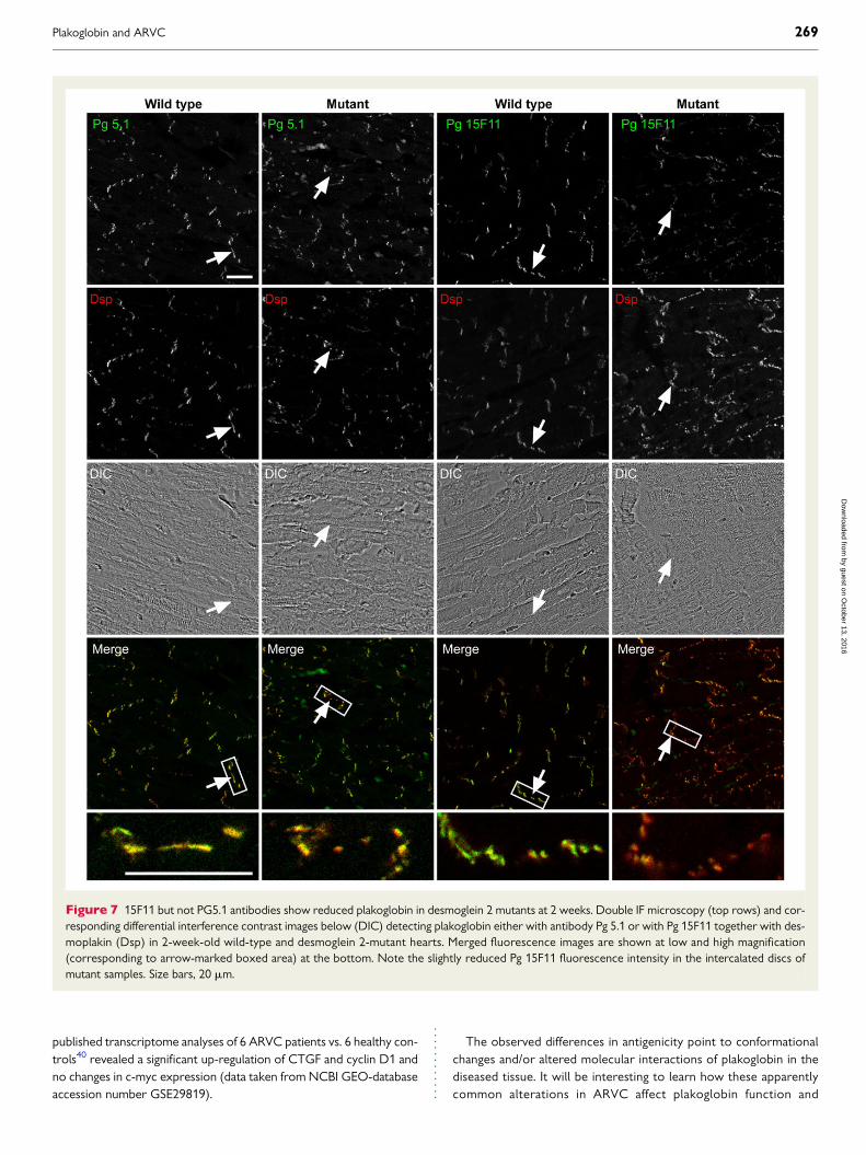
Figure 7 15F11 but not PG5.1 antibodies show reduced plakoglobin in desmoglein 2 mutants at 2 weeks. Double IF microscopy (top rows) and cor-responding differential interference contrast images below (DIC) detecting plakoglobin either with antibody Pg 5.1 or with Pg 15F11 together with des-moplakin (Dsp) in 2-week-old wild-type and desmoglein 2-mutant hearts. Merged fluorescence images are shown at low and high magnification(corresponding to arrow-marked boxed area) at the bottom. Note the slightly reduced Pg 15F11 fluorescence intensity in the intercalated discs ofmutant samples. Size bars, 20 mm.
Plakoglobin and ARVC 269by guest on O
ctober 13, 2016D
ownloaded from

whether they are related to ARVC pathogenesis. It is attractive tospeculate that the reduction and mutation of desmoglein 2 in ourmouse model lead to alteration of the conformation of plakoglobinand/or liberate plakoglobin binding sites. These sites would, in turn,be available for either intramolecular or intermolecular interactionswithin the intercalated disc resulting in masking of the circumscribedepitope recognized by antibody 15F11.34 This scenario is not withoutprecedence: it has been shown that desmogleins compete witha-catenin for the same aminoterminal binding site of plakoglobin35,41
and with classical cadherins for other binding sites.35,42,43 Similar ef-fects may occur in other desmosome-related forms of ARVC giventhe association of plakoglobin with all major desmosomal compo-nents.44,45 The outcome in all instances could be that signallingand/or mechanical linker functions of intercalated disc-localized pla-koglobin are altered.
Another surprising outcome of our study is that the reduction ofdesmosomal cadherins does not affect the localization of any majordesmosomal plaque proteins, i.e. besides plakoglobin, desmoplakin,and plakophilin 2. The same was true for ARVC patients carrying othermutations. Furthermore, we cannot detect obvious alterations in des-min intermediate filament distribution (data not shown). These findingsare in agreement with observations by others,23,24 although contrastingresults have been reported elsewhere.16
Taken together, our findings suggest that ARVC is either a muchmore heterogeneous disease than hitherto assumed with multiplepathogenic mechanisms or that the observed alterations in signall-ing are not triggered by relocalization of desmosomal and non-desmosomal arm-repeat proteins but by other cues. Given thefrequent involvement of desmosomal proteins in ARVC, we favourthe second possibility. Protein modification may be induced which donot result in redistribution but in altered protein interactions. Thus,while b-catenin was reported to be properly localized to intercalateddiscs in heterozygous plakoglobin-deficient mice,46 a reduction inb-catenin phosphorylation implicating altered PI3K-AKT signallingwas reported for mice with a cardiac tissue-restricted depletion ofplakoglobin.47 Recent publications point to involvement of otherpathways besides canonical wnt signalling, most notably the Hippopathway.48,49 In addition, desmosome-specific dysfunctions such ascompromised mechanotransduction, mechanosensation, or vesicletrafficking may either by themselves or through resulting increase inmechanical stress induce these pathways as has been shown for the in-duction of the MRTF/SRF pathway.50
Supplementary materialSupplementary material is available at Cardiovascular Research online.
AcknowledgementsWe thank Marina Lurkens-Weber and Claudia Schmitz for experttechnical assistance.
Conflict of interest: none declared.
FundingThis work was supported by the German Research Council (LE 566/11–1),the Interdisciplinary Center for Clinical Research (IZKF) within the Facultyof Medicine at RWTH Aachen University, the Erich & Hanna Klessmann-Foundation, and the Heart Valve Bank of the University Hospital Rotterdam(control tissue of rejected donor hearts).
References1. Marcus FI, McKenna WJ, Sherrill D, Basso C, Bauce B, Bluemke DA, Calkins H,
Corrado D, Cox MG, Daubert JP, Fontaine G, Gear K, Hauer R, Nava A, Picard MH,Protonotarios N, Saffitz JE, Sanborn DM, Steinberg JS, Tandri H, Thiene G, Towbin JA,Tsatsopoulou A, Wichter T, Zareba W. Diagnosis of arrhythmogenic right ventricularcardiomyopathy/dysplasia: proposed modification of the Task Force Criteria. Eur HeartJ 2010;31:806–814.
2. Saguner AM, Brunckhorst C, Duru F. Arrhythmogenic ventricular cardiomyopathy: aparadigm shift from right to biventricular disease. World J Cardiol 2014;6:154–174.
3. Kant S, Krull P, Eisner S, Leube RE, Krusche CA. Histological and ultrastructural abnor-malities in murine desmoglein 2-mutant hearts. Cell Tissue Res 2012;348:249–259.
4. Krusche CA, Holthofer B, Hofe V, van de Sandt AM, Eshkind L, Bockamp E, Merx MW,Kant S, Windoffer R, Leube RE. Desmoglein 2 mutant mice develop cardiac fibrosis anddilation. Basic Res Cardiol 2011;106:617–633.
5. Li D, Liu Y, Maruyama M, Zhu W, Chen H, Zhang W, Reuter S, Lin SF, Haneline LS,Field LJ, Chen PS, Shou W. Restrictive loss of plakoglobin in cardiomyocytes leads toarrhythmogenic cardiomyopathy. Hum Mol Genet 2011;20:4582–4596.
6. Pilichou K, Remme CA, Basso C, Campian ME, Rizzo S, Barnett P, Scicluna BP, Bauce B,van den Hoff MJ, de Bakker JM, Tan HL, Valente M, Nava A, Wilde AA, Moorman AF,Thiene G, Bezzina CR. Myocyte necrosis underlies progressive myocardial dystrophy inmouse dsg2-related arrhythmogenic right ventricular cardiomyopathy. J Exp Med 2009;206:1787–1802.
7. Gerull B, Heuser A, Wichter T, Paul M, Basson CT, McDermott DA, Lerman BB,Markowitz SM, Ellinor PT, MacRae CA, Peters S, Grossmann KS, Drenckhahn J,Michely B, Sasse-Klaassen S, Birchmeier W, Dietz R, Breithardt G, Schulze-Bahr E,Thierfelder L. Mutations in the desmosomal protein plakophilin-2 are common in ar-rhythmogenic right ventricular cardiomyopathy. Nat Genet 2004;36:1162–1164.
8. Basso C, Corrado D, Bauce B, Thiene G. Arrhythmogenic right ventricular cardiomy-opathy. Circ Arrhythm Electrophysiol 2012;5:1233–1246.
9. Bao JR, Wang JZ, Yao Y, Wang YL, Fan XH, Sun K, Zhang S, Hui RT, Song L. Screening ofpathogenic genes in Chinese patients with arrhythmogenic right ventricular cardiomy-opathy. Chin Med J (Engl) 2013;126:4238–4241.
10. Fressart V, Duthoit G, Donal E, Probst V, Deharo JC, Chevalier P, Klug D, Dubourg O,Delacretaz E, Cosnay P, Scanu P, Extramiana F, Keller D, Hidden-Lucet F, Simon F,Bessirard V, Roux-Buisson N, Hebert JL, Azarine A, Casset-Senon D, Rouzet F,Lecarpentier Y, Fontaine G, Coirault C, Frank R, Hainque B, Charron P. Desmosomalgene analysis in arrhythmogenic right ventricular dysplasia/cardiomyopathy: spectrumof mutations and clinical impact in practice. Europace 2010;12:861–868.
11. Romero J, Mejia-Lopez E, Manrique C, Lucariello R. Arrhythmogenic right ventricularcardiomyopathy (ARVC/D): a systematic literature review. Clin Med Insights Cardiol2013;7:97–114.
12. Otten E, Asimaki A, Maass A, van Langen IM, van der Wal A, de Jonge N, van denBerg MP, Saffitz JE, Wilde AA, Jongbloed JD, van Tintelen JP. Desmin mutations as acause of right ventricular heart failure affect the intercalated disks. Heart Rhythm2010;7:1058–1064.
13. Rickelt S, Pieperhoff S. Mutations with pathogenic potential in proteins located in or atthe composite junctions of the intercalated disk connecting mammalian cardiomyo-cytes: a reference thesaurus for arrhythmogenic cardiomyopathies and for Naxosand Carvajal diseases. Cell Tissue Res 2012;348:325–333.
14. van der Zwaag PA, van Rijsingen IA, Asimaki A, Jongbloed JD, van Veldhuisen DJ,Wiesfeld AC, Cox MG, van Lochem LT, de Boer RA, Hofstra RM, Christiaans I, vanSpaendonck-Zwarts KY, Lekanne dit Deprez RH, Judge DP, Calkins H,Suurmeijer AJ, Hauer RN, Saffitz JE, Wilde AA, van den Berg MP, van Tintelen JP. Phos-pholamban R14del mutation in patients diagnosed with dilated cardiomyopathy or ar-rhythmogenic right ventricular cardiomyopathy: evidence supporting the concept ofarrhythmogenic cardiomyopathy. Eur J Heart Fail 2012;14:1199–1207.
15. Asimaki A, Tandri H, Huang H, Halushka MK, Gautam S, Basso C, Thiene G,Tsatsopoulou A, Protonotarios N, McKenna WJ, Calkins H, Saffitz JE. A new diagnostictest for arrhythmogenic right ventricular cardiomyopathy. N Engl J Med 2009;360:1075–1084.
16. Gehmlich K, Asimaki A, Cahill TJ, Ehler E, Syrris P, Zachara E, Re F, Avella A,Monserrat L, Saffitz JE, McKenna WJ. Novel missense mutations in exon 15 ofdesmoglein-2: role of the intracellular cadherin segment in arrhythmogenic right ven-tricular cardiomyopathy? Heart Rhythm 2010;7:1446–1453.
17. Gehmlich K, Syrris P, Reimann M, Asimaki A, Ehler E, Evans A, Quarta G, Pantazis A,Saffitz JE, McKenna WJ. Molecular changes in the heart of a severe case of arrhythmo-genic right ventricular cardiomyopathy caused by a desmoglein-2 null allele. CardiovascPathol 2012;21:275–282.
18. Noorman M, Hakim S, Kessler E, Groeneweg JA, Cox MG, Asimaki A, van Rijen HV, vanStuijvenberg L, Chkourko H, van der Heyden MA, Vos MA, de Jonge N, van der Smagt JJ,Dooijes D, Vink A, de Weger RA, Varro A, de Bakker JM, Saffitz JE, Hund TJ, Mohler PJ,Delmar M, Hauer RN, van Veen TA. Remodeling of the cardiac sodium channel,connexin43, and plakoglobin at the intercalated disk in patients with arrhythmogeniccardiomyopathy. Heart Rhythm 2013;10:412–419.
19. Munkholm J, Christensen AH, Svendsen JH, Andersen CB. Usefulness of immunostain-ing for plakoglobin as a diagnostic marker of arrhythmogenic right ventricular cardio-myopathy. Am J Cardiol 2012;109:272–275.
S. Kant et al.270by guest on O
ctober 13, 2016D
ownloaded from

20. Kwon YS, Park TI, Cho Y, Bae MH, Kim S. Clinical usefulness of immunohistochemistryfor plakoglobin, N-cadherin, and connexin-43 in the diagnosis of arrhythmogenic rightventricular cardiomyopathy. Int J Clin Exp Pathol 2013;6:2928–2935.
21. Ermakov S, Ursell PC, Johnson CJ, Meadows A, Zhao S, Marcus GM, Scheinman M. Pla-koglobin immunolocalization as a diagnostic test for arrhythmogenic right ventricularcardiomyopathy. Pacing Clin Electrophysiol 2014;37:1708–1716.
22. Asimaki A, Tandri H, Duffy ER, Winterfield JR, Mackey-Bojack S, Picken MM,Cooper LT, Wilber DJ, Marcus FI, Basso C, Thiene G, Tsatsopoulou A,Protonotarios N, Stevenson WG, McKenna WJ, Gautam S, Remick DG, Calkins H,Saffitz JE. Altered desmosomal proteins in granulomatous myocarditis and potentialpathogenic links to arrhythmogenic right ventricular cardiomyopathy. Circ ArrhythmElectrophysiol 2011;4:743–752.
23. Vite A, Gandjbakhch E, Prost C, Fressart V, Fouret P, Neyroud N, Gary F, Donal E,Varnous S, Fontaine G, Fornes P, Hidden-Lucet F, Komajda M, Charron P, Villard E.Desmosomal cadherins are decreased in explanted arrhythmogenic right ventriculardysplasia/cardiomyopathy patient hearts. PLoS One 2013;8:e75082.
24. Tavora F, Zhang M, Cresswell N, Li L, Fowler D, Franco M, Burke A. Quantitative im-munohistochemistry of desmosomal proteins (plakoglobin, desmoplakin and plakophi-lin), connexin-43, and N-cadherin in arrhythmogenic cardiomyopathy: an autopsystudy. Open Cardiovasc Med J 2013;7:28–35.
25. Garcia-Gras E, Lombardi R, Giocondo MJ, Willerson JT, Schneider MD, Khoury DS,Marian AJ. Suppression of canonical Wnt/beta-catenin signaling by nuclear plakoglobinrecapitulates phenotype of arrhythmogenic right ventricular cardiomyopathy. J Clin In-vest 2006;116:2012–2021.
26. Asimaki A, Saffitz JE. Remodeling of cell-cell junctions in arrhythmogenic cardiomyop-athy. Cell Commun Adhes 2014;21:13–23.
27. Lombardi R, da Graca Cabreira-Hansen M, Bell A, Fromm RR, Willerson JT, Marian AJ.Nuclear plakoglobin is essential for differentiation of cardiac progenitor cells to adipo-cytes in arrhythmogenic right ventricular cardiomyopathy. Circ Res 2011;109:1342–1353.
28. Awad MM, Calkins H, Judge DP. Mechanisms of disease: molecular genetics of arrhyth-mogenic right ventricular dysplasia/cardiomyopathy. Nat Clin Pract Cardiovasc Med 2008;5:258–267.
29. Klauke B, Kossmann S, Gaertner A, Brand K, Stork I, Brodehl A, Dieding M, Walhorn V,Anselmetti D, Gerdes D, Bohms B, Schulz U, Zu Knyphausen E, Vorgerd M, Gummert J,Milting H. De novo desmin-mutation N116S is associated with arrhythmogenic rightventricular cardiomyopathy. Hum Mol Genet 2010;19:4595–4607.
30. Brodehl A, Hedde PN, Dieding M, Fatima A, Walhorn V, Gayda S, Saric T, Klauke B,Gummert J, Anselmetti D, Heilemann M, Nienhaus GU, Milting H. Dual color photo-activation localization microscopy of cardiomyopathy-associated desmin mutants. J BiolChem 2012;287:16047–16057.
31. Groeneweg JA, van der Zwaag PA, Olde Nordkamp LR, Bikker H, Jongbloed JD,Jongbloed R, Wiesfeld AC, Cox MG, van der Heijden JF, Atsma DE, de Boer K,Doevendans PA, Vink A, van Veen TA, Dooijes D, van den Berg MP, Wilde AA, vanTintelen JP, Hauer RN. Arrhythmogenic right ventricular dysplasia/cardiomyopathy ac-cording to revised 2010 task force criteria with inclusion of non-desmosomal phospho-lamban mutation carriers. Am J Cardiol 2013;112:1197–1206.
32. Schlegel N, Meir M, Heupel WM, Holthofer B, Leube RE, Waschke J. Desmoglein2-mediated adhesion is required for intestinal epithelial barrier integrity. Am J PhysiolGastrointest Liver Physiol 2010;298:G774–G783.
33. Mertens C, Kuhn C, Franke WW. Plakophilins 2a and 2b: constitutive proteins of duallocation in the karyoplasm and the desmosomal plaque. J Cell Biol 1996;135:1009–1025.
34. Sacco PA, McGranahan TM, Wheelock MJ, Johnson KR. Identification of plakoglobindomains required for association with N-cadherin and alpha-catenin. J Biol Chem1995;270:20201–6.
35. Wahl JK, Sacco PA, McGranahan-Sadler TM, Sauppe LM, Wheelock MJ, Johnson KR.Plakoglobin domains that define its association with the desmosomal cadherins andthe classical cadherins: identification of unique and shared domains. J Cell Sci 1996;109(Pt 5):1143–1154.
36. Noorman M, Hakim S, Asimaki A, Vreeker A, van Rijen HV, van der Heyden MA, deJonge N, de Weger RA, Hauer RN, Saffitz JE, van Veen TA. Reduced plakoglobin immu-noreactivity in arrhythmogenic cardiomyopathy: methodological considerations. Cardi-ovasc Pathol 2013;22:314–318.
37. Munkholm J, Andersen CB, Ottesen GL. Plakoglobin: a diagnostic marker of arrhyth-mogenic right ventricular cardiomyopathy in forensic pathology? Forensic Sci Med Pathol2015;11:47–52.
38. Lombardi R, Dong J, Rodriguez G, Bell A, Leung TK, Schwartz RJ, Willerson JT,Brugada R, Marian AJ. Genetic fate mapping identifies second heart field progenitorcells as a source of adipocytes in arrhythmogenic right ventricular cardiomyopathy.Circ Res 2009;104:1076–1084.
39. Hofe V. Murine Desmoglein 2-Mutanten als Tiermodell zur Untersuchung der Arrhythmogen-en Rechtsventrikularen Kardiomyopathie. VVB Laufersweiler Verlag: Gießen; 2009.p1–124.
40. Gaertner A, Schwientek P, Ellinghaus P, Summer H, Golz S, Kassner A, Schulz U,Gummert J, Milting H. Myocardial transcriptome analysis of human arrhythmogenicright ventricular cardiomyopathy. Physiol Genomics 2012;44:99–109.
41. Ozawa M, Terada H, Pedraza C. The fourth armadillo repeat of plakoglobin (gamma-catenin) is required for its high affinity binding to the cytoplasmic domains of E-cadherinand desmosomal cadherin Dsg2, and the tumor suppressor APC protein. J Biochem1995;118:1077–1082.
42. Al-Jassar C, Bikker H, Overduin M, Chidgey M. Mechanistic basis of desmosome-targeted diseases. J Mol Biol 2013;425:4006–4022.
43. Choi HJ, Gross JC, Pokutta S, Weis WI. Interactions of plakoglobin and beta-cateninwith desmosomal cadherins: basis of selective exclusion of alpha- and beta-cateninfrom desmosomes. J Biol Chem 2009;284:31776–31788.
44. Holthofer B, Windoffer R, Troyanovsky S, Leube RE. Structure and function of desmo-somes. Int Rev Cytol 2007;264:65–163.
45. Swope D, Li J, Radice GL. Beyond cell adhesion: the role of armadillo proteins in theheart. Cell Signal 2013;25:93–100.
46. Fabritz L, Hoogendijk MG, Scicluna BP, van Amersfoorth SC, Fortmueller L, Wolf S,Laakmann S, Kreienkamp N, Piccini I, Breithardt G, Noppinger PR, Witt H, Ebnet K,Wichter T, Levkau B, Franke WW, Pieperhoff S, de Bakker JM, Coronel R,Kirchhof P. Load-reducing therapy prevents development of arrhythmogenic right ven-tricular cardiomyopathy in plakoglobin-deficient mice. J Am Coll Cardiol 2011;57:740–750.
47. Li J, Swope D, Raess N, Cheng L, Muller EJ, Radice GL. Cardiac tissue-restricted dele-tion of plakoglobin results in progressive cardiomyopathy and activation of{beta}-catenin signaling. Mol Cell Biol 2011;31:1134–1144.
48. Chen SN, Gurha P, Lombardi R, Ruggiero A, Willerson JT, Marian AJ. The hippo path-way is activated and is a causal mechanism for adipogenesis in arrhythmogenic cardio-myopathy. Circ Res 2014;114:454–468.
49. Hu Y, Pu WT. Hippo activation in arrhythmogenic cardiomyopathy. Circ Res 2014;114:402–405.
50. Ho CY, Jaalouk DE, Vartiainen MK, Lammerding J. Lamin A/C and emerin regulateMKL1-SRF activity by modulating actin dynamics. Nature 2013;497:507–511.
Plakoglobin and ARVC 271by guest on O
ctober 13, 2016D
ownloaded from