mutations in tetratricopeptide repeat domain 7a result...
TRANSCRIPT

Gastroenterology 2014;146:1028–1039
BASICAND
TRANSLATIONALAT
Mutations in Tetratricopeptide Repeat Domain 7A Result in a SevereForm of Very Early Onset Inflammatory Bowel DiseaseYaron Avitzur,1,2,3,* Conghui Guo,2,* Lucas A. Mastropaolo,2 Ehsan Bahrami,4 Hannah Chen,5
Zhen Zhao,2 Abdul Elkadri,2,3,6 Sandeep Dhillon,2 Ryan Murchie,2 Ramzi Fattouh,2
Hien Huynh,7 Jennifer L. Walker,8 Paul W. Wales,1 Ernest Cutz,9 Yoichi Kakuta,10
Joel Dudley,11 Jochen Kammermeier,12 Fiona Powrie,13 Neil Shah,12 Christoph Walz,14
Michaela Nathrath,15 Daniel Kotlarz,4 Jacek Puchaka,4 Jonathan R. Krieger,2 Tomas Racek,4
Thomas Kirchner,14 Thomas D. Walters,2,3 John H. Brumell,2,3,6 Anne M. Griffiths,2,3
Nima Rezaei,16,17 Parisa Rashtian,18 Mehri Najafi,18 Maryam Monajemzadeh,19
Stephen Pelsue,8 Dermot P. B. McGovern,10 Holm H. Uhlig,5 Eric Schadt,11
Christoph Klein,4,§ Scott B. Snapper,20,21,§ and Aleixo M. Muise2,3,6,§
1Group for Improvement of Intestinal Function and Treatment (GIFT), Hospital for Sick Children, Toronto, Ontario, Canada;2SickKids Inflammatory Bowel Disease Center and Cell Biology Program, Research Institute, Hospital for Sick Children,Toronto, Ontario, Canada; 3Division of Gastroenterology, Hepatology, and Nutrition, Department of Pediatrics, University ofToronto, Hospital for Sick Children, Toronto, Ontario, Canada; 4Department of Pediatrics, Dr von Hauner Children’s Hospital,Ludwig-Maximilians-University, Munich, Germany; 5Translational Gastroenterology Unit and Paediatric Gastroenterology,University of Oxford, Oxford, UK; 6Institute of Medical Science, University of Toronto, Toronto, Ontario, Canada; 7Division ofPediatric Gastroenterology, Stollery Children’s Hospital, Edmonton, Ontario, Canada; 8Department of Immunology andMolecular Biology, University of Southern Maine, Portland, Maine; 9Division of Pathology, The Hospital for Sick Children,Toronto, Ontario, Canada; 10F. Widjaja Foundation Inflammatory Bowel Disease Center and Immunobiology Research Instituteat Cedars-Sinai Medical Center, Los Angeles, California; 11Icahn Institute for Genomics and Multiscale Biology, Department ofGenetics and Genomics Sciences at Mount Sinai, New York, New York; 12Gastroenterology Department, Great Ormond StreetHospital, London, UK; 13Translational Gastroenterology Unit, Nuffield Department Clinical Medicine-Experimental MedicineDivision, University of Oxford, John Radcliffe Hospital, Oxford, UK; 14Institute for Pathology, Ludwig-Maximilians University,Munich, Germany; 15Department of Pediatric Oncology, Kassel and CCG Osteosarcoma, Helmholtz Center Munich, Munich,Germany; 16Research Center for Immunodeficiencies, Children’s Medical Center, Tehran University of Medical Sciences,Tehran, Iran; 17Molecular Immunology Research Center and Department of Immunology, School of Medicine, Tehran Universityof Medical Sciences, Tehran, Iran; 18Department of Pediatric Gastroenterology, Children’s Medical Center, Tehran University ofMedical Sciences, Tehran, Iran; 19Department of Pathology, Children’s Medical Center, Tehran University of Medical Sciences,Tehran, Iran; 20Division of Pediatric Gastroenterology, Hepatology, and Nutrition, Department of Medicine, Children’s HospitalBoston, Massachusetts; and 21Division of Gastroenterology and Hepatology, Brigham & Women’s Hospital, Department ofMedicine, Harvard Medical School, Boston, Massachusetts
*Authors share co-first authorship; §Authors share co-senior authorship.
Abbreviations used in this paper: co-IP, co-immunoprecipitate; EFR3B,EFR3 homolog B; MIA, multiple intestinal atresia; PI4KIIIa, phosphatidyli-nositol 4-kinase IIIa; SCID, severe combined immunodeficiency; shRNA,short hairpin RNA; TPR, tetratricopeptide repeat; TTC7A, tetratricopeptiderepeat domain 7; VEOIBD, very early onset inflammatory bowel diseases;WT, wild type.
© 2014 by the AGA Institute0016-5085/$36.00
http://dx.doi.org/10.1053/j.gastro.2014.01.015
See Covering the Cover synopsis on page 876.
BACKGROUND & AIMS: Very early onset inflammatory boweldiseases (VEOIBD), including infant disorders, are a diversegroup of diseases found in children younger than 6 years of age.They have been associated with several gene variants. Our aimwas to identify the genes that cause VEOIBD. METHODS: Weperformed whole exome sequencing of DNA from 1 infant withsevere enterocolitis and her parents. Candidate gene mutationswere validated in 40 pediatric patients and functional studieswere carried out using intestinal samples and human intestinalcell lines. RESULTS: We identified compound heterozygotemutations in the Tetratricopeptide repeat domain 7 (TTC7A)gene in an infant from non-consanguineous parents with severeexfoliative apoptotic enterocolitis; we also detected TTC7Amutations in 2 unrelated families, each with 2 affected siblings.TTC7A interacts with EFR3 homolog B to regulate phosphati-dylinositol 4-kinase at the plasma membrane. Functionalstudies demonstrated that TTC7A is expressed in human
enterocytes. The mutations we identified in TTC7A result ineither mislocalization or reduced expression of TTC7A. Phos-phatidylinositol 4-kinase was found to co-immunoprecipitatewith TTC7A; the identified TTC7A mutations reduced thisbinding. Knockdown of TTC7A in human intestinal-like celllines reduced their adhesion, increased apoptosis, anddecreased production of phosphatidylinositol 4-phosphate.CONCLUSIONS: In a genetic analysis, we identified loss offunction mutations in TTC7A in 5 infants with VEOIBD.

April 2014 TTC7A Mutations Cause VEOIBD 1029
Functional studies demonstrated that the mutations cause de-fects in enterocytes and T cells that lead to severe apoptoticenterocolitis. Defects in the phosphatidylinositol 4-kinase�TTC7A�EFR3 homolog B pathway are involved in thepathogenesis of VEOIBD.
Keywords: IBD; Intestinal Atresia; Autoimmunity; Intestine.
ery early onset inflammatory bowel diseases
BASICAN
DTR
ANSLAT
IONA
LAT
V(VEOIBD), including forms of infantile disease, are adiverse group of diseases that are diagnosed before 6 years ofage.1 In contrast to adult-onset IBD, VEOIBD frequently en-compasses a unique clinical presentationwith severe, colonicdisease that often has a poor response to standard therapies,including biologic agents.2,3 Recently, several groups,including our own, demonstrated thatmutations in IL10RA/Bgenes4 cause a severe form of VEOIBD, with symptomsconsistently developing in infancy.5 Subsequently, causativevariants in IL10,6 XIAP,7 ADAM17,8 and NCF4,9 and associa-tion variants in the nicotinamide adenine dinucleotidephosphate oxidase genes NCF2/RAC210 were identified inVEOIBD patients, suggesting that severe infantile colitisfrequently starting immediately after birth might represent agroup of heterogeneous monogenetic diseases.
Recently, mutations in the tetratricopeptide repeatdomain 7 (TTC7A) gene were found to cause multiple in-testinal atresia (MIA) with severe combined immunodefi-ciency (SCID), although no details about the intestinalphenotype or function of the TTC7A gene were pro-vided.11,12 In this report, we describe novel human muta-tions in the TTC7A gene (we termed TTC7A deficiency)identified independently by whole exome sequencing thatresult in severe infantile apoptotic enterocolitis with andwithout MIA and define the intestinal defects associatedwith this novel form of VEOIBD.
Materials and MethodsWhole Exome Sequencing
Genetic studies were carried out with approval from theresearch ethics board at the Hospital for Sick Children, Uni-versity of Oxford, Cedars-Sinai Medical Center, and Dr vonHauner Children’s Hospital, LMU Munich. In the index case,whole exome sequencing was performed using the SureSelectHuman All Exon 50 Mb kit (Agilent, Santa Clara, CA) with high-throughput sequencing conducted using the Solid 4 System atThe Center for Applied Genomics through the Hospital for SickChildren (Toronto, ON) on the complete parent�child trio set.Sanger sequencing was used to verify variant genotypes in theindex patient and her family, and 40 infantile patients from theinstitutions named here were screened for TTC7A mutations.
Histologic methods are presented in the SupplementaryMaterial.
Tandem Mass SpectrometryDetailed methods are presented in the Supplementary
Materials. Briefly, to identify potential interactors of TTC7A,M2 anti-FLAG-agarose FLAG-agarose FLAG-tagged wild type
(WT), E71K, or Q526X TTC7A were transiently overexpressedin HEK293T, immunoprecipitated with FLAG-agarose, andbound proteins were trypsin digested and analyzed by tandemmass spectrometry as described previously.13
Knockdown of Endogenous TTC7A by ShortHairpin RNA
GIPZ human TTC7A short hairpin RNA (shRNA) (greenfluorescent protein tagged) targeting coding regions and greenfluorescent protein tagged control shRNA (Thermo Scientific,Logan, UT) were transfected into Henle-407 cells with Lip-ofectamine 2000 (Life Technologies, Carlsbad, CA). Detailedmethods are provided in the Supplementary Materials.
Apoptosis AnalysisConfluent cells were starved for indicated time points.
Apoptosis was assessed by both measured caspase-3 usingWestern blotting and cytoplasmic DNA fragments using flowcytometric analysis of Annexin V. Cells were stained withAnnexin V-phycoerythrin and 7-aminoactinomycin D (BDBiosciences, San Jose, CA) according to manufacturer’s in-structions, and samples were run on a BD LSR II analyzer.Apoptotic cells were identified as Annexin Vþ
7-aminoactinomycin D� cells.
Cell Adhesion AssayTo evaluate cellular adhesion, approximately 5 � 104 cells
were seeded on 96-well plates precoated with fibronectin (20mg/mL; Sigma-Aldrich, St Louis, MO), collagen type I (50 mg/mL; Life Technologies), or bovine serum albumin (5% inphosphate-buffered saline; Sigma) for 60 minutes at 37�C.The wells were subsequently washed with phosphate-buffered saline twice to remove nonadherent cells. Afterfixation with 4% paraformaldehyde, attached cells werevisualized by staining with 1% crystal violet dissolved in 33%acetic acid and were quantified by measuring the absorbanceat 570 nm on a Versamax microplate reader (MolecularDevices, Sunnyvale, CA).
Constructs, Western Blot, Cell Culture, andImmunoprecipitation
Details of constructs, antibodies, and methods used can befound in the Supplementary Materials.
Statistical AnalysisData are presented as mean � SD. Experiments were
performed with a minimum of 3 replications. Statistical signif-icance between groups was established at P < .05 using a2-tailed Student t test. P values are indicated in the figurelegends and text.
ResultsIdentification of Apoptotic Enterocolitis in aVEOIBD Patient
In Family 1 (index case), a female patient born at term toa Caucasian mother and Sudanese father presented with

1030 Avitzur et al Gastroenterology Vol. 146, No. 4
BASICAND
TRANSLATIONALAT
high-output secretory diarrhea and hematochezia thatstarted almost immediately after birth, requiring totalparenteral nutrition. Colonoscopy demonstrated chronicinflammation with severe friability, exfoliative mucosalchanges, and sloughed mucosa within the colonic lumen(Figure 1A). Biopsies taken from the duodenum showedvillous atrophy and the duodenum and colon showedglandular dropout with crypt apoptosis and explodingcrypts (Figure 1B and C). The severity of the epithelial injurywas strikingly reminiscent of acute gastrointestinal graft-vs-host disease and intestine allograft rejection. There was noevidence of perianal disease or dermatological disease. Thepatient had clinical features of immunodeficiency, including
lymphopenia and hypogammaglobulinemia. The patient wastreated with 2 mg/kg methylprednisone without significantresponse. At 11 months of age, she developed respiratoryfailure and succumbed shortly afterward (seeSupplementary Material for details). Autopsy did not showany evidence of bowel atresia, but did confirm widespreadsevere apoptotic enterocolitis as identified previously byendoscopy.
In Family 2, an infant male was born at 36 weeks’gestation to non-consanguineous Caucasian parents. Shortlyafter birth, the infant presented with symptoms of smallbowel obstruction due to short-segment jejunal atresia.Despite surgical resection, the intestinal disease progressed
Figure 1. Histologic andendoscopic characteristicsof intestine of patients withTTC7A mutations. (A) Co-lonoscopy showed severeinflammation characterizedby continuous grade 2 co-litis and multiple areas ofexfoliation and sloughingof the surface epitheliumfrom Family 1. (B) Low-magnification electronmicrograph of crypt epi-thelium from the same bi-opsy shown in panel Cfrom Family 1. Amongregenerating crypt cells,apoptotic cell (whiteasterisk), enteroendocrinecell (EC), and Paneth cell(PC) are present. Cryptenterocytes showed spa-rse brush border microvilli(arrow). (C) High magnifi-cation of duodenal cryptepithelium showed exten-sive apoptosis from Family1 (arrows) (H&E stain). Low(D) and high (E) magnifica-tion of cecum epitheliumshowed extensive apo-ptosis from Family 2 (arrow)(H&E stain). (F) Highmagnification of cecumepithelium showed exten-sive apoptosis from Family3 (H&E stain).
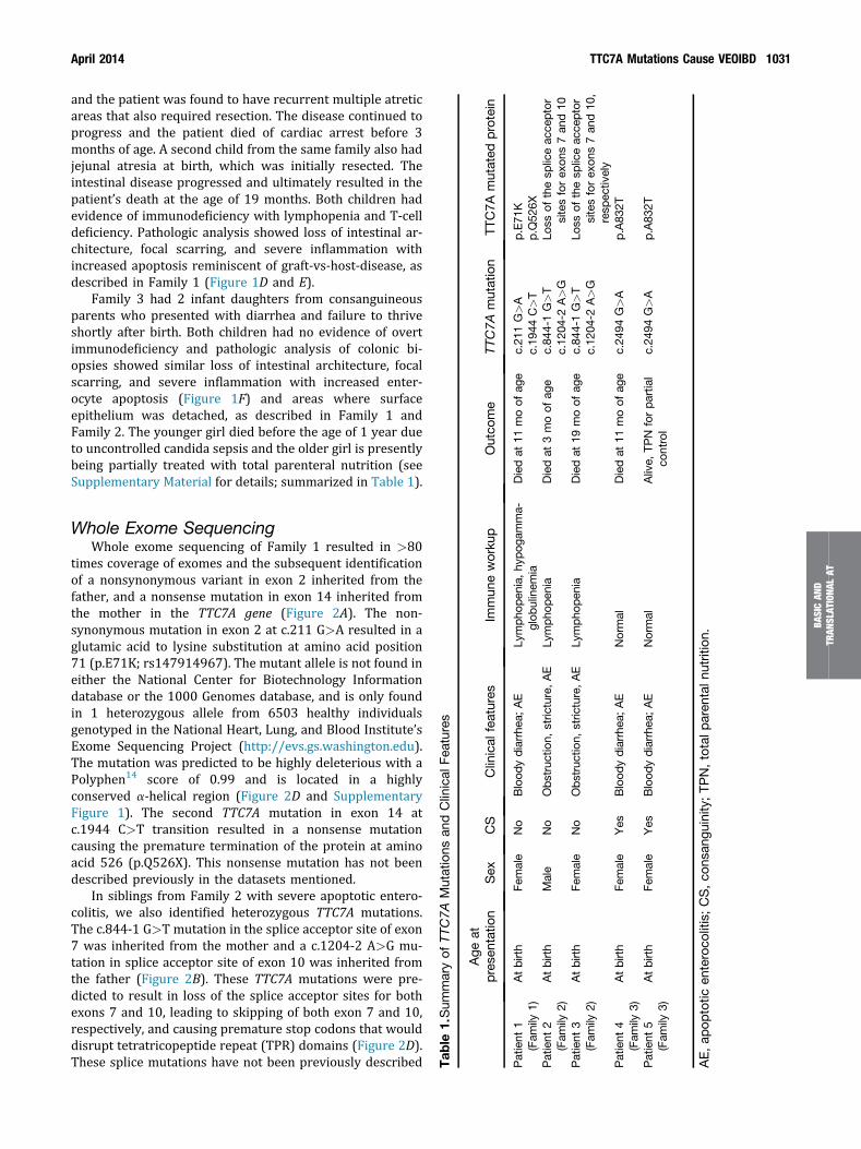
Outco
me
TTC7A
mutation
TTC7A
mutated
protein
Diedat
11moof
age
c.21
1G>A
c.19
44C>T
p.E71
Kp.Q
526X
Diedat
3moof
age
c.84
4-1G>T
c.12
04-2
A>G
Loss
ofthesp
liceac
ceptor
sitesforex
ons7an
d10
Diedat
19moof
age
c.84
4-1G>T
c.12
04-2
A>G
Loss
ofthesp
liceac
ceptor
sitesforex
ons7an
d10
,resp
ectiv
ely
Diedat
11moof
age
c.24
94G>A
p.A83
2T
Alive,
TPN
forpa
rtial
control
c.24
94G>A
p.A83
2T
April 2014 TTC7A Mutations Cause VEOIBD 1031
and the patient was found to have recurrent multiple atreticareas that also required resection. The disease continued toprogress and the patient died of cardiac arrest before 3months of age. A second child from the same family also hadjejunal atresia at birth, which was initially resected. Theintestinal disease progressed and ultimately resulted in thepatient’s death at the age of 19 months. Both children hadevidence of immunodeficiency with lymphopenia and T-celldeficiency. Pathologic analysis showed loss of intestinal ar-chitecture, focal scarring, and severe inflammation withincreased apoptosis reminiscent of graft-vs-host-disease, asdescribed in Family 1 (Figure 1D and E).
Family 3 had 2 infant daughters from consanguineousparents who presented with diarrhea and failure to thriveshortly after birth. Both children had no evidence of overtimmunodeficiency and pathologic analysis of colonic bi-opsies showed similar loss of intestinal architecture, focalscarring, and severe inflammation with increased enter-ocyte apoptosis (Figure 1F) and areas where surfaceepithelium was detached, as described in Family 1 andFamily 2. The younger girl died before the age of 1 year dueto uncontrolled candida sepsis and the older girl is presentlybeing partially treated with total parenteral nutrition (seeSupplementary Material for details; summarized in Table 1).
Tab
le1.Sum
maryof
TTC7A
Mutations
andClinical
Features
Age
atprese
ntation
Sex
CS
Clinical
features
Immun
eworku
p
Patient
1(Fam
ily1)
Atbirth
Female
No
Blood
ydiarrhea
;AE
Lympho
pen
ia,hy
pog
amma-
glob
ulinem
iaPatient
2(Fam
ily2)
Atbirth
Male
No
Obstruction,
stric
ture,AE
Lympho
pen
ia
Patient
3(Fam
ily2)
Atbirth
Female
No
Obstruction,
stric
ture,AE
Lympho
pen
ia
Patient
4(Fam
ily3)
Atbirth
Female
Yes
Blood
ydiarrhea
;AE
Normal
Patient
5(Fam
ily3)
Atbirth
Female
Yes
Blood
ydiarrhea
;AE
Normal
AE,ap
optotic
enteroco
litis;CS,co
nsan
guinity
;TP
N,totalp
aren
taln
utrition.
BASICAN
DTR
ANSLAT
IONA
LAT
Whole Exome SequencingWhole exome sequencing of Family 1 resulted in >80
times coverage of exomes and the subsequent identificationof a nonsynonymous variant in exon 2 inherited from thefather, and a nonsense mutation in exon 14 inherited fromthe mother in the TTC7A gene (Figure 2A). The non-synonymous mutation in exon 2 at c.211 G>A resulted in aglutamic acid to lysine substitution at amino acid position71 (p.E71K; rs147914967). The mutant allele is not found ineither the National Center for Biotechnology Informationdatabase or the 1000 Genomes database, and is only foundin 1 heterozygous allele from 6503 healthy individualsgenotyped in the National Heart, Lung, and Blood Institute’sExome Sequencing Project (http://evs.gs.washington.edu).The mutation was predicted to be highly deleterious with aPolyphen14 score of 0.99 and is located in a highlyconserved a-helical region (Figure 2D and SupplementaryFigure 1). The second TTC7A mutation in exon 14 atc.1944 C>T transition resulted in a nonsense mutationcausing the premature termination of the protein at aminoacid 526 (p.Q526X). This nonsense mutation has not beendescribed previously in the datasets mentioned.
In siblings from Family 2 with severe apoptotic entero-colitis, we also identified heterozygous TTC7A mutations.The c.844-1 G>T mutation in the splice acceptor site of exon7 was inherited from the mother and a c.1204-2 A>G mu-tation in splice acceptor site of exon 10 was inherited fromthe father (Figure 2B). These TTC7A mutations were pre-dicted to result in loss of the splice acceptor sites for bothexons 7 and 10, leading to skipping of both exon 7 and 10,respectively, and causing premature stop codons that woulddisrupt tetratricopeptide repeat (TPR) domains (Figure 2D).These splice mutations have not been previously described

Figure 2. TTC7A genetic analysis. (A) Pedigree and TTC7Amutations in Family 1. Patient from Family 1 was heterozy-gous for 211G>A (p.E71K) inherited from her father andc.1944 C>T (p.Q526X) inherited from her mother. (B) Pedi-gree and TTC7A mutations in Family 2. Siblings from Family 2were heterozygous for a novel c.844-1 G>T TTC7A mutationin splice acceptor site of exon 7 inherited from the mother anda novel c.1204-2 A>G TTC7A mutation in splice acceptor siteof exon 10 inherited from the father. A third sibling was foundto be negative for both mutations. (C) Pedigree and TTC7Amutations in Family 3. Siblings from Family 2 were homozy-gous for a nonsynonymous mutation in exon 20 at c.G2494Aresulted in a alanine acid to threonine substitution at aminoacid position 832 (p.A832T). (D) Location of TTC7A mutationsin patients. Illustration of TTC7A protein with TPR domains inred and identified mutations are highlighted.
1032 Avitzur et al Gastroenterology Vol. 146, No. 4
BASICAND
TRANSLATIONALAT
in the datasets mentioned and it is likely that these muta-tions will result in nonsense-mediated decay of the TTC7Amessenger RNA.
In siblings from Family 3 with severe apoptotic entero-colitis, we identified a homozygous nonsynonymous muta-tion in exon 20 at c.2494 G>A (Figure 2C) that resulted inan alanine to threonine substitution at amino acid position832 (p.A832T). The mutation was predicted to be highlydeleterious with a Polyphen14 score of 0.99 and is located ina highly conserved region of the ninth TPR domain(Figure 2D; Supplementary Figure 1) and has not beendescribed previously in the datasets mentioned (see Table 1for a summary).
Functional Analysis of TTC7A Mutationsin Enterocytes
Immunostaining of TTC7A from healthy human con-trol intestinal tissue (duodenum, ileum, and colon)showed that TTC7A was strongly expressed in enter-ocytes with areas of discreet localization at the plasmamembrane and only few lamina propria cells stainedpositive (Figure 3A). This observed pattern of intestinalexpression suggests a primary role for TTC7A in enter-ocyte homeostasis. To determine if the mutation identi-fied in Family 1 and Family 3 resulted in abnormalTTC7A cellular localization, we transiently transfectedCaco-2 cells with Myc-tagged WT, E71K, Q526X, andA832T TTC7A. Immunostaining using anti-Myc antibodydemonstrated that E71K, Q526X, and A832T TTC7A mu-tants appeared to accumulate in cytoplasmic puncta, andthe WT-TTC7A localized diffusely in the cytoplasm(Figure 3B). In addition, biopsies from the Family 2 pa-tient with the TTC7A splice acceptor site mutations pre-dicted to result in complete loss of the protein showedloss of TTC7A in enterocytes, as expected, indicating thatthese mutations might result in nonsense-mediated decayof the TTC7A messenger RNA (Figure 3C).
Knockdown of TTC7A by shRNA resulted in loss ofcobblestone morphology, typical of human Henle-407 cellswith the development of fibroblastoid morphology withspindle-like features (Figure 3D and SupplementaryFigure 2). In addition, overexpression of E71K, A832T, andQ526X TTC7A in Caco-2 cells demonstrated cytoplasmicaccumulations of Myc-TTC7A in addition to disruptedcortical actin staining suggestive of adhesion defects or lossof cellular polarity (Supplementary Figure 3). Reducedexpression of TTC7A in the enterocytes also resulted indetachment during trypsinization (Figure 4A), impairedadhesion to collagen and fibronectin (Figure 4B), andincreased apoptosis, as measured by caspase-3 (Figure 4C)and Annexin V (Figure 4D). These cellular changes arereminiscent of the apoptosis and mucosal exfoliationdescribed in our patient.
TTC7A Binding PartnersTandem mass spectrometry was performed on proteins
co-immunoprecipitated (co-IP) from HEK293T cells ex-pressing human TTC7A, with the aim of identifying TTC7Abinding partners. Isolated proteins were digested withtrypsin to generate peptide fragments and analyzed bytandem mass spectrometry (Supplementary Figure 4). To

April 2014 TTC7A Mutations Cause VEOIBD 1033
BASICAN
DTR
ANSLAT
IONA
LAT
refine this list to TTC7A binding partners, spectral hitcounts were compared between WT TTC7A and the E71Kmutation samples, and determined that phosphatidylino-sitol 4-kinase IIIa (PI4KIIIa) protein fragments wereable to co-IP with WT TTC7A, but were significantlyreduced with E71K TTC7A mutation (Figure 5A andSupplementary Figure 4). This PI4KIIIa and TTC7A inter-action was supported by a weighted coexpressionnetwork15 from small bowel gene expression datademonstrating that Ttc7a falls within a subnetwork (mod-ule) of the mouse small bowel network that included Pi4kca(murine form of PI4KIIIa) (Supplementary Tables 1–4;Supplementary Figure 5). The additional hits identified inthe tandem mass spectrometry screen (SupplementaryFigure 4) implicated several proteins associated withubiquitination pathways, including E3 ligases (HUWE1,HECTD1, UBR5), and proteins that function in theubiquitin-proteasome system (USP9X, PSMD1, VCP).
Loss of TTC7A Results in PI4KIIIaDysfunction
As TTC7A has been implicated previously in PI4KIIIaregulation in yeast,16,17 and confirmed through the tandemmass spectrometry and network analysis of mouse smallbowel, we next confirmed through co-IP experiments thatTTC7A and PI4KIIIa interacted in human cell lines. We foundthat Myc-Flag-tagged WT TTC7A was able to co-IP PI4KIIIa,indicating that these proteins interact either directly orindirectly in a larger complex (Figure 5B). We also observedreduced co-IP of PI4KIIIa with the TTC7A Q526X and E71Kmutated proteins identified in Family 1 and the A832T TTC7mutation identified in Family 3 (Figure 5B). As the splicevariants identified in Family 2 were assumed to be unstable,we would predict that the gene product of these TTC7Amutations would also not bind to PI4KIIIa.
We next examined human PI4KIIIa in intestinal tissue ofhealthy controls and found that PI4KIIIa was abundantlyexpressed in both enterocytes and immune cells, includinglymphocytes (Figure 5C, left panel). In our patients withTTC7A deficiency, the severe disruption of the bowel ar-chitecture with sloughing of the majority of enterocytesmade interpretation of PI4KIIIa localization difficult; how-ever, in areas with relatively preserved epithelial architec-ture, we observed overall reduced PI4KIIIa expression in apatient from Family 2 (while lamina propria expression waspreserved; Figure 5C). To confirm these results, we tran-siently co-transfected TTC7A and TTC7A shRNA into Henle-407 cells and observed a reduction in PI4KIIIa (Figure 5Dand E). These results indicate that loss of TTC7A resulted inaberrant subcellular localization of PI4KIIIa in enterocytes.
Finally, we determined that knockdown of TTC7A inhuman Henle-407 cell lines resulted in decreased phos-phatidylinositol 4-phosphate, the end product of PI4KIIIaenzyme, in both the cytoplasm (Figure 5E) and at theplasma membrane (Figure 5F). Together these resultsindicate that TTC7A is required for PI4KIIIa localization tothe plasma membrane and that TTC7A deficiency results inloss of PI4KIIIa signaling in enterocytes.
DiscussionOur index case (Family 1) had severe infantile apoptotic
enterocolitis with a presentation significantly different frompreviously described cases of VEOIBD with IL10 and IL10Rmutations that are invariably present with colitis and peri-anal disease.4,5,18–20 The severe enterocolitis with friabilityand exfoliative mucosal changes along with villous atrophy,gland dropout, and crypt apoptosis led to our geneticexploration through whole exome sequencing and identifi-cation of TTC7A as the causative gene.
The TPR domain is defined by a degenerate consensussequence of 34 amino acids21 and 4 of the 5 TTC7A muta-tions found in our patients resulted in disruption of theseTPR domains. TPR domains mediate protein�protein in-teractions and the assembly of multi-protein complexes thatare involved in the regulation of cell cycle, transcription, andprotein transport.22 Our tandem mass spectrometry andintestinal network experiments demonstrated an associa-tion between TTC7A and PI4KIIIa that was previously onlydescribed in yeast.16,17 In yeast, the TTC7 ortholog, YPP1, isessential and has been shown to rescue a lethal a-synuclein(aSyn-A53T) yeast mutant.23 Ypp1 (TTC7) directly binds toStt4 (PI4KIIIa), and this binding is critical to maintainingphosphatidylinositol 4-phosphate levels and PI4KIIIa sta-bility at the plasma membrane.16,17 In addition, in yeast, thephenotypes of YPP1 (TTC7) and STT4 (PI4KIIIa) conditionalmutants are identical and both mutants result in cell walldestabilization and defective organization of actin. Over-expression of STT4 (PI4KIIIa) also suppresses thetemperature-sensitive growth defect observed in YPP1(TTC7) mutants.17 The role of TTC7A in PI4KIIIa recruit-ment to the plasma membrane was also recently confirmedin mammalian cell lines24 and we demonstrate for the firsttime that TTC7A and PI4KIIIa directly interact in human celllines. Because TTC7A is required for proper localization ofPI4KIIIa at the plasma membrane,24 we propose that TTC7Amutations result in disease through loss of PI4KIIIa at theplasma membrane and subsequent reduction of phosphati-dylinositol 4-phosphate that is required for cell polarity andsurvival. In support of this model, down-regulation ofPI4KIIIa results in increased apoptosis25 and, in addition,intestinal-specific murine knockout of Pi4kca (PI4KIIIa)results in a strikingly severe intestinal phenotype withwidespread mucosal epithelial degeneration26 reminiscentof our patients with TTC7Amutations. Therefore, our resultsdemonstrate a direct interaction between PI4KIIIa andTTC7A. And similar to the phenotype observed in TTC7A-deficient patients, TTC7A knockdown in human intestinal-like cell lines resulted in decreased adhesion andincreased apoptosis. These results indicate that disruptionof the PI4KIIIa�TTC7A pathway results in a combinedT-cell and enterocyte defect that results in the intestinalphenotype described here (Figure 6).
The EFR3 homolog B (EFR3B; ENSG00000084710) geneproduct EFR3B tethers TTC7A (and TTC7B) to the plasmamembrane and is essential for both TTC7A and PI4KIIIafunction.24 In addition, knockdown of EFR3B results in theloss of both TTC7A and PI4KIIIa at the plasma membrane

1034 Avitzur et al Gastroenterology Vol. 146, No. 4
BASICAND
TRANSLATIONALAT
and is critical for PI4KIIIa signaling.24 Interesting, severalEFR3B single nucleotide polymorphisms located both in theEFR3B gene and its flanking regions were reported to beassociated with Crohn’s disease (http://www.ibdgenetics.org; Supplementary Table 5; lead single nucleotide poly-morphism rs1077492; P ¼ 1.9 � 10�14, odds ratio, 1.11).This locus on chromosome 2 at 25.12 Mb was recently
reported in the International Inflammatory Bowel DiseaseGenetics Consortium meta-analysis as an IBD locus.27 Insilico analyses carried out by the International Inflamma-tory Bowel Disease Genetics Consortium suggested ADCY3as a potential candidate at this locus27; however, EFRB3’srole in regulating PI4KIIIa-TTC7A implicates EFRB3 as aplausible causative IBD gene at this locus and that this

Figure 4. (A) Impaired cell adhesion in TTC7A-depleted cells. The total dissociation time, defined as the time required forcomplete dissociation of all cells from the tissue culture plate, was markedly reduced in TTC7A-depleted Henle-407 cellscompared with control cells. Dissociation assay (n ¼ 6, biological replicates; Student t test, *P ¼ .0022). (B) Impaired celladhesion to collagen and fibronectin in TTC7A-depleted cells. Cell adhesion assays were performed using crystal violetstaining. Control and TTC7A-depleted Henle-407 cells were seeded on 96-well plates coated with either collagen or fibro-nectin. Adhesion was assessed on the basis of optical density (OD) at 570 nm. n ¼ 3; adhesion assay (n ¼ 3) biologicalreplicates, Student t test, **collagen: P ¼ .027 and *fibronectin: P ¼ .0077. (C) TTC7A-depletion in Henle-407 cells results ingreater caspase-dependent apoptosis. To investigate the impact of TTC7A suppression on the induction of apoptosis, theactivation of caspase-3 was measured by Western blot. Specific cleavage of pro-caspase 3 (32 kDa) into the active caspase-3fragments (17 kDa) was increased in cells serum starved for 24 and 48 hours. n ¼ 3; P ¼ .012, analysis of variance. (D) TTC7A-depletion in Henle-407 cells results in greater apoptosis measured by flow cytometric analysis of Annexin V. To examine thesignificance of TTC7A suppression, after loss of attachment, flow cytometric analysis was conducted to quantity the extent ofapoptosis in cells starved for 24 and 48 hours. Cells were stained with Annexin V-phycoerythrin and 7-aminoactinomycin D(viability marker); apoptotic cells were identified as Annexin Vþ 7-aminoactinomycin D� cells. In TTC7A-depleted cells, theproportion of cells in early apoptosis increased to approximately 4.5% at 24 hours and 11.2% at 48 hours of serum-starvationcompared to 1.3% (24 hours) and 4.1% (48 hours) in control cells. Annexin V apoptosis assay n ¼ 3 biological replicates,Student t test, fetal bovine serum (FBS), 24 hours: *P ¼ .0018; FBS, 48 hours: *P ¼ .0076; serum starved, 24 hours: *P ¼ .021;serum starved, 48 hours: *P ¼ .0034. BSA, bovine serum albumin.
April 2014 TTC7A Mutations Cause VEOIBD 1035
BASICAN
DTR
ANSLAT
IONA
LAT
PI4KIIIa-TTC7A-EFRB3 pathway plays a broader role inadult-onset IBD.
Mutations in a Ttc7a TPR domain28–30 result in flakyskin (fsn) mice and the associated pleiotropic abnormalities,
=Figure 3. Functional TTC7A enterocyte studies. (A) TTC7A expscopy performed on human tissue sections immunostained us[DAPI]) demonstrates TTC7A expression in enterocytes withinsent zoomed images of the corresponding panel above. ScaleTTC7A. Caco-2 cells were transiently transfected with Myc-tagusing anti-Myc antibody and visualized using confocal microscoantibody only. The control plasmid represents an empty vectorocytes from patient cecum (Family 2). Immunofluorescence micrcecal tissues sections from both control and patient (Family 2expression is reduced in the patient sample (right panel). Scalemorphological changes in Henle-407 cells. Expression of TTC780%) compared with Henle-407 cells stably transfected with conTTC7A shRNA knockdown was examined in Henle-407 cellsculture conditions. Knockdown of TTC7A resulted in a loss ofmorphology with spindle-like features. GAPDH, glyceraldehyde
including severe weight loss with diarrhea and intestinalapoptosis, being reported infrequently.31,32 In addition, theintestinal-specific knockout of Pi4kca (PI4KIIIa) in miceresulted in a severe intestinal phenotype with widespread
ression in intestinal enterocytes. Immunofluorescence micro-ing anti-TTC7A antibody (and 40,6-diamidino-2-phenylindolethe duodenum, ileum, and colon. Lower inset panels repre-bars ¼ 100 mm. (B) E71K, Q526X, and A832T mutations inged WT, E71K, Q526X, and A832T TTC7A, immunostainedpy. Negative control panels represent staining with secondarysham transfection. Scale bars ¼ 25 mm. (C) TTC7A in enter-oscopy was performed on TTC7A-immunostained (and DAPI)) biopsies. Compared to control staining (left panel), TTC7Abar ¼ 100 mm. (D) Stable knockdown of TTC7A resulted inA in stably transfected Henle-407 cells was reduced (70%�trol shRNA. The impact on cellular morphology of control andby contrast microscopy (100� magnification) under normalcobblestone morphology and development of fibroblastoid
-3-phosphate dehydrogenase.

1036 Avitzur et al Gastroenterology Vol. 146, No. 4
BASICAND
TRANSLATIONALAT
mucosal epithelial degeneration.26 The intestinal phenotypeobserved in these 2 mouse models is reminiscent of thephenotype seen in our infantile patients who had massiveshedding of enterocytes with increased apoptosis; however,none of the patients developed psoriasis or other skin ab-normalities like the fsnmice. TTC7A has been investigated inhuman psoriasis and found not to be associated with humandermatological disease.30 Therefore, psoriasis might only beobserved in the fsnmice and might not be part of the humandisorder.
We have shown that TTC7A is expressed in enterocytesand has a role in enterocyte survival and function, sug-gesting that the physiological abnormalities observed inboth mice and humans with TTC7A mutations result, at leastin part, from epithelial dysfunction. However, as Chen et al11
also demonstrated, TTC7A is expressed in the thymus with amarked reduction of thymocytes and lymphoid depletion in1 patient with TTC7A deficiency, TTC7A plays a critical rolein modulating immune homeostasis and the immunodefi-ciency also contributes to the pathogenesis of TTC7A

Figure 6. Summary of TTC7 mutations. Schematic representation of the role of TTC7A in the trafficking of PI4KIIIa to theplasma membrane from the trans-Golgi network. The left panel represents WT TTC7A in enterocytes wherein TTC7A binds toand facilitates the transport of PI4KIIIa from the trans-Golgi to the plasma membrane. At the membrane, PI4KIIIa can catalyzethe production of PtdIns-4P(PI-4P). PI-4P levels at the plasma membrane have been implicated in cell survival and themaintenance of cell polarity. In the right panel, the various TTC7A mutations identified in the patients are depicted. E71K,Q526X, and A832T TTC7A all demonstrated reduced binding to PI4KIIIa, which could reduce the interaction between TTC7Aand PI4KIIIa, hindering transport to the plasma membrane (PM). Consequently, this will lead to reduced plasma membranelevels of PI-4P, a dysregulation that would affect downstream signaling pathways.
=Figure 5. (A) Tandem mass spectrometry. E71K and Q526X mutations reduce the ability of TTC7A to immunoprecipitatePI4KA. Selected peptides from PI4KA and TTC7A were analyzed to determine the area under their MS1 peaks to assess therelative abundance of each peptide. The PI4KA present in each sample was normalized to the total TTC7A in each technicalreplicate to allow comparisons among biological replicates (n ¼ 3). These normalized values were averaged over all experi-ments. Error bars represent the standard error. (B) PI4KIIIa-TTC7A co-immunoprecipitate. HEK293T cells were transientlytransfected with Myc-tagged WT (WT-TTC7A), E71K, Q526X, and A832T TTC7A constructs. Lysates were immunoprecipatedwith anti-Myc antibody, and then immunoblotted using anti-PI4KIIIa and anti-Myc (for TTC7A) antibodies. The control lanerepresents transfection with an empty vector. (C) Expression and localization of PI4KIIIa is altered in patients with TTC7Adeficiency. Immunofluorescence microscopy was performed on both control and patient colonic tissue sections immuno-stained with anti-PI4KIIIa antibodies. In the left panel, immunohistochemistry demonstrated that PI4KIIIa is highly expressed inenterocytes and immune cells from healthy human intestine. Inset panel depicts zoomed view of left panel, demonstratingPI4KIIIa expression at the plasma membrane of enterocytes. In the patient tissues, immunohistochemistry demonstrated thatPI4KIIIa is dysregulated in enterocytes. Inset panel (representing region indicated by white arrow) demonstrates loss of PI4KIIIaat the plasma membrane of enterocytes bordering the intestinal crypt. Scale bar ¼ 100 mm. (D) shRNA-mediated knockdown ofTTC7A expression leads to decreased PI4KIIIa levels. To test the efficacy of the TTC7A shRNA, Henle-407 cells were tran-siently co-transfected with WT TTC7A and the various knock-down constructs, labeled #1 through #4, including a scrambledshRNA control and sham transfection. shRNA #1 and #3 showed reduction in TTC7A expression (left panels). shRNA con-taining the same targeting sequences were used to lentivirally infect Henle-407 cells where expression of PI4KIIIa wasassessed in cell lysates by Western blot (right panels). Glyceraldehyde-3-phosphate dehydrogenase (GAPDH) was stained asloading control for all blots. The intensity of each PI4KIIIa band was normalized to the GADPH loading control by densitometry.Quantitation of band intensities (listed below each lane) demonstrates a statistically significant reduction in PI4KIIIa expressionafter TTC7A knockdown (Student t test, n ¼ 3, P ¼ .0234). Each normalized band intensity is presented as �SEM. (E) TTC7Adepletion results in decreased cytoplasmic phosphatidylinositol 4-phosphate (PI-4P) production. TTC7A knockdown andcontrol Henle-407 cells were stained with antibodies against Ptdlns4P (in red; Z-P004, IgM, Cedarlane, Burlington, NC) and40,6-diamidino-2-phenylindole (DAPI) (in blue) to visualize nuclei. (F) TTC7A knockdown Henle-407 cells have reduced plasmamembrane immunostaining for PI-4P compared with controls. For control and TTC7A knockdown (KD), Henle-407 cells Z-stack images were generated at 0.2-mm intervals and recapitulated using Volocity to generate a 3-dimensional model toillustrate cell surface levels of PIP. Unconjugated green fluorescent protein, expressed from the control and knockdownplasmids, was visualized and used to approximate the morphology of the cells. Each pair of images represents 2 views of thesame cell according to axes depicted.
April 2014 TTC7A Mutations Cause VEOIBD 1037
BASICAN
DTR
ANSLAT
IONA
LAT

1038 Avitzur et al Gastroenterology Vol. 146, No. 4
BASICAND
TRANSLATIONALAT
deficiency, as seen in Family 1 and Family 2. These resultsare consistent with those seen in the MIA patients describedwith SCID,11,12,33 and points to a severe defect in bothenterocyte and T-cell function; however, patients fromFamily 3 did not have an overt T-cell defect and patientswith MIA described previously11,12,33 had varying degrees ofimmunodeficiency, with some patients exhibiting mild T-celllymphopenia in Chen et al,11 who also suggested an enter-ocyte defect based on the high frequency of bloodstreaminfections with intestinal microbes.
Therefore, our studies also suggest that mutations in theTTC7A gene can result in a spectrum of intestinal diseaseranging from VEOIBD with apoptotic enterocolitis, as firstdescribed here, to MIA with SCID, as described here andpreviously.11,12,33 In support of this, TTC7A mutations werefound to cause hereditary MIA with SCID11,12,33; however,apoptotic enterocolitis has not been reported. The patientsfrom Family 1 and Family 3, with apoptotic enterocolitiswith no evidence of MIA or stricturing disease on autopsy,had mutations that would be predicted to reduce TTC7Aexpression but not completely abolish function. In support,we also demonstrated that the mutations identified inFamily 1 and Family 3 reduced TTC7A binding to PI4KIIIa.Therefore, it is possible that the disease observed in patientsfrom Family 1 and Family 3 represents a hypomorphic state,where some residual TTC7A activity in both enterocytes andthe thymus results in severe enterocolitis without MIA andwith or without lymphopenia.
All TTC7A-deficiency patients, including the patientsdescribed here, died in infancy due to their progressive boweldisease, failed allogeneic hematopoietic stem cell trans-plantation, or survived with short gut and total parenteralnutrition.11,12 Interestingly, both Chen et al11 and Samuelset al12 described an MIA TTC7A-deficiency patient who hadhematopoietic stem cell transplantation and developed se-vere recurrence of MIA post transplantation. The recurrenceof MIA after resection in our Family 2 patients and thosepresented by Chen et al and Samuels et al,11,12 suggests thatTTC7A deficiency results in a severe intestinal inflammatoryprocess driven by a combined epithelial and T-cell defect thatcontinues post resection of atretic regions, and the enterocytedefect will not respond to hematopoietic stem cell transplant.Therefore, as we have demonstrated that an enterocytedefect is also found in patients with TTC7A deficiency,transplantation of allogeneic hematopoietic stem cells mightnot be warranted in TTC7A-deficient patients. However, ourstudy opens the possibility of pharmacologically targeting thePI4KIIIa�TTC7A�EFR3B pathway as a potential therapeuticapproach. The identification of TTC7A as a candidate gene fora unique and unrecognized variant of severe apoptoticenterocolitis expands the genetic diversity of VEOIBD and theneed to tailor therapeutic approaches for individual subtypes.
Supplementary MaterialNote: To access the supplementary material accompanyingthis article, visit the online version of Gastroenterology atwww.gastrojournal.org, and at http://dx.doi.org/10.1053/j.gastro.2014.01.015.
References
1. Muise AM, Snapper SB, Kugathasan S. The age of genediscovery in very early onset inflammatory bowel dis-ease. Gastroenterology 2012;143:285–288.
2. Griffiths AM. Specificities of inflammatory bowel diseasein childhood. Best Pract Res Clin Gastroenterol 2004;18:509–523.
3. Heyman MB, Kirschner BS, Gold BD, et al. Children withearly-onset inflammatory bowel disease (IBD): analysis of apediatric IBDconsortium registry. JPediatr 2005;146:35–40.
4. Glocker EO, Kotlarz D, Boztug K, et al. Inflammatorybowel disease and mutations affecting the interleukin-10receptor. N Engl J Med 2009;361:2033–2045.
5. Kotlarz D, Beier R, Murugan D, et al. Loss of interleukin-10 signaling and infantile inflammatory bowel disease:implications for diagnosis and therapy. Gastroenterology2012;143:347–355.
6. Glocker EO, Frede N, Perro M, et al. Infant colitis—it’s inthe genes. Lancet 2010;376(9748):1272.
7. Worthey EA, Mayer AN, Syverson GD, et al. Making adefinitive diagnosis: successful clinical application ofwhole exome sequencing in a child with intractable in-flammatory bowel disease. Genet Med;13:255–262.
8. Blaydon DC, Biancheri P, Di WL, et al. Inflammatory skinand bowel disease linked to ADAM17 deletion. N Engl JMed;365:1502–1508.
9. Matute JD, Arias AA, Wright NA, et al. A new geneticsubgroup of chronic granulomatous disease with auto-somal recessive mutations in p40 phox and selectivedefects in neutrophil NADPH oxidase activity. Blood2009;114:3309–3315.
10. Muise AM, Xu W, Guo CH, et al. NADPH oxidase com-plex and IBD candidate gene studies: identification of arare variant in NCF2 that results in reduced binding toRAC2. Gut 2012;61:1028–1035.
11. ChenR, Giliani S, Lanzi G, et al.Whole-exome sequencingidentifies tetratricopeptide repeat domain 7A (TTC7A)mutations for combined immunodeficiency with intestinalatresias. J Allergy Clin Immunol 2013;132:656–664.e17.
12. Samuels ME, Majewski J, Alirezaie N, et al. Exomesequencing identifies mutations in the gene TTC7A inFrench-Canadian cases with hereditary multiple intesti-nal atresia. J Med Genet 2013;50:324–329.
13. Krieger JR, Taylor P, Gajadhar AS, et al. Identificationand selected reaction monitoring (SRM) quantification ofendocytosis factors associated with Numb. Mol CellProteomics 2013;12:499–514.
14. Dixon AL, Liang L, Moffatt MF, et al. A genome-wideassociation study of global gene expression. Nat Genet2007;39:1202–1207.
15. Zhang B, Horvath S. A general framework for weightedgene co-expression network analysis. Stat Appl GenetMol Biol 2005;4:Article17.
16. Baird D, Stefan C, Audhya A, et al. Assembly of thePtdIns 4-kinase Stt4 complex at the plasma membranerequires Ypp1 and Efr3. J Cell Biol 2008;183:1061–1074.
17. Zhai C, Li K, Markaki V, et al. Ypp1/YGR198w plays anessential role in phosphoinositide signalling at theplasma membrane. Biochem J 2008;415:455–466.

April 2014 TTC7A Mutations Cause VEOIBD 1039
BASICAN
DTR
ANSLAT
IONA
LAT
18. Begue B, Verdier J, Rieux-Laucat F, et al. Defective IL10signalingdefiningasubgroupofpatientswith inflammatorybowel disease. Am J Gastroenterol 2011;106:1544–1555.
19. Engelhardt KR, Shah N, Faizura-Yeop I, et al. Clinicaloutcome in IL-10- and IL-10 receptor-deficient patientswith or without hematopoietic stem cell transplantation.J Allergy Clin Immunol 2013;131:825–830.
20. Moran CJ, Walters TD, Guo CH, et al. IL-10R poly-morphisms are associated with very-early-onset ulcera-tive colitis. Inflamm Bowel Dis 2013;19:115–123.
21. Cortajarena AL, Regan L. Ligand binding by TPR do-mains. Protein Sci 2006;15:1193–1198.
22. D’Andrea LD, Regan L. TPR proteins: the versatile helix.Trends Biochem Sci 2003;28:655–662.
23. Flower TR, Clark-Dixon C, Metoyer C, et al. YGR198w(YPP1) targets A30P alpha-synuclein to the vacuole fordegradation. J Cell Biol 2007;177:1091–1104.
24. Nakatsu F, Baskin JM, Chung J, et al. PtdIns4P synthesisby PI4KIIIalpha at the plasma membrane and its impacton plasma membrane identity. J Cell Biol 2012;199:1003–1016.
25. Ma H, Blake T, Chitnis A, et al. Crucial role of phospha-tidylinositol 4-kinase IIIalpha in development of zebrafishpectoral fin is linked to phosphoinositide 3-kinase andFGF signaling. J Cell Sci 2009;122:4303–4310.
26. Vaillancourt FH, Brault M, Pilote L, et al. Evaluation ofphosphatidylinositol-4-kinase IIIalpha as a hepatitis Cvirus drug target. J Virol 2012;86:11595–11607.
27. Jostins L, Ripke S, Weersma RK, et al. Host-microbeinteractions have shaped the genetic architecture of in-flammatory bowel disease. Nature 2012;491:119–124.
28. White RA, McNulty SG, Nsumu NN, et al. Positionalcloning of the Ttc7 gene required for normal iron ho-meostasis and mutated in hea and fsn anemia mice.Genomics 2005;85:330–337.
29. Takabayashi S, Iwashita S, Hirashima T, et al. The noveltetratricopeptide repeatdomain7mutation,Ttc7fsn-Jic,withdeletion of the TPR-2B repeat causes severe flaky skinphenotype. Exp Biol Med (Maywood) 2007;232:695–699.
30. Helms C, Pelsue S, Cao L, et al. The Tetratricopeptiderepeat domain 7 gene is mutated in flaky skin mice: amodel for psoriasis, autoimmunity, and anemia. Exp BiolMed (Maywood) 2005;230:659–667.
31. Sundberg JP, France M, Boggess D, et al. Developmentand progression of psoriasiform dermatitis and systemic
lesions in the flaky skin (fsn) mouse mutant. Pathobiology1997;65:271–286.
32. Nüesch U, Seger R, Pachlopnik Schmid J. Clinical andhistological features of flaky skin (fsn) mice. Presented atthe XXIX Meeting of the Swiss Immunology PhD Stu-dents, Schloss Wolfsberg, April 2–4, 2012.
33. Bigorgne AE, Farin HF, Lemoine R, et al. TTC7A muta-tions disrupt intestinal epithelial apicobasal polarity.J Clin Invest 2013 Dec 2 [Epub ahead of print].
Received October 2, 2013. Accepted January 3, 2014.
Reprint requestsAddress requests for reprints to: Aleixo Muise, MD, PhD, The Hospital for SickChildren, 555 University Avenue, Toronto, Ontario, Canada, M5G 1X8. e-mail:[email protected]; fax: (416) 813-6531.
AcknowledgmentsThe authors thank the families of the patients described here from Canada,Germany, and Iran. International Early Onset Pediatrics IBD Cohort Studyparticipants (www.NEOPICS.org): Yaron Avitzur, Conghui Guo, Lucas A.Mastropaolo, Abdul Elkadri, Sandeep Dhillon, Ryan Murchie, Ramzi Fattouh,Hien Huynh, Fiona Powrie, Thomas D. Walters, John H. Brumell, Anne M.Griffiths, Dermot P. B. McGovern, Holm H. Uhlig, Eric Schadt, ChristophKlein, Scott B. Snapper, and Aleixo M. Muise.
Conflicts of interestThe authors disclose no conflicts.
FundingAMM is supported by an Early Researcher Award from the Ontario Ministry ofResearch and Innovation and funded by a Canadian Institute of HealthResearch Operating Grant (MOP119457). SBS is supported by NationalInstitutes of Health grants HL59561, DK034854, and AI50950 and theWolpow Family Chair in IBD Treatment and Research. SP is supported byLupus Research Institute Novel Grant Program and University of SouthernMaine/Maine Economic Improvement Fund Development Grant program.This work was supported by DFG SFB1054, the Gottfried-Wilhelm-Leibnizprogram, BaySysNet (CK) and the Care-for-Rare Foundation (EB, DK). IBDResearch at Cedars-Sinai is supported by US Public Health Service grantPO1DK046763 and the Cedars-Sinai F. Widjaja Foundation InflammatoryBowel and Immunobiology Research Institute Research Funds. Genotypingat Cedars-Sinai Medical Center is supported in part by the National Centerfor Research Resources grant M01-RR00425, UCLA/Cedars-Sinai/Harbor/Drew Clinical and Translational Science Institute grant (UL1 TR000124-01),the Southern California Diabetes and Endocrinology Research Grant(DK063491). In addition, DPBM is supported by The Leona M. and Harry B.Helmsley Charitable Trust, The European Union, the Crohn’s and ColitisFoundation of America, and The Joshua L. and Lisa Z. Greer Chair in IBDGenetics. Subject ascertainment and data and sample processing was alsosupported by DK062413 and DK084554 and supplements to activities of theNational Institute of Diabetes and Digestive and Kidney Diseases IBDGenetics Consortium. Partial funding is provided Harvard Institute ofTranslational Immunology (HITI) to AMM and SBS. This work was funded inpart by a Leona M. and Harry B. Helmsley Charitable Trust grant to AMM,SBS, CK, DPBM, FP, and ES.