nanoplasmonic sensing using metal nanoparticles · pdf fileen biosensor är en...
TRANSCRIPT

Linköping Studies in Science and Technology Dissertation No. 1624
Nanoplasmonic Sensing using
Metal Nanoparticles
Erik Martinsson
Division of Molecular Physics Department of Physics, Chemistry and Biology
Linköping University, Sweden
Linköping 2014

Cover: An illustration of surface immobilized spherical gold nanoparticles
illuminated with a ray of light.
During the course of the research underlying this thesis, Erik Martinsson was
enrolled in Forum Scientium, a multidisciplinary doctoral programme at
Linköping University, Sweden.
© Copyright 2014 ERIK MARTINSSON, unless otherwise noted
Martinsson, Erik
Nanoplasmonic Sensing using Metal Nanoparticles
ISBN: 978-91-7519-223-9
ISSN: 0345-7524
Linköping Studies in Science and Technology, Dissertation No. 1624
Electronic publication: http://www.ep.liu.se
Printed in Sweden by LiU-Tryck, Linköping 2014.

“Everything happens for a reason
and that reason is usually physics”


V
Abstract
In our modern society, we are surrounded by numerous sensors, constantly
feeding us information about our physical environment. From small, wearable
sensors that monitor our physiological status to large satellites orbiting around
the earth, detecting global changes. Although, the performance of these sensors
have been significantly improved during the last decades there is still a demand
for faster and more reliable sensing systems with improved sensitivity and
selectivity. The rapid progress in nanofabrication techniques has made a
profound impact for the development of small, novel sensors that enables
miniaturization and integration. A specific area where nanostructures are
especially attractive is biochemical sensing, where the exceptional properties of
nanomaterials can be utilized in order to detect and analyze biomolecular
interactions.
The focus of this thesis is to investigate plasmonic nanoparticles composed of
gold or silver and optimize their performance as signal transducers in optical
biosensors. Metal nanoparticles exhibit unique optical properties due to
excitation of localized surface plasmons, which makes them highly sensitive
probes for detecting small, local changes in their surrounding environment, for
instance the binding of a biomolecule to the nanoparticle surface. This is the
basic principle behind nanoplasmonic sensing based on refractometric detection,
a sensing scheme that offers real-time and label-free detection of molecular
interactions.
This thesis shows that the sensitivity for detecting local refractive index changes
is highly dependent on the geometry of the metal nanoparticles, their interaction
with neighboring particles and their chemical composition and functionalization.
An increased knowledge about how these parameters affects the sensitivity is
essential when developing nanoplasmonic sensing devices with high
performance based on metal nanoparticles.

VI

VII
Populärvetenskaplig sammanfattning
I dagens samhälle finns en påtaglig vilja och strävan efter att kunna mäta,
övervaka, analysera och reglera vardagliga funktioner och beteenden. Detta har
lett till en explosionsartad utveckling av nya, funktionella sensorer som numera
finns överallt i våra liv - i hemmet, på arbetsplatsen, i våra fickor och till och
med inuti våra kroppar. En biosensor är en särskild typ av sensor som används
för att detektera specifika kemiska eller biologiska ämnen genom att omvandla
förekomsten av dessa ämnen till en mätbar signal. Biosensorer kan användas
inom flera olika områden som livsmedelindustri, processövervakning,
kriminalteknologi samt inom medicinsk diagnostik. Ofta är dock
koncentrationen av de eftersökta ämnena mycket låg vilket ställer stora krav på
sensorernas känslighet och precision.
I denna avhandling beskrivs hur nanopartiklar av guld och silver kan användas
som signalomvandlare i optiska biosensorer. Metalliska nanopartiklar har unika
optiska egenskaper vilket gör dem väldigt känsliga mot små förändringar i dess
absoluta närhet. Sådana förändringar ger upphov till en färgförändring hos
partiklarna vilket gör det möjligt att optiskt detektera interaktionen med
intressanta biomolekyler och därmed utveckla extremt känsliga biosensorer.
Nanopartiklarnas minimala storlek underlättar vid integrering i befintliga
instrument och möjliggör även skapandet av små, portabla sensoriska system.
Resultaten från detta arbete visar att känsligheten för att detektera förändringar i
partiklarnas direkta omgivning kan förbättras avsevärt genom att modifiera
nanopartiklarnas storlek, form och kemiska sammansättning. En ökad känslighet
medför att lägre koncentrationer av de eftersökta ämnena kan bestämmas, något
som är väldigt viktigt för utvecklandet av framtidens bioanalytiska sensorer.

VIII

IX
List of publications
This thesis is based on the following papers, which are referred to in the text by
their Roman numerals (I-IV).
Martinsson E, Shahjamali MM, Enander K, Boey F, Xue C, Aili D
and Liedberg B. Local Refractive Index Sensing Based on Edge Gold-Coated
Silver Nanoprisms Journal of Physical Chemistry C 2013, 117, 23148-23154
Author’s contribution:
EM was responsible for planning, performing and evaluating the refractive
index sensitivity measurements. EM wrote the main part of the paper.
MMS synthesized the nanoparticles.
Martinsson E, Sepulveda B, Chen P, Elfwing A, Liedberg B and
Aili D.
Optimizing the Refractive Index Sensitivity of Plasmonically
Coupled Gold Nanoparticles
Plasmonics 2014, 9, 773-780
Author’s contribution:
EM was responsible for planning, performing and evaluating all of the
experimental work. EM wrote the main part of the manuscript. BS performed
the theoretical calculations.

X
Martinsson E, Otte MA, Shahjamali MM, Sepulveda B and Aili D.
Substrate Effect on the Refractive Index Sensitivity of Silver
Nanoparticles
Journal of Physical Chemistry C 2014, In Press
Author’s contribution:
EM was responsible for planning, performing and evaluating the refractive
index sensitivity measurements. EM wrote the main part of the manuscript.
MMS synthesized the nanoparticles. MAO and BS performed the theoretical
calculations.
Martinsson E, Shahjamali MM, Large N, Zaraee N, Schatz GC,
Mirkin CA and Aili D.
Influence of Surfactant Bilayers and Substrate Immobilization
on the Refractive Index Sensitivity of Anisotropic Gold
Nanoparticles
In manuscript
Author’s contribution:
EM was responsible for planning, performing and evaluating the refractive
index sensitivity measurements. MMS synthesized the nanoparticles. EM and
MMS wrote the main part of the manuscript. NL performed the theoretical
calculations.

XI
Papers not included in the thesis
Jönsson C, Aronsson M, Rundström G, Pettersson C, Mendel-
Hartvig I, Bakker J, Martinsson E, Liedberg B, MacCraith B,
Öhman O and Melin J.
Silane-Dextran Chemistry on Lateral Flow Polymer Chips for
Immunoassays
Lab on a Chip 2008, 8, 1191-1197
Shahjamali MM, Bosman M, Cao S, Huang X, Saadat S,
Martinsson E, Aili D, Tay YY, Liedberg B, Chye S, Loo J, Zhang H,
Boey F and Xue C.
Gold Coating of Silver Nanoprisms
Advanced Functional Materials 2012, 22, 849-854
Shahjamali MM, Martinsson E, Marcello W, Yin L, Liedberg B,
Boey F and Xue C.
Edge Gold-Coated Silver Nanoprisms [Ag@(Au Nanoframe)]
for H2O2 Detection
Asia Pacific Optical Sensors Conference 2012, 83511S-5

XII
Abbreviations
AFM Atomic Force Microscopy
APTES (3-Aminopropyl) Triethoxysilane
BSPP bis(p-Sulfonatophenyl) Phenylphosphine Dihydrate
Dipotassium
CO Carbon Monoxide
CTAC Cetyltrimethylammonium Chloride
CTAB Cetyltrimethylammonium Bromide
DDA Discrete Dipole Approximation
FDTD Finite-Difference Time-Domain
FOM Figure of Merit
FWHM Full-Width-Half-Maximum
IR Infrared
LBL Layer-By-Layer
LSPR Localized Surface Plasmon Resonance
MPTES (3-Mercaptopropyl) Triethoxysilane
NSL Nanosphere Lithography
PAH Polyallylamine Hydrochloride
PEF Plasmon-Enhanced Fluorescence
PEG Polyethylene Glycol
PSS Polystyrene Sulfonate
QCM Quartz Crystal Microbalance
RI Refractive Index
RIU Refractive Index Unit
SAM Self-Assembled Monolayer
SEM Scanning Electron Microscopy
SERS Surface-Enhanced Raman Spectroscopy
SP Surface Plasmon
SPR Surface Plasmon Resonance
TEM Transmission Electron Microscopy

XIII
Contents
Preface ........................................................................................................ 1
1. Introduction ............................................................................................... 3
Nanoplasmonic sensing ............................................................................... 5
2. Synthesis and fabrication of metal nanoparticles
and nanostructures ................................................................................... 9
Top-down vs bottom-up ............................................................................ 10Citrate reduction ........................................................................................ 12Shape-controlled seed-mediated growth ................................................... 13Photoinduced conversion synthesis ........................................................... 15
3. Optical properties of metal nanoparticles ............................................17
Optical properties of metals ...................................................................... 17Surface plasmons ...................................................................................... 18Localized surface plasmons ...................................................................... 20Optical response of anisotropic nanoparticles – beyond the Mie theory ... 23Plasmonic coupling ................................................................................... 24
4. Functionalization and immobilization of metal nanoparticles ...........27
Functionalization of metal nanoparticles .................................................. 27Immobilization of metal nanoparticles ...................................................... 28Controlling the surface coverage of metal nanoparticles .......................... 29Nanoparticle assembly on silanized surfaces ............................................ 31Nanoparticle assembly on polyelectrolytes ............................................... 31Nanoparticle multilayers ........................................................................... 33
5. Plasmonic biosensing ..............................................................................35
Biosensors – general introduction ............................................................. 35SPR-based biosensing ............................................................................... 38LSPR-based refractometric biosensing ..................................................... 41Refractive index sensitivity ....................................................................... 43Figure of Merit (FOM) .............................................................................. 46Substrate effect .......................................................................................... 48

XIV
Sensing volume ......................................................................................... 51
6. Summary of papers..................................................................................55
Paper I ....................................................................................................... 56Paper II ...................................................................................................... 57Paper III .................................................................................................... 58Paper IV .................................................................................................... 59
7. Future perspective ...................................................................................61
References ................................................................................................65

1
Preface
Nanoplasmonics explores the formation and application of surface plasmons in
metal nanostructures. This topic, which belongs to the field of nanophotonics,
enables a way to confine and control light at a nanoscale and promise new,
exciting applications within a broad range of technological areas ranging from
molecular diagnostics to quantum computers. Nanoplasmonics is indeed a
fascinating and intriguing scientific field and it has been a pleasure to enter and
explore this world during my years as a Ph.D. student. I am convinced that we
only have seen the beginning of this emerging topic and that the future will
include a great number of technical inventions and products based on
nanoplasmonics.
This thesis focuses on nanoplasmonic sensing and is divided into seven chapters.
The first chapter provides an introduction to the world of plasmonics and
nanoplasmonic sensing. The synthesis and fabrication of plasmonic
nanomaterials is covered in chapter 2 and the optical properties of these
materials are described in chapter 3. Chapter 4 focuses on functionalization and
surface immobilization of metal nanoparticles. Optical biosensing, which is the
central topic of this thesis, is covered in chapter 5. A short summary of the

2
included papers are presented in chapter 6. Finally, chapter 7 is devoted to future
perspective and challenges for nanoplasmonic sensing.
The main work presented in this thesis has been conducted at the Department of
Physics, Chemistry, and Biology (IFM) at Linköping University within the
group of Molecular Physics. The work that has resulted in this thesis could not
been achieved without the help and encouragement from numerous people. I
would like express my sincere gratitude and appreciation to some particular
people who have helped me throughout my years at IFM.
First, I would like to acknowledge my main supervisors Bo Liedberg for
accepting me as a Ph.D. student and giving me the freedom to pursue my own
ideas and Daniel Aili for the excellent guidance and enthusiasm during the last
hectic time. Thanks also to Karin Enander and Thomas Ederth for chairing some
of their great wisdom with me.
Many thanks to my closest collaborators and co-authors of the papers, especially
Mohammad Shahjamali for providing me with all the magnificent nanoparticles.
Thanks also to Borja Sepulveda, Marinus Otte and Nicolas Large for their
invaluable assistance with the theoretical calculations.
All past and present members in the group of Molecular Physics are also
acknowledged for fruitful discussions in the lab and at the group meetings.
Many thanks to all my wonderful friends within the orienteering community for
helping me to “relax” during my spare time.
Finally, but most importantly, I would like to thank my family especially Sara
for unconditional support and for always believing in me no matter what.
Linköping, October 2014

3
Introduction
When matter is reduced into nanoscale structures, new unique physical
properties emerge that are not seen in bulk material. This has fascinated mankind
for centuries and forms the basis of nanotechnology a scientific and
technological field that has grown tremendously during the last decades.
Nanotechnology deals with materials, systems, and devices on the nanometer
length scale (1 – 100 nanometer) and the concept was first described in 1959 by
Richard Feynman in his famous lecture entitled “there’s plenty of room at the
bottom”.1 In his talk he accurately predicted the direction of modern
nanotechnology and nanoscience, playing with the idea of miniaturization and
atomic engineering. Today, many of the things he anticipated have become
reality and we now possess the ability to create and manipulate material at
nanoscale dimensions. This has made a significant impact on modern society and
the number of applications and products that include nanomaterials increases
steadily. Nowadays, nanotechnology is well-established within the
manufacturing industry and a respectable scientific field that involves numerous
researchers from a wide range of areas including physics, chemistry, biology,
medicine, electronics, and engineering.
1

4
Nanoplasmonics is a field within nanotechnology that utilizes the unique
physical and optical properties of metals.2,3 These properties are strongly
associated with a phenomena known as localized surface plasmon resonance
(LSPR),4-6 which arises when free electrons in metal nanostructures are excited,
creating collective electron oscillations confined in the nanostructures (more
information about plasmons in chapter 3). Excitation of localized surface
plasmons can be induced by electromagnetic radiation (light) which results in
strong scattering and absorption at specific wavelengths which gives metallic
nanomaterial distinctive colours. The vibrant colours of nanoplasmonic materials
have captivated humans for a long time and have been used to decorate glass,
ceramics, and mosaics for centuries. One of the most famous artifacts utilizing
the optical properties of noble metal nanostructures is the Lycurgus cup (Figure
1.1), which is a Roman glass cup dated from the fourth century, currently
exhibited at the British Museum in London. The cup is of dichroic nature,
exhibiting a green jade colour in ambient light and a deep ruby red colour when
illuminated from inside. Several analyses have been conducted on the cup in
order to understand its optical features.7-9 These studies have revealed that the
Figure 1.1: The Lycurgus cup illuminated with ambient lighting (left) and illuminated
from inside (right) (from the British Museum free image service).
5
cup contains gold-silver (30:70) alloy nanoparticles, 50-100 nm in diameter,
embedded in the glass and which absorbs and scatters light at around 515 nm.
Although, there are several other examples of glass stained with metal
nanoparticles from this era, the Roman glassmakers most certainly produced
these objects without knowing that it was actually metal nanoparticles that gave
rise to these wonderful colours. Today, nanoplasmonic materials are not
primarily used to stain glass anymore but they have, however, found several
other applications in many different areas such as optical waveguides,6,10-12
photovoltaics,13-15 catalysis16,17 and finally within chemical and biological
sensing.5,18-26
Nanoplasmonic sensing
The excitation of localized surface plasmons in metallic nanostructures using
visible light makes nanoplasmonic materials especially attractive for optical
sensing applications. When incident light interacts with metal nanostructures, the
photons are either absorbed or scattered which is greatly enhanced at the
resonance frequency which can be monitored using optical spectroscopy based
either on extinction or scattering measurements. When this interaction occurs
and light is confined and the energy is converted into a localized surface
plasmon, a strong electromagnetic field is created in the direct vicinity of the
nanostructures. This highly localized field induced by the LSPR excitation
makes metal nanostructures sensitive probes for detecting small, local variations
in the surrounding environment.5,18,20 A local change in the refractive index (RI),
e.g. due to adsorption of proteins or other biomolecules, can be detected since
spectral changes occurs in the light used for LSPR excitation. This forms the
basis of nanoplasmonic sensing a simple yet sensitive strategy for detecting
biological or chemical interactions. Nanoplasmonic sensing offers real-time,
label-free molecular detection and shows a great potential for miniaturization
and multiplexing due to the small dimensions of the metal nanoparticles.
Refractometric sensing using plasmonic nanostructures have for example been
used to detect biomarkers for Alzheimer’s disease27,28 as well as for cancer.29
However, most clinically relevant biomarkers are present at very low
concentrations, which mean that the sensitivity is a very important aspect to
consider when developing bioanalytical sensing devices, in particular for

6
diagnostics. Nanoplasmonic sensors are also employed for a wide number of
non-medical applications, such as environmental pollution control, food testing
and detection of chemical warfare agents and explosives. In all of these sensing
applications, the sensitivity is of critical importance for the overall performance
of the sensor system. Methods to improve the sensitivity in LSPR-based sensors
have therefore been an area of intense research.
In nanoplasmonic refractometric sensing, the sensitivity for detecting small,
local changes in the RI close to the metal nanostructures is commonly defined
and measured as changes in either the plasmon peak position or intensity per
refractive index unit (RIU). These measurements are normally performed by
monitoring the spectral changes that occurs when plasmonic materials are
exposed to solutions with a known RI. The RI sensitivity is highly dependent on
the nature of the plasmonic nanoparticles i.e. their size, shape, and metal
composition but also on the interaction with other metal nanoparticles.30,31 Thus,
there are several parameters that influence the sensitivity and by varying the
structure, morphology, and surroundings of the plasmonic nanomaterial it is
possible to tune the sensitivity and hence, improve the performance of a
nanoplasmonic sensing system based on RI detection.
The aim of the work presented in this thesis has been to explore the RI
sensitivity of metal nanoparticles made by gold and/or silver, two commonly
used plasmonic materials. Both the bulk and surface RI sensitivity of several
different types of nanoparticles has been examined as well as how interparticle
coupling and surface immobilization affects the sensitivity. A comprehensive
overview on factors influencing the RI sensitivity and possibilities to optimize
the RI sensitivity is presented, providing valuable information for the production
of sensing devices utilizing plasmonic nanoparticles as transducer elements.
This thesis is based on four papers, which all focus on the RI sensitivity of metal
nanoparticles. In Paper I, we studied both the bulk and local RI sensitivity of
edge-gold coated silver nanoprisms and compared them to both conventional
surface plasmon resonance (SPR) and spherical gold nanoparticles. Paper II
focuses on how the RI sensitivity of gold nanospheres can be increased by
utilizing plasmonic coupling between nanoparticles separated by
polyelectrolytes layers. In Paper III, we examined both experimentally and

7
theoretically, how a surface immobilization to a solid support (glass) affects the
sensitivity of three types of silver nanoparticles of different shapes. This is an
important aspect to consider since most nanoplasmonic sensing devices are
based on immobilized nanoparticles rather than suspended particles. Finally, the
influence of a common stabilizing agent (CTAX) on the RI sensitivity is
explored in Paper IV, where a simple method based on oxygen plasma
treatment is employed in order to increase the RI sensitivity of nanoparticles
immobilized on a solid substrate.

8

9
Synthesis and fabrication of metal
nanoparticles and nanostructures
Although, metal nanomaterial have been used for a long time it was not until the
mid-19th century that the properties of metal nanoparticles was first examined
and described by Michael Faraday.32 He was fascinated by the colours of metal
nanoparticles and was able to produce an aqueous solution of gold particles by
reducing gold salt with phosphorus. He suspected that the colour of the
suspension was caused by the presence of very fine particles of gold that were
“very minute in their dimensions” and he also concluded that “a mere variation
in the size of its particles gave rise to a variety of resultant colours” but he could
not confirm their presence or explain the colour alterations. Some decades later,
Richard Zsigmondy continued the work with colloidal nanoparticles and
together with Henry Siedentopf he developed the ultramicroscope where single
gold nanoparticles could be observed and their nanosize dimensions could be
verified.33 After the pioneering work of Faraday and Zsigmondy, colloidal
chemistry and the production of nanomaterials have advanced considerably and
during the past decades, a variety of different methods have been developed, to
2

10
create well-defined nanostructures with various properties.34-36 The
advancements in nanofabrication techniques have enabled production of
nanoparticles and nanopatterns with great control over their size, shape, and
separation. Combined with an increasing knowledge in surface chemistry and
functionalization, metal nanostructures have become versatile and functional
building blocks for a large variety of applications.
Although the progress in this area has been substantial in the recent years there is
still need for improvements, in particular with respect to the production of
highly-defined metal nanostructures using large-scale, high-throughput
fabrications techniques. The sensitivity and performance of nanoplasmonic
sensing devices can be significantly improved if nanoparticles with high
uniformity and stability can be synthesized since a narrow size distribution will
result in a sharper LSPR band, which means that smaller spectral shifts can be
detected. Also, since any structural modification of metal nanoparticles will
affect their RI sensitivity, it is crucial that the nanoparticles exhibit a high
morphological stability and remains unaltered after functionalization and when
exposed to a complex media. Thus, further development in the synthesis and
fabrication of metal nanomaterials are necessary in order to develop
commercially viable and highly sensitive nanoplasmonic sensing platforms.
Top-down vs bottom-up
The construction of nanomaterials can be divided into two major production
techniques – top-down fabrication and bottom-up assembly.37 Top-down
fabrication techniques utilize different lithographic methods in order to produce
patterned nanostructures whereas bottom-up methods are based on the assembly
of atoms, molecules, and other components into nanostructures either on
substrates or in solution. Top-down lithography methods, such as electron beam
lithography, can produce plasmonic materials with great control over the size,
shape, and spacing of metal nanostructures.38 This enables a production of highly
reproducible plasmonic substrates where the optical properties can be tuned,
which allows for systematic investigations of various plasmonic applications
such as Surface-Enhanced Raman Spectroscopy (SERS),39-41 Plasmon-Enhanced
Fluorescence (PEF),42,43 and refractometric sensing.44,45 However, a major

11
disadvantage of conventional lithography techniques is the problem to realize
large areas with nanopatterns at a reasonable cost. Because of this reason, Van
Duyne and his group developed a technique called nanosphere lithography
(NSL), which is an inexpensive nanofabrication method that enables production
of larger nanopatterned areas.46,47 NSL utilize micron sized beads, made for
instance by silica or polystyrene, that are immobilized on a substrate and act as a
mask during metal deposition. When removing the beads, a nanopattern is
developed from the metal that was deposited in the voids between the beads.
NSL can produce well-ordered arrays of nanostructures with uniform size and
shape in a large format (>1 cm2) at relativity low cost.48 There are, however,
apparent limitations to the structural features that can be fabricated using this
technique because of the geometry of the beads used as mask.
NSL is considered to be a hybrid method that utilizes both top-down fabrication
and bottom-up assembly and is good example on how these two nanofabrication
techniques have converged during the last decades (Figure 2.1). Today, both
techniques can be used to create structures with nanometer precision in
essentially the same size regime.
Figure 2.1: The convergence of top-down and bottom-up approaches. Adopted from 49.

12
Compared to top-down lithographic techniques the bottom-up assembly
approach is generally a faster and cheaper way to produce desirable
nanomaterials for various applications. The bottom-up approach provides a huge
flexibility and a large variety of structures can be created. The power of self-
assembly becomes very evident when looking at the structural and compositional
complexity of natural materials and nanostructures. The challenge is thus, to
control the forces and physical boundary conditions involved in the assembly
when molecules and nanostructures form larger aggregates, in order to produce
functional and well-defined materials. This in fact also applies to synthesis of
plasmonic nanoparticles, where elaborate procedures have been developed to
control the arrangement of metal atoms to realize nanoparticles of various sizes,
shapes, and compositions.35,50,51
The work included in this thesis is based solely on metal nanoparticles fabricated
by bottom-up solution phase synthesis where the particles either have been kept
in suspension or immobilized on a solid support using self-assembly.
Citrate reduction
A common strategy to synthesize metal nanoparticles is to dissolve and reduce
metal salt in the presence of molecules that can prevent the nanoparticles from
aggregating. The concentration ratio of reduction agent to metal salt affects the
growth kinetics of the nanoparticles and thus affects their size. Often the
reduction agent also stabilizes the nanoparticles thus filling a dual role. One
example is the method first described by Turkevich et al. in 1951,52 where gold
chloride (HAuCl4) was reduced by sodium citrate generating fairly
monodispersed gold nanoparticles suspended in an aqueous solution. This
method was later improved by Frens who produced gold particles with a
diameter of ~10-150 nm.53 In this procedure, citrate acts both as a reducing agent
and electrostatic stabilizer where the size of the particles can be controlled by
varying the ratio between the gold salt and the citrate (Figure 2.2). The reaction
temperature is also important for the formation of the metal nanoparticles since
both the reaction kinetics and oxidation potential are dependent upon the
temperature. The excess of citrate anions forms a complex multilayer around the
particles which prevent aggregation and gives the particles a net negative surface

13
charge.50 The moderately weak coordination of citrate makes the particles
amenable for surface functionalization with for example proteins or other
biomolecules. However, the citrate capping makes the particles sensitive to
changes in pH, ionic strength in the medium, and the presence of other organic
materials which complicates the functionalization procedure significantly.34,54
Figure 2.2: (A) Extinction spectra and (B-D) transmission electron microscopy (TEM)
images of gold nanoparticles of different sizes produced by citrate reduction.
Shape-controlled seed-mediated growth
Generally, the growth mechanism for metal nanoparticles can be divided into
two steps, a nucleation followed by a controllable growth of the existing nuclei.
If only metal salt and a reducing agent are added to the reaction, spherical
nanoparticles will be formed since they possess the lowest surface energy.55 In
order to produce anisotropic nanoparticles the growth reaction needs to be
directed to specific crystal facets which promote growth in a particular direction.
A popular technique for shape-controlled synthesis of metal nanoparticles is the
seed-mediated growth method.26,34,50,56-58 In this method, tiny seeds are first
created that act as nucleation sites. In the second step, a mild reducing agent (e.g.
ascorbic acid) is used to facilitate further growth without initiating further

14
nucleation, i.e. formation of new seeds. By adding various capping agents to the
initial seeds that preferentially bind to specific crystal facets, the growth rate is
reduced at these facets, guiding the growth of the nanoparticle into certain
geometries. The size and shape of the particles are controlled by varying the type
and concentration of the reagents used in the reaction as well as the reaction
conditions. One of the most common capping agents used in order to control the
shape of gold nanoparticles is an ionic surfactant called
cetyltrimethylammonium (CTAX) where X=Cl- or Br- (Figure 2.3). CTAX has
been extensively employed for the synthesis of various gold nanoparticles with
different geometries and thus different optical properties, including nanorods,58
nanocubes,59 nanostars,60 nanoprism61 etc. However, a major disadvantage with
CTAX as a capping agent is that it binds very strongly to the metal nanoparticles,
making it particularly hard to chemically replace the CTAX with other more
biocompatible and biofunctional molecules. Moreover, CTAX is known to be
highly cytotoxic.62-64 Various strategies have, however been examined in order to
exchange the CTAX with different ligands to increase the biocompatibility and
reduce the cytotoxicity.62-65 For instance, Niidome et al. replaced the CTAX on
gold nanorods with thiol-PEG (Polyethylene glycol), which resulted in a
significantly lower cytotoxicity, thus enabling the usage of gold nanorods for in
vivo applications.62 Gold nanoparticles stabilized with CTAX has been
synthesized and were exploited in Paper IV

15
Figure 2.3: (A) Illustration of CTAX-capped nanoparticles and (B) TEM images of four
differently-shaped gold nanoparticles produced with CTAX.
Photoinduced conversion synthesis
Anisotropic nanoparticles are particularly attractive for optical biosensing due to
the presence of enhanced electromagnetic near-fields, especially around sharp
edges and tips.66,67 Due to this reason, triangular nanoparticles or nanoprisms
have gained a great deal of interest from the sensing community. A versatile
synthesis route for producing triangular silver nanoparticles in high yield was
reported by Mirkin et al. in 2001.68 They showed that silver nanospheres could
be transformed into triangular nanoprisms using a photoinduced conversion
procedure. By irradiating a solution containing small silver nanospheres (~8 nm
in diameter) with visible light in the presence of sodium citrate and bis(p-
sulfonatophenyl) phenylphosphine dihydrate dipotassium salt (BSPP), triangular
nanoprisms were formed with an edge length of about 100 nm. Later, it was
shown that the size of the nanoprisms could be controlled by varying the
wavelength of the irradiation light.69 Consequently, since the optical properties
are highly dependent on the structure and morphology of the nanoparticles,
colloids with tunable LSPR bands and colors ranging from blue to red could be
prepared.
16
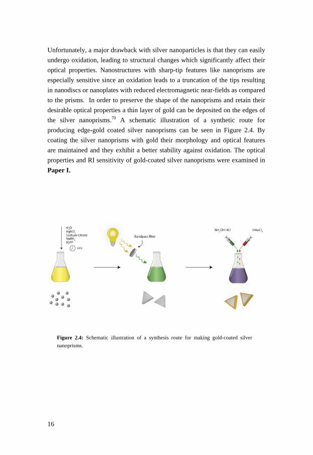
Unfortunately, a major drawback with silver nanoparticles is that they can easily
undergo oxidation, leading to structural changes which significantly affect their
optical properties. Nanostructures with sharp-tip features like nanoprisms are
especially sensitive since an oxidation leads to a truncation of the tips resulting
in nanodiscs or nanoplates with reduced electromagnetic near-fields as compared
to the prisms. In order to preserve the shape of the nanoprisms and retain their
desirable optical properties a thin layer of gold can be deposited on the edges of
the silver nanoprisms.70 A schematic illustration of a synthetic route for
producing edge-gold coated silver nanoprisms can be seen in Figure 2.4. By
coating the silver nanoprisms with gold their morphology and optical features
are maintained and they exhibit a better stability against oxidation. The optical
properties and RI sensitivity of gold-coated silver nanoprisms were examined in
Paper I.
Figure 2.4: Schematic illustration of a synthesis route for making gold-coated silver
nanoprisms.

17
Optical properties of metal
nanoparticles
Optical properties of metals
The optical properties of bulk materials can be characterized by the dielectric
function which describes how materials interact with electromagnetic
radiation. For metals, which have partially filled electron bands, the dielectric
function can be described with a simple, free-electron model.
1Γ
(3.1)
This is known as the Drude model which assumes that metals consists of a free
electron gas also known as plasma that moves within a lattice of fixed, positively
charged ion cores.
3

18
If the electron damping factor, Γ is neglected, the dielectric function can be
further simplified to
1 (3.2)
where is the plasma oscillation frequency of the free electron gas relative to
the positively charged ion cores. The plasma oscillation can be calculated using
the equation
(3.3)
where is the density of electrons, is the electric charge, the effective
electron mass, and is the permittivity of vacuum.
A quantum of plasma oscillation is known as a plasmon, or plasmon polariton
when induced by photon excitation. Plasmons can be created in metal bulk
material, at metal surfaces or in metal nanostructures which all possess different
optical properties.
Surface plasmons
Plasmons are longitudinal collective electron density fluctuations that when
formed at a metal surface, create a surface-bound electromagnetic wave, known
as a surface plasmon (SP).71,72 This SP wave propagates at the interface between
the metal and a dielectric, which is schematically illustrated in Figure 3.1 A. The
propagating SP wave has an accompanying electric field that decays
exponentially both in the direction of propagation, due to energy losses to the
metal, and perpendicular to the interface into both the metal and the dielectric
medium. The decay length depends on the dielectric properties of the metal and
the surrounding media.

19
The energy for an SP wave is always smaller than that of bulk plasmons and the
plasmon frequency is /√2.
A propagating SP wave carries momentum and by solving Maxwell’s equations
with specific boundary conditions, an expression for the dispersion relation of an
SP can be derived
(3.4)
where kSP is the wave vector for the SP and and is the dielectric function
of the metal and the dielectric surrounding, respectively.
For an SP to be excited both the frequency (energy) and the wave vector
(momentum) need to conserved, which cannot be achieved by simply
illuminating a metal surface with light passing through air. Figure 3.1 B shows
the dispersion relation for an SP compared to photons in vacuum and since the
curves do not coincide, the excitation requirements cannot be fulfilled. However,
in 1968 Kretschmann and Raether showed that SPs can be excited in flat metal
films using ordinary light by guiding the incident light through a coupling
medium e.g. a quartz prism.73 If the metal film is thin enough the photons can
penetrate through the metal film and excite an SP wave on the opposite side of
the film at the metal-dielectric interface, where the excitation of SPs is
dependent on the thickness of the metal film.72 SPs can however be generated at
a thick metal film by introducing a spacer layer between the metal and the prism
where the RI of the spacer material needs to be lower than that of the prism. The
electromagnetic field from the incident light can couple with the metal through
the spacer and excite an SP at the metal interface. This is known as the Otto
configuration.74 Grating coupling is another technique developed in order to
excite SPs using electromagnetic waves, where a roughened metal surface is
used instead of a smooth surface which allows for direct excitation without
coupling through a prism.75

20
Figure 3.1: (A) Schematic illustration of a surface plasmon propagating along the x-axis
on the interface between a metal and dielectric with electric field lines in the opposite
direction. (B) Dispersion relation for a surface plasmon and for photons in vacuum.
If the RI of the dielectric medium ( at the metal interface is altered, the
resonance condition for exciting the SP changes, as seen from Equation 3.4. This
is the basic principle behind SPR-based biosensing which is further explored in
chapter 5.
Localized surface plasmons
Discrete metal nanostructures cannot support propagating SPs since there is
simply no room for the electrons to propagate. Instead, free electrons in metal
nanostructures can undergo excitation and create a collective oscillation
restricted by the geometrical boundaries of the nanostructure. These oscillations
are known as localized surface plasmons and can be excited by an external
electromagnetic field. Since localized SPs do not carry any momentum, only the
energy needs to be matched in order to excite the electrons which mean that free
photons can be used for excitation and no momentum matching is needed.
Consequently, exploration of localized SPs can be done by rather simple optical
equipment.
The interaction between light and small metal nanoparticles was first described
by Gustav Mie over a century ago.76 He analytically solved Maxwell’s equations
and derived an expression for metal nanoparticles in a homogenous medium
interacting with an electromagnetic field. For spherical metal nanoparticles much

21
smaller than the wavelength of light (2r<<λ) the optical extinction cross section
( can be expressed with the following relationship:5,25
24 /
ln 10 2(3.5)
where is the radius of the nanoparticle, is the dielectric constant of the
surrounding medium, is the electron density, and and are the real and
imaginary part of the complex dielectric function of the bulk metal, respectively.
In this quasi-static approximation, the small nanoparticles are assumed to
experience a uniform static electric field and they can thereby be considered as
electrical dipoles. The polarizability ( for such a dipole can be expressed as:
42
(3.6)
As seen from Equation 3.6, maximum polarizability occurs when the term in the
denominator approaches zero, hence when 2 . The dielectric
permittivity of the surrounding medium can normally be considered as a
constant and real parameter, which means that the real part in the dielectric
function of the metal nanoparticle is required to possess a negative value in
order for strong polarization to occur, if the imaginary part of the dielectric
function is small (low losses). From Equation 3.5, it is clear that maximum
extinction arises when this condition is fulfilled, and plasmon resonance occurs.
Introducing the Drude dielectric function gives the resonance frequency for a
dipolar localized surface plasmon.77
1 2
(3.7)
For silver and gold nanoparticles, this frequency falls in the visible region of the
spectrum. Consequently, these nanoparticles exhibit bright colours both in
transmitted and reflected light (see chapter 1).

22
When plasmon resonance occurs, the conduction electrons are displaced relative
to the nuclei, moving them away from their equilibrium position. The coulombic
attraction between the relocated electrons and the atomic cores acts as a restoring
force on the free electrons, which gives rise to an oscillating electron motion at a
specific frequency. This oscillation typically decays within a few
femtoseconds,78 but when an alternating electric field is introduced a continuous
coherent plasmon oscillation can be maintained (Figure 3.2). The plasmon
excitation gives rise to a strongly enhanced electric field in close vicinity of the
nanoparticles, which is utilized in several field-enhanced plasmonic
applications.71,77
Figure 3.2: Schematic illustration of localized surface plasmon resonance induced by an
external electrical field.
When the dielectric medium changes close to the nanoparticles e.g. by
biomolecular adsorption to the metal surface, the condition for resonance is
altered. An increase in the dielectric function of the environment, gives rise to an
increased screening of the surface charges, which weakens the restoring force on
the electrons. As a result, less energy is required to excite the electrons which
red-shifts (longer wavelength) the plasmon resonance frequency. This is also
obvious from Equation 3.7, where an increase in results in a lower plasmon
frequency hence, a longer resonance wavelength. This makes metal
nanoparticles sensitive probes for detecting local RI changes, where the
sensitivity depends on the polarizability and the strength of the restoring force,
which is a function of the material and the geometry of the nanoparticles.77
Refractometric sensing based on metal nanoparticles is covered in chapter 5.

23
Optical response of anisotropic nanoparticles – beyond the Mie theory
For small spherical nanoparticles, the Mie theory can be considered as a simple
but efficient model to describe the optical response. However, when the particle
size increases the quasi-static approximation is no longer valid since the
electrons do not experience a homogenous electric field. This causes a dephasing
of the conduction electrons and a retardation of the dipolar field.79 The
retardation effect lowers the excitation energy which causes a red-shift of the
plasmon resonance wavelength for larger nanoparticles. Additionally, radiative
losses increase when the particle size gets larger which broadens the plasmon
band width and reduces the intensity.
Another way to tune the plasmon frequency is to change the geometry of the
nanoparticles. A modification of the nanoparticle shape has a significant impact
on the spectral position of the plasmon resonance. Spherical nanoparticles with
high symmetry only possess one dipolar resonance, but when the shape is
modified and the particles become more asymmetric, multiple dipolar modes can
arise which makes the optical response more complex.80 For instance, elongated
metal nanoparticles like nanorods display both a transverse and a longitudinal
localized surface plasmon mode which gives rise to two distinct plasmon
resonance peaks at different spectral positions. Several other geometrical
features in addition to elongation, affects the actual plasmon frequency such as
curvature, asymmetry and intracoupling.71 Furthermore, higher-order plasmon
modes can be created due to an inhomogeneous distribution of the surface
charges in the nanoparticles. Such multimodes (e.g. quadropoles, octopoles) are
normally created in larger nanoparticles and are always located at shorter
wavelengths (higher energy) compared to the dipolar excitation.81-83
Consequently, by varying the size and/or shape of the nanoparticles, plasmon
peaks over the entire visible and near-infrared region can be generated.
The optical response from anisotropic nanoparticles of different shape, size, and
material can be calculated with the help of various numerical methods. Two of
the most commonly used methods are discrete dipole approximation (DDA) and
finite-difference time-domain (FDTD).84 DDA calculations are based on a three-
dimensional array of small, polarizable elements considered as point dipoles.
The dipole moment induced by an external electric field and through interaction

24
with other dipoles, is calculated for each element. The contribution from each
dipole is then used to calculate the overall optical response, i.e. the scattering
and absorption of the incident light caused by the metal nanoparticles.85,86 FDTD
simulations are based on solving Maxwell’s differential equations in a three-
dimensional cubic lattice over time. The optical properties of the material are
defined in each cell and the electric and magnetic fields are calculated at a given
instant of time. FDTD enables simulation of the electromagnetic response from
heterogeneous materials of arbitrary geometries.87-89 Computational simulation
techniques are important in order to increase the knowledge of the physics
behind plasmonics and to verify and explain experimentally obtained results.
Plasmonic coupling
So far, only single nanoparticles have been considered. However,
electromagnetic interactions between adjacent nanoparticles give rise to an
additional spectral shift of the plasmon resonance position due to Coulomb
interactions. This is known as plasmonic coupling and results in new hybridized
plasmon modes.90 The interaction can either form a “bonding” plasmon mode
(lower energy) or an “anti-bonding” plasmon mode (higher energy) depending
on the polarization of the surface charges. The simplest model is to consider two
closely spaced spherical metal nanoparticles separated with a distance (d). As
seen in Figure 3.3 A, the spectral position of the plasmonic coupling mode is
highly dependent on the interparticle separation and red-shifts significantly when
the distance between the particles is reduced. Utilization of plasmonic coupling
is hence another powerful way to tune the plasmon resonance frequency, which
is employed in Paper II.31,91
Plasmonic coupling between closely spaced metal nanostructures generates large
near-field enhancement in the gap between the particles, usually called “hot
spots”. The induced local electrical field can be enhanced by a factor of x100
compared to the surrounding electric field.92 Figure 3.3 shows the formation of a
hot spot in a gold nanoparticle dimer separated by 3 nm (B) and 10 nm (C).
Clearly, a significantly higher field strength occurs when the particles are closely
spaced, separated by only 3 nm. These hot spots are especially interesting for
SERS, where the large field strengths can be used to increase the Raman

25
scattering signals from specific molecules placed inside the enhanced near-
fields.93,94
Figure 3.3: The plasmonic coupling between individual nanoparticles is
significantly dependent on the interparticle separation (d). (A) Scattering spectra
acquired with FDTD calculations of two gold nanoparticles (10 nm in diameter)
at different particle separations. Corresponding field plots for (B) d = 3 nm and
(C) d = 10 nm. Courtesy of Dr. Borja Sepulveda (CIN2, Barcelona)
The large resonance shift created when metal nanoparticles are brought in close
proximity is used in a biosensing method called colorimetric sensing. This
method was pioneered by Mirkin et al. who used DNA to facilitate an
aggregation of gold nanoparticles.95 Several other sensing strategies, based on
metal nanoparticle aggregation have later been developed, detecting a large
variety of molecular species.96,97

26

27
Functionalization and
immobilization of metal
nanoparticles
Functionalization of metal nanoparticles
In order to convert metal nanoparticles to useful, hybrid nanomaterials, they
typically need to be modified with some organic material that provides them
with a desired functionality.98 A chemical modification of the metal surface is
normally required in order to introduce specificity and to increase the biological
compatibility of the metal nanoparticles in sensing applications.
Functionalization of metal nanoparticles generally requires a modification or
replacement of the stabilizing agent that is used in the synthesis to prevent the
particles from aggregating. Thus, the new surface chemistry must maintain
particle stability and at the same time provide the particles with new chemical
properties and functionalities. Biomolecules can be conjugated to nanoparticles
through physisorption (noncovalent coupling) or by chemisorption (covalent
coupling).98 This can be done directly on the particle surface or with the
4

28
assistance of a bifunctional crosslinker. Regardless of the choice of conjugation
method, it is a great challenge to control the amount of material bound to the
surface as well as the orientation of the biomolecules in order to achieve an
accessible surface chemistry with high specificity for binding of analytes.
Extensive work has been conducted on metal nanoparticle functionalization
which has resulted in a huge amount of protocols for introducing different
chemical moieties using both covalent and non-covalent approaches. The most
frequently employed method to functionalize gold nanoparticles is indisputably
the usage of thiol chemistry. Thiol groups (-SH) form covalent bonds with gold
atoms, forming a self-assembled monolayer (SAM) on gold surfaces.99 As thiols
are present in, or can be introduced into several different classes of molecules
(e.g. DNA, polypeptides etc.), gold nanoparticles can easily be functionalized
with a large variety of biomolecules. Physisorption of, for instance, polymers or
proteins is another commonly applied method to functionalize nanoparticles.98 In
addition, they can also be coated with inorganic materials like silica in order to
make them more chemically inert (silver nanoparticles) and suspendable in
various solvents.100,101
Immobilization of metal nanoparticles
Dispersed nanoparticles need to be stabilized in order to prevent them from
aggregating. However, if the particles are tethered to a solid support, it is no
longer necessary to uphold a chemical or steric repulsion between them since
strongly attached nanoparticles are unable to move laterally across the surface
and thereby incapable of aggregating. For this reason, substrates with
nanoparticles organized into ordered structures like arrays or other controlled
assemblies, has found their way into many electronic, optical, and sensor
applications.102
The optical properties of metal nanoparticles are normally studied using optical
spectroscopy. However, an exploration of immobilized nanoparticles generally
requires the use of high magnification imaging techniques. Electron microscopy
techniques like scanning electron microscopy (SEM) or transmission electron

29
microscopy (TEM) are especially useful for studying metal nanoparticles since
their high electron densities gives a very high contrast in the images. For this
reason, metal nanoparticles can be used as contrast agents in various microscopy
techniques.103 Atomic force microscopy (AFM) is also routinely used to study
particle assembly and morphology on various substrates.
Controlling the surface coverage of metal nanoparticles
Immobilization of nanoparticles on a solid support usually involves surface
modification followed by particle adsorption using either pre-functionalized or
non-treated particles, where the particle adhesion is driven by electrostatic forces
and van der Waals dispersion forces. Controlling the surface density of particles
attached on the substrate is very important in order to achieve an appropriate
surface morphology with desired functionalities.
If the particles are exposed to a surface that enables adsorption, the binding
kinetics primarily depends on the properties of the particle solution.104 In the
initial adsorption process, we can assume that every nanoparticle that touches the
surface will adhere and stick to it, irreversibly. Then the number density
( can be calculated using the following expression:104
2 / (4.1)
where is the bulk concentration of nanoparticles, is the incubation time and
is the diffusion constant given by the well-known Stokes-Einstein
equation:
6
(4.2)
where is Boltsmann’s constant, is the absolute temperature, is the
viscosity of the medium, and is the radius of a spherical particle. As seen from

30
Equation 4.1, the surface density of particles on the surface depends on the
particle concentration and the incubation time ( ) and can hence be
considered as a first-order rate process.105 The particle concentration and the
incubation time can rather easily be varied in order to control the surface
coverage of nanoparticles. A high surface coverage with short interparticle
distances, gives rise to plasmonic coupling between the closely packed metal
nanoparticles. This is normally an unwanted effect since the distinct plasmon
resonance band originating from individual nanoparticles can be significantly
broadened.
Most metal nanoparticles suspended in aqueous media are stabilized by charged
species at their surface, such as citrate, and this charge repulsion will also affect
the surface coverage. If the repulsion is decreased (shorter Debye length) the
nanoparticles can form a closer packing and hence, a higher surface coverage
can be obtained. The electrostatic repulsion between particles can be controlled
by varying the ionic strength in the solution.105 A density gradient of
immobilized gold nanoparticles was created by Lundgren et al. by changing the
ionic strength in the nanoparticle solution by ion diffusion.106
Figure 4.1 shows SEM images of gold nanoparticles immobilized on substrates
with different surface coverages. Different surface coverages were obtained
because of variations in the particle concentration in the different particle
suspensions.
Figure 4.1: SEM images of substrates covered with gold nanoparticles of
various sizes with different surface coverage. (A) 12% (25 nm), (B) 7 % (50 nm)
and (C) 3% (100 nm).

31
Nanoparticle assembly on silanized surfaces
Silanization is a straightforward method to introduce functional chemical groups
onto silica-based substrates like glass. Thus, silanization is a frequently used
surface modification in order to immobilize nanoparticles or biomolecules onto
glass substrates.105 A self-assembled monolayer of silanes is created by the
interaction with hydroxyl group on the surface, forming a covalent Si-O-Si
bond.107 Prior to the silanization process, the surfaces must be thoroughly
cleaned in order to remove organic material and increase the amount of hydroxyl
groups present on the surface. A well-defined silanized monolayer is crucial for
obtaining a successful colloidal immobilization.
Two frequently used organosilanes utilized for nanoparticle attachment is (3-
aminopropyl) triethoxysilane (APTES) and (3-mercaptopropyl) triethoxysilane
(MPTES) (Figure 4.2). In Paper II and III, silanization with APTES was used
for surface immobilization of metal nanoparticles on glass substrates.
Figure 4.2: Surfaces functionalized with two different silanes, (A) APTES and
(B) MPTES.
Nanoparticle assembly on polyelectrolytes
Polyelectrolyte multilayer formation is a versatile layer-by-layer (LBL)
technique which enables a nanoscale arrangement of organic molecules where
materials are sequentially adsorbed on top of each other.108 LBL methods can be
used to sequentially build-up thin films with nanometer-scale precision which
has several applications in a large variety of fields, including nanoparticle
functionalization and immobilization. Polyelectrolyte multilayers are formed by
exploiting the electrostatic attraction between oppositely charged polymer chains.

32
The adsorption of molecules with different charge has two important
consequences: i) repulsion of equally charged molecules limits the adsorption to
the formation of a single monolayer in each sequence and ii) the ability of an
oppositely charged molecule to be adsorbed on top of the first one.108 This
allows for the formation of a multilayer molecular structure if these steps are
repeated in a cyclic manner.
The formation of a multilayered structure comprised of oppositely charged
polyelectrolytes give rise to a charged surface, where the top polyelectrolyte
layer determines the surface charge. This enables immobilization of charged
nanoparticles using electrostatic interactions between the surface charges on the
nanoparticles and the top layer on the polyelectrolyte coated surface. Since the
surface charge can be altered by simply changing the top polyelectrolyte layer,
both negatively and positively charged nanoparticles can be immobilized using
this method.
Two specific polyelectrolytes have been extensively used in the work included
in this thesis, polyallylamine hydrochloride (PAH) which is a cationic
polyelectrolyte and polystyrene sulfonate (PSS) which is anionic. These are two
of the most frequently used polyelectrolytes for LBL formation and their
properties have been comprehensively examined by others.109-112 One bilayer of
PAH/PSS results in a thickness in air of about 3 nm (Figure 4.3), which was
determined using null ellipsometry. However, the thickness of the polymer film
significantly depends on the ionic strength in the deposition solution, since an
increase of the electrolyte concentration results in a more effective screening of
the polyelectrolyte charges, which changes the conformation of the
polymers.111,112 Also, polyelectrolytes multilayers are known to swell and
increase in thickness when exposed to water. A swelling of ~30% for a polymer
film composed of PAH/PSS has been reported by others.113

33
Figure 4.3: A polyelectrolyte bilayer composed of polyallylamine
hydrochloride (PAH) and polystyrene sulfonate (PSS). The thickness (3 nm)
was obtained using null ellipsometry in air.
As each layer deposition enables a thickness control on the nanometer-scale, a
layer-by-layer assembly of polyelectrolytes can also be used for probing the
surface sensitivity and sensing depth of a refractometric sensing system, which
was exploited in Paper I. Also, polyelectrolyte multilayers can be used to
control and fine-tune interparticle distances (Paper II).
Nanoparticle multilayers
In order to create a multilayer structure of nanoparticles, the initial monolayer
must be modified in such a way that adsorption of a second particle layer is
possible. This can be achieved by using a variety of different surface chemistry
modifications with molecular components such as DNA,114 peptides,115 or
polymers.116,117 Widely separated layers of nanoparticles can significantly
increase the extinction of light (Figure 4.4), which can be useful for applications
where surfaces with high extinction (absorption and scattering) are desired.
Hence, 3D-assembly is yet another way to tune the optical response from
plasmonic nanoparticles.
If the distance between the layers is reduced, plasmonic coupling can occur
which gives rise to new plasmon bands. If the distance between the layers is not

34
precisely defined, many plasmon resonance modes will be present which gives a
broad and undefined plasmon band. The spacing between each monolayer of
particles depends solely on the material used to achieve the multilayer formation.
Plasmonic coupling with distinct plasmon bands was achieved in Paper II, using
a protocol based on polyelectrolytes as spacer material.
Figure 4.4: Light extinction increases when gold nanoparticle multilayers are
formed. A rather thick polymer film made by spincoating was used to separate
the nanoparticle layers.

35
Plasmonic biosensing
Biosensors – general introduction
In general, a sensor is defined as a device that detects physical or chemical
changes (e.g. temperature, pressure, light, concentration etc.) in our environment
and converts them into measurable signals. An example of a historical sensor is
the usage of canary birds in coal mines. They were used to detect the presence of
carbon monoxide (CO), a toxic gas both for birds and humans. Since the birds
are more sensitive than humans to lethal doses of CO, the mine workers kept an
eye on the bird while they were working and if the bird passed out, they knew it
was time to get out of the mine. Today, this rather cruel method has been
replaced with modern sensors which have significantly higher sensitivity and
faster response time compared to the canary bird.
A biosensor is a specific type of sensor that utilizes a biological reagent in the
sensing system. A frequently used definition of a biosensor is that it is a “self-
contained integrated device, which is capable of providing specific quantitative
or semi-quantitative analytical information using a biological recognition
element (biochemical receptor) which is retained in direct spatial contact with a
5

36
transduction element”.118 However, in this definition, bioanalytical systems are
not included and they are also considered to be biosensors by some. A broader
definition would therefore be that it is “a tool used for analyzing biomolecular
interactions”.119
The most successful and exploited biosensor is the glucose sensor. It is used
daily by millions of diabetics all over the world to monitor their blood glucose
level. The first glucose sensor was developed in 1962 by Clark and Lyon, using
an enzyme-coated electrode.120 Modern glucose sensors are significantly smaller
compared to the first prototype and are based on the enzyme glucose oxidase
which generates a measurable current upon glucose oxidation.121 Biosensors are
today important devices used in several different areas including industrial
process monitoring, food quality control, drug development, environmental
analysis and medical healthcare. Another classical example of a frequently used,
commercially available biosensor is the home pregnancy test.
Biosensors are composed of two central components; a detector, consisting of
biorecognition elements used for analyte identification, and a transducer which
converts biological interactions into a measurable signal. Figure 5.1 is a
schematic illustration of the components used in a biosensor.
The primary function of the biorecognition layer is to provide selectivity for the
targets or analytes of interest. The biological elements incorporated in the
biorecognition layer interact with specific analytes in the surrounding medium.
Various classes of molecules have been exploited as biorecognition elements
including enzymes, antibodies, nucleic acids, receptors etc. In addition to
specificity, these elements need to possess several other important properties in
order to assure a high performance of the biosensing device including stability,
functionality, availability and, in some applications, also reversibility.

37
Figure 5.1: A schematic illustration of the components used in a biosensor. A
biorecognition layer detects specific analytes from a complex solution and a
transducer transforms the binding event to a measurable signal.
The transducer responds to the biochemical interaction and transforms it into a
measurable signal. The choice of transduction mechanism party depends on the
signal that is generated by the interaction between the biorecognition element
and the analyte. A large variety of transduction mechanisms have been exploited
in biosensors, including electrochemical, mechanical, thermal, and optical. If
quantification is required, the signal output from the transducer should be
correlated to the concentration of the analyte of interest where the concentration
span depends on the dynamic range of the sensing system. Many biosensing
systems can also provide information about the binding affinity and kinetics for
the interaction.
The work included in this thesis focuses exclusively on optical transduction
where visible light has been used for signal detection. Optical transducers offer a
broad range of signal parameters that can be utilized for detection including the
frequency, phase, polarization, and intensity of the light. Optical transduction
techniques can be exploited for many applications and can generally be
constructed by rather simple and inexpensive components.122,123 Optical fibers
enable miniaturization and optical imaging can be used for multiplex analysis of
arrays.

38
Biosensors can be divided into different classes depending on: i) type of
biochemical event, ii) the transduction mechanism or iii) the application area.
However, they can also generally be divided into two groups, those that require
labelling and those that do not. Label-free techniques such as SPR and Quartz
Crystal Microbalance (QCM) enable a real-time, quantitative analysis of
molecular binding events while label-based sensing requires tagging of the
biomolecules in order to detect the interactions. Methods using labelling are
often very sensitive, but a major drawback with these techniques is that a
molecular labelling with, for instance, a fluorescent, enzymatic, or radioactive
probe can affect the functionality of the molecule and induce molecular
aggregation. For this reason there is an increased interest in label-free techniques
for high-throughput screening of biomolecular interactions.
Most biosensors are based on the interaction of biomolecules bound to a solid
support, since this facilitates washing, regeneration of the reagents, handling,
and storage. Thus, an appropriate surface chemistry is of critical importance
when producing biosensing devices with high performance. The surface
chemistry should enable an immobilization of the biorecognition elements
without affecting the functionality and at the same time be resistant to non-
specific adsorption. Much work has been invested into surface chemistry
development, both with 2D-structures based on for example thiols124,125 or
silanes126 and 3D-structures like hydrogels composed of various polymers (e.g.
sugars and ethylene glycol).127,128 Hydrogels are particularly attractive since they
can provide a suitable environment for the biomolecules, maintaining their
structure and functionality.129
SPR-based biosensing
SPR-based sensing is one of the most exploited biosensing techniques and has
been extensively used to monitor and characterize biomolecular interactions in
real-time without the need for labelling. The first demonstration of SPR-based
biosensing was performed by Liedberg et al. in 1983, where they studied the
interaction between an antigen and an antibody on a silver surface.130 After their
pioneering work, the technique was commercialized and is today routinely used

39
by biochemists, pharmacologists and molecular biologists in order to determine
biomolecular binding affinities and interaction kinetics.131
SPR-based sensors are based on refractometric detection using the excitation of
a surface plasmon (see chapter 3). A typical setup for performing SPR
biosensing experiments based on the Kretschmann configuration can be seen in
Figure 5.2. In general, one interaction partner (ligand) is immobilized on a thin
metal film, usually gold, followed by the introduction of the second interaction
partner in solution (analyte). The analyte binding results in a change in the RI
close to the metal surface, which changes the resonance condition for SP
excitation. This can be monitored in real-time by measuring changes in the
reflected light used for excitation. SPR-based sensors can be based on different
interrogation modes including detection of angular, wavelength, intensity, or
phase alterations.131
Figure 5.2: Setup for biosensing using SPR based on the Kretschmann
configuration. Polarized light is guided through a glass prism and is totally
internally reflected on the backside of a thin metal film creating an evanescent
field through the metal film. At a specific angle or wavelength of the incident
light the evanescent field can excite surface plasmons at the interface between
the metal and the sample medium, resulting in a sharp decrease in the reflected
light (upper right). If the refractive index changes at the metal interface the
condition for SPR excitation is altered, thus changing the SPR angle or
wavelength (from I to II in the figure). This allows for monitoring biomolecular
events at the metal surface in real-time (lower right).
40
SPR is generally considered to be a surface sensitive technique since the
excitation of SPs creates an evanescent field that reaches out into the sample
medium. The penetration depth is defined as the distance when the intensity of
the electric field has decayed to about 1/е of the value at the immediate surface.
Any change of the RI within this region will alter the condition for SP excitation.
The penetration depth depends on the excitation wavelength and the optical
properties of the metal and the ambient medium and falls in the range of 150-300
nm in most SPR setups. This prompts a surface chemistry composed of a
polymer 3D matrix which allows for an increased ligand surface concentration
and utilization of the entire sensing volume.127
The overall performance of an SPR-based biosensor depends on several
parameters including sensitivity, resolution, dynamic range, and limit of
detection. The sensitivity is defined as the change in sensor response ( ) (angle,
wavelength or intensity) caused by a change in the refractive index ( ), which
can be expressed as:
(5.1)
The term is the RI sensitivity and this value depends on the choice of
wavelength and the incident angle but is independent on the interrogation mode.
The RI sensitivity can be divided into two different sensitivity expressions –
bulk and surface RI sensitivity. The bulk RI sensitivity refers to any RI
changes in the entire sensing region. However, as SPR is routinely used to study
molecular interactions at surfaces where only a fraction of the evanescent field is
employed, it is also appropriate to introduce a term for the surface sensitivity
( . This refers to the sensitivity for detecting a RI change ( caused by the
adsorption of a thin film of thickness . The surface sensitivity term is especially
relevant when comparing SPR with LSPR-based sensors, which is explored in
Paper I.

41
LSPR-based refractometric biosensing
Similar to conventional SPR-based sensing, nanostructures supporting a LSPR
excitation can be used for refractometric sensing. As described in chapter 3, the
condition for exiting electrons in metal nanostructures and creating localized
surface plasmon resonance, depends on the dielectric properties of the
surrounding medium. This allows for a spectroscopic detection of molecular
interactions that occurs in the local proximity of the metal nanostructures (Figure
5.3). When the energy of the incident light matches the excitation energy of the
electrons in the metal, a distinct peak known as the plasmon peak, can be seen in
the optical extinction spectrum. By introducing molecules that induce a change
of the RI in the surrounding environment, the resonance condition will be altered
which results in a shift of the plasmon peak. The amount of material introduced
around the metal nanostructures can be estimated by measuring the quantity of
the plasmon peak shift.
Figure 5.3: Schematic illustration showing the basic principle behind
nanoplasmonic sensing based on refractometric detection. A shift of the
plasmon peak is observed in the extinction spectra when an analyte interacts
with a ligand on the surface of a metal nanoparticle.

42
The first biosensing experiments using propagating surface plasmons were
conducted in 1983 by Liedberg et al.130, but it took an additional 15 years before
Englebienne demonstrated experimentally that also localized surface plasmons
could be utilized for biosensing by detecting protein interactions using
immobilized gold nanoparticles.132 Numerous of different LSPR-based sensing
systems have been demonstrated after this, for detection of a large variety of
biomolecular interactions such as biotin-streptavidin,133,134 DNA-DNA,135,136
antibody-antigen,27,28,132 etc.
The detection principle in LSPR-based refractometric sensing can be based on
monitoring different spectral changes including intensity variations, either in the
plasmon peak maxima or at a single wavelength,134,137,138 peak curvature
changes,139 or by detecting the position of the plasmon resonance peak.27,28 The
work included in this thesis focuses exclusively on the detection of plasmon
peak wavelength shifts since this detection principle is favorable for randomly
adsorbed nanoparticles onto substrates, where the surface coverage can vary
somewhat across the surface. The surface coverage, i.e. the amount of particles
used in the analysis will influence the intensity of the signal, but the plasmon
peak position is not affected by the surface density of particles as long as there is
no interparticle coupling occurring. However, this strategy demands an accurate
determination of the spectral position of the plasmon peak maxima which
requires a relatively high signal-to-noise ratio.
LSPR-based sensing show a great potential for miniaturized sensing and much
work has been conducted on single nanoparticle detection, especially by the
group of Van Duyne.140,141 Single nanoparticle spectroscopy has some
advantages since it offers the ability to explore the optical properties of a
specific nanostructure and how this is affected by the particle geometry. Also
single nanoparticle detection enables multiplexed sensing schemes and a
significant reduction of the absolute detection limit, which is required for
achieving the ultimate sensitivity goal i.e. single molecule resolution. However,
even if plasmonic nanostructures have large extinction cross sections the signal
generated from a single nanoparticle is generally very weak which results in a
low signal-to-noise ratio. Therefore, spectroscopic measurements on an
ensemble of nanoparticles generally results in a better overall sensing
performance.

43
Refractometric sensing using localized surface plasmons is mainly focused on
plasmonic nanostructures composed of either gold or silver. However, other
materials such as aluminum, copper, platinum, and palladium, also display
plasmonic properties which can be exploited for sensing applications.142-144
Recently, also non-metallic materials like semiconductor nanocrystals have been
shown to exhibit plasmonic properties by introducing charge carriers through
doping.145
Refractive index sensitivity
Over a narrow range of RI changes, the bulk RI sensitivity ( can be
approximated as a linear function connecting the RI change (Δ ) and the LSPR
peak shift Δ :
Δ Δ (5.2)
Thus, in order to maximize the sensing response i.e. the peak shift that occurs for
a specific change in the surrounding RI, the bulk RI sensitivity should be as
large as possible. Miller and Lazarides have shown using theoretical calculations
that the bulk RI sensitivity for gold nanoparticles with a plasmon resonance
below 800 nm, solely depends on the plasmon peak position.146 Consequently,
this indicates that even though the plasmon resonance depends on the shape and
the size of the nanoparticles, the bulk RI sensitivity is not directly dependent on
the particle geometry. Hence, two nanoparticles that give rise to an equivalent
plasmon frequency but has totally different structures will exhibit a similar bulk
RI sensitivity. Several studies have been reported with the aim of finding
nanoparticles with high bulk RI sensitivities,30,147 but this has only been a quest
for synthesizing nanoparticles displaying plasmon resonance bands at longer
wavelengths. However, a longer resonance wavelength will not per se, give rise
to a better sensing performance, which will be discussed in the next section. The
correlation between the plasmon peak position and the bulk RI sensitivity has
been verified experimentally by several groups including ours.147-149 In Paper IV,
the RI sensitivity was investigated for a large variety of gold nanoparticles with

44
different plasmon frequencies, obtaining a clearly linear relationship between the
plasmon peak resonance position and the bulk RI sensitivity (Figure 5.4).
However, it is important to notice that this correlation only is valid for dispersed
nanoparticles in solution. Immobilization to a solid support or a surface
functionalization will significantly affect the bulk RI sensitivity which will be
addressed later in this chapter.
Figure 5.4: Experimentally obtained bulk RI sensitivity values for different
types of gold nanoparticles plotted against the spectral position of their plasmon
resonance peak. The straight line demonstrates the linear relationship between
the two parameters.
Also, the correlation between the plasmon frequency and the bulk RI sensitivity
is not applicable when comparing nanoparticles composed of different materials.
For instance, silver nanoparticles exhibit plasmon bands located at shorter
wavelengths compared to gold nanoparticles, but despite this fact, they exhibit
higher bulk RI sensitivities.150 This can be explained by the different dielectric
properties of the two materials. Despite favorable RI sensitivities, silver
nanoparticles suffer from chemical instability both in air and aqueous
environments which limits their practical use.
Table 5.1 summaries some of the experimentally obtained properties of all metal
nanoparticle structures used in the work included in this thesis. The highest bulk
RI sensitivity is seen for gold-coated silver nanoprisms which were used in
Paper I.

45
Table 5.1: Summary of the physical properties of the metal nanoparticles used
in the work included in this thesis.
Geometry
Size (nm)
ʎLSPR
(nm) ƞB
(nm/RIU) FOM
Gold nanosphere 25 525 60 1.2
Gold nanosphere 50 532 80 1.5
Gold nanosphere 100 578 180 1.6
Gold-coated silver
nanoprism 30 - 40 660 425 3.0
Silver nanosphere 35 398 144 3.0
Silver nanocube 40 444 158 3.3
Silver nanoplate 30 - 40 552 315 3.6
Gold nanocube 40 535 91 2.0
Concave gold
nanocube 50 615 266 2.6
Gold nanorod 65 x 20 740 385 3.6
Gold nanosprism 50 - 60 775 405 3.8

46
Figure of Merit (FOM)
As plasmonic materials give rise to peak features in their extinction spectrum, it
is convenient to analyze the sensing data based on the spectral changes that
occurs for these peaks. However, since spectroscopic measurements only
provide the relative extinction intensity of light at different wavelengths, the
discrete data points acquired needs to be converted into a parameter that
accurately can represent the sensing result.77. If the intensity is measured and
compared at a single wavelength the post signal processing required is rather
small, but normally it is necessary to include a larger spectral region in the data
analysis. As discussed in the previous section, the RI sensitivity is defined by
detecting either the wavelength or the intensity shift of the plasmon peak
maxima that occurs upon a change in the RI close to the nanostructures.
However, a determination of the plasmon peak maxima requires a mathematical
calculation that depends on the width of the plasmon peak and the intensity of
the signal, or more accurately the noise level in the signal. It is therefore
necessary to introduce a term that includes resolution in order to make a fair
comparison between different plasmonic nanostructures since the peak width can
vary significantly. This has motivated the introduction of a new parameter
known as figure of merit (FOM) that includes both sensitivity and resolution.
FOM is defined as the ratio between the bulk RI sensitivity and the peak width:
FOM (5.3)
where fwhm is an acronym for full-width-half-maximum and is defined as the
width (in nm) of the plasmon peak at half of its maximum value. Calculated
FOM values for all nanoparticles used within this work can be seen in Table 5.1.
As discussed in the previous section, the bulk RI sensitivity generally depends
on the spectral position of the plasmon resonance peak. However, shifting the
LSPR peak position to longer wavelength into the infrared (IR) region can result
in a significant broadening of the plasmon peak due to dephasing and radiative
damping effects. Consequently, an increase in the FOM value is not expected for

47
longer wavelengths since the higher bulk RI sensitivity will be compensated by a
broader plasmon peak i.e. a larger fwhm value. Thus, tuning the LSPR position
into the IR region will increase the sensitivity but reduce the resolution and will
therefore not give any additional improvements of the sensing performance. This
is also attractive for practical reasons since relocation into the IR region requires
the usage of more advanced optical equipment.
Motivated by the issue discussed above, Otte and Sepulveda calculated the
optimum spectral region where the FOM value is maximized.77,151 They used the
dipolar approximation to calculate the optical response and RI sensitivity of
ellipsoidal gold nanoparticles, by varying the aspect ratio but keeping the
volume constant. They obtained, unsurprisingly, a nearly linear increase of the
bulk RI sensitivity with increasing wavelength of the LSPR peak position
(Figure 5.5 A). However, a distinct minimum was observed for the fwhm value
at λmax ~ 700 nm. By calculating the ratio between the sensitivity and the fwhm
values they obtained maximum FOM values in the spectral region comprised
between 700 and 900 nm (Figure 5.5 B). These calculations clearly show that
longer resonance wavelengths can result in lower FOM values, hence a reduction
of the sensing performance.
Figure 5.5: (A) Wavelength dependence of the bulk RI sensitivity and the
fwhm value for ellipsoidal gold nanoparticles with different aspect ratio. (B)
Wavelength dependence of the FOM value calculated by the ratio between the
bulk RI sensitivity and the fwhm value. Data kindly provided by Dr. Borja
Sepulveda (CIN2, Barcelona)

48
Substrate effect
Since refractometric sensing is solely based upon the detection of RI changes,
any variation of the RI within the sensing volume will give rise to changes in the
output signal used for detection. Thus, in order to maximize the sensitivity the
effective RI change caused by, for instance, the introduction of a biomolecule
inside the sensing volume, should be as large as possible. Consequently, a larger
response will be attained by either detecting molecules that gives a larger RI
response or by decreasing the RI of the surrounding medium within the sensing
volume before introducing the target molecules. For example, the addition of a
biomolecular layer (n~1.46) that occupies the entire sensing volume will give a
3.5 times higher response, if air (n~1.0) is the surrounding medium rather than
water (n~1.33), assuming a linear response. However, for practical reasons,
biomolecular detection is normally carried out in liquid. The largest response
possible is thus achieved when the ambient medium in the vicinity of the
nanoparticle surface is completely replaced by the target molecules.
As discussed in chapter 4, tethering nanoparticles to a solid support has many
advantages when it comes to detecting biomolecular interactions. However, one
obvious disadvantage is that the substrate will reduce the RI sensitivity in a
nanoplasmonic refractometric sensing system. When introducing a substrate in
the direct vicinity of the metal nanoparticles it will occupy a certain space within
the sensing volume of the nanoparticles thereby eliminating the ability to detect
any RI changes within this region. Hence, the substrate influence on the RI
sensitivity depends on how much space the substrate occupies. This is known as
the substrate effect and it is connected with the surface area of the nanoparticles
that is in direct contact with the underlying substrate (Paper III and IV).

49
The substrate effect can be estimated using the effective medium approximation
which is used for calculating the effective RI value ( around a structure
with a specific material like for instance a metal nanoparticle.
1 (5.4)
where is the fractional surface area connected to the substrate and and is
the RI of the substrate and the surrounding medium, respectively. This is
schematically illustrated in Figure 5.6 where a gold nanocube is immobilized
onto a substrate where one side of the nanocube (1/6) is in direct contact with the
substrate i.e. =0.167. Thus, the bulk RI sensitivity of a metal nanocube is
expected to be reduced around 17% upon a surface immobilization using the
effective medium approximation. Sensitivity experiments verify that this
approximation is an effective way to estimate the surface effect on the bulk RI
sensitivity of metal nanoparticles (Paper III and IV). However, it should be
noted that this approximation is not that good for estimating the surface effect
for complex, anisotropic nanoparticles and for nanoparticle batches with high
polydispersity.
Figure 5.6: A schematic illustration of a gold nanocube immobilized on a solid
support with a surface contact fractional area .

50
If the material of the substrate has a higher RI value than the surrounding
medium ( ) the plasmon resonance peak position will be red-shifted to a
longer wavelength since the effective RI around the nanoparticles will increase.
The quantity of this shift will also depend on the fractional surface area in
contact with the substrate since this will affect the effective RI around the
particles.
In addition, the introduction of a substrate with a higher RI value can cause a
redistribution of the electromagnetic field around the nanoparticles, shifting it
towards and into the substrate.152,153 This will increase the substrate effect on the
sensitivity since RI changes cannot be detected within this electromagnetic field.
However, this effect can be minimized by decreasing the RI of the underlying
substrate since a larger portion of the electromagnetic field then will be
comprised in an accessible sensing region. Brian et al. showed that the substrate
effect can be reduces for nanoplasmonic structures using substrates made by
Teflon which has a low RI value (n=1.32).154
Another obvious effect caused by nanoparticle immobilization onto a substrate is
that the surface accessible for biofunctionalization on the nanoparticles reduces
i.e. the maximum amount of recognition molecules is decreased. Consequently,
this affects the overall sensing performance since a lower quantity of analytes
can interact with the capturing molecules within the sensing region thereby
reducing the maximum response.
So in conclusion, in order to achieve maximum sensitivity in refractometric
sensing systems based on surface-bound nanoplasmonic material the contact
area with the underlying substrate should be minimized and the RI of the
substrate should be as low as possible. The substrate effect for various metal
nanoparticles has been explored in both Paper III and Paper IV.

51
Sensing volume
So far, only the bulk RI sensitivity of nanoparticles has been discussed which
concerns RI changes within the entire sensing volume. However, in many
sensing applications only a fraction of the sensing volume is exploited.
Therefore, it is essential to also consider how the RI sensitivity varies with the
distance from the metal surface i.e. how the electromagnetic field propagates
into the ambient medium. For metal nanoparticles, the electromagnetic field
created upon excitation is highly localized to the proximity of the nanoparticles
where the decay length (L) is considered to be around ~10-20 nm155,156 (Figure
5.7). However, it is important to notice that the field distribution can vary
significantly between different nanostructures but also around a single
nanoparticle.45,157 It has been shown that the RI sensitivity can be extensively
increased in areas where the electromagnetic field is enhanced.133,158,159 Thus, if
molecular detection can be addressed to these specific “hot spots” the overall
sensitivity could be significantly improved. However, this is very complicated to
implement in practice.
Figure 5.7: The sensing volume is highly localized to the vicinity of the metal
nanoparticles and depends on the decay length (L) of the electromagnetic field.

52
Instead of trying to quantify the binding of a specific molecule it is much more
convenient to consider the adsorption of a single molecular layer to the surface
of a metal nanostructure. If the electromagnetic field is assumed to decay
exponentially from the particle surface, the spectral wavelength shift that occurs
when a thin film is added to a metal nanostructure can be estimated using the
following expression:
Δ Δ 1 (5.5)
where is the bulk RI sensitivity, Δ is the difference in RI between the
ambient medium and the adsorbed material, is the thickness of the adsorbed
film, and is the decay length of the electromagnetic field. This expression is
largely determined based on empirical measurements, but it provides a decent
approximation of the maximum sensing response that can be obtained upon a
molecular adsorption. From Equation 5.5 it is obvious that the peak shift can be
maximized by increasing either the bulk RI sensitivity or the induced RI change
and by optimizing the thickness of the adsorbed film and the decay length of the
electromagnetic field.
Since the actual decay length of the electromagnetic field is difficult to
determine experimentally it is more appropriate to report about the sensing
distance i.e. the average distance from the surface of a metal nanostructure
where a change in the RI can be detected. The sensing distance from numerous
metal nanostructures has been explored experimentally using a variety of surface
layer coatings including polymers,160-162 silica,163,164 or alkanethiols.155,165 all
reporting a sensing distance of around 20-30 nm. Hence, molecules further away
than the sensing distance, i.e. outside the sensing volume, will not cause a shift
of the plasmon peak resonance position. So in order to maximize the sensing
response, the sensing volume should be optimized so it only includes the
molecular events that are supposed to be detected.

53
In Paper I, polyelectrolyte layers were used to compare the sensing distance
between metal nanoparticles and conventional SPR based on propagating surface
plasmons. It is shown that the sensing distance for LSPR is significantly smaller
compared to SPR (by roughly one order of magnitude) however, the sensitivity
for detecting RI changes close to the metal surface can actually be higher for
LSPR-based sensors compared to sensors based on SPR. Others have report on
similar sensing performance between the two sensing schemes when it comes to
detecting molecular events close to the metal surface.166 The performance is
comparable since the sensing volume around the nanostructures better matches
the volume of the molecular interaction compared to ordinary SPR-based
sensing, even though the bulk RI sensitivity is significantly higher for
propagating plasmons compared to localized plasmons.
Detection of small analytes with low molecular weights is problematic in
conventional SPR-based sensing since the induced RI change is so small
compared to the large background signal, thus a careful sample preparation and
background subtraction is extremely important in order to attain a detectable
response. As the sensing volume in LSPR-based sensing system is evidently
small, the background signal, also known as the bulk response, can be
significantly reduced as compared to SPR-based systems. Sensing based on
LSPR thus show a higher potential for detecting small molecules since the bulk
contribution is considerably lower and the induced RI change is not obscured by
the background noise. For example, Kabashin et al. were able to detect biotin
(244 Da) instead of Streptavidin, using localized surface plasmons created in
gold nanopillars.167 Also, studies of protein conformational changes has been
demonstrated using LSPR detection.168
The highly localized sensing volume around metal nanostructures is not only
advantageous since this limits the usage of larger recognition molecules as well
as the usage of various surface functionalization chemistries such as the well-
established dextran matrix often used in SPR-based biosensors.127 For example,

54
when introducing one of the most frequently used biorecognition elements i.e.
antibodies, to the surface of a metal nanoparticle, they will occupy a large
fraction of the sensing volume thereby reducing the analyte sensitivity
significantly. Thus, it is preferable to use small biorecognition elements, like
antibody fragments or peptides in LSPR-based sensing in order to fully exploit
the small sensing region around the metal nanostructures.
A sensing device produced for detecting specific analytes in a complex media
needs to resistant towards undesired molecular interactions. There are typically
three different types of interactions that needs to be avoided, i) unspecific
binding of other molecules present in the sample directly to or in the close
proximity of the nanoparticle surface, ii) binding of other molecules to the
biorecognition element i.e. problem with specificity and iii) the analyte of
interest can itself bind unspecifically direct or close to the particle surface. All of
the binding events described above will result in a false positive response. Thus,
minimizing undesired molecular binding is crucial in order to attain a high
sensing performance. However, due to the small sensing volume around
plasmonic nanostructures, the possibility for incorporating various surface
chemistries that exhibit low non-specific binding e.g. PEG chains or a dextran
matrix is highly limited since such a modification will occupy almost the entire
sensing volume. Consequently, this will reduce the ability for detecting specific
target molecules. Functionalizing nanostructures with a surface chemistry that
both exhibit specificity and at the same time has a resistance towards non-
specific binding of other molecules is thus a great challenge.

55
Summary of papers
Four papers are included in this thesis. Three has been published in peer-review
journals and one is in manuscript.
This chapter provides a short summary of the included papers.
6
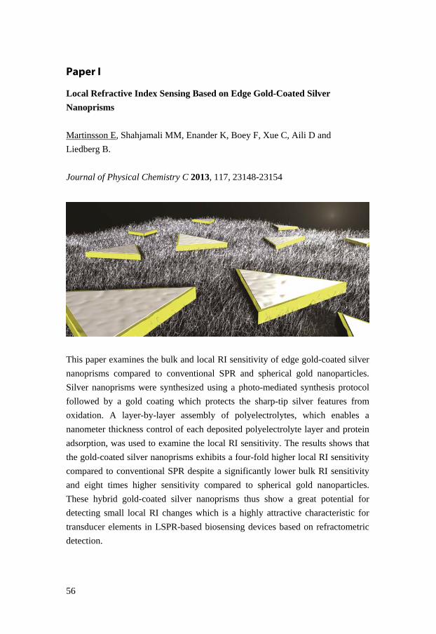
56
Paper I
Local Refractive Index Sensing Based on Edge Gold-Coated Silver
Nanoprisms
Martinsson E, Shahjamali MM, Enander K, Boey F, Xue C, Aili D and
Liedberg B.
Journal of Physical Chemistry C 2013, 117, 23148-23154
This paper examines the bulk and local RI sensitivity of edge gold-coated silver
nanoprisms compared to conventional SPR and spherical gold nanoparticles.
Silver nanoprisms were synthesized using a photo-mediated synthesis protocol
followed by a gold coating which protects the sharp-tip silver features from
oxidation. A layer-by-layer assembly of polyelectrolytes, which enables a
nanometer thickness control of each deposited polyelectrolyte layer and protein
adsorption, was used to examine the local RI sensitivity. The results shows that
the gold-coated silver nanoprisms exhibits a four-fold higher local RI sensitivity
compared to conventional SPR despite a significantly lower bulk RI sensitivity
and eight times higher sensitivity compared to spherical gold nanoparticles.
These hybrid gold-coated silver nanoprisms thus show a great potential for
detecting small local RI changes which is a highly attractive characteristic for
transducer elements in LSPR-based biosensing devices based on refractometric
detection.

57
Paper II
Optimizing the Refractive Index Sensitivity of Plasmonically Coupled Gold
Nanoparticles
Martinsson E, Sepulveda B, Chen P, Elfwing A, Liedberg B and Aili D.
Plasmonics 2014, 9, 773-780
Herein, a protocol for controlling the self-assembly and separation of spherical
gold nanoparticles has been developed in order to explore the plasmonic
coupling effect on the RI sensitivity. Polyelectrolyte layers were used to control
the interparticle distance between spherical gold nanoparticles of different sizes.
A strong coupling was observed between the gold particles generating distinct
plasmon peaks where the frequency could be tuned by varying either the size of
the nanoparticles or the interparticle spacing. Examination of the bulk RI
sensitivity showed a significantly higher sensitivity for plasmonic coupling
modes compared to plasmon bands from single nanoparticles. The presented
strategy is simple and scalable and increases the rather modest RI sensitivity of
spherical gold nanoparticles.

58
Paper III
Substrate Effect on the Refractive Index Sensitivity of Silver Nanoparticles
Martinsson E, Otte MA, Shahjamali MM, Sepulveda B and Aili D.
Journal of Physical Chemistry C 2014, In Press
The aim of this paper was to explore how the RI sensitivity is affected when
metal nanoparticles are immobilized on a solid support. Silver nanoparticles with
three different shapes (spheres, cubes and plates) were chemically synthesized.
The bulk RI sensitivity of dispersed nanoparticles was determined and compared
to the sensitivity of immobilized nanoparticles. Both experimental and
theoretical results showed a strong correlation between the fractional surface
area in direct contact with the underlying substrate and the reduction of the RI
sensitivity. Nanoplates showed the largest decrease in sensitivity (-36%) upon
immobilization, drastically reducing their advantage over other nanoparticles.
This works shows that the substrate effect is important to consider when
developing nanoplasmonic sensing devices based on metal nanoparticles.

59
Paper IV
Influence of Surfactant Bilayers and Substrate Immobilization on the
Refractive Index Sensitivity of Anisotropic Gold Nanoparticles
Martinsson E, Shahjamali MM, Large N, Zaraee N, Schatz GC, Mirkin CA and
Aili D.
2014, In Manuscript
This work examines the influence of surfactant bilayers and substrate
immobilization on the RI sensitivity of gold nanoparticles. Surfactants like
CTAX are commonly used to control the shape of gold nanoparticles. During
synthesis, the gold nanoparticles are covered with a 3-4 nm thick bilayer of
CTAX, which significantly affects their possibility to detect local RI changes. In
this work, four types of gold nanoparticles with different geometry were
synthesized using CTAX. Their bulk RI sensitivity was examined both
theoretically and experimentally as well as the effect of a substrate
immobilization. In addition, it is shown that the RI sensitivity can be
significantly increased by exposing the CTAX-covered nanoparticles to a short
(30 sec) oxygen plasma treatment, where some of the surface-bound CTAX is
etched away from the surface. A longer plasma exposure (30 min), however,
leads to a reduction in the RI sensitivity caused by morphological changes of the
nanoparticles. It is shown that a removal of CTAX using oxygen plasma
treatment can increase the RI sensitivity with up to 40 %.

60

61
Future perspective
Metal nanoparticles are indeed promising transducer elements for biosensing
devices based on refractometric detection. Localized surface plasmons can be
excited by light in the visible or near-infrared wavelength range and highly
sensitive devices can be constructed using rather simple instrumentation.
Therefore, LSPR-based methods are especially suitable for miniaturized sensors
in, for example, point-of-care diagnostics or in other applications outside core
lab facilities. However, much work is required before they can be practically
implemented and utilized in biomolecular sensing. Several challenges need to be
addressed in order to realize LSPR-based sensing devices that are sensitive,
specific, and robust, while also being cost-effective. Some of these challenges
are discussed below:
Biofunctionalization: A reliable and biofunctional surface chemistry is a key
aspect in order to produce a successful biosensing device that enables molecular
recognition in complex samples. The surface chemistry should exhibit a high
selectivity as well as a satisfactory resistance against unspecific adsorption. To
compress all these functionalities onto the surface of nanoparticles without
7

62
significantly reducing their sensitivity is thus a great challenge and will require a
substantial combined research effort. Focus should be on the development of
thin surface coatings that display low non-specific adsorption together with the
production of small recognition elements that provides specificity without
occupying the entire sensing volume.
Reproducibility: The techniques for fabrication of metal nanostructures have
advanced significantly during the last decade, but there is room for improvement
in this area especially when it comes to reproducibility. Nanostructures made by
various lithography techniques can yield high reproducibility, although generally
at higher production costs and with problems with up-scaling. Self-assembly of
nanoparticles made by wet-synthesis strategies offers a simple and cost effective
way to produce nanoplasmonic substrates. However, the reproducibility needs to
be improved, in particular the polydispersity in the size and shape of the
synthesized nanoparticles and thereby obtaining narrow plasmon bands. Also, a
better control of the self-assembly process is critical in order to produce
substrates with identical surface nanoparticle coverages that give rise to similar
intensities in the output signal. This could be achieved by combining self-
assembly with soft lithography techniques in order to guide the surface
adsorption of the nanoparticles. This will also enable the creation of
nanopatterns which is important for multiplexed detection.
Stability: Nanoparticle robustness is another key aspect to consider when
developing sensing devices with reliable output signals. Morphological changes
of the metal nanoparticles can give rise to spectral changes that can interfere
with the sensing response and hence, produce negative results. The possibility to
introduce various chemical and biological substances in the close proximity of
metal nanoparticles without affecting their structure is of critical importance.
Localization: A higher sensing performance can be achieved by controlling the
orientation of nanoparticles since the sensitivity for detecting local RI changes
around or in between anisotropic nanoparticles can be significantly improved if
the molecular interactions can be guided into areas with enhanced
electromagnetic fields. Thus, it is better to focus on the nanoparticle assembly
rather than trying to optimize the structure of individual nanoparticles since a
good control of the interparticle separation and orientation will provide higher

63
sensitivities compared to single nanoparticles. It is, however, a great challenge to
simultaneously control the immobilization of recognition molecules into these
areas with enhanced sensitivity.
The work presented in this thesis provides comprehensive information about
how metal nanoparticles can be utilized for nanoplasmonic sensing, especially
regarding nanoparticle immobilization and optimization of the RI sensitivity.
However, several challenges remain in order to fully demonstrate their potential
as sensing transducers and take the next step in the development. A joint
multidisciplinary effort is undoubtedly needed in order to produce
nanoplasmonic sensing platforms that could compete with existing sensing
techniques.

64

65
References
1. Feynman, P. R., There's Plenty of Room at the Bottom. Caltech Engineering and Science 1960, 23, 22-36.
2. Stockman, M. I., Nanoplasmonics: past, present, and glimpse into future. Optics Express 2011, 19, 22029-22106.
3. Giannini, V.; Fernandez-Dominguez, A. I.; Sonnefraud, Y.; Roschuk, T.; Fernandez-Garcia, R.; Maier, S. A., Controlling Light Localization and Light-Matter Interactions with Nanoplasmonics. Small 2010, 6, 2498-2507.
4. Hutter, E.; Fendler, J. H., Exploitation of localized surface plasmon resonance. Advanced Materials 2004, 16, 1685-1706.
5. Willets, K. A.; Van Duyne, R. P., Localized surface plasmon resonance spectroscopy and sensing. Annual Review of Physical Chemistry 2007, 58, 267-297.
6. Lal, S.; Link, S.; Halas, N. J., Nano-optics from sensing to waveguiding. Nature Photonics 2007, 1, 641-648.
7. Brill, R. H., The Chemistry of the Lycurgus Cup. Proc 7th Internat. Cong. Glass, comptes rendus 2 1965, Paper 223, 1-13.
8. Barber, D. J.; Freestone, I. C., An investigation of the origin of the color of the lycurgus cup by analytical transmission electron-microscopy. Archaeometry 1990, 32, 33-45.
9. Hornyak, G. L.; Patrissi, C. J.; Oberhauser, E. B.; Martin, C. R.; Valmalette, J.-C.; Lemaire, L.; Dutta, J.; Hofmann, H., Effective Medium Theory Characterization of Au/Ag Nanoalloy-porous Alumina Composites. NanoStructured Materials 1997, 9, 571-574.
10. Maier, S. A.; Brongersma, M. L.; Kik, P. G.; Meltzer, S.; Requicha, A. A. G.; Atwater, H. A., Plasmonics - A route to nanoscale optical devices. Advanced Materials 2001, 13, 1501-1505.
11. Barnes, W. L.; Dereux, A.; Ebbesen, T. W., Surface plasmon subwavelength optics. Nature 2003, 424, 824-830.
12. Maier, S. A.; Kik, P. G.; Atwater, H. A., Observation of coupled plasmon-polariton modes in Au nanoparticle chain waveguides of different lengths: Estimation of waveguide loss. Applied Physics Letters 2002, 81, 1714-1716.
13. Atwater, H. A.; Polman, A., Plasmonics for improved photovoltaic devices. Nature Materials 2010, 9, 205-213.
14. Wu, J. L.; Chen, F. C.; Hsiao, Y. S.; Chien, F. C.; Chen, P. L.; Kuo, C. H.; Huang, M. H.; Hsu, C. S., Surface Plasmonic Effects of Metallic Nanoparticles on the Performance of Polymer Bulk Heterojunction Solar Cells. Acs Nano 2011, 5, 959-967.
15. Pillai, S.; Catchpole, K. R.; Trupke, T.; Green, M. A., Surface plasmon enhanced silicon solar cells. Journal of Applied Physics 2007, 101, 8.

66
16. Christopher, P.; Xin, H. L.; Linic, S., Visible-light-enhanced catalytic oxidation reactions on plasmonic silver nanostructures. Nature Chemistry 2011, 3, 467-472.
17. Langhammer, C.; Larsson, E. M.; Kasemo, B.; Zoric, I., Indirect Nanoplasmonic Sensing: Ultrasensitive Experimental Platform for Nanomaterials Science and Optical Nanocalorimetry. Nano Letters 2010, 10, 3529-3538.
18. Anker, J. N.; Hall, W. P.; Lyandres, O.; Shah, N. C.; Zhao, J.; Van Duyne, R. P., Biosensing with Plasmonic Nanosensors. Nature Materials 2008, 7, 442-453.
19. Jain, P. K.; Huang, X. H.; El-Sayed, I. H.; El-Sayed, M. A., Noble Metals on the Nanoscale: Optical and Photothermal Properties and Some Applications in Imaging, Sensing, Biology, and Medicine. Accounts of Chemical Research 2008, 41, 1578-1586.
20. Sepulveda, B.; Angelome, P. C.; Lechuga, L. M.; Liz-Marzan, L. M., LSPR-based nanobiosensors. Nano Today 2009, 4, 244-251.
21. Haes, A. J.; Haynes, C. L.; McFarland, A. D.; Schatz, G. C.; Van Duyne, R. R.; Zou, S. L., Plasmonic materials for surface-enhanced sensing and spectroscopy. Mrs Bulletin 2005, 30, 368-375.
22. Yonzon, C. R.; Stuart, D. A.; Zhang, X. Y.; McFarland, A. D.; Haynes, C. L.; Van Duyne, R. P., Towards advanced chemical and biological nanosensors - An overview. Talanta 2005, 67, 438-448.
23. Haes, A. J.; Stuart, D. A.; Nie, S. M.; Van Duyne, R. P., Using solution-phase nanoparticles, surface-confined nanoparticle arrays and single nanoparticles as biological sensing platforms. Journal of Fluorescence 2004, 14, 355-367.
24. Szunerits, S.; Boukherroub, R., Sensing using localised surface plasmon resonance sensors. Chemical Communications 2012, 48, 8999-9010.
25. Petryayeva, E.; Krull, U. J., Localized surface plasmon resonance: Nanostructures, bioassays and biosensing-A review. Analytica Chimica Acta 2011, 706, 8-24.
26. Stewart, M. E.; Anderton, C. R.; Thompson, L. B.; Maria, J.; Gray, S. K.; Rogers, J. A.; Nuzzo, R. G., Nanostructured plasmonic sensors. Chemical Reviews 2008, 108, 494-521.
27. Haes, A. J.; Hall, W. P.; Chang, L.; Klein, W. L.; Van Duyne, R. P., A localized surface plasmon resonance biosensor: First steps toward an assay for Alzheimer's disease. Nano Letters 2004, 4, 1029-1034.
28. Haes, A. J.; Chang, L.; Klein, W. L.; Van Duyne, R. P., Detection of a biomarker for Alzheimer's disease from synthetic and clinical samples using a nanoscale optical biosensor. Journal of the American Chemical Society 2005, 127, 2264-2271.
29. Acimovic, S. S.; Ortega, M. A.; Sanz, V.; Berthelot, J.; Garcia-Cordero, J. L.; Renger, J.; Maerkl, S. J.; Kreuzer, M. P.; Quidant, R., LSPR Chip for Parallel, Rapid, and Sensitive Detection of Cancer Markers in Serum. Nano Letters 2014, 14, 2636-2641.
30. Chen, H. J.; Kou, X. S.; Yang, Z.; Ni, W. H.; Wang, J. F., Shape- and size-dependent refractive index sensitivity of gold nanoparticles. Langmuir 2008, 24, 5233-5237.

67
31. Jain, P. K.; El-Sayed, M. A., Plasmonic coupling in noble metal nanostructures. Chemical Physics Letters 2010, 487, 153-164.
32. Faraday, M., The Bakerian Lecture: Experimental Relations of Gold (and Other Metals) to Light. Philos. T. Roy. Soc. 1857, 147, 145-181.
33. Mappes, T.; Jahr, N.; Csaki, A.; Vogler, N.; Popp, J.; Fritzsche, W., The Invention of Immersion Ultramicroscopy in 1912-The Birth of Nanotechnology? Angewandte Chemie-International Edition 2012, 51, 11208-11212.
34. Dahl, J. A.; Maddux, B. L. S.; Hutchison, J. E., Toward greener nanosynthesis. Chemical Reviews 2007, 107, 2228-2269.
35. Rycenga, M.; Cobley, C. M.; Zeng, J.; Li, W. Y.; Moran, C. H.; Zhang, Q.; Qin, D.; Xia, Y. N., Controlling the Synthesis and Assembly of Silver Nanostructures for Plasmonic Applications. Chemical Reviews 2011, 111, 3669-3712.
36. Henzie, J.; Lee, J.; Lee, M. H.; Hasan, W.; Odom, T. W., Nanofabrication of Plasmonic Structures. In Annual Review of Physical Chemistry, Annual Reviews: Palo Alto, 2009; Vol. 60, pp 147-165.
37. Stewart, M. E.; Motala, M. J.; Yao, J.; Thompson, L. B.; Nuzzo, R. G., Unconventional Methods for Forming Nanopatterns. Proceedings of the Institution of Mechanical Engineers, Part N: Journal of Nanoengineering and Nanosystems 2006, 220, 81-138.
38. Pease, R. F.; Chou, S. Y., Lithography and other patterning techniques for future electronics. Proceedings of the Ieee 2008, 96, 248-270.
39. Gunnarsson, L.; Bjerneld, E. J.; Xu, H.; Petronis, S.; Kasemo, B.; Kall, M., Interparticle coupling effects in nanofabricated substrates for surface-enhanced Raman scattering. Applied Physics Letters 2001, 78, 802-804.
40. Felidj, N.; Aubard, J.; Levi, G.; Krenn, J. R.; Salerno, M.; Schider, G.; Lamprecht, B.; Leitner, A.; Aussenegg, F. R., Controlling the optical response of regular arrays of gold particles for surface-enhanced Raman scattering. Physical Review B 2002, 65, 9.
41. Abu Hatab, N. A.; Oran, J. M.; Sepaniak, M. J., Surface-enhanced Raman spectroscopy substrates created via electron beam lithography and nanotransfer printing. Acs Nano 2008, 2, 377-385.
42. Shankar, S. S.; Rizzello, L.; Cingolani, R.; Rinaldi, R.; Pompa, P. P., Micro/Nanoscale Patterning of Nanostructured Metal Substrates for Plasmonic Applications. Acs Nano 2009, 3, 893-900.
43. Corrigan, T. D.; Guo, S.; Phaneuf, R. J.; Szmacinski, H., Enhanced fluorescence from periodic arrays of silver nanoparticles. Journal of Fluorescence 2005, 15, 777-784.
44. Acimovic, S. S.; Kreuzer, M. P.; Gonzalez, M. U.; Quidant, R., Plasmon Near-Field Coupling in Metal Dimers as a Step toward Single-Molecule Sensing. Acs Nano 2009, 3, 1231-1237.
45. Piliarik, M.; Kvasnicka, P.; Galler, N.; Krenn, J. R.; Homola, J., Local refractive index sensitivity of plasmonic nanoparticles. Optics Express 2011, 19, 9213-9220.

68
46. Haynes, C. L.; Van Duyne, R. P., Nanosphere lithography: A versatile nanofabrication tool for studies of size-dependent nanoparticle optics. Journal of Physical Chemistry B 2001, 105, 5599-5611.
47. Hulteen, J. C.; Vanduyne, R. P., Nanosphere lithography - a materials general fabrication process for periodic particle array surfaces. Journal of Vacuum Science & Technology a-Vacuum Surfaces and Films 1995, 13, 1553-1558.
48. Ormonde, A. D.; Hicks, E. C. M.; Castillo, J.; Van Duyne, R. P., Nanosphere lithography: Fabrication of large-area Ag nanoparticle arrays by convective self-assembly and their characterization by scanning UV-visible extinction spectroscopy. Langmuir 2004, 20, 6927-6931.
49. Whatmore, R. W., Nanotechnology: big prospects for small engineering Ingenia 2001, 28-34.
50. Zhao, P. X.; Li, N.; Astruc, D., State of the art in gold nanoparticle synthesis. Coordination Chemistry Reviews 2013, 257, 638-665.
51. Sun, Y. G.; Xia, Y. N., Shape-controlled synthesis of gold and silver nanoparticles. Science 2002, 298, 2176-2179.
52. Turkevich, J.; Stevenson, P. C.; Hillier, J., A study of the nucleation and growth processes in the synthesis of colloidal gold. Discussions of the Faraday Society 1951, 11, 55-75.
53. Frens, G., Controlled Nucleation for the Regulation of the Particle Size in Monodisperse Gold Suspensions. Nature Physical Science 1973, 241, 20-22.
54. Ji, X. H.; Song, X. N.; Li, J.; Bai, Y. B.; Yang, W. S.; Peng, X. G., Size control of gold nanocrystals in citrate reduction: The third role of citrate. Journal of the American Chemical Society 2007, 129, 13939-13948.
55. Hu, M.; Chen, J. Y.; Li, Z. Y.; Au, L.; Hartland, G. V.; Li, X. D.; Marquez, M.; Xia, Y. N., Gold nanostructures: engineering their plasmonic properties for biomedical applications. Chemical Society Reviews 2006, 35, 1084-1094.
56. Jana, N. R.; Gearheart, L.; Murphy, C. J., Wet chemical synthesis of high aspect ratio cylindrical gold nanorods. Journal of Physical Chemistry B 2001, 105, 4065-4067.
57. Jana, N. R.; Gearheart, L.; Murphy, C. J., Seed-mediated growth approach for shape-controlled synthesis of spheroidal and rod-like gold nanoparticles using a surfactant template. Advanced Materials 2001, 13, 1389-1393.
58. Nikoobakht, B.; El-Sayed, M. A., Preparation and growth mechanism of gold nanorods (NRs) using seed-mediated growth method. Chemistry of Materials 2003, 15, 1957-1962.
59. Zhang, J. A.; Langille, M. R.; Personick, M. L.; Zhang, K.; Li, S. Y.; Mirkin, C. A., Concave Cubic Gold Nanocrystals with High-Index Facets. Journal of the American Chemical Society 2010, 132, 14012-14014.
60. Nehl, C. L.; Liao, H. W.; Hafner, J. H., Optical properties of star-shaped gold nanoparticles. Nano Letters 2006, 6, 683-688.

69
61. Ha, T. H.; Koo, H. J.; Chung, B. H., Shape-controlled syntheses of gold nanoprisms and nanorods influenced by specific adsorption of halide ions. Journal of Physical Chemistry C 2007, 111, 1123-1130.
62. Niidome, T.; Yamagata, M.; Okamoto, Y.; Akiyama, Y.; Takahashi, H.; Kawano, T.; Katayama, Y.; Niidome, Y., PEG-modified gold nanorods with a stealth character for in vivo applications. Journal of Controlled Release 2006, 114, 343-347.
63. Takahashi, H.; Niidome, Y.; Niidome, T.; Kaneko, K.; Kawasaki, H.; Yamada, S., Modification of gold nanorods using phospatidylcholine to reduce cytotoxicity. Langmuir 2006, 22, 2-5.
64. Hauck, T. S.; Ghazani, A. A.; Chan, W. C. W., Assessing the effect of surface chemistry on gold nanorod uptake, toxicity, and gene expression in mammalian cells. Small 2008, 4, 153-159.
65. Gole, A.; Murphy, C. J., Polyelectrolyte-coated gold nanorods: Synthesis, characterization and immobilization. Chemistry of Materials 2005, 17, 1325-1330.
66. Hrelescu, C.; Sau, T. K.; Rogach, A. L.; Jackel, F.; Laurent, G.; Douillard, L.; Charra, F., Selective Excitation of Individual Plasmonic Hotspots at the Tips of Single Gold Nanostars. Nano Letters 2011, 11, 402-407.
67. Guedje, F. K.; Giloan, M.; Potara, M.; Hounkonnou, M. N.; Astilean, S., Optical properties of single silver triangular nanoprism. Physica Scripta 2012, 86, 6.
68. Jin, R. C.; Cao, Y. W.; Mirkin, C. A.; Kelly, K. L.; Schatz, G. C.; Zheng, J. G., Photoinduced conversion of silver nanospheres to nanoprisms. Science 2001, 294, 1901-1903.
69. Millstone, J. E.; Hurst, S. J.; Metraux, G. S.; Cutler, J. I.; Mirkin, C. A., Colloidal Gold and Silver Triangular Nanoprisms. Small 2009, 5, 646-664.
70. Shahjamali, M. M.; Bosman, M.; Cao, S. W.; Huang, X.; Saadat, S.; Martinsson, E.; Aili, D.; Tay, Y. Y.; Liedberg, B.; Loo, S. C. J.; Zhang, H.; Boey, F.; Xue, C., Gold Coating of Silver Nanoprisms. Advanced Functional Materials 2012, 22, 849-854.
71. Enoch, S.; Bonod, N., Plasmonics: From Basics to Advanced Topics. Springer: Heidelberg, 2012; Vol. 167.
72. Raether, H., Surface Plasmons on Smooth and Rough Surfaces and on Gratings. Springer-Verlag: 1988.
73. Kretschmann, E.; Raether, H., Radiative decay of non radiative surface plasmons excited by light. Zeitschrift für Naturforschung A 1968, 23a, 2135-2136.
74. Otto, A., Excitation of Nonradiative Surface Plasma Waves in Silver by the Method of Frustrated Total Reflection. Zeitschrift für Physik. A, Hadrons and Nuclei 1968, 216, 398-410.
75. Teng, Y. Y.; Stern, E. A., Plasma Radiation from Metal Grating Surfaces. Physical Review Letters 1967, 19, 511-514.
76. Mie, G., Beiträge zur Optik trüber Medien, speziell kolloidaler Metallösungen. Annalen der Physik, 1908; Vol. 25, pp 377-445.

70
77. Dmitriev, A., Nanoplasmonic Sensors. Springer: New York, 2012.
78. Sonnichsen, C.; Franzl, T.; Wilk, T.; von Plessen, G.; Feldmann, J.; Wilson, O.; Mulvaney, P., Drastic reduction of plasmon damping in gold nanorods. Physical Review Letters 2002, 88, 4.
79. Meier, M.; Wokaun, A., Enhanced fields on large metal particles: dynamic depolarization Optics Letters 1983, 8, 581-583.
80. Zhang, J. Z.; Noguez, C., Plasmonic Optical Properties and Applications of Metal Nanostructures. Plasmonics 2008, 3, 127-150.
81. Shuford, K. L.; Ratner, M. A.; Schatz, G. C., Multipolar excitation in triangular nanoprisms. Journal of Chemical Physics 2005, 123, 9.
82. Wei, H.; Reyes-Coronado, A.; Nordlander, P.; Aizpurua, J.; Xu, H. X., Multipolar Plasmon Resonances in Individual Ag Nanorice. Acs Nano 2010, 4, 2649-2654.
83. Krenn, J. R.; Schider, G.; Rechberger, W.; Lamprecht, B.; Leitner, A.; Aussenegg, F. R.; Weeber, J. C., Design of multipolar plasmon excitations in silver nanoparticles. Applied Physics Letters 2000, 77, 3379-3381.
84. Schatz, G. C., Electrodynamics of nonspherical noble metal nanoparticles and nanoparticle aggregates. Journal of Molecular Structure-Theochem 2001, 573, 73-80.
85. Jain, P. K.; Lee, K. S.; El-Sayed, I. H.; El-Sayed, M. A., Calculated absorption and scattering properties of gold nanoparticles of different size, shape, and composition: Applications in biological imaging and biomedicine. Journal of Physical Chemistry B 2006, 110, 7238-7248.
86. Kelly, K. L.; Coronado, E.; Zhao, L. L.; Schatz, G. C., The optical properties of metal nanoparticles: The influence of size, shape, and dielectric environment. Journal of Physical Chemistry B 2003, 107, 668-677.
87. Krug, J. T.; Sanchez, E. J.; Xie, X. S., Design of near-field optical probes with optimal field enhancement by finite difference time domain electromagnetic simulation. Journal of Chemical Physics 2002, 116, 10895-10901.
88. Oubre, C.; Nordlander, P., Optical properties of metallodielectric nanostructures calculated using the finite difference time domain method. Journal of Physical Chemistry B 2004, 108, 17740-17747.
89. Chang, S. H.; Gray, S. K.; Schatz, G. C., Surface plasmon generation and light transmission by isolated nanoholes and arrays of nanoholes in thin metal films. Optics Express 2005, 13, 3150-3165.
90. Prodan, E.; Radloff, C.; Halas, N. J.; Nordlander, P., A hybridization model for the plasmon response of complex nanostructures. Science 2003, 302, 419-422.
91. Ghosh, S. K.; Pal, T., Interparticle coupling effect on the surface plasmon resonance of gold nanoparticles: From theory to applications. Chemical Reviews 2007, 107, 4797-4862.
92. Esteban, R.; Borisov, A. G.; Nordlander, P.; Aizpurua, J., Bridging quantum and classical plasmonics with a quantum-corrected model. Nature Communications 2012, 3, 9.

71
93. Camden, J. P.; Dieringer, J. A.; Wang, Y. M.; Masiello, D. J.; Marks, L. D.; Schatz, G. C.; Van Duyne, R. P., Probing the structure of single-molecule surface-enhanced Raman scattering hot spots. Journal of the American Chemical Society 2008, 130, 12616-12617.
94. Li, W. Y.; Camargo, P. H. C.; Lu, X. M.; Xia, Y. N., Dimers of Silver Nanospheres: Facile Synthesis and Their Use as Hot Spots for Surface-Enhanced Raman Scattering. Nano Letters 2009, 9, 485-490.
95. Mirkin, C. A.; Letsinger, R. L.; Mucic, R. C.; Storhoff, J. J., A DNA-based method for rationally assembling nanoparticles into macroscopic materials. Nature 1996, 382, 607-609.
96. Aili, D.; Selegard, R.; Baltzer, L.; Enander, K.; Liedberg, B., Colorimetric Protein Sensing by Controlled Assembly of Gold Nanoparticles Functionalized with Synthetic Receptors. Small 2009, 5, 2445-2452.
97. Chen, P.; Selegard, R.; Aili, D.; Liedberg, B., Peptide functionalized gold nanoparticles for colorimetric detection of matrilysin (MMP-7) activity. Nanoscale 2013, 5, 8973-8976.
98. Kumar, C., Biofunctionalization of Nanomaterials. Wiley–VCH Verlag: Weinheim, 2005.
99. Nuzzo, R. G.; Allara, D. L., Adsorption of bifunctional organic disulfides on gold surfaces. Journal of Chemical Society 1983, 5, 4481-4483.
100. Pastoriza-Santos, I.; Perez-Juste, J.; Liz-Marzan, L. M., Silica-coating and hydrophobation of CTAB-stabilized gold nanorods. Chemistry of Materials 2006, 18, 2465-2467.
101. Xue, C.; Chen, X.; Hurst, S. J.; Mirkin, C. A., Self-assembled monolayer mediated silica coating of silver triangular nanoprisms. Advanced Materials 2007, 19, 4071-4074.
102. Shipway, A. N.; Katz, E.; Willner, I., Nanoparticle arrays on surfaces for electronic, optical, and sensor applications. Chemphyschem 2000, 1, 18-52.
103. Hahn, M. A.; Singh, A. K.; Sharma, P.; Brown, S. C.; Moudgil, B. M., Nanoparticles as contrast agents for in-vivo bioimaging: current status and future perspectives. Analytical and Bioanalytical Chemistry 2011, 399, 3-27.
104. Schmitt, J.; Machtle, P.; Eck, D.; Mohwald, H.; Helm, C. A., Preparation and optical properties of colloidal gold monolayers. Langmuir 1999, 15, 3256-3266.
105. Grabar, K. C.; Smith, P. C.; Musick, M. D.; Davis, J. A.; Walter, D. G.; Jackson, M. A.; Guthrie, A. P.; Natan, M. J., Kinetic control of interparticle spacing in Au colloid-based surfaces: Rational nanometer-scale architecture. Journal of the American Chemical Society 1996, 118, 1148-1153.
106. Lundgren, A.; Hulander, M.; Brorsson, J.; Hermansson, M.; Elwing, H.; Andersson, O.; Liedberg, B.; Berglin, M., Gold-Nanoparticle-Assisted Self-Assembly of Chemical Gradients with Tunable Sub-50 nm Molecular Domains. Particle & Particle Systems Characterization 2014, 31, 209-218.
107. Silberzan, P.; Leger, L.; Ausserre, D.; Benattar, J. J., Silanation of silica surfaces - a new method of constructing pure or mixed monolayers. Langmuir 1991, 7, 1647-1651.
108. Decher, G., Fuzzy nanoassemblies: Toward layered polymeric multicomposites. Science 1997, 277, 1232-1237.

72
109. Decher, G.; Hong, J. D.; Schmitt, J., Buildup of ultrathin multilayer films by a self-assembly process. III Consecutively alternating adsorption of anionic and cationic polyelectrolytes on charged surfaces. Thin Solid Films 1992, 210, 831-835.
110. Smith, R. N.; McCormick, M.; Barrett, C. J.; Reven, L.; Spiess, H. W., NMR studies of PAH/PSS polyelectrolyte multilayers adsorbed onto silica. Macromolecules 2004, 37, 4830-4838.
111. Feldoto, Z.; Varga, I.; Blomberg, E., Influence of Salt and Rinsing Protocol on the Structure of PAH/PSS Polyelectrolyte Multilayers. Langmuir 2010, 26, 17048-17057.
112. Losche, M.; Schmitt, J.; Decher, G.; Bouwman, W. G.; Kjaer, K., Detailed structure of molecularly thin polyelectrolyte multilayer films on solid substrates as revealed by neutron reflectometry. Macromolecules 1998, 31, 8893-8906.
113. Wong, J. E.; Rehfeldt, F.; Hanni, P.; Tanaka, M.; Klitzing, R. V., Swelling behavior of polyelectrolyte multilayers in saturated water vapor. Macromolecules 2004, 37, 7285-7289.
114. Taton, T. A.; Mucic, R. C.; Mirkin, C. A.; Letsinger, R. L., The DNA-mediated formation of supramolecular mono- and multilayered nanoparticle structures. Journal of the American Chemical Society 2000, 122, 6305-6306.
115. Aili, D.; Han, H. S.; Enander, K.; Baltzer, L.; Liedberg, B. In Controlled assembly of gold nanoparticles using de novo designed polypeptide scaffolds, Conference on MEMS/MOEMS Components and Their Applications V, San Jose, CA, Jan 21-22; Spie-Int Soc Optical Engineering: San Jose, CA, 2008; pp 88506-88506.
116. Schmitt, J.; Decher, G.; Dressick, W. J.; Brandow, S. L.; Geer, R. E.; Shashidhar, R.; Calvert, J. M., Metal nanoparticle/polymer superlattice films: Fabrication and control of layer structure. Advanced Materials 1997, 9, 61-65.
117. Jiang, C. Y.; Markutsya, S.; Tsukruk, V. V., Collective and individual plasmon resonances in nanoparticle films obtained by spin-assisted layer-by-layer assembly. Langmuir 2004, 20, 882-890.
118. Thevenot, D. R.; Toth, K.; Durst, R. A.; Wilson, G. S., Electrochemical biosensors: recommended definitions and classification. Biosensors & Bioelectronics 2001, 16, 121-131.
119. Dahlin, A. B., Plasmonic Biosensors: An Integrated View of Refractometric Detection. IOS Press: 2012.
120. Clark Jr, L. C.; Lyons, C., Electrode Systems for Continuous Monitoring in Cardiovascular Surgery. Annals New York Academy of Sciences 1962, 102, 29-45.
121. Wang, J., Glucose biosensors: 40 years of advances and challenges. Electroanalysis 2001, 13, 983-988.
122. Filippini, D.; Svensson, S. P. S.; Lundstrom, I., Computer screen as a programmable light source for visible absorption characterization of (bio)chemical assays. Chemical Communications 2003, 240-241.
123. Preechaburana, P.; Gonzalez, M. C.; Suska, A.; Filippini, D., Surface Plasmon Resonance Chemical Sensing on Cell Phones. Angewandte Chemie-International Edition 2012, 51, 11585-11588.

73
124. Harder, P.; Grunze, M.; Dahint, R.; Whitesides, G. M.; Laibinis, P. E., Molecular conformation in oligo(ethylene glycol)-terminated self-assembled monolayers on gold and silver surfaces determines their ability to resist protein adsorption. Journal of Physical Chemistry B 1998, 102, 426-436.
125. Love, J. C.; Estroff, L. A.; Kriebel, J. K.; Nuzzo, R. G.; Whitesides, G. M., Self-assembled monolayers of thiolates on metals as a form of nanotechnology. Chemical Reviews 2005, 105, 1103-1169.
126. Wang, Z. H.; Jin, G., Silicon surface modification with a mixed silanes layer to immobilize proteins for biosensor with imaging ellipsometry. Colloids and Surfaces B-Biointerfaces 2004, 34, 173-177.
127. Lofas, S.; Johnsson, B., A novel hydrogel matrix on gold surfaces in surface-plasmon resonance sensors for fast and efficient covalent immobilization of ligands. Journal of the Chemical Society-Chemical Communications 1990, 1526-1528.
128. Larsson, A.; Ekblad, T.; Andersson, O.; Liedberg, B., Photografted poly(ethylene glycol) matrix for affinity interaction studies. Biomacromolecules 2007, 8, 287-295.
129. Zhang, S. G., Hydrogels - Wet or let die. Nature Materials 2004, 3, 7-8.
130. Liedberg, B.; Nylander, C.; Lundstrom, I., Surface-plasmon resonance for gas-detection and biosensing. Sensors and Actuators 1983, 4, 299-304.
131. Homola, J., Surface plasmon resonance sensors for detection of chemical and biological species. Chemical Reviews 2008, 108, 462-493.
132. Englebienne, P., Use of colloidal gold surface plasmon resonance peak shift to infer affinity constants from the interactions between protein antigens and antibodies specific for single or multiple epitopes. Analyst 1998, 123, 1599-1603.
133. Ferreira, J.; Santos, M. J. L.; Rahman, M. M.; Brolo, A. G.; Gordon, R.; Sinton, D.; Girotto, E. M., Attomolar Protein Detection Using in-Hole Surface Plasmon Resonance. Journal of the American Chemical Society 2009, 131, 436-437.
134. Nath, N.; Chilkoti, A., Label-free biosensing by surface plasmon resonance of nanoparticles on glass: Optimization of nanoparticle size. Analytical Chemistry 2004, 76, 5370-5378.
135. Otte, M. A.; Estevez, M. C.; Carrascosa, L. G.; Gonzalez-Guerrero, A. B.; Lechuga, L. M.; Sepulveda, B., Improved Biosensing Capability with Novel Suspended Nanodisks. Journal of Physical Chemistry C 2011, 115, 5344-5351.
136. Olofsson, L.; Rindzevicius, T.; Pfeiffer, I.; Kall, M.; Hook, F., Surface-based gold-nanoparticle sensor for specific and quantitative DNA hybridization detection. Langmuir 2003, 19, 10414-10419.
137. Dahlin, A. B.; Tegenfeldt, J. O.; Hook, F., Improving the instrumental resolution of sensors based on localized surface plasmon resonance. Analytical Chemistry 2006, 78, 4416-4423.
138. Satija, J.; Punjabi, N. S.; Sai, V. V. R.; Mukherji, S., Optimal Design for U-bent Fiber-optic LSPR Sensor Probes. Plasmonics 2014, 9, 251-260.

74
139. Chen, P.; Liedberg, B., Curvature of the Localized Surface Plasmon Resonance Peak. Analytical Chemistry 2014, 86, 7399-7405.
140. McFarland, A. D.; Van Duyne, R. P., Single silver nanoparticles as real-time optical sensors with zeptomole sensitivity. Nano Letters 2003, 3, 1057-1062.
141. Sherry, L. J.; Jin, R. C.; Mirkin, C. A.; Schatz, G. C.; Van Duyne, R. P., Localized surface plasmon resonance spectroscopy of single silver triangular nanoprisms. Nano Letters 2006, 6, 2060-2065.
142. Chan, G. H.; Zhao, J.; Schatz, G. C.; Van Duyne, R. P., Localized surface plasmon resonance spectroscopy of triangular aluminum nanoparticles. Journal of Physical Chemistry C 2008, 112, 13958-13963.
143. Zoric, I.; Zach, M.; Kasemo, B.; Langhammer, C., Gold, Platinum, and Aluminum Nanodisk Plasmons: Material Independence, Subradiance, and Damping Mechanisms. Acs Nano 2011, 5, 2535-2546.
144. Lu, X. M.; Rycenga, M.; Skrabalak, S. E.; Wiley, B.; Xia, Y. N., Chemical Synthesis of Novel Plasmonic Nanoparticles. In Annual Review of Physical Chemistry, Annual Reviews: Palo Alto, 2009; Vol. 60, pp 167-192.
145. Faucheaux, J. A.; Stanton, A. L. D.; Jain, P. K., Plasmon Resonances of Semiconductor Nanocrystals: Physical Principles and New Opportunities. Journal of Physical Chemistry Letters 2014, 5, 976-985.
146. Miller, M. M.; Lazarides, A. A., Sensitivity of metal nanoparticle surface plasmon resonance to the dielectric environment. Journal of Physical Chemistry B 2005, 109, 21556-21565.
147. Charles, D. E.; Aherne, D.; Gara, M.; Ledwith, D. M.; Gun'ko, Y. K.; Kelly, J. M.; Blau, W. J.; Brennan-Fournet, M. E., Versatile Solution Phase Triangular Silver Nanoplates for Highly Sensitive Plasmon Resonance Sensing. Acs Nano 2010, 4, 55-64.
148. Sannomiya, T.; Sahoo, P. K.; Mahcicek, D. I.; Solak, H. H.; Hafner, C.; Grieshaber, D.; Voros, J., Biosensing by Densely Packed and Optically Coupled Plasmonic Particle Arrays. Small 2009, 5, 1889-1896.
149. Tesler, A. B.; Chuntonov, L.; Karakouz, T.; Bendikov, T. A.; Haran, G.; Vaskevich, A.; Rubinstein, I., Tunable Localized Plasmon Transducers Prepared by Thermal Dewetting of Percolated Evaporated Gold Films. Journal of Physical Chemistry C 2011, 115, 24642-24652.
150. Jakab, A.; Rosman, C.; Khalavka, Y.; Becker, J.; Trugler, A.; Hohenester, U.; Sonnichsen, C., Highly Sensitive Plasmonic Silver Nanorods. Acs Nano 2011, 5, 6880-6885.
151. Otte, M. A.; Sepulveda, B.; Ni, W. H.; Juste, J. P.; Liz-Marzan, L. M.; Lechuga, L. M., Identification of the Optimal Spectral Region for Plasmonic and Nanoplasmonic Sensing. Acs Nano 2010, 4, 349-357.
152. Chen, H. J.; Ming, T.; Zhang, S. R.; Jin, Z.; Yang, B. C.; Wang, J. F., Effect of the Dielectric Properties of Substrates on the Scattering Patterns of Gold Nanorods. Acs Nano 2011, 5, 4865-4877.

75
153. Valamanesh, M.; Borensztein, Y.; Langlois, C.; Lacaze, E., Substrate Effect on the Plasmon Resonance of Supported Flat Silver Nanoparticles. Journal of Physical Chemistry C 2011, 115, 2914-2922.
154. Brian, B.; Sepulveda, B.; Alaverdyan, Y.; Lechuga, L. M.; Kall, M., Sensitivity enhancement of nanoplasmonic sensors in low refractive index substrates. Optics Express 2009, 17, 2015-2023.
155. Haes, A. J.; Zou, S. L.; Schatz, G. C.; Van Duyne, R. P., A nanoscale optical biosensor: The long range distance dependence of the localized surface plasmon resonance of noble metal nanoparticles. Journal of Physical Chemistry B 2004, 108, 109-116.
156. Brolo, A. G., Plasmonics for future biosensors. Nature Photonics 2012, 6, 709-713.
157. Rang, M.; Jones, A. C.; Zhou, F.; Li, Z. Y.; Wiley, B. J.; Xia, Y. N.; Raschke, M. B., Optical Near-Field Mapping of Plasmonic Nanoprisms. Nano Letters 2008, 8, 3357-3363.
158. Feuz, L.; Jonsson, P.; Jonsson, M. P.; Hook, F., Improving the Limit of Detection of Nanoscale Sensors by Directed Binding to High-Sensitivity Areas. Acs Nano 2010, 4, 2167-2177.
159. Zhang, N.; Liu, Y. J.; Yang, J.; Su, X. D.; Deng, J.; Chum, C. C.; Hong, M. H.; Teng, J. H., High sensitivity molecule detection by plasmonic nanoantennas with selective binding at electromagnetic hotspots. Nanoscale 2014, 6, 1416-1422.
160. Okamoto, T.; Yamaguchi, I.; Kobayashi, T., Local plasmon sensor with gold colloid monolayers deposited upon glass substrates. Optics Letters 2000, 25, 372-374.
161. Kedem, O.; Tesler, A. B.; Vaskevich, A.; Rubinstein, I., Sensitivity and Optimization of Localized Surface Plasmon Resonance Transducers. Acs Nano 2011, 5, 748-760.
162. Tian, L. M.; Chen, E.; Gandra, N.; Abbas, A.; Singamaneni, S., Gold Nanorods as Plasmonic Nanotransducers: Distance-Dependent Refractive Index Sensitivity. Langmuir 2012, 28, 17435-17442.
163. Evanoff, D. D.; White, R. L.; Chumanov, G., Measuring the distance dependence of the local electromagnetic field from silver nanoparticles. Journal of Physical Chemistry B 2004, 108, 1522-1524.
164. Bukasov, R.; Ali, T. A.; Nordlander, P.; Shumaker-Parry, J. S., Probing the Plasmonic Near-Field of Gold Nanocrescent Antennas. Acs Nano 2010, 4, 6639-6650.
165. Malinsky, M. D.; Kelly, K. L.; Schatz, G. C.; Van Duyne, R. P., Chain length dependence and sensing capabilities of the localized surface plasmon resonance of silver nanoparticles chemically modified with alkanethiol self-assembled monolayers. Journal of the American Chemical Society 2001, 123, 1471-1482.
166. Svedendahl, M.; Chen, S.; Dmitriev, A.; Kall, M., Refractometric Sensing Using Propagating versus Localized Surface Plasmons: A Direct Comparison. Nano Letters 2009, 9, 4428-4433.
167. Kabashin, A. V.; Evans, P.; Pastkovsky, S.; Hendren, W.; Wurtz, G. A.; Atkinson, R.; Pollard, R.; Podolskiy, V. A.; Zayats, A. V., Plasmonic nanorod metamaterials for biosensing. Nature Materials 2009, 8, 867-871.

76
168. Hall, W. P.; Anker, J. N.; Lin, Y.; Modica, J.; Mrksich, M.; Van Duyne, R. P., A calcium-modulated plasmonic switch. Journal of the American Chemical Society 2008, 130, 5836-5837.

Publications
The articles associated with this thesis have been removed for copyright reasons. For more details about these see: http://urn.kb.se/resolve?urn=urn:nbn:se:liu:diva-111841