nih public access 1,2,# marianela candolfi thomas j ... · maria g. castro1,2,#, marianela...
TRANSCRIPT
Adenoviral Vector-Mediated Gene Therapy for Gliomas: Comingof Age
Maria G. Castro1,2,#, Marianela Candolfi3, Thomas J. Wilson1, Alexandra Calinescu1,Christopher Paran1, Neha Kamran1, Carl Koschmann4, Mariela A. Moreno-Ayala3, HikmatAssi1, and Pedro R. Lowenstein1,2
1 Department of Neurosurgery, University of Michigan Medical School, Ann Arbor, MI, USA
2 Department of Cell and Developmental Biology, University of Michigan Medical School, AnnArbor, MI, USA
3 Instituto de Investigaciones Biomédicas (INBIOMED), University of Buenos Aires, School ofMedicine, CABA, Argentina
4 Division of Pediatric Hematology and Oncology, University of Michigan Hospital, Ann Arbor, MI,USA
Abstract
Introduction—Glioblastoma multiforme (GBM) is the most common primary brain tumor in
adults; it carries a dismal prognosis. Adenoviral vector (Ad)-mediated gene transfer is being
developed as a promising therapeutic strategy for GBM. Preclinical studies have demonstrated
safety and efficacy of adenovirus administration into the brain and tumor mass in rodents and into
the non-human primates’ brain. Importantly Ads have been safely administered within the tumor
resection cavity in humans.
Areas Covered—Background on GBM and Ad vectors; we describe gene therapy strategies for
GBM and discuss the value of combination approaches. Finally we discuss the results of the
human clinical trials for GBM that have used adenoviral vectors.
Expert Opinion—The transduction characteristics of Ad vectors, and their safety profile, added
to their capacity to achieve high levels of transgene expression have made them powerful vectors
for the treatment of GBM. Recent gene therapy successes in the treatment of retinal diseases and
systemic brain metabolic diseases, encourages the development of gene therapy for malignant
glioma. Exciting clinical trials are currently recruiting patients; although it is large randomized
phase III controlled clinical trials that will provide the final decision on the success of gene
therapy for the treatment of GBM.
# Corresponding Author: Maria G. Castro, Ph.D., Department of Neurosurgery, Department of Cell and Developmental Biology,University of Michigan School of Medicine, 4570 MSRB II, 1150 West Medical Center Drive, Ann Arbor, MI 48109-5689, USA Tel:(734) 764-0850 Fax (734) 764-7051 [email protected].
Declaration of Interest The authors declare no competing interests.
NIH Public AccessAuthor ManuscriptExpert Opin Biol Ther. Author manuscript; available in PMC 2014 September 01.
Published in final edited form as:Expert Opin Biol Ther. 2014 September ; 14(9): 1241–1257. doi:10.1517/14712598.2014.915307.
NIH
-PA
Author M
anuscriptN
IH-P
A A
uthor Manuscript
NIH
-PA
Author M
anuscript
Keywords
High-capacity adenovirus; dendritic cells; immunotherapy; glioblastoma multiforme; Flt3L;HSV1-TK
1 Introduction - Overview of Gliomas
The term “malignant glioma” encompasses a heterogeneous group of glial based, high-grade
tumors including anaplastic astrocytoma, GBM, mixed oligoastrocytoma, and anaplastic
oligodendroglioma. The World Health Organization (WHO) classifies all of these tumors as
either grade III (anaplastic astrocytoma, oligoastrocytoma, and anaplastic
oligodendroglioma) or grade IV (GBM). The WHO has adopted the St. Anne-Mayo system
for histologic diagnosis. According to this system, two of the following four features must
be present for the diagnosis of anaplastic astrocytoma: 1) nuclear atypia, 2) mitoses, 3)
endothelial proliferation, and 4) necrosis. When at least three of these features are present,
the diagnosis is glioblastoma multiforme. Of note, the grading system for these glial
malignancies is purely histologic due to the high concordance of the prognosis with these
histologic features. A systemic survey is not performed due to the extremely low risk of
dissemination of this tumor outside the central nervous system.
Glioblastoma multiforme is the most common primary malignant tumor of the central
nervous system. Between 2004 and 2008, the Central Brain Tumor Registry of the United
States (CBTRUS) reported that glioblastoma multiforme accounted for 16.3 % of the
primary central nervous system tumors. The only well-established risk factor for developing
a glioblastoma multiforme is exposure to ionizing radiation, though GBMs occur more
frequently in males, whites, and with increasing age (1). There is no pathognomonic
presentation of the disease and most clinical presentations are due to irritation or destruction
of local central nervous system structures or due to increased intracranial pressure. Thus,
common clinical presentation includes focal neurologic deficits, altered mental status,
seizures, personality changes, and headaches.
There has been extensive preclinical and clinical research into the pathophysiology of
malignant gliomas, but at this point the prognosis remains extremely poor. Based on work
published by Stupp and colleagues, the current standard of care includes maximal surgical
resection, radiation therapy, and temozolomide chemotherapy (2). The median survival
remains between 12 and 15 months (1). A wide variety of factors have been postulated to
account for continued therapeutic failure, including neurotoxicity of therapeutic agents that
is dose-limiting, difficulty accessing the central nervous system with therapeutic agents
secondary to the blood-brain barrier, resistance of glioblastoma to radiotherapy, and the
inability to achieve complete surgical resection due to the invasive nature of the disease.
Despite extensive research, little headway has been made in combating this devastating
disease. Thus, novel therapeutic approaches are necessary to escape the current limitations
to therapy. These novel therapeutic approaches include a wide variety of adenoviral-based
therapies, including both the use of adenoviral-vectors for gene delivery and the use of
modified oncolytic adenoviruses.
Castro et al. Page 2
Expert Opin Biol Ther. Author manuscript; available in PMC 2014 September 01.
NIH
-PA
Author M
anuscriptN
IH-P
A A
uthor Manuscript
NIH
-PA
Author M
anuscript
2. Adenoviral Vectors for Gene Therapy Applications
Adenoviruses are non-enveloped, icosahedral viruses approximately 90 nm in diameter.
Complexes composed of the hexon protein, along with a number of hexon-associated
proteins, comprise the 20 sides of the icosahedron (3). A complex called the penton base is
located on the surface of the capsid at each of the 12 vertices, and another protein complex,
the fiber, extends outward from the surface of the capsid (3). The end of the fiber complex,
known as the knob domain, binds to the coxsackie and adenovirus receptor (CAR), thus
mediating cell tropism (4,5). Also, the interaction of the adenoviral penton protein with cell
surface integrins (INT), such as INT αvβ3 and αvβ5, leads to the internalization of the virus
by endocytosis (6). Alternatively, the Major Histocompatibility Complex Class I (MHCI) α2
domain has been proposed to be involved in the initial binding of adenoviruses to cell
membranes (7). The genome is a linear double stranded DNA, covalently bound on either
end to a protein called the terminal protein; there are other numerous associated DNA-
binding proteins (4). The total length of the genome is approximately 35 thousand bases.
Upon infection, the genome does not integrate into the host genome, but remains episomal
while expressing viral genes.
Gene therapy applications using adenoviruses have typically used serotypes 2 and 5,
classified under adenovirus species type C (4). Progress in this area has proceeded in two
stages. Initially, first-generation adenoviral vectors (Ads) were produced by deletion of the
E1, and sometimes E3, region of the virus genome, thereby largely eliminating the ability of
the virus to express viral genes within infected cells. This was a necessary step to reduce the
toxicity associated with adenovirus infection. The therapeutic transgene was inserted into
the E1 region. Drawbacks of first generation Ads included residual expression of viral
proteins, which was associated with a significant immune response and loss of therapeutic
transgene expression (4,8-15). Additionally, their insertion capacity for therapeutic
transgenes is limited to approximately 8 kbp. The latest generation Ads, i.e. high-capacity
helper dependent adenoviral vectors (HCAds), have been engineered to delete all
endogenous viral coding regions from the vector's genome (16). The immune response
generated by HC-Ads is not as significant as the first generation vectors, and they also allow
for much larger inserts (~35 kbp maximum cloning capacity) (8,17). Importantly, HC-Ad
are capable of eliciting long-term transgene expression, even in the presence of an anti-Ad
systemic immune response, which has been shown to curtail transgene expression from first
generation Ads (13,14,18). With established methods for modifying viral genomes along
with the technology for cell culture-based production and amplification of the vectors, it is
possible to remove nearly all native viral DNA and insert novel DNA sequences for
therapeutic purposes. This flexibility allows for the production of non-replicating Ad viruses
with completely engineered genomes, suitable for gene therapy applications for GBM in
human patients (19-21).
Conditionally replicative adenoviruses (CRAds) were developed as an alternative
therapeutic strategy. CRAds are recombinant adenoviruses that can selectively replicate
within and kill tumor cells. CRAds have several advantages, such as: i) replication of the Ad
itself allows amplification of the input dose of the virus, ii) high levels of expression of
therapeutic transgenes, as a result of the replication of the virus DNA, iii) spreading of the
Castro et al. Page 3
Expert Opin Biol Ther. Author manuscript; available in PMC 2014 September 01.
NIH
-PA
Author M
anuscriptN
IH-P
A A
uthor Manuscript
NIH
-PA
Author M
anuscript
therapeutic effect to other adjacent tumor cells. Oncolytic Ads can kill the cells by direct
lysis as the result of the replicative cycle, but they can also express cytotoxic proteins,
stimulate the production of inflammatory cytokines, activate T-cell-mediated immunity and
sensitize tumor cells to chemotherapy. To specifically replicate in tumor cells, the E1A gene
has been encoded under the control of tissue/tumor-specific promoters or enhancers (22).
Transcriptional specificity has been achieved using C-X-C chemokine receptor 4 (CXCR4),
which is active in human mesenchymal stem cells (hMSC) and human glioma cells. Thus,
hMSC could provide replicative-oncolytic Ad to distant glioma cells (23). The oncolytic Ad
delta-24 the E1A gene has a small deletion that restricts the interaction of E1A with the
retinoblastoma protein (Rb). Thus, Delta-24 can only replicate in cancer cells that have
disrupted Rb function, without affecting normal cells (24). Using a similar approach,
ONYX-015 Ad has a deletion in the E1B gene, thus when the virus infects normal cells with
active p53, it cannot replicate. However, when this virus infects tumor cells with an aberrant
p53 function, it replicates and cause cellular lysis (25). The role of p53 in the ONYX-015
selectivity is not clearly understood as it has been shown that this vector could still replicate
in tumor cells with wild type p53 (26). O'Shea et al demonstrated that the loss of E1B
induces the expression of p53, but not its activation (27). Instead, the selective replication in
tumor cells seems to be determined by the capacity of these cells to export late viral RNA in
the absence of E1B (27). Another strategy to improve clinical efficacy of these vectors is to
augment their infectivity. The oncolytic potency of the Ad5-delta24 adenovirus was
enhanced by the incorporation into the HI loop of the fiber knob domain of a sequence
encoding for Arg- Gly-Asp peptide, that its known to interact with αv integrins (28). This
modification allows the virus to utilize αv integrins as an alternative receptor during the cell
entry process (29) and has enhanced infectivity in in vitro and in vivo GBM models (30).
An important problem that remains to be solved for the use of these vectors in the clinical
setting is the development of anti-Ad neutralizing antibodies that hampers the infectivity of
replication competent vectors upon their repeated administration (31). Nevertheless, the
replication competent adenoviruses have proven to be safe for the treatment of patients (32).
The possibility of genetic manipulations of these vectors, joined with the accelerated
increase in the knowledge of the mechanisms and the proteins involved in tumor progression
will enable researchers to achieve more effective and specific cancer therapies.
3. Gene Therapeutic Targets for Glioblastoma
3.1 Conditional Cytotoxic Therapy
Suicide genes encode for enzymes that activate non-toxic compounds into cytotoxic
molecules. When prodrugs are incorporated by transduced cells, they are converted into
cytotoxic compounds by the conditionally cytotoxic enzymes encoded within Ads. These
cytotoxic compounds can freely diffuse into neighboring cells or migrate through cell-to-cell
contacts, amplifying the cytotoxic effect. Herpes Simplex Virus Type 1-Thymidine Kinase
(TK) phosphorylates the prodrug ganciclovir (GCV) to GCV-monophosphate, which after
conversion into a tri-phosphorylated GCV by cellular kinases becomes 2-deoxyguanosine
triphosphate (33). When GCV-triphosphate is incorporated into duplicating DNA, it inhibits
DNA polymerase and leads to DNA chain termination (Figure 1) (33). Other suicide gene/
Castro et al. Page 4
Expert Opin Biol Ther. Author manuscript; available in PMC 2014 September 01.
NIH
-PA
Author M
anuscriptN
IH-P
A A
uthor Manuscript
NIH
-PA
Author M
anuscript
nucleoside analog systems include E. coli cytosine deaminase, which catalyzes the
conversion of the prodrug 5-fluorocytosine to 5-fluorouracil, resulting in DNA breaks (34).
Cytochrome P450 converts cyclophosphamide into a toxin that leads to DNA crosslinking
and protein alkylation (35). E. coli purine nucleoside phosphorylase converts nontoxic
purine nucleoside analogs, such as 6-methylpurine and F-araAMP into toxic adenine analogs
that block mRNA and protein synthesis (36). Carboxypeptidase G2 is combined with the
prodrug 4-benzoyl-L-glutamic acid to produce a mustard alkylating agent that does not
require further enzymatic processing to promote cell-cycle independent DNA-crosslinking
(37).
The suicide gene that has been exploited the most for the treatment of GBM is TK (38). TK
mutants have also been developed that exhibit high affinity for the prodrugs GCV and
acyclovir, SR39 and SR26, respectively (39,40). These transgenes allow for the use of
suicide gene therapy with lower systemic concentrations of the prodrugs, reducing toxicity
(41). A novel tomato plant-derived TK (toTK), which exhibits high affinity and specificity
for the nucleoside analogue azidothymidine (AZT), shows a robust cytotoxic effect in
human GBM cells in vitro (42). The advantages of this system are that AZT easily
penetrates the blood-brain barrier and phosphorylates AZT to AZT-monophosphate (42).
TK+GCV sensitizes GBM cells to the cytotoxic effect of radiotherapy and chemotherapeutic
agents (43). Treatment of intracranial human GBM xenografts with Ad-TK increased the
efficacy of radiotherapy and reduced the occurrence of neurological side effects in irradiated
mice (44). It has been suggested that synergistic effects of TK and radiation may also
involve a mechanism of TK-mediated immune-stimulation (45). Considering the many
preclinical and clinical studies that show TK synergism with cytotoxic agents as well as with
immune-stimulants, it appears worth further developing this strategy.
3.2 Toxins
Genes encoding proteins that inhibit protein synthesis, such as Pseudomonas exotoxin A
(PE), Diphtheria toxin (DT), and saporin have been encoded in gene therapy vectors to kill
tumor cells. However, gene therapy-mediated delivery of these types of highly cytotoxic
proteins in the brain may induce neurotoxic side effects, by inhibition of protein synthesis in
non-neoplastic cells that can become infected in the tissue surrounding the brain tumor.
Gene therapy vectors encoding these highly cytotoxic proteins can be more specific and less
potentially neurotoxic if transgene expression is driven by inducible or tumor cell-specific
promoters.
In order to control the expression of ribosome-inactivating toxins, inducible plasmids
encoding native or attenuated DT have been constructed and tested for efficacy and
expression in vitro (46,47). Native DT was examined using either the tetracycline-dependent
Tet-Off promoter or a lactose-dependent promoter, and DT expression was found to be more
tightly regulated by the Tet-Off system (47). Attenuated DT was tested using the Tet-Off
system; however, toxicity was not totally abolished (46), perhaps due to the leakiness of the
Tet-Off system (48).
Castro et al. Page 5
Expert Opin Biol Ther. Author manuscript; available in PMC 2014 September 01.
NIH
-PA
Author M
anuscriptN
IH-P
A A
uthor Manuscript
NIH
-PA
Author M
anuscript
Another strategy used to reduce the non-specific toxic effects of cytotoxins consists of
fusing them with ligands that bind to receptors overexpressed specifically on the surface of
GBM cells, such as receptors for cytokines, growth factors, ephrins, urokinase-type
plasminogen activator, or transferrin. For example, IL13Rα2, a mutated receptor for IL-13
that is expressed in GBM specimens but is not present in normal brain tissue, has been
targeted by using IL-13 fused to PE toxin (49,50). Unfortunately, a Phase III trial for GBM
using intracranial delivery of hIL-13-PE showed dose-related neurotoxicity in several of the
patients, possibly related to the expression in normal brain tissue of IL4αR, which binds
native hIL-13 (51,52). In fact, findings from our lab indicate that a single injection of
hIL-13-PE into the mouse brain leads to severe neurotoxicity (53). Additionally, intra-
tumoral hIL-13-PE requires multiple injections or continuous infusion to be effective
(54,55).
To overcome these limitations, we constructed an adenoviral vector
(Ad.mIL-4.TRE.mIL-13-PE, Figure 2) encoding a mutated form of hIL13 that does not bind
to the physiological IL-13/IL-4R fused to PE toxin (53,56). This vector also encoded for a
mutated form of IL-4 (mIL4, IL-4.Y124D) that blocks the physiological IL13R/IL4R
without binding to IL-13α2R (56,57). Intra-tumoral administration of
Ad.mIL-4.TRE.mIL-13-PE provided long-term expression of mIL-13-PE and a robust
cytotoxic response in IL-13Rα2 expressing-GBM cells, leading to tumor regression and
long-term survival without neurotoxicity (53). Our findings indicate that Ads can be
excellent tools to deliver cytotoxic genes to the brain.
3.3 Gene Therapy-Mediated Immune Stimulatory Strategies
Although the central nervous system (CNS) is an immune-privileged organ, immune
responses against glioma cells can be effectively mounted as activated immune cells can
traffic in and out of the central nervous system (58). A strategy to mediate dendritic cell (45)
activation in the CNS is by intra-tumoral administration of cytokines to influence either (1)
DC differentiation and recruitment to the tumor site or (59) increase DC activation and
antigen presenting capacity. Key cytokines that act in these pathways are summarized below
(Figure 3).
3.3.1 DC differentiation and recruitment
3.3.1.1 Flt3L: Fms-like tyrosine kinase 3 ligand (Flt3L) was initially characterized as a
cytokine that promoted myelopoiesis and B lymphopoiesis. Subsequently, it was shown that
Flt3L promoted the proliferation of both myeloid- and lymphoid-derived DC populations in
mice (60). The anti-tumor potential of Flt3L has been tested in murine models of breast
cancer, prostate cancer, and metastatic lung carcinoma (61-63). In these studies,
recombinant Flt3L was administered systemically and enhanced tumor regression with
increased survival. Our laboratory has tested adenoviral delivery of Flt3L in rodent models
of intracranial glioma (64-67). In these experiments, Flt3L administration inhibited tumor
growth, increased survival, and increased the expression of the DC markers OX62 and MHC
II in rats and CD11c, 33D1, MHC II and F4/80 in mice (68).
Castro et al. Page 6
Expert Opin Biol Ther. Author manuscript; available in PMC 2014 September 01.
NIH
-PA
Author M
anuscriptN
IH-P
A A
uthor Manuscript
NIH
-PA
Author M
anuscript
3.3.2 DC activation
3.3.2.1 CD40L: The CD40/CD40 ligand (CD40L) interaction plays an essential role in cell-
mediated anti-tumor immune responses. Tada et al. showed that T-cell-dependent antitumor
effects in a model of lung carcinoma were mediated through CD40L-induced maturation of
DCs that upregulated CD86 expression and produced IL-23, IL-12p35 and IL-18, among
others (69). In a model of CT-26 colon cancer, intra-tumoral administration of adenovirus
expressing CD40L resulted in the production of IL-12 and IFN-γ and with an increased
expression of chemokines such as MIP-1α, MIP-1β, MIP-2, RANTES and eotaxin, thus
inducing tumor regression and protection against re-challenge. Therapeutic efficacy of
CD40L administration was also observed in a murine melanoma model (70). In glioma
however, the effect of CD40L cytokine therapy has not been extensively tested. Our data
have shown that adenovirus expressing CD40L fails to show therapeutic efficacy in the RG2
glioma model, which is also refractory to several other therapies (40) .
3.3.2.2 IFNs: The contribution of IFN-γ was confirmed in primary tumorigenesis models,
where mice that were either insensitive to or deficient in IFN-γ developed tumors more
rapidly than wild type controls (71,72). Since then, other tumor models have confirmed the
importance of IFN-γ in controlling tumor growth (73). Mice with syngeneic GL26 gliomas
were treated with intratumoral administration of IFN-γ-expressing adenovirus (Ad-IFN-γ),
which resulted in increased MHC I expression on tumor cells, enhanced infiltration of CD4+
and CD8+ T cells within the tumor, and increased survival (74). The role of Type I IFNs in
tumor immunosurveillance was confirmed in IFNAR1-/- and IFNAR2-/- mice. In both cases,
lack of Type I IFNs made the mice more susceptible to developing tumors, and the tumors
also progressed at a faster rate (73). In studies using glioma cell lines and human glioma
xenografts in mice, INF-β treatment induced tumor cell apoptosis and regression (75). To
avoid the toxicity associated with the systemic administration of IFN-β, adenoviral vectors
expressing IFN-β (Ad-IFN-β) were used to deliver IFN-β directly to the tumor site. Data
from our lab has shown that rats treated with Flt3L/TK that have overexpression of IFN-γ
within the tumor microenvironment show an enhanced anti-tumor immune response that is
predominantly T cell mediated. Ultimately, these rats showed increased survival .
Administration of an Ad vector encoding IFN-α in combination with Ad.TK led to tumor
regression and long-term survival in rats bearing intracranial GBM (76). However, the
neurotoxicity induced by Ad-IFN-α when injected in the normal rat brain makes this vector
unsuitable for the treatment of GBM (76).
3.4 Stimulating T-cell Function
Glioma cells, like many tumor cells, have mechanisms to evade the immune system that
affect several cell types, including CD4+ helper T cells and CD8+ T cells. Studies have
shown that patients with malignant gliomas exhibit severe impairment of T cell-mediated
immunity (77). T cells purified from glioma patients often have impaired mitogenic
responses, including decreased IL-2 production and signaling and decreased IFN-γ
production (78). Immune-stimulatory cytokines have thus been considered as adjuvant
therapy for glioma in an effort to counter tumor-induced immunosuppression (Figure 4).
Castro et al. Page 7
Expert Opin Biol Ther. Author manuscript; available in PMC 2014 September 01.
NIH
-PA
Author M
anuscriptN
IH-P
A A
uthor Manuscript
NIH
-PA
Author M
anuscript
3.4.1 Costimulatory molecules—T cell activation proceeds first by binding of antigens
on antigen presenting cells (APCs) and second by interaction of T cell CD28 with CD80 and
CD86 on the APC. Tumor cells can express homologues of CD80 and CD86 to block T cell
activation and induce apoptosis (79). Adenoviral vector expression of B7.1 in malignant
astrocytomas increased survival in a mouse orthotopic xenograft model, and this was
correlated with increased lymphocyte infiltration in the tumor (80). Viral administration of
IL-18, IL-12, and B7.1 led to increased therapeutic efficacy and protective immunity
attributed to T cells and possibly NK cells (81).
3.4.2 IL-2—IL-2 has been considered for therapeutic use as a cytokine to enhance
proliferation of lymphocytes and induce the production of cells such as natural killer (NK)
cells and cytotoxic T lymphocytes (CTLs). Studies in rodent models of glioma have shown
that IL-2 can inhibit tumor formation and increase survival alone or in combination with
other therapies (40,82), and increased numbers of cytotoxic T cells and memory T cells
along with decreased numbers of Tregs within the tumor mass have been demonstrated (40).
3.4.3 IL-12—In a rat glioma model, administration of IL-12 with a tumor cell vaccine
showed a significant decrease in tumor growth and provided protective immunity to tumor
challenge (83). Intracerebral administration of a conditionally replication-competent HSV
virus engineered to express IL-12 improved survival of a mouse model of glioblastoma, and
this was correlated with increased intra-tumoral infiltration of CD4+ and CD8+ T cells and
macrophages (84). Adenovirus-mediated transfer of IL-12 led to 50% of the animals
surviving more than 60 days after tumor implantation, and this was accompanied by
significant tumor infiltration with CD4+ and CD8+ lymphocytes (85). Intra-tumoral
injection of mesenchymal stem cells expressing a more potent IL-12 led to increased
survival and rejection of tumors when rechallenged (86). This study also showed
pronounced tumor infiltration with CD4+ and CD8+ cells.
3.4 Combination Therapies
In an effort to overcome the shortcomings of single therapies, combination therapies have
been developed. Our lab has pioneered the combination of Ad-TK and Ad-Flt3L. The theory
behind this combination is that expression of TK results in phosphorylation of GCV
ultimately resulting in tumor cell death (87). Cell death induces the release of tumor antigens
into the tumor microenvironment and damage-associated molecular pattern molecules
(DAMPs), which are nuclear or cytosolic molecules that when released or exposed in the
cell membrane during apoptosis or necrosis trigger an immune response against self-
antigens. Tumor cells release a wide variety of DAMPS, such as DNA, heat-shock proteins
or (high-mobility group box 1, HMGB1) and ATP that could mediate antitumor immunity
(88). Our results indicate that release of HMGB1 from Ad-TK infected tumor cells is
required for the efficacy of Ad.TK+Ad.Flt3L-mediated immunotherapy (64,65). Flt3L
increases the number of infiltrating dendritic cells into the tumor microenvironment
(58,65,76), which are able to phagocytose the antigens that were released during the TK-
induced cell death. Soluble HMGB1 activates DCs through a TLR2-mediated mechanism
(65) and then, activated dendritic cells transport the antigens to the draining lymph nodes
generating a T cell-mediated, antigen-specific cytotoxic immune response (65,66,76). This
Castro et al. Page 8
Expert Opin Biol Ther. Author manuscript; available in PMC 2014 September 01.
NIH
-PA
Author M
anuscriptN
IH-P
A A
uthor Manuscript
NIH
-PA
Author M
anuscript
combination therapy elicits long-term survival and immunological memory in multiple rat
and murine glioma models, including both unifocal and multifocal GBM as well as primary
and recurrent GBM (Figure 5) (89-91). We have subsequently described the use of a novel
bi-cistronic high-capacity Ad vectors (HC-Ad) driving expression of both thymidine kinase
and inducible expression of Flt3L in a single vector platform (21,92). These studies suggest
this may be a promising avenue for treatment of GBM in human patients. We recently
assessed whether temozolomide (TMZ)—the current standard of care chemotherapeutic
agent—affects the response to treatment with Ad-TK and Ad-Flt3L. We found that while
TMZ reduced the number of T cells found within the tumor, the therapeutic efficacy
remained the same (67). This finding makes translation into a clinical trial where Ad-TK and
Ad-Flt3L are combined with current standard of care, very favorable.
In addition to combining adenoviral vectors, we have also combined adenoviral-vector
mediated gene therapy with dendritic cell vaccination. We found that the combination of
intratumoral Ad-Flt3L/Ad-TK with dendritic cell vaccination resulted in long-term survival
in ~90% of animals. Compared to either therapy alone, this represented a significant
increase in long-term survival (93). We believe that modification of the microenvironment
by Ad-Flt3L/Ad-TK enhances the efficacy of dendritic cell vaccination by potentiating the
anti-tumor immunity generated by dendritic cell vaccination. This promising finding is
under further investigation with a view towards translation to a phase 1 clinical trial.
It has been previously shown that expression of human melanoma differentiation-associated
gene-7 (MDA-7), also known as interleukin (IL)-24, can induce tumor cells’ death and Ad
bearing the mda-7 gene (Ad-mda-7) produced antitumor effects to a number of human
tumors, but not in non-transformed cells (94). Ad-mda-7-mediated cytotoxicity was
attributable to various mechanisms including endoplasmic stresses-induced apoptosis,
autophagy, anti-angiogenesis and immune stimulation (95). In GBM models, both in vitro
and in vivo, the anti-proliferative effects of Ad.mda-7 were enhanced by radiation in a
greater than additive fashion (96,97). Due partly to insufficient adenovirus serotype 5 gene
delivery this therapeutic approach has shown limited success in GBM. To address this
problem, a recombinant adenovirus that comprises the tail and shaft domains of a serotype 5
virus and the knob domain of a serotype 3 virus expressing MDA-7/IL-24 (Ad.5/3-mda-7)
was generated. Ad.5/3-mda-7 more effectively infected and killed GBM cells in vitro and in
vivo when compared to Ad.5-mda-7 (98). It has also recently been shown that histone
deacetylase inhibitors (HDACIs) increase MDA-7/IL-24 lethality through mechanisms
involving ER stress and activation of the extrinsic apoptosis pathway (94). Adenoviral
delivery of mda-7/IL-24 to GBM cells and tumors can be enhanced by a serotype 3 tropism
modification and by engineering of the virus to conditionally replicate in tumor cells (99).
This approach constitutes an attractive adjuvant therapeutic strategy for GBM.
4. Clinical Trials for Gliomas using Adenoviral Vectors
A number of adenoviral-based therapies have been tested in clinical trials. Therapeutic
approaches have included the use of oncolytic viruses as well as gene therapy with gene
targets including TK, p53, and IFN-β. To date, there has been no significant extension of
Castro et al. Page 9
Expert Opin Biol Ther. Author manuscript; available in PMC 2014 September 01.
NIH
-PA
Author M
anuscriptN
IH-P
A A
uthor Manuscript
NIH
-PA
Author M
anuscript
survival or progression-free survival using adenoviral-based therapies, but additional trials
are forthcoming.
Table 1 shows an overview of adenovirus-based gene therapy clinical trials for glioma. The
first trial was published in 2000 by Trask and colleagues (100). They performed a dose-
escalation in 13 patients with recurrent malignant gliomas injecting replication-defective
adenoviral vector carrying herpes simplex thymidine kinase (Ad-TK) via intratumoral
injection. Four escalating doses were administered with the highest dose showing
neurotoxicity. While this study was not powered for survival analysis and overall survival
was similar to the historical norm, 3 of the 13 patients survived longer than 25 months,
representing a significant survival advantage in those 3 patients. Thus, this initial Phase I
study suggested that intratumoral injection of adenoviral vector-based therapy is safe but at
higher doses does have dose-limiting toxicity and suggested that some patients may respond
to therapy (100). Following publication of this initial study, four additional Phase I studies
were published using Ad-TK vectors (101-105). These additional trials yielded similar
results, finding that delivery of thymidine kinase is safe, with higher doses showing
neurotoxicity. Similarly, while not powered to analyze survival, each study showed overall
survival and progression-free survival similar to historical norms but had a small subset of
longer-term survivors. The authors concluded that the safety and potential efficacy justified
further trials. Three additional studies are either ongoing or completed but not published
(106).
Another approach has been the delivery of p53 (107). This Phase I study was a dose
escalation including 15 patients with recurrent GBM. During this study, a maximum
tolerated dose was not reached. The authors found that intra-tumoral Ad-p53 led to
expression of functional p53 protein but only within close proximity to the injection site.
This phase I study was not powered to examine overall survival; however, survival in these
patients was similar to historical norms. One patient remained alive at the time of
publication, more than 3 years following treatment. Thus, Ad-p53 administration was shown
to be safe with demonstrated expression near the injection site. The authors felt that future
research should focus on generating more widespread expression (107).
The final gene therapy target that has made it to clinical trial is IFN-β (108). This Phase I
trial involved 11 patients, and dose-limiting toxicity was reached. Detectable levels of IFN-β
were found within the tumor, and at the highest dose the authors found induction of
apoptosis within the resected tumor specimen. Overall and progression-free survival were
similar to historical norms, although this study was not powered to compare survival. The
reproducible induction of apoptosis within the tumor specimens was a promising finding and
suggested that further research is warranted (108).
In addition to gene-based adenoviral therapies, one clinical trial has utilized an oncolytic
adenovirus (109). Chiocca and colleagues generated a replication-conditional adenovirus
mutant called ONYX-015. This replication-conditional virus is thought to replicate
efficiently in cells with disruptions in the p53 tumor suppressor pathway, though the exact
conditional mechanism is controversial (110). This Phase I, dose-escalation trial included 24
patients with recurrent malignant glioma. During this study, the maximum tolerated dose
Castro et al. Page 10
Expert Opin Biol Ther. Author manuscript; available in PMC 2014 September 01.
NIH
-PA
Author M
anuscriptN
IH-P
A A
uthor Manuscript
NIH
-PA
Author M
anuscript
was not reached. Progression-free and overall survival was similar to historical norms.
Similar to previous studies using other adenoviral-based therapies, a small subset of longer-
term survivors was seen. The authors concluded that administration of ONYX-015 was safe
at reported doses and that future research should focus on the efficacy of this treatment
(109). The DNX-2401 or Delta-24-RGD-4C for recurrent malignant gliomas is currently
being tested in an ongoing clinical trial.
Taken together, these trials suggest that administration of adenovirus-based gene therapy is
safe, though there do appear to be dose-limiting side effects at higher doses. The main
purpose of these Phase I trials was to establish a safety profile and to determine the
maximum tolerated dose for each therapy. Though not powered to examine survival, no
survival benefit was seen in any of the studies, although most of the studies had a small
subset of longer-term survivors. Understanding why some patients enrolled in the trials
responded very favorably to therapy and others did not will be important to improving these
therapies in order to generate a survival advantage. Our team has also started enrolling
patients in a phase I trial at our institution using Ad-CMV-Flt3L and Ad-CMV-TK co-
administered to patients in the resection cavity (106). Additional trials are ongoing and we
await the next set of results to aid in further development of promising adenovirus-mediated
gene therapies for GBM.
5. Conclusions
In conclusion, GBM patients still have a dismal prognosis with current standard of care
including resection, chemotherapy, and radiotherapy. Adenovirus-based gene transfer
represents an attractive option for the delivery of a wide range of beneficial genes for
therapeutic purposes, including conditional cytotoxic enzymes, toxins, cytokines, and
shRNAs. Adenoviruses can also be engineered to selectively replicate within and kill tumor
cells. Given the ability to engineer the adenovirus genome in addition to the demonstrated
safety of adenoviral vector administration to GBM patients in the clinic, adenoviral-
mediated gene therapy should prove to be a valuable therapeutic tool for the treatment of
GBM.
6. Expert Opinion
Adv-mediated gene therapy, specifically, for the treatment of malignant glioma
The ability to utilize Ads to transfer genes followed studies on the capacity of adenoviruses
to induce tumors in rodents. Of note, in humans the most common Ad serotypes are not
oncogenic, even if the protein E1a encodes various functions that classify it as an oncogene.
Deletion of genomic regions predicted to initiate the replication cycle led to the discovery of
the early transcription region 1, which contains the E1a and E1b genes, which are expressed
very early after viral infection (111,112) In the process of doing so, Frank Graham and
colleagues developed the calcium-phosphate transfection method, a robust gene transfer
method that ushered widespread genetic studies. Deletions in the adenoviral genome then
led to their replacement by alternative genetic material, giving birth to Ad-mediated gene
transfer (113). As the idea of gene transfer gained hold, a search for ideal vector systems
began. Thirty years later, the practical outcome of this search can be gleaned from the
Castro et al. Page 11
Expert Opin Biol Ther. Author manuscript; available in PMC 2014 September 01.
NIH
-PA
Author M
anuscriptN
IH-P
A A
uthor Manuscript
NIH
-PA
Author M
anuscript
frequency of viral systems within the current clinical trials’ database, ClinicalTrials.gov.
Searching this data base for “gene therapy/transfer and the viral delivery system”,
adenovirus returns 69 studies, AAV 41, Herpes Simplex virus 8, retrovirus 61, and lentivirus
20 trials; plasmid delivery returns 19 studies. When these current trials are compared to the
number of historical trials, an interesting pattern appears with adenovirus being employed in
23% of trials, retrovirus in 20%, AAV in 5%, Herpes simplex virus in 3%, and lentivirus in
3% of trials overall. Thus, the use of AAV and lentiviral vectors is on the increase, while
adenovirus’ use appears stable over time. The numbers give an overview of “winners” and
“losers” in the translation to clinical trials. However, the trends noted need to be dissected to
understand what the numbers actually mean, and where the largest successes can be found.
While a handful of gene therapeutics have made it into the market in Asia, no gene
therapeutic product is yet available for general patient use outside of their experimental use
in North America and Europe.
AAV vectors have enjoyed recent success in the treatment of inherited eye diseases (114),
while lentiviral vectors, building on the strong record of retrovirus, have made gene transfer
into the bone marrow safer and more effective (115,116) . In summary, rather than winner
takes all, vectors have found their ideal niches. Just like in experimental gene transfer,
lentiviral vectors are most powerful when used to transduce cells with a high division rate,
such as the bone marrow, while adenoviral and AAV vectors are more powerful when
transducing non-dividing cells such as those found in the retina and the brain. Lentiviral
vector gene transfer to the bone marrow and AAV gene delivery to the retina, have found
their most efficient niche in treating inherited diseases of the bone marrow and retina,
respectively. The high level and robust gene delivery achieved by adenovirus has found its
niche in gene therapy for cancer.
Adenoviruses expressing various potentially therapeutic targets have been developed. In the
clinic, this comes down to just a few approaches. While expression of interferon-ß was tried
earlier, these studies did not progress to larger phase III clinical trials (108). Most trials
using adenovirus have utilized the HSV1-TK + prodrug (i.e., ganciclovir, valganciclovir)
approach. This has progressed to a Phase III randomized controlled clinical trial throughout
Europe, which has been recently completed and published (117). Though safe, the efficiency
in prolonging the life of patients was deemed too low to warrant commercial approval by the
European Medicines Agency (European equivalent of FDA). Nevertheless, a general faith in
this approach's robustness is leading other scientists and companies in the US to repeat these
trials within a North American context (45,100-103).
An approach that has made much progress is the use of replication competent adenoviruses,
with or without additional therapeutic payloads. Of significance, currently there are two
trials open for the treatment of malignant gliomas with adenoviral gene therapy. Active
studies (‘Safety Study of Replication-competent Adenovirus (Delta-24-rgd) in patients With
Recurrent Glioblastoma’ [NCT01582516], and ‘Virus DNX2401 and Temozolomide in
Recurrent Glioblastoma (D24GBM)’ [NCT01956734], are currently active for the treatment
of patients with recurrent gliobalstoma, are complemented by the study ‘DNX-2401
(Formerly Known as Delta-24-RGD-4C) for Recurrent Malignant Gliomas’
Castro et al. Page 12
Expert Opin Biol Ther. Author manuscript; available in PMC 2014 September 01.
NIH
-PA
Author M
anuscriptN
IH-P
A A
uthor Manuscript
NIH
-PA
Author M
anuscript
[NCT00805376] which is currently analyzing data from another trial on the use of this same
replicating adenovirus for recurrent glioma.
The other trial currently open, i.e., ‘Combined Cytotoxic and Immune-Stimulatory Therapy
for Glioma’, sponsored by PRL at The University of Michigan [NCT01811992], will
explore the use of two adenoviruses given in combination, as described above, to patients
undergoing primary resection of malignant glioma grade IV. One adenovirus expresses the
cytokine Flt3L which recruits immune cells, i.e., DCs to the tumor microenvironment, while
the second virus, Ad-TK expresses the conditionally cytotoxic enzyme HSV1-TK which in
combination with valganciclovir will kill tumor cells and release powerful TLR2 agonists,
i.e., HMGB1(65). Pre-clinical work on this approach has been published over the last ten
years, in over twenty publications, which have shown the overall efficiency of this approach.
In this approach, the therapeutic intent is to reconstruct those aspects of the immune system
normally not present in the brain, i.e., the capacity to stimulate immune responses against
antigens located within the brain parenchyma proper.
It has been shown over the last fifty years that what is usually described as the brain's
“immune privilege” is the selective inability for the immune system to recognize particulate
(non-diffusible) antigens located within the brain parenchyma. In 2002 we postulated that
these responses were absent due to the lack of a cell located within the brain that would be
able to pick up foreign antigen, leave the brain parenchyma and migrate to the lymph nodes
to carry out the antigen presentation and stimulation of the full blown adaptive immune
response (118,119). The missing cell type was postulated to be the dendritic cells, and we
developed the direct Ad-mediated delivery of Flt3L to the brain as a way to attract dendritic
cells to the brain, and thus, reconstruct the immune circuits left behind during the co-
evolution of the brain and the immune system (58,89). Regarding the safety of this
approach, examination of all our models has failed to show the induction of brain
autoimmunity (89,120). In summary, the transduction characteristics of adenoviral vectors,
and their overall safety profile, added to their capacity to achieve high levels of transgene
expression in the brain have made them the vector of choice to treat GBM. Exciting clinical
trials are now recruiting patients, and we will look forward to the opportunity to continue to
update the scientific community on their progress. In the long run, it is only large
randomized phase III controlled clinical trials that will provide the final decision on the
overall success of gene therapy for the treatment of patients suffering from malignant brain
tumors.
Acknowledgments
We are grateful to Dr. Karin Murasko for her academic leadership, to M. Dahlgren for superb administrativesupport, and for her exceptional editing skills, and to R. Lemons and M. Dzaman for superb technical assistance.This work was supported by National Institutes of Health/National Institute of Neurological Disorders & Stroke(NIH/NINDS) Grants U01-NS052465, U01-NS052465-S1, R01-NS074387, R01-NS057711, MICHR Pilot R14U040007, and BioInterfaces Institute, University of Michigan U042841 to M.G.C.; NIH/NINDS Grants R01-NS054193, R01-NS061107, R01-NS082311, R21-NS084275, and M-Cube U036756 University of Michigan toP.R.L.; the Department of Neurosurgery, University of Michigan School of Medicine; the Michigan Institute forClinical and Health Research, NIH UL1-TR000433; University of Michigan Cancer Biology Training Grant,NIH/NCI (National Cancer Institute) T32-CA009676; University of Michigan Training in Clinical and BasicNeuroscience, NIH/NINDS T32-NS007222; the University of Michigan Medical Scientist Training Program, NIH/NIGMS (National Institute of General Medicine Sciences) T32-GM007863, and the National Institutes of Healththrough the University of Michigan's Cancer Center Support Grant P30-CA046592. M.C. and M.A.M.A were
Castro et al. Page 13
Expert Opin Biol Ther. Author manuscript; available in PMC 2014 September 01.
NIH
-PA
Author M
anuscriptN
IH-P
A A
uthor Manuscript
NIH
-PA
Author M
anuscript
supported by the Consejo Nacional de Ciencia y Tecnologia (CONICET PIP 114-201101-00353) and the AgenciaNacional de Promocion Cientifica y Tecnologica (PICT-2012-0830).
References
1. Fisher JL, Schwartzbaum JA, Wrensch M, et al. Epidemiology of brain tumors. Neurol Clin. 2007;25:867–890. vii. [PubMed: 17964019]
2. Stupp R, Hegi ME, Mason WP, et al. Effects of radiotherapy with concomitant and adjuvanttemozolomide versus radiotherapy alone on survival in glioblastoma in a randomised phase IIIstudy: 5-year analysis of the EORTC-NCIC trial. Lancet Oncol. 2009; 10:459–466. [PubMed:19269895]
3. Rux JJ, Burnett RM. Adenovirus structure. Hum Gene Ther. 2004; 15:1167–1176. [PubMed:15684694]
4*. Volpers C, Kochanek S. Adenoviral vectors for gene transfer and therapy. J Gene Med. 2004;6(Suppl 1):S164–171. Review of adenoviral vectors including high capacity, gutless vectors.[PubMed: 14978759]
5. Bergelson JM, Cunningham JA, Droguett G, et al. Isolation of a common receptor for Coxsackie Bviruses and adenoviruses 2 and 5. Science. 1997; 275:1320–1323. [PubMed: 9036860]
6. Zhang Y, Bergelson JM. Adenovirus receptors. J Virol. 2005; 79:12125–12131. [PubMed:16160140]
7. Hong SS, Karayan L, Tournier J, et al. Adenovirus type 5 fiber knob binds to MHC class I alpha2domain at the surface of human epithelial and B lymphoblastoid cells. Embo J. 1997; 16:2294–2306. [PubMed: 9171344]
8. Lowenstein PR, Thomas CE, Umana P, et al. High-capacity, helper-dependent, “gutless” adenoviralvectors for gene transfer into brain. Methods Enzymol. 2002; 346:292–311. [PubMed: 11883074]
9. Zirger JM, Puntel M, Bergeron J, et al. Immune-mediated loss of transgene expression from virallytransduced brain cells is irreversible, mediated by IFNgamma, perforin, and TNFalpha, and due tothe elimination of transduced cells. Mol Ther. 2012; 20:808–819. [PubMed: 22233583]
10. Puntel M, Barrett R, Sanderson NS, et al. Identification and visualization of CD8+ T cell mediatedIFN-gamma signaling in target cells during an antiviral immune response in the brain. PloS ONE.2011; 6:e23523. [PubMed: 21897844]
11. Barcia C, Gerdes C, Xiong WD, et al. Immunological thresholds in neurological gene therapy:highly efficient elimination of transduced cells might be related to the specific formation ofimmunological synapses between T cells and virus-infected brain cells. Neuron Glia Biol. 2006;2:309–322. [PubMed: 18084640]
12. Lowenstein PR, Mandel RJ, Xiong WD, et al. Immune responses to adenovirus and adeno-associated vectors used for gene therapy of brain diseases: the role of immunological synapses inunderstanding the cell biology of neuroimmune interactions. Curr Gene Ther. 2007; 7:347–360.[PubMed: 17979681]
13*. Barcia C, Jimenez-Dalmaroni M, Kroeger KM, et al. One-year expression from high-capacityadenoviral vectors in the brains of animals with pre-existing anti-adenoviral immunity: clinicalimplications. Mol Ther. 2007; 15:2154–2163. Long -term expression mediated by high capacityadenoviral vectors in the presence of pre-existing anti/adenoviral immunity. [PubMed:17895861]
14. Thomas CE, Schiedner G, Kochanek S, et al. Preexisting antiadenoviral immunity is not a barrierto efficient and stable transduction of the brain, mediated by novel high-capacity adenovirusvectors. Hum Gene Ther. 2001; 12:839–846. [PubMed: 11339900]
15**. Thomas CE, Schiedner G, Kochanek S, et al. Peripheral infection with adenovirus causesunexpected long-term brain inflammation in animals injected intracranially with first-generation,but not with high-capacity, adenovirus vectors: toward realistic long-term neurological genetherapy for chronic diseases. Proc Natl Acad Sci USA. 2000; 97:7482–7487. Brain immuneresponses elicited by first generation and high capacity adenovirus vectors. [PubMed: 10840055]
16. Kochanek S. High-capacity adenoviral vectors for gene transfer and somatic gene therapy. HumGene Ther. 1999; 10:2451–2459. [PubMed: 10543611]
Castro et al. Page 14
Expert Opin Biol Ther. Author manuscript; available in PMC 2014 September 01.
NIH
-PA
Author M
anuscriptN
IH-P
A A
uthor Manuscript
NIH
-PA
Author M
anuscript
17. Lowenstein PR, Castro MG. Genetic engineering within the adult brain: implications for molecularapproaches to behavioral neuroscience. Physiol Behav. 2001; 73:833–839. [PubMed: 11566216]
18*. Xiong W, Goverdhana S, Sciascia SA, et al. Regulatable gutless adenovirus vectors sustaininducible transgene expression in the brain in the presence of an immune response againstadenoviruses. J Virol. 2006; 80:27–37. High capacity adenoviral vectors mediated regulatabletransgene expression in the presence of anti-adenoviral immunity. [PubMed: 16352528]
19. VanderVeen N, Paran C, Krasinkiewicz J, et al. Effectiveness and preclinical safety profile ofdoxycycline to be used “off-label” to induce therapeutic transgene expression in a phase I clinicaltrial for glioma. Hum Gene Ther Clin Dev. 2013; 24:116–126. [PubMed: 24007469]
20. Muhammad AK, Puntel M, Candolfi M, et al. Study of the efficacy, biodistribution, and safetyprofile of therapeutic gutless adenovirus vectors as a prelude to a phase I clinical trial forglioblastoma. Clin Pharmacol Ther. 2010; 88:204–213. [PubMed: 20164833]
21. Puntel M, Muhammad AKMG, Farrokhi C, et al. Safety profile, efficacy, and biodistribution of abicistronic high-capacity adenovirus vector encoding a combined immunostimulation andcytotoxic gene therapy as a prelude to a phase I clinical trial for glioblastoma. Toxicol ApplPharmacol. 2013; 268:318–330. [PubMed: 23403069]
22. Rodriguez R, Schuur ER, Lim HY, et al. Prostate attenuated replication competent adenovirus(ARCA) CN706: a selective cytotoxic for prostate-specific antigen-positive prostate cancer cells.Cancer Res. 1997; 57:2559–2563. [PubMed: 9205053]
23. Sonabend AM, Ulasov IV, Tyler MA, et al. Mesenchymal stem cells effectively deliver anoncolytic adenovirus to intracranial glioma. Stem Cells. 2008; 26:831–841. [PubMed: 18192232]
24*. Fueyo J, Gomez-Manzano C, Alemany R, et al. A mutant oncolytic adenovirus targeting the Rbpathway produces anti-glioma effect in vivo. Oncogene. 2000; 19:2–12. Description of oncolyticadenoviral vectors targeting the Rb pathway. [PubMed: 10644974]
25. Gomez-Manzano C, Yung WK, Alemany R, et al. Genetically modified adenoviruses againstgliomas: from bench to bedside. Neurology. 2004; 63:418–426. [PubMed: 15304571]
26. Harada JN, Berk AJ. p53-Independent and -dependent requirements for E1B-55K in adenovirustype 5 replication. J Virol. 1999; 73:5333–5344. [PubMed: 10364280]
27. O'Shea CC, Johnson L, Bagus B, et al. Late viral RNA export, rather than p53 inactivation,determines ONYX-015 tumor selectivity. Cancer Cell. 2004; 6:611–623. [PubMed: 15607965]
28. Suzuki K, Fueyo J, Krasnykh V, et al. A conditionally replicative adenovirus with enhancedinfectivity shows improved oncolytic potency. Clin Cancer Res. 2001; 7:120–126. [PubMed:11205899]
29. Dmitriev I, Krasnykh V, Miller CR, et al. An adenovirus vector with genetically modified fibersdemonstrates expanded tropism via utilization of a coxsackievirus and adenovirus receptor-independent cell entry mechanism. J Virol. 1998; 72:9706–9713. [PubMed: 9811704]
30*. Lamfers ML, Idema S, Bosscher L, et al. Differential effects of combined Ad5- delta 24RGD andradiation therapy in in vitro versus in vivo models of malignant glioma. Clin Cancer Res. 2007;13:7451–7458. Radiation therapy used in combination with an oncolytic adenoviral vector inmodels of glioma. [PubMed: 18094429]
31. Hemminki O, Bauerschmitz G, Hemmi S, et al. Oncolytic adenovirus based on serotype 3. CancerGene Ther. 2011; 18:288–296. [PubMed: 21183947]
32. Yu W, Fang H. Clinical trials with oncolytic adenovirus in China. Curr Cancer Drug Targets.2007; 7:141–148. [PubMed: 17346105]
33**. Moolten FL. Tumor chemosensitivity conferred by inserted herpes thymidine kinase genes:paradigm for a prospective cancer control strategy. Cancer Res. 1986; 46:5276–5281. Tumorspecific cytotoxicity conferred by herpes thymidine kinase genes and ganciclovir. [PubMed:3019523]
34. Kurozumi K, Tamiya T, Ono Y, et al. Apoptosis induction with 5-fluorocytosine/cytosinedeaminase gene therapy for human malignant glioma cells mediated by adenovirus. J Neuro-Oncol. 2004; 66:117–127.
35. Dachs GU, Tupper J, Tozer GM. From bench to bedside for gene-directed enzyme prodrug therapyof cancer. Anti-Cancer Drugs. 2005; 16:349–359. [PubMed: 15746571]
Castro et al. Page 15
Expert Opin Biol Ther. Author manuscript; available in PMC 2014 September 01.
NIH
-PA
Author M
anuscriptN
IH-P
A A
uthor Manuscript
NIH
-PA
Author M
anuscript
36. Gadi VK, Alexander SD, Waud WR, et al. A long-acting suicide gene toxin, 6-methylpurine,inhibits slow growing tumors after a single administration. J Pharmacol Exp Ther. 2003;304:1280–1284. [PubMed: 12604707]
37. Springer CJ, Niculescu-Duvaz I. Prodrug-activating systems in suicide gene therapy. J Clin Invest.2000; 105:1161–1167. [PubMed: 10791987]
38*. Maatta AM, Samaranayake H, Pikkarainen J, et al. Adenovirus mediated herpes simplex virus-thymidine kinase/ganciclovir gene therapy for resectable malignant glioma. Curr Gene Ther.2009; 9:356–367. Review on adenovirus mediated conditional cytotoxic gene therapy for glioma.[PubMed: 19860650]
39. Kokoris MS, Black ME. Characterization of herpes simplex virus type 1 thymidine kinase mutantsengineered for improved ganciclovir or acyclovir activity. Protein Sci. 2002; 11:2267–2272.[PubMed: 12192082]
40. Mineharu Y, Muhammad AK, Yagiz K, et al. Gene therapy-mediated reprogramming tumorinfiltrating T cells using IL-2 and inhibiting NF-kappaB signaling improves the efficacy ofimmunotherapy in a brain cancer model. Neurotherapeutics. 2012; 9:827–843. [PubMed:22996231]
41*. Black ME, Kokoris MS, Sabo P. Herpes simplex virus-1 thymidine kinase mutants created bysemi-random sequence mutagenesis improve prodrug-mediated tumor cell killing. Cancer Res.2001; 61:3022–3026. Improved herpes simplex virus-1 thymidine kinase mutants for tumorkilling. [PubMed: 11306482]
42. Khan Z, Knecht W, Willer M, et al. Plant thymidine kinase 1: a novel efficient suicide gene formalignant glioma therapy. Neuro-Oncol. 2010; 12:549–558. [PubMed: 20154339]
43. Valerie K, Brust D, Farnsworth J, et al. Improved radiosensitization of rat glioma cells withadenovirus-expressed mutant herpes simplex virus-thymidine kinase in combination withacyclovir. Cancer Gene Ther. 2000; 7:879–884. [PubMed: 10880018]
44. Nestler U, Wakimoto H, Siller-Lopez F, et al. The combination of adenoviral HSV TK genetherapy and radiation is effective in athymic mouse glioblastoma xenografts without increasingtoxic side effects. J Neuro-Oncol. 2004; 67:177–188.
45**. Chiocca EA, Aguilar LK, Bell SD, et al. Phase IB study of gene-mediated cytotoxicimmunotherapy adjuvant to up-front surgery and intensive timing radiation for malignant glioma.J Clin Oncol. 2011; 29:3611–3619. Results from a Phase Ib study of gene therapy for malignantglioma. [PubMed: 21844505]
46. Keyvani K, Baur I, Paulus W. Tetracycline-controlled expression but not toxicity of an attenuateddiphtheria toxin mutant. Life Sci. 1999; 64:1719–1724. [PubMed: 10353625]
47. Paulus W, Baur I, Oberer DM, et al. Regulated expression of the diphtheria toxin A gene in humanglioma cells using prokaryotic transcriptional control elements. J Neurosurg. 1997; 87:89–95.[PubMed: 9202271]
48. Curtin JF, Candolfi M, Xiong W, et al. Turning the gene tap off; implications of regulating geneexpression for cancer therapeutics. Mol Cancer Ther. 2008; 7:439–448. [PubMed: 18347132]
49. Debinski W, Gibo DM. Molecular expression analysis of restrictive receptor for interleukin 13, abrain tumor-associated cancer/testis antigen. Mol Med. 2000; 6:440–449. [PubMed: 10952023]
50**. Debinski W, Obiri NI, Pastan I, et al. A novel chimeric protein composed of interleukin 13 andPseudomonas exotoxin is highly cytotoxic to human carcinoma cells expressing receptors forinterleukin 13 and interleukin 4. J Biol Chem. 1995; 270:16775–16780. Cytotoxicity interleukin13 - Pseudomonas exotoxin in human cancer cells. [PubMed: 7622490]
51. Kunwar S, Prados MD, Chang SM, et al. Direct intracerebral delivery of cintredekin besudotox(IL13-PE38QQR) in recurrent malignant glioma: a report by the Cintredekin BesudotoxIntraparenchymal Study Group. J Clin Oncol. 2007; 25:837–844. [PubMed: 17327604]
52. Liu H, Prayson RA, Estes ML, et al. In vivo expression of the interleukin 4 receptor alpha byastrocytes in epilepsy cerebral cortex. Cytokine. 2000; 12:1656–1661. [PubMed: 11052816]
53**. Candolfi M, Xiong W, Yagiz K, et al. Gene therapy-mediated delivery of targeted cytotoxins forglioma therapeutics. Proc Natl Acad Sci USA. 2010; 107:20021–20026. Gene therapy mediateddelivery of IL13 - Pseudomonas exotoxin in models of glioma. [PubMed: 21030678]
Castro et al. Page 16
Expert Opin Biol Ther. Author manuscript; available in PMC 2014 September 01.
NIH
-PA
Author M
anuscriptN
IH-P
A A
uthor Manuscript
NIH
-PA
Author M
anuscript
54. Kawakami K, Kawakami M, Kioi M, et al. Distribution kinetics of targeted cytotoxin in glioma bybolus or convection-enhanced delivery in a murine model. J Neurosurg. 2004; 101:1004–1011.[PubMed: 15597761]
55. Mintz A, Gibo DM, Madhankumar AB, et al. Molecular targeting with recombinant cytotoxins ofinterleukin-13 receptor alpha2-expressing glioma. J Neuro-Oncol. 2003; 64:117–123.
56. Debinski W, Gibo DM, Obiri NI, et al. Novel anti-brain tumor cytotoxins specific for cancer cells.Nat Biotechnol. 1998; 16:449–453. [PubMed: 9592393]
57. Madhankumar AB, Mintz A, Debinski W. Interleukin 13 mutants of enhanced avidity toward theglioma-associated receptor, IL13Ralpha2. Neoplasia. 2004; 6:15–22. [PubMed: 15068667]
58. Curtin JF, King GD, Barcia C, et al. Fms-like tyrosine kinase 3 ligand recruits plasmacytoiddendritic cells to the brain. J Immunol. 2006; 176:3566–3577. [PubMed: 16517725]
59. Sanchez-Perez L, Choi BD, Reap EA, et al. BLyS levels correlate with vaccine-induced antibodytiters in patients with glioblastoma lymphodepleted by therapeutic temozolomide. CancerImmunol Immunother. 2013; 62:983–987. [PubMed: 23591978]
60. Maraskovsky E, Brasel K, Teepe M, et al. Dramatic increase in the numbers of functionally maturedendritic cells in Flt3 ligand-treated mice: multiple dendritic cell subpopulations identified. J ExpMed. 1996; 184:1953–1962. [PubMed: 8920882]
61. Chakravarty PK, Alfieri A, Thomas EK, et al. Flt3-ligand administration after radiation therapyprolongs survival in a murine model of metastatic lung cancer. Cancer Res. 1999; 59:6028–6032.[PubMed: 10626784]
62. Chen K, Braun S, Lyman S, et al. Antitumor activity and immunotherapeutic properties of Flt3-ligand in a murine breast cancer model. Cancer Res. 1997; 57:3511–3516. [PubMed: 9270021]
63. Somers KD, Brown RR, Holterman DA, et al. Orthotopic treatment model of prostate cancer andmetastasis in the immunocompetent mouse: efficacy of flt3 ligand immunotherapy. Int J Cancer.2003; 107:773–780. [PubMed: 14566827]
64. Candolfi M, Yagiz K, Foulad D, et al. Release of HMGB1 in response to proapoptotic gliomakilling strategies: efficacy and neurotoxicity. Clin Cancer Res. 2009; 15:4401–4414. [PubMed:19570774]
65**. Curtin JF, Liu N, Candolfi M, et al. HMGB1 mediates endogenous TLR2 activation and braintumor regression. PLoS Med. 2009; 6:e10. First report on HMGB1 as an endogenous TLR2activator in cancer models. [PubMed: 19143470]
66*. Ghulam Muhammad AK, Candolfi M, King GD, et al. Antiglioma immunological memory inresponse to conditional cytotoxic/immune-stimulatory gene therapy: humoral and cellularimmunity lead to tumor regression. Clin Cancer Res. 2009; 15:6113–6127. Conditionalcytotoxic/immune stimulation elicits anti-tumor immunological memory in glioma models.[PubMed: 19789315]
67*. Candolfi M, Yagiz K, Wibowo M, et al. Temozolomide does not impair gene therapy-mediatedantitumor immunity in syngeneic brain tumor models. Clin Cancer Res. 2014; 20:1555–1565.Temozolomide does not impair conditional cytotoxic/immune-stimulatory gene therapy.[PubMed: 24501391]
68. Ali S, Curtin JF, Zirger JM, et al. Inflammatory and anti-glioma effects of an adenovirusexpressing human soluble Fms-like tyrosine kinase 3 ligand (hsFlt3L): treatment with hsFlt3Linhibits intracranial glioma progression. Mol Ther. 2004; 10:1071–1084. [PubMed: 15564139]
69. Tada Y, J OW, Yu L, et al. T-cell-dependent antitumor effects produced by CD40 ligand expressedon mouse lung carcinoma cells are linked with the maturation of dendritic cells and secretion of avariety of cytokines. Cancer Gene Ther. 2003; 10:451–456. [PubMed: 12768190]
70. Peter I, Nawrath M, Kamarashev J, et al. Immunotherapy for murine K1735 melanoma:combinatorial use of recombinant adenovirus expressing CD40L and other immunomodulators.Cancer Gene Ther. 2002; 9:597–605. [PubMed: 12082460]
71. Kaplan DH, Shankaran V, Dighe AS, et al. Demonstration of an interferon gamma-dependenttumor surveillance system in immunocompetent mice. Proc Natl Acad Sci USA. 1998; 95:7556–7561. [PubMed: 9636188]
72. Street SE, Cretney E, Smyth MJ. Perforin and interferon-gamma activities independently controltumor initiation, growth, and metastasis. Blood. 2001; 97:192–197. [PubMed: 11133760]
Castro et al. Page 17
Expert Opin Biol Ther. Author manuscript; available in PMC 2014 September 01.
NIH
-PA
Author M
anuscriptN
IH-P
A A
uthor Manuscript
NIH
-PA
Author M
anuscript
73. Dunn GP, Koebel CM, Schreiber RD. Interferons, immunity and cancer immunoediting. Nat RevImmunol. 2006; 6:836–848. [PubMed: 17063185]
74. Ehtesham M, Samoto K, Kabos P, et al. Treatment of intracranial glioma with in situ interferon-gamma and tumor necrosis factor-alpha gene transfer. Cancer Gene Ther. 2002; 9:925–934.[PubMed: 12386831]
75. Arko L, Katsyv I, Park GE, et al. Experimental approaches for the treatment of malignant gliomas.Pharmacol Ther. 2010; 128:1–36. [PubMed: 20546782]
76. Candolfi M, King GD, Yagiz K, et al. Plasmacytoid dendritic cells in the tumor microenvironment:immune targets for glioma therapeutics. Neoplasia. 2012; 14:757–770. [PubMed: 22952428]
77. Dix AR, Brooks WH, Roszman TL, et al. Immune defects observed in patients with primarymalignant brain tumors. J Neuroimmunol. 1999; 100:216–232. [PubMed: 10695732]
78. Mahaley MS Jr. Brooks WH, Roszman TL, et al. Immunobiology of primary intracranial tumors.Part 1: studies of the cellular and humoral general immune competence of brain-tumor patients. JNeurosurg. 1977; 46:467–476. [PubMed: 191575]
79. Dong H, Strome SE, Salomao DR, et al. Tumor-associated B7-H1 promotes T-cell apoptosis: apotential mechanism of immune evasion. Nat Med. 2002; 8:793–800. [PubMed: 12091876]
80. Morioka J, Kajiwara K, Yoshikawa K, et al. Adenovirus-mediated gene transfer of B7.1 inducesimmunological anti-tumor effects in a murine brain tumor. J Neuro-Oncol. 2002; 60:13–23.
81. Ino Y, Saeki Y, Fukuhara H, et al. Triple combination of oncolytic herpes simplex virus-1 vectorsarmed with interleukin-12, interleukin-18, or soluble B7-1 results in enhanced antitumor efficacy.Clin Cancer Res. 2006; 12:643–652. [PubMed: 16428511]
82. Chen B, Timiryasova TM, Haghighat P, et al. Low-dose vaccinia virus-mediated cytokine genetherapy of glioma. J Immunother. 2001; 24:46–57. [PubMed: 11211148]
83. Jean WC, Spellman SR, Wallenfriedman MA, et al. Interleukin-12-based immunotherapy againstrat 9L glioma. Neurosurgery. 1998; 42:850–856. discussion 856-857. [PubMed: 9574650]
84. Parker JN, Gillespie GY, Love CE, et al. Engineered herpes simplex virus expressing IL-12 in thetreatment of experimental murine brain tumors. Proc Natl Acad Sci USA. 2000; 97:2208–2213.[PubMed: 10681459]
85. Liu Y, Ehtesham M, Samoto K, et al. In situ adenoviral interleukin 12 gene transfer confers potentand long-lasting cytotoxic immunity in glioma. Cancer Gene Ther. 2002; 9:9–15. [PubMed:11916248]
86. Ryu CH, Park SH, Park SA, et al. Gene therapy of intracranial glioma using interleukin 12-secreting human umbilical cord blood-derived mesenchymal stem cells. Hum Gene Ther. 2011;22:733–743. [PubMed: 21261460]
87. Fischer U, Steffens S, Frank S, et al. Mechanisms of thymidine kinase/ganciclovir and cytosinedeaminase/ 5-fluorocytosine suicide gene therapy-induced cell death in glioma cells. Oncogene.2005; 24:1231–1243. [PubMed: 15592511]
88. Tang D, Kang R, Coyne CB, et al. PAMPs and DAMPs: signal 0s that spur autophagy andimmunity. Immunol Rev. 2012; 249:158–175. [PubMed: 22889221]
89**. Ali S, King GD, Curtin JF, et al. Combined immunostimulation and conditional cytotoxic genetherapy provide long-term survival in a large glioma model. Cancer Res. 2005; 65:7194–7204.Combined cytotoxic/immune-stimulatory gene therapy elicits long-term survival in gliomamodels. [PubMed: 16103070]
90. King GD, Muhammad AK, Curtin JF, et al. Flt3L and TK gene therapy eradicate multifocal gliomain a syngeneic glioblastoma model. Neuro-Oncol. 2008; 10:19–31. [PubMed: 18079358]
91. King GD, Muhammad AK, Larocque D, et al. Combined Flt3L/TK gene therapy inducesimmunological surveillance which mediates an immune response against a surrogate brain tumorneoantigen. Mol Ther. 2011; 19:1793–1801. [PubMed: 21505426]
92. Puntel M, Muhammad AK, Candolfi M, et al. A novel bicistronic high-capacity gutless adenovirusvector that drives constitutive expression of herpes simplex virus type 1 thymidine kinase and tet-inducible expression of Flt3L for glioma therapeutics. J Virol. 2010; 84:6007–6017. [PubMed:20375153]
93**. Mineharu Y, King GD, Muhammad AK, et al. Engineering the brain tumor microenvironmentenhances the efficacy of dendritic cell vaccination: implications for clinical trial design. Clin
Castro et al. Page 18
Expert Opin Biol Ther. Author manuscript; available in PMC 2014 September 01.
NIH
-PA
Author M
anuscriptN
IH-P
A A
uthor Manuscript
NIH
-PA
Author M
anuscript
Cancer Res. 2011; 17:4705–4718. Combined cytotoxic/immune-stimulatory gene therapyenhances dendritic cell vaccinaiton efficacy in glioma models. [PubMed: 21632862]
94. Dent P, Yacoub A, Hamed HA, et al. MDA-7/IL-24 as a cancer therapeutic: from bench tobedside. Anti-Cancer Drugs. 2010; 21:725–731. [PubMed: 20613485]
95. Emdad L, Lebedeva IV, Su ZZ, et al. Historical perspective and recent insights into ourunderstanding of the molecular and biochemical basis of the antitumor properties of mda-7/IL-24.Cancer Biol Ther. 2009; 8:391–400. [PubMed: 19276652]
96. Yacoub A, Mitchell C, Lebedeva IV, et al. mda-7 (IL-24) Inhibits growth and enhancesradiosensitivity of glioma cells in vitro via JNK signaling. Cancer Biol Ther. 2003; 2:347–353.[PubMed: 14508103]
97. Yacoub A, Mitchell C, Lister A, et al. Melanoma differentiation-associated 7 (interleukin 24)inhibits growth and enhances radiosensitivity of glioma cells in vitro and in vivo. Clin Cancer Res.2003; 9:3272–3281. [PubMed: 12960112]
98. Hamed HA, Yacoub A, Park MA, et al. Inhibition of multiple protective signaling pathways andAd.5/3 delivery enhances mda-7/IL-24 therapy of malignant glioma. Mol Ther. 2010; 18:1130–1142. [PubMed: 20179672]
99. Hamed HA, Yacoub A, Park MA, et al. Histone deacetylase inhibitors interact with melanomadifferentiation associated-7/interleukin-24 to kill primary human glioblastoma cells. MolPharmacol. 2013; 84:171–181. [PubMed: 23661648]
100**. Trask TW, Trask RP, Aguilar-Cordova E, et al. Phase I study of adenoviral delivery of theHSV-tk gene and ganciclovir administration in patients with current malignant brain tumors. MolTher. 2000; 1:195–203. Results on a Phase I study using Ad-TK and ganciclovir in recurrentglioma. [PubMed: 10933931]
101. Germano IM, Fable J, Gultekin SH, et al. Adenovirus/herpes simplex-thymidine kinase/ganciclovir complex: preliminary results of a phase I trial in patients with recurrent malignantgliomas. J Neuro-Oncol. 2003; 65:279–289.
102*. Sandmair AM, Loimas S, Puranen P, et al. Thymidine kinase gene therapy for human malignantglioma, using replication-deficient retroviruses or adenoviruses. Hum Gene Ther. 2000;11:2197–2205. Comparison of adenoviral and retroviral vectors encoding HSV1-TK in humanglioma. [PubMed: 11084677]
103. Smitt PS, Driesse M, Wolbers J, et al. Treatment of relapsed malignant glioma with an adenoviralvector containing the herpes simplex thymidine kinase gene followed by ganciclovir. Mol Ther.2003; 7:851–858. [PubMed: 12788659]
104. Smith JG, Raper SE, Wheeldon EB, et al. Intracranial administration of adenovirus expressingHSV-TK in combination with ganciclovir produces a dose-dependent, self-limiting inflammatoryresponse. Hum Gene Ther. 1997; 8:943–954. [PubMed: 9195217]
105. Eck SL, Alavi JB, Alavi A, et al. Treatment of advanced CNS malignancies with the recombinantadenovirus H5.010RSVTK: a phase I trial. Hum Gene Ther. 1996; 7:1465–1482. [PubMed:8844206]
106. Clinicaltrials.gov. Clinicaltrialsgov. US National Institutes of Health; 2013. Combined Cytotoxicand Immune-Stimulatory Therapy for Glioma (NCT01811992).. wwwclinicaltrialsgov
107. Lang FF, Bruner JM, Fuller GN, et al. Phase I trial of adenovirus-mediated p53 gene therapy forrecurrent glioma: biological and clinical results. J Clin Oncol. 2003; 21:2508–2518. [PubMed:12839017]
108. Chiocca EA, Smith KM, McKinney B, et al. A phase I trial of Ad.hIFN-beta gene therapy forglioma. Mol Ther. 2008; 16:618–626. [PubMed: 18180770]
109*. Chiocca EA, Abbed KM, Tatter S, et al. A phase I open-label, dose-escalation, multi-institutional trial of injection with an E1B-Attenuated adenovirus, ONYX-015, into theperitumoral region of recurrent malignant gliomas, in the adjuvant setting. Mol Ther. 2004;10:958–966. Effects of oncolytic adenoviruses in recurrent glioma. [PubMed: 15509513]
110. Edwards SJ, Dix BR, Myers CJ, et al. Evidence that replication of the antitumor adenovirusONYX-015 is not controlled by the p53 and p14(ARF) tumor suppressor genes. J Virol. 2002;76:12483–12490. [PubMed: 12438574]
Castro et al. Page 19
Expert Opin Biol Ther. Author manuscript; available in PMC 2014 September 01.
NIH
-PA
Author M
anuscriptN
IH-P
A A
uthor Manuscript
NIH
-PA
Author M
anuscript
111**. Hitt MM, Graham FL. Adv Virus Res. 2000; 55:479–505. Adenovirus vectors for human genetherapy. Development of adenoviruses as gene therapy vectors. [PubMed: 11050953]
112. Graham FL. Adenovirus vectors for high-efficiency gene transfer into mammalian cells. ImmunolToday. 2000; 21:426–428. [PubMed: 10953094]
113. Graham FL, van der Eb AJ. A new technique for the assay of infectivity of human adenovirus 5DNA. Virology. 1973; 52:456–467. [PubMed: 4705382]
114. Maguire AM, Simonelli F, Pierce EA, et al. Safety and efficacy of gene transfer for Leber'scongenital amaurosis. N Engl J Med. 2008; 358:2240–2248. [PubMed: 18441370]
115. Biffi A, Montini E, Lorioli L, et al. Lentiviral hematopoietic stem cell gene therapy benefitsmetachromatic leukodystrophy. Science. 2013; 341:1233158. [PubMed: 23845948]
116**. Aiuti A, Biasco L, Scaramuzza S, et al. Lentiviral hematopoietic stem cell gene therapy inpatients with Wiskott-Aldrich syndrome. Science. 2013; 341:1233151. Lentiviral-mediated genetherapy in Wiskott-Aldrich syndrome patients. [PubMed: 23845947]
117**. Westphal M, Yla-Herttuala S, Martin J, et al. Adenovirus-mediated gene therapy withsitimagene ceradenovec followed by intravenous ganciclovir for patients with operable high-grade glioma (ASPECT): a randomised, open-label, phase 3 trial. Lancet Oncol. 2013; 14:823–833. Report of Phase 3 clinical trial results of Ad-TK mediated gene therapy for human gliomapatients. [PubMed: 23850491]
118. Lowenstein PR. Immunology of viral-vector-mediated gene transfer into the brain: anevolutionary and developmental perspective. Trends Immunol. 2002; 23:23–30. [PubMed:11801451]
119. Lowenstein PR. Dendritic cells and immune responses in the central nervous system. TrendsImmunol. 2002; 23:70. [PubMed: 11956012]
120. Larocque D, Sanderson NS, Bergeron J, et al. Exogenous fms-like tyrosine kinase 3 ligandoverrides brain immune privilege and facilitates recognition of a neo-antigen without causingautoimmune neuropathology. Proc Natl Acad Sci USA. 2010; 107:14443–14448. [PubMed:20660723]
121. Clinicaltrials.gov. Clinicaltrialsgov. US National Institutes of Health; 2008. DNX-2401 (FormerlyKnown as Delta-24-RGD-4C) for Recurrent Malignant Gliomas (NCT00805376)..wwwclinicaltrialsgov
122. Clinicaltrials.gov. Clinicaltrialsgov. US National Institutes of Health; 2012. Safety Study ofReplication-competent Adenovirus (Delta-24-rgd) in Patients With Recurrent Glioblastoma(NCT01582516).. wwwclinicaltrialsgov
123. Clinicaltrials.gov. Clinicaltrialsgov. US National Institutes of Health; 2008. Phase 1b Study ofAdV-tk + Valacyclovir Combined With Radiation Therapy for Malignant Gliomas(NCT00751270).. wwwclinicaltrialsgov
124. Clinicaltrials.gov. Clinicaltrialsgov. US National Institutes of Health; 2007. Phase 2a Study ofAdV-tk With Standard Radiation Therapy for Malignant Glioma (BrTK02) (NCT00589875)..wwwclinicaltrialsgov
Castro et al. Page 20
Expert Opin Biol Ther. Author manuscript; available in PMC 2014 September 01.
NIH
-PA
Author M
anuscriptN
IH-P
A A
uthor Manuscript
NIH
-PA
Author M
anuscript
Article Highlights
• Glioblastoma multiforme (GBM) is the most common primary brain tumor, and
novel therapies are greatly needed for this disease.
• Replication deficient first-generation or high-capacity adenoviruses can be used
as gene therapy vectors to effectively deliver therapeutic genes to treat GBM.
• Various therapeutic paradigms, including conditional cytotoxicity, toxins,
immunostimulation, and RNA interference can be employed to treat GBM.
• Replication-competent adenoviruses that directly lyse tumor cells can also be
used for GBM treatment.
• A number of clinical trials for GBM utilizing first-generation adenoviral vectors
have been safely performed, demonstrating the feasibility of adenoviral vector
administration in humans.
Castro et al. Page 21
Expert Opin Biol Ther. Author manuscript; available in PMC 2014 September 01.
NIH
-PA
Author M
anuscriptN
IH-P
A A
uthor Manuscript
NIH
-PA
Author M
anuscript
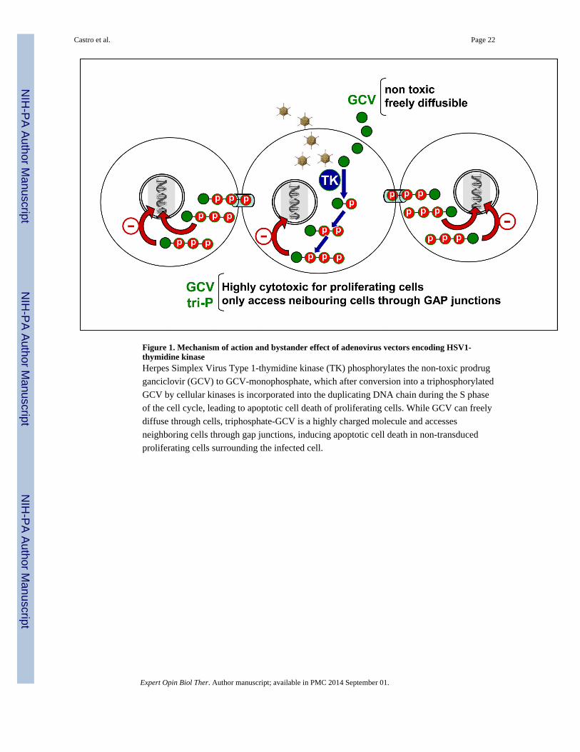
Figure 1. Mechanism of action and bystander effect of adenovirus vectors encoding HSV1-thymidine kinaseHerpes Simplex Virus Type 1-thymidine kinase (TK) phosphorylates the non-toxic prodrug
ganciclovir (GCV) to GCV-monophosphate, which after conversion into a triphosphorylated
GCV by cellular kinases is incorporated into the duplicating DNA chain during the S phase
of the cell cycle, leading to apoptotic cell death of proliferating cells. While GCV can freely
diffuse through cells, triphosphate-GCV is a highly charged molecule and accesses
neighboring cells through gap junctions, inducing apoptotic cell death in non-transduced
proliferating cells surrounding the infected cell.
Castro et al. Page 22
Expert Opin Biol Ther. Author manuscript; available in PMC 2014 September 01.
NIH
-PA
Author M
anuscriptN
IH-P
A A
uthor Manuscript
NIH
-PA
Author M
anuscript
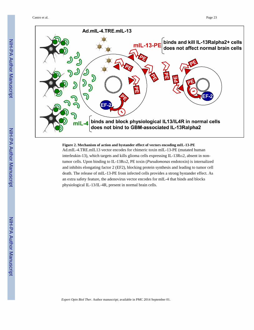
Figure 2. Mechanism of action and bystander effect of vectors encoding mIL-13-PEAd.mIL-4.TRE.mIL13 vector encodes for chimeric toxin mIL-13-PE (mutated human
interleukin-13), which targets and kills glioma cells expressing IL-13Rα2, absent in non-
tumor cells. Upon binding to IL-13Rα2, PE toxin (Pseudomonas endotoxin) is internalized
and inhibits elongating factor 2 (EF2), blocking protein synthesis and leading to tumor cell
death. The release of mIL-13-PE from infected cells provides a strong bystander effect. As
an extra safety feature, the adenovirus vector encodes for mIL-4 that binds and blocks
physiological IL-13/IL-4R, present in normal brain cells.
Castro et al. Page 23
Expert Opin Biol Ther. Author manuscript; available in PMC 2014 September 01.
NIH
-PA
Author M
anuscriptN
IH-P
A A
uthor Manuscript
NIH
-PA
Author M
anuscript
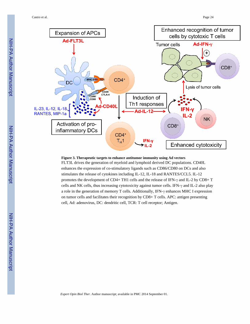
Figure 3. Therapeutic targets to enhance antitumor immunity using Ad vectorsFLT3L drives the generation of myeloid and lymphoid derived DC populations. CD40L
enhances the expression of co-stimulatory ligands such as CD86/CD80 on DCs and also
stimulates the release of cytokines including IL-12, IL-18 and RANTES/CCL5. IL-12
promotes the development of CD4+ TH1 cells and the release of IFN-γ and IL-2 by CD8+ T
cells and NK cells, thus increasing cytotoxicity against tumor cells. IFN-γ and IL-2 also play
a role in the generation of memory T cells. Additionally, IFN-γ enhances MHC I expression
on tumor cells and facilitates their recognition by CD8+ T cells. APC: antigen presenting
cell, Ad: adenovirus, DC: dendritic cell, TCR: T cell receptor; Antigen.
Castro et al. Page 24
Expert Opin Biol Ther. Author manuscript; available in PMC 2014 September 01.
NIH
-PA
Author M
anuscriptN
IH-P
A A
uthor Manuscript
NIH
-PA
Author M
anuscript
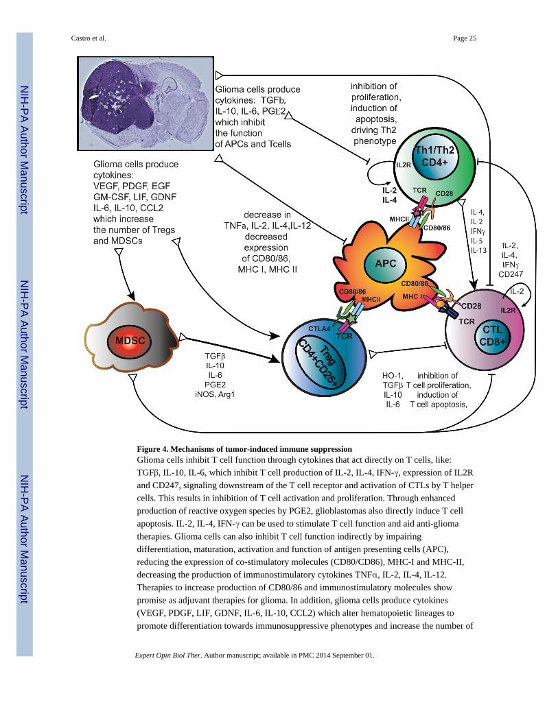
Figure 4. Mechanisms of tumor-induced immune suppressionGlioma cells inhibit T cell function through cytokines that act directly on T cells, like:
TGFβ, IL-10, IL-6, which inhibit T cell production of IL-2, IL-4, IFN-γ, expression of IL2R
and CD247, signaling downstream of the T cell receptor and activation of CTLs by T helper
cells. This results in inhibition of T cell activation and proliferation. Through enhanced
production of reactive oxygen species by PGE2, glioblastomas also directly induce T cell
apoptosis. IL-2, IL-4, IFN-γ can be used to stimulate T cell function and aid anti-glioma
therapies. Glioma cells can also inhibit T cell function indirectly by impairing
differentiation, maturation, activation and function of antigen presenting cells (APC),
reducing the expression of co-stimulatory molecules (CD80/CD86), MHC-I and MHC-II,
decreasing the production of immunostimulatory cytokines TNFα, IL-2, IL-4, IL-12.
Therapies to increase production of CD80/86 and immunostimulatory molecules show
promise as adjuvant therapies for glioma. In addition, glioma cells produce cytokines
(VEGF, PDGF, LIF, GDNF, IL-6, IL-10, CCL2) which alter hematopoietic lineages to
promote differentiation towards immunosuppressive phenotypes and increase the number of
Castro et al. Page 25
Expert Opin Biol Ther. Author manuscript; available in PMC 2014 September 01.
NIH
-PA
Author M
anuscriptN
IH-P
A A
uthor Manuscript
NIH
-PA
Author M
anuscript
Myeloid Derived Suppressor Cells (MDSCs) and regulatory T cells (Tregs). These are
recruited to the tumor microenvironment and circulate back to lymphoid organs to suppress
anti-tumor immune responses further. Abbreviations used: Arg1, arginase 1; CCL2,
Chemokine ligand 2 (also known as monocyte chemotactic protein -1 MCP-1); EGF,
epidermal growth factor; GDNF, glial derived neurotrophic factor; GM-CSF, granulocyte
macrophage colony stimulating factor; HO-1 heme oxygenase 1; iNOS, inducible nitric
oxide synthase (also known as NOS2); LIF, leukemia inhibitory factor; PGE2, Prostaglandin
E2; PDGF, platelet-derived growth factor; TGFβ, Transforming growth factor beta; TNFα,
Tumor necrosis factor alpha, VEGF, vascular endothelial growth factor.
Castro et al. Page 26
Expert Opin Biol Ther. Author manuscript; available in PMC 2014 September 01.
NIH
-PA
Author M
anuscriptN
IH-P
A A
uthor Manuscript
NIH
-PA
Author M
anuscript
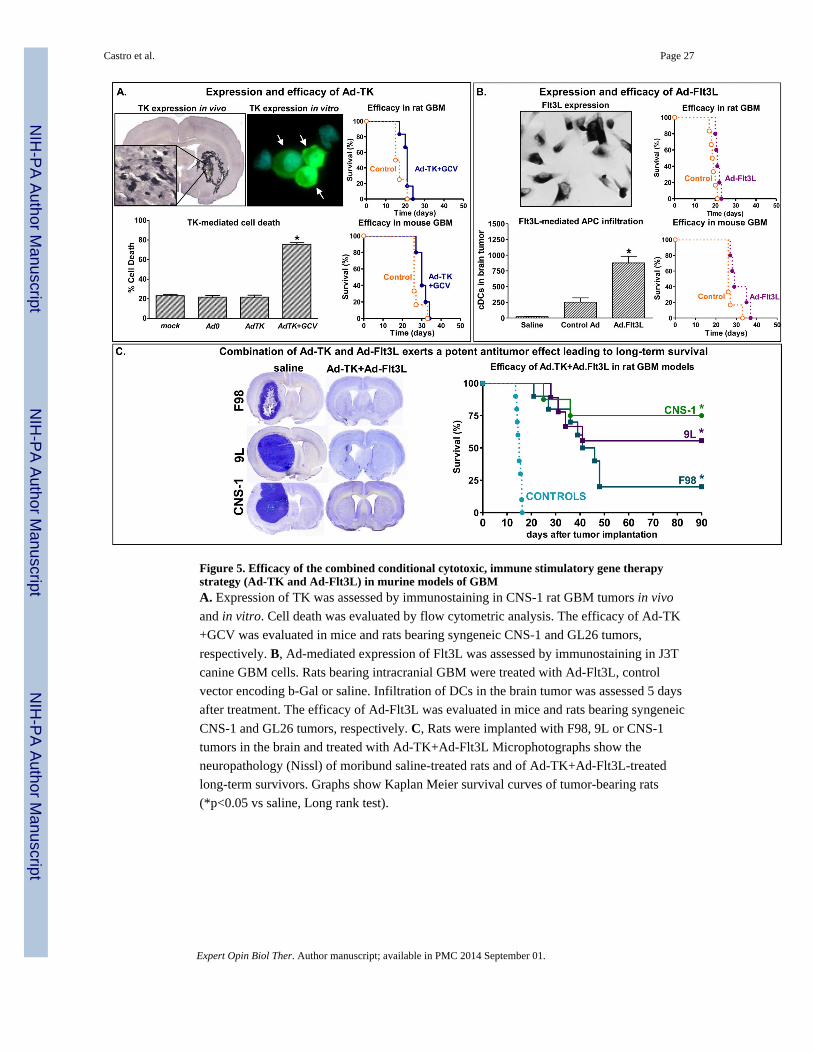
Figure 5. Efficacy of the combined conditional cytotoxic, immune stimulatory gene therapystrategy (Ad-TK and Ad-Flt3L) in murine models of GBMA. Expression of TK was assessed by immunostaining in CNS-1 rat GBM tumors in vivo
and in vitro. Cell death was evaluated by flow cytometric analysis. The efficacy of Ad-TK
+GCV was evaluated in mice and rats bearing syngeneic CNS-1 and GL26 tumors,
respectively. B, Ad-mediated expression of Flt3L was assessed by immunostaining in J3T
canine GBM cells. Rats bearing intracranial GBM were treated with Ad-Flt3L, control
vector encoding b-Gal or saline. Infiltration of DCs in the brain tumor was assessed 5 days
after treatment. The efficacy of Ad-Flt3L was evaluated in mice and rats bearing syngeneic
CNS-1 and GL26 tumors, respectively. C, Rats were implanted with F98, 9L or CNS-1
tumors in the brain and treated with Ad-TK+Ad-Flt3L Microphotographs show the
neuropathology (Nissl) of moribund saline-treated rats and of Ad-TK+Ad-Flt3L-treated
long-term survivors. Graphs show Kaplan Meier survival curves of tumor-bearing rats
(*p<0.05 vs saline, Long rank test).
Castro et al. Page 27
Expert Opin Biol Ther. Author manuscript; available in PMC 2014 September 01.
NIH
-PA
Author M
anuscriptN
IH-P
A A
uthor Manuscript
NIH
-PA
Author M
anuscript
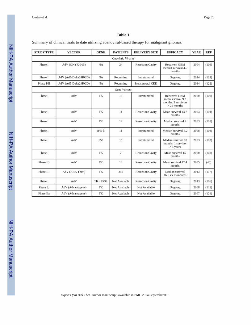
NIH
-PA
Author M
anuscriptN
IH-P
A A
uthor Manuscript
NIH
-PA
Author M
anuscript
Castro et al. Page 28
Table 1
Summary of clinical trials to date utilizing adenoviral-based therapy for malignant gliomas.
STUDY TYPE VECTOR GENE PATIENTS DELIVERY SITE EFFICACY YEAR REF
Oncolytic Viruses
Phase I AdV (ONYX-015) NA 24 Resection Cavity Recurrent GBMmedian survival 4.9
months
2004 (109)
Phase I AdV (Ad5-Delta24RGD) NA Recruiting Intratumoral Ongoing 2014 (121)
Phase I/II AdV (Ad5-Delta24RGD) NA Recruiting Intratumoral CED Ongoing 2014 (122)
Gene Vectors
Phase I AdV TK 13 Intratumoral Recurrent GBMmean survival 9.2
months. 3 survivors> 25 months
2000 (100)
Phase I AdV TK 11 Resection Cavity Mean survival 13.7months
2003 (101)
Phase I AdV TK 14 Resection Cavity Median survival 4months
2003 (103)
Phase I AdV IFN-β 11 Intratumoral Median survival 4.2months
2008 (108)
Phase I AdV p53 15 Intratumoral Median survival 10months. 1 survivor
> 3 years
2003 (107)
Phase I AdV TK 7 Resection Cavity Mean survival 15months
2000 (102)
Phase IB AdV TK 13 Resection Cavity Mean survival 12.4months
2005 (45)
Phase III AdV (ARK Ther.) TK 250 Resection Cavity Median survival16.5 vs 15 months
2013 (117)
Phase I AdV TK+ Flt3L Not Available Resection Cavity Ongoing 2013 (106)
Phase Ib AdV (Advantagene) TK Not Available Not Available Ongoing 2008 (123)
Phase IIa AdV (Advantagene) TK Not Available Not Available Ongoing 2007 (124)
Expert Opin Biol Ther. Author manuscript; available in PMC 2014 September 01.