nih public accessmona batish salvatore marras lev
TRANSCRIPT

Highly Bright Avidin-based Affinity Probes Carrying MultipleLanthanide Chelates
Laura Wirpsza†,‡, Shyamala Pillai†,‡, Mona Batish‡, Salvatore Marras‡, Lev Krasnoperov†,and Arkady Mustaev‡,*
†Department of Chemistry and Environmental Sciences, New Jersey Institute of Technology, 151Tiernan Hall, University Heights, Newark, New Jersey 07102‡PHRI Center, New Jersey Medical School, Department of Microbiology and Molecular Genetics,University of Medicine and Dentistry of New Jersey, 225 Warren Street, Newark, New Jersey07103
AbstractLanthanide ion luminescence has a long lifetime enabling highly sensitive detection in time-gatedmode. The sensitivity can be further increased by using multiple luminescent labels attached to acarrier molecule, which can be conjugated to an object of interest. We found that up to 30lanthanide chelates can be attached to avidin creating highly bright constructs. These constructswith Eu3+ chelates display synergistic effect that enhance the brightness of heavily modifiedsamples, while the opposite effect was observed for Tb3+ chelates thereby significantly reducingtheir light emission. This undesirable quenching of Tb3+ luminophores was completely suppressedby the introduction of an aromatic spacer between the chelate and the protein attachment site. Theestimated detection limit for the conjugates is in the 10-14 – 10-15 M range. We demonstrated ahigh sensitivity for the new probes by using them to label living cells of bacterial and mammalianorigin.
KeywordsLanthanide; luminescense; multiple; chelates; labeling; synthesis
1. IntroductionLong lifetime of lanthanide luminescence allows its highly sensitive detection in time-gatedmode [1-9], making luminescent probes an attractive alternative to radioisotopes. Tocompensate for the low inherent absorbance of lanthanide ions, the luminescent probescontain an antenna fluorophore, which absorbs the light and transfers the energy to atethered Ln3+ ion that finally emits the light [3 and references therein]. One of the ways tosignificantly increase the detection sensitivity of light-emitting probes is to bundle themonto a carrier molecule, which then can be attached to an object of interest [10-11]. Withconventional fluorophores this approach is complicated due to self-quenching, which isfacilitated by the fluorescence resonance energy transfer (FRET) from an excited to a nearbynon-excited dye molecule that efficiently absorbs the energy [10-11]. The degree of
*To whom correspondence should be addressed. [email protected], Phone: 973-854-3442, Fax: 973-854-3101.
Publisher's Disclaimer: This is a PDF file of an unedited manuscript that has been accepted for publication. As a service to ourcustomers we are providing this early version of the manuscript. The manuscript will undergo copyediting, typesetting, and review ofthe resulting proof before it is published in its final citable form. Please note that during the production process errors may bediscovered which could affect the content, and all legal disclaimers that apply to the journal pertain.
NIH Public AccessAuthor ManuscriptJ Photochem Photobiol B. Author manuscript; available in PMC 2013 November 05.
Published in final edited form as:J Photochem Photobiol B. 2012 November 5; 116: 22–29. doi:10.1016/j.jphotobiol.2012.07.001.
$waterm
ark-text$w
atermark-text
$waterm
ark-text

quenching is highly dependent on the spectral overlap between the excitation (absorption)and emission of a particular fluorophore. The majority of conventional fluorophores have asmall (10-30 nm) Stokes shift (the spectral separation between the emission and absorptionmaxima) causing a significant spectral overlap. High molar extinction of the commonfluorescent dyes also contributes to quenching. On the contrary, lanthanide luminescentprobes possess an extremely large Stokes shift (150-250 nm), which prevents efficientenergy transfer between the excited and non-excited fluorophore molecules [12]. Previously,this approach was explored on streptavidin with Eu3+ chelate [12]. Parent protein, avidinpossesses 32 lysine residues at which luminescent labels can be attached, which makes it asuperior scaffold for multiple label attachment comparing to streptavidin (which has 12lysine residues). In the present study, we obtained avidin conjugates with a new generationof high-quantum-yield lanthanide chelates of Eu3+ and Tb3+ containing cs124 and cs124-CF3 antennae-fluorophores (Chart 1) synthesized by us in the course of current and previousstudies [13]. We find that unlike typical fluorophore BODIPY, the light emission efficiencyof the Eu3+ probes was not affected by self-quenching. In fact, the cumulative luminescenceof the conjugate as a function of the number of the attached residues displayed a super-linearbehavior, suggesting synergistic effect [12]. We found that this effect was due to theenhanced antenna-to-lanthanide energy transfer. We tested the same approach with Tb3+-based luminescent probes, which possess higher quantum yield compared to the cs124 Eu3+
chelates. Significant self-quenching was observed when these multiple Tb3+ probes wereattached to avidin. However, introduction of a biphenyl spacer between the chelate and thecrosslinking group completely suppressed the quenching, yielding highly bright conjugates.The obtained luminescent avidin constructs were used for labeling bacterial and mammaliancells giving highly contrast images in time-resolved detection mode. These new probes canfind a broad range of applications in the biological and biomedical fields that rely on highdetection sensitivity.
2. MATERIALS AND METHODSThe following reagents were purchased from Sigma Aldrich: Avidin,diethylenetriaminepentaacetic acid dianhydride (DTPA), triethylamine; butylamine; 1,3-phenylenediamine; ethyl 4,4,4-trifluoroacetoacetate; ethylacetoacetate, 1,3-dicyclohexylcarbodiimide (DCC), ethylenedianime; methylbromacetate; anhydrousdimethylformamide and dimethylsulfoxide; 1-butanol, ethylacetate, chloroform; acetonitrile;ethanol; sodium and potassium hydroxide; TbCl3 and EuCl3; silica gel TLC plates onaluminum foil (200 μm layer thick with a fluorescent indicator). Distilled and deionizedwater (18 MΩ cm-1) was used. All experiments including lanthanide complexes preparationand using thereof were performed either in glassware washed with mixed acid solution andrinsed with metal-free water, or in metal-free plasticware purchased from Biorad. Allchemicals were the purest grade available. Probes I and II were synthesized as previouslydescribed [13]. Biotinylated oligonucleotide containing BHQ had a structure: 5’NH2 –ACCTGGTGCCTCGTCGCCGCAGCTCAGG dT (BHQ2) TT-3’ – biotin. NHS-dPEG12-biotin was purchased from Quanta Biodesign.
2.1 Synthesis2.1.1 Probe 4 (Fig. 1)2.1.1.1. Product I: To a solution of 106 mg (0.6 mmol) of cs124 in 0.8 ml of DMF 72 μl of10 M NaOH was added followed by rigorous agitation until the water phase disappeared.This solution was mixed with a 300 mg 4, 4’-bis (chloromethyl) biphenyl dissolved in 2 mlof DMF. After 20 min incubation at room temperature the TLC analysis in hexane-acetone(1:1) revealed the formation of a single reaction product. The mixture was supplementedwith 100 mg of lithium azide and heated for 20 min at 50 °C followed by precipitation with
Wirpsza et al. Page 2
J Photochem Photobiol B. Author manuscript; available in PMC 2013 November 05.
$waterm
ark-text$w
atermark-text
$waterm
ark-text
20 ml of water. The residue was collected by centrifugation, washed with water anddissolved in 20 ml of hot acetonitrile. The acetonitrile was removed by evaporation underreduced pressure and the residue was washed a few times with hot hexane and subjected tosilica gel chromatography in hexane-acetone (1:1) developing system. Yield-120 mg. 1HNMR in DMSO: 7.65 (dd, 4H, o,o’biphenyl H, J1 = 11.1, J2 = 8.4), 7.45 (dd overlapped, 1H,5H), 7.45 (dd,2H, biphenyl m-H, J1 = 8.25, J2 = 5.1), 7.25 (d, 2H, biphenyl-m’- H, J – 8.1),6.49 (d, 1H, 6H), 6.44 (dd, 1H, 3H, J= 1.8), 6.21 (s, 1H, 8H), 5.8 (s, 2H, 7-amino), 5.38(s,2H, N-CH2), 4.4 (s, 2H, -CH2-N3), 2.36 (d, 3H, 4-methyl, J = 0.9).
2.1.1.2. Product II: Solution of 68 mg of product I in 0.5 ml of DMF was supplementedwith 1.5 molar excess of triphenylphosphine, incubated for 1 h at 50 °C and 0.13 ml of 25 %aqueous ammonium hydroxide was added. The mixture was incubated for 1 h at 50 °C andleft for 20 min at - 20 °C. The precipitate was collected by centrifugation, washed by etherand dried in vacuo affording 36 mg of product II.
2.1.1.3. Product III: The solution of 30 mg of product II in 0.5 ml of DMSO was titratedwith thiocarbonyldiimidazole dissolved in 0.1 ml of chloroform. Addition was continueduntil the subsequent portion of C(S) Im2 stopped decolorizing. The reaction mixture wasanalyzed by TLC in hexane-acetone (1:1) developing system revealing nearly completeconversion of the original cs124 derivative. Small excess of C(S) Im2 was required tocomplete the reaction. The mixture was supplemented with 5 μl of TFA and left for 1 h at45 °C. The reaction was monitored by TLC. The product was precipitated by water (13 ml),collected by centrifugation and washed by water two more times. Most of the remainingresidue was dissolved in 10 ml of acetonitrile and the remaining material was removed bycentrifugation. Acetonitrile solution was evaporated to dryness in vacuo affording 20 mg ofproduct III. 1H NMR in DMSO: 7.66 (m, 4H, o,o’biphenyl H), 7.48 (dd overlapped, 1H,5H, J=2.1), 7.45 (d, 2H, biphenyl m-H, J = 8.1), 7.3 (d, 2H, biphenyl-m’- H, J – 8.4), 6.7 (s,1H, 8H), 6.62 (dd, 1H, 6H, J1=9.0, J2 = 1.8), 6.55 (d, 1H, 3H, J=1.8), 6.25 (s, 2H, 7 amino),5.5 (s, 2H, -CH2-NCS), 4.48 (s, 2H, N-CH2).
2.1.1.4. Probe 4: Solution of 35 mg (0.1 mmol) of DTPA dianhydride in 0.3 ml of DMSOobtained under heating to 60-80 °C was cooled down to room temperature and added to 20mg (0.048 mmol) of compound III. The reaction was carried on for 15 min at 20 °C. Themixture was supplemented with 4 ml of water, left for 20 min at room temperature and pHwas adjusted to 5.0 by LiOH. The product was purified by preparative C-18 HPLC column(20 × 250 mm) using linear gradient (0.5: l) of acetonitrile in water (0 – 70 %). The elutionrate was 2 ml/min. The fractions containing desired product were combined andsupplemented with one equivalent of a lanthanide salt. The resulting solutions wereconcentrated in vacuo by co-evaporation with acetonitrile under gentle heating (25-30 °C) tofinal concentration 20 mM.
2.2 Modification of avidin with light-emitting probesThe reaction cocktails (10-16 μl) were composed by mixing of 7 μl of avidin (20 mg/ml), 1μl of 1 M sodium borate buffer pH 10.0, and 1-8 μl of a reactive light-emitting probe atconcentrations specified in figure legends. After incubation for 4 h at 56 °C the mixtureswere diluted to 100 μl by water and subjected to size-exclusion chromatography onSephadex G-50 “medium” in 10 mM Hepes-HCl buffer pH 8.0 containing 50 mM NaCl.The fractions corresponding to modified avidin were collected by visual detection using UVmonitor (365 nm light).
Wirpsza et al. Page 3
J Photochem Photobiol B. Author manuscript; available in PMC 2013 November 05.
$waterm
ark-text$w
atermark-text
$waterm
ark-text

2.3 Preparation of E. coli cells labeled with modified avidinLB broth (100 ml) was inoculated with suspension of 10 μl of E. coli cells (RL721 strain)and incubated in a 500 mL Erlenmeyer flask overnight at 37 °C. The cells were harvested bycentrifugation (4000 rpm, 5 min), washed with PBS and re-suspended in the same buffercontaining 50 % glycerol at a final density of 32 mg ml-1. Thirty microliters of thissuspension containing ca. 1 mg of cells was washed 3 times with 1 ml of 0.1 M sodiumborate buffer, pH 8.5, and each time collected by centrifugation. After the last wash, thecells were suspended in 50 μl of the same buffer and 4 μl of 100 mM DMSO solution ofNHS-dPEG12-biotin was added. After incubation at room temperature for 30 minutes thecells were washed 4 times with 500 μl of PBS. After the final wash, cells were suspended in15 μl of PBS buffer and supplemented with 15 μl of 5 μM avidin modified with one of thelanthanide labels [AV -Probe 4 -Tb3+ (n=15) and AV- Probe 1-Eu3+ (n=19)]. After 25minutes of the incubation at room temperature cells were washed by PBS (4 × 500 μl) andsuspended in 100 μl of the same buffer.
2.4 Preparation of Chinese hamster ovarian cells (CHO) labeled with modified avidinCHO cells were grown in Dulbecco’s modified Eagle’s medium, supplemented with 10%fetal bovine serum, 200mM L-glutamine and 100 μg/ml Penicillin/streptomycin solution.Once the cells reach 80-90% confluency, they were trypsinized and collected bycentrifugation (1000 rpm for 5 min), washed with 0.1 M Na-borate buffer pH 8.5 (3 × 0.5ml) and spun down at 3,000 rpm for 30 seconds. The cells were suspended in 500 μl ofsodium-borate buffer, divided into 5 equivalent volumes 100 μl each, and supplementedwith various volumes (0.5 μl, 1 μl, 2 μl, and 4 μl) DMSO solution of 100 mM NHS-dPEG12-biotin. 100 μl of the suspension was kept for auto fluorescence reference. The cellsin each tube were washed with 150 μl of PBS buffer three times, suspended in 20 μL ofPBS and supplemented with 15 μl of 5 μM AV- Probe 1-Eu3+. After incubation at roomtemperature for 20 minutes, the cells were washed with 100 μl of PBS buffer and suspendedin 50 μl of the same buffer.
2.5 Preparation of the cells for microscopic detectionOne microliter of Poly-L-Lysine was spread onto a fused silica microscope substrate into anarea of 0.3 cm2 and removed. One microliter of the cell suspension of labeled cells (E. colior CHO cells) containing 109-1010 cells cm-3 in PBS buffer was spread into the same areaand left to air dry for 15 minutes.
2.6 Physical MethodsExcitation and emission fluorescence spectra in the continuous excitation mode wererecorded using QuantaMaster 1 (Photon Technology International) digital fluorometer atambient temperature. Time-resolved and gated luminescence measurements were performedusing the previously described home-built experimental set-up [13]. A Hacker InstrumentsZetopan microscope was equipped with an ICCD Camera (PI-MAX, Princeton Instruments).In the experiments, the images were taken in luminescence light using evanescent waveexcitation at 351 nm as well as in scattered light using standard top illumination by xenonlamp. In the evanescent excitation, a right angle fused silica prism was illuminated withlaser light (351 nm) from a XeF laser (OPTEX, Lambda Physik). The sample was located onthe hypotenuse side of the prism positioned horizontally. Images taken in scattered lightfrom a xenon lamp were taken before and after the luminescent images collected in thephoton counting mode. Online thresholding mode was used to discriminate photon pulsesfrom the readout noise as well as the “cosmic events”. The 1024 × 1024 camera pixels were8 × 8 binned resulting in 128 × 128 pixel2 images. The microscope used an objective withthe magnification of ×56 and the numerical aperture of 0.90. Combined with the
Wirpsza et al. Page 4
J Photochem Photobiol B. Author manuscript; available in PMC 2013 November 05.
$waterm
ark-text$w
atermark-text
$waterm
ark-text
intermediate “ocular” lens with the magnification of ×10 it provided the field of view of 14× 14 μm2. In some experiments, an ×5 intermediate “ocular” lens was used resulting in the28 × 28 μm2 field of view.
2.6.1 Microscopic detection of living cells utilizing avidin labeled with multipleprobes using Total Internal Reflection Fluorescence Microscopy (TIRFM)—Thecells labeled with avidin carrying multiple probes (Probe 1-Eu3+ and Probe 4-Tb3+) wereplaced on the hypotenuse side of the prism mounted at the microscope base. The excitationof probes occurred in the evanescent wave by laser light totally internally reflected from thehypotenuse side inside the prism. The probes with Eu3+ have emission lifetime of ca. 0.5ms, while the probes with Tb3+ have emission lifetime of ca. 1.5 ms. Therefore, for sampleslabeled with Eu3+ probes we used a gate width of 1 msec and the gate delay of 50 μsec andfor samples labeled with Tb3+ probes 2 msec gate width and 100 μsec gate delay were used.Images in luminescence light were acquired in the photon counting mode; typically 1000pulses were accumulated (at the repetition rate of 2 Hz). The number of probes per cell wascalculated based on the total photon count with the subtraction of the background count.
The calibration of the set-up was performed by collection of luminescence light from a thinlayer of the probes solution excited directly by the laser beam at the right angle from thebottom of a thin fused silica substrate. The microscope field of view in these experimentswas 14 × 14 μm2. To achieve homogeneity of the excitation beam, the beam was passedthrough a 0.32 cm2 diaphragm. The pulse energy was measured after the diaphragm (0.32mJ pulse-1). This allowed a reliable determination of the laser light fluence. Measuredvolume of the probes solutions (1.12 mM Probe 1-Eu3+ or 0.107 mM Probe 4-Tb3+) inglycerol was placed on the top of the substrate and spread upon the surface with a cover slip(the spot area of 3.80 cm2 and the thickness of the layer of 2.63 μm). The luminescence lightintensity was calculated based on the photon fluence, the absorption cross-sections of theprobes at 351 nm (2.1 × 10⁻¹⁷ cm2 molecule-1 and 3.6 × 10⁻¹⁷ cm2 molecule-1 for probesEu3+ and Tb3+respectively), the luminescence quantum yield (0.167 for Eu3+ [14], and ca.0.45 for Tb3+ probe), and the total number of probes in the field-of-view area. This wascompared with the total number of photons counted in the image. This procedure alloweddetermination of the calibration coefficients, which lump sum the solid angle of lightcollection of the objective lens, the microscope throughput coefficient, the photocathodequantum efficiency, as well as the photon counting efficiency. The average number of theprobes per externally labeled E. coli cells determined in this way was 2.1×10⁵ and 2.9×10⁵
for Eu3+ and Tb3+ probes, respectively.
Externally labeled CHO cells were prepared in a similar manner. The cells were labeled withavidin conjugates carrying multiple Eu3+ chelates of probe 1 with an average 1.6 × 10⁷
probes per cell.
2.6.2 Measurement of light emission of lanthanide chelates in steady-statemode—The detection of light emission of a lanthanide chelates and their conjugates withavidin as well as of BODIPY-modified avidin was performed in a measuring cell (150 μl) ina buffer containing 10 mM Hepes pH 8.0. Water-based or deuterium oxide-based solutionswere used.
3. RESULTS3.1 The synthesis and properties of reactive luminescent lanthanide probe 4
In our previous study [15], we found a convenient modification reaction for the cs124CF3fluorophore, which allows introduction of the crosslinking groups at N1 position. Here weperformed the same reaction with parent cs124 compound in order to obtain probe 4 (Fig. 1).
Wirpsza et al. Page 5
J Photochem Photobiol B. Author manuscript; available in PMC 2013 November 05.
$waterm
ark-text$w
atermark-text
$waterm
ark-text
Similarly to corresponding trifluoro-derivative, alkylation of cs124 fluorophore bybifunctional biphenyl compound produced alkylation product at N1 with high yield (Fig. 2).Notably, alkylation proceeded almost exclusively at N-1 of the quinolone ring, while thesame reactions with ethyl ester of 4-toluenesulfonic acid or with 1-iodo-3-azidopropaneyielded detectable amount of O-alkylated products (15). This can be accounted for differentstructure of the reactive groups. Employing high molar excess of alkylating agentsuppressed the formation of crosslinked quinolone adducts. After the alkylation, theremaining chloromethylene group was quantitatively converted to an azido derivative(compound I) by incubation with LiN3. The later was reduced to corresponding amino-compound II by treatment with triphenylphosphine and ammonium hydroxide. Reactiveisothiocyano-derivative III was obtained by subsequent incubation of II withthiocarbonyldiimidazole and TFA. Acylation of compound III with DTPA dianhydrideproduced final product, which was chelated with Tb3+ ion by addition of TbCl3 to yieldprobe 4.
3.2 Modification of avidin by reactive light emitting labelsAs expected, incubation of various reactive fluorophores with avidin resulted in covalentattachment to the protein as judged by size-exclusion chromatography. The dependence ofthe number of attached fluorophore residues of probe 1, 2, and 4 as well as BODIPYfluorophore per avidin molecule on probes concentration is shown in Fig. 3. Since theprobes are amine-reactive it is expected that they will predominantly attach to lysineresidues. It can be seen that at a high concentration 24 to 31 out of 32 lysine residues of theprotein can be modified by the probes. Attempt to attach more then 4 BODIPY residues peravidin was not successful due to precipitation of the modified protein.
3.3 Light-absorbing and light-emitting properties of the lanthanide probes and theirconjugates with avidin
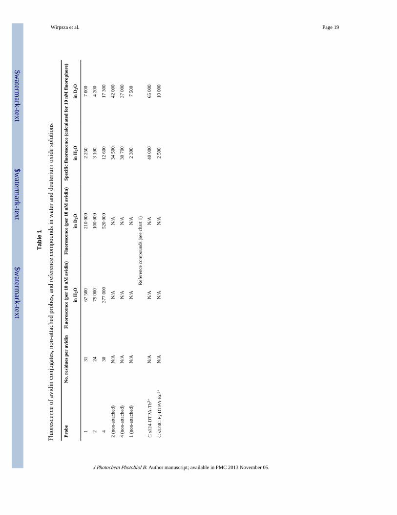
As seen from Fig. 4, in comparison to probe 2, probe 4 possesses a significant absorption inthe range of 240-300 nm, which is obviously due to the presence of the biphenylchromophore. Also, modification of the cs124 moiety at N1 causes a small (6 nm)batochromic shift of the absorption in the region of 320-360 nm. Biphenyl modification onlyslightly affects the brightness of the chelate as compared to the brightness of previouslydesigned probe 2 (Table 1 and Fig. 5A, B), which makes this position a convenient site forthe introduction of crosslinking or other functional groups. Strong light absorption of thebiphenyl group in the region 240-300 nm does not interfere with the light absorptionproperties of the antenna and antenna-to-lanthanide energy transfer, as biphenyl- andquinolone moieties do not form a common light-absorbing unit, being separated bymethylene group. As seen from Fig. 5A, a shift in the light absorption of probe 4 results inthe same shift of the fluorescence excitation spectrum. Also, the excitation spectrum ofprobe 4 displays a significant maximum in the region 240-300 nm where the biphenyl groupabsorbs the light. This is indicative for energy transfer from the excited state of the biphenylgroup to the cs124 chromophore, favored by close proximity of the moieties. Heavy watercaused a significant enhancement of lanthanide emission (Table 1) due to the elimination ofthe excitation energy dissipation by coordinated water molecule through O-H bondvibration.
Avidin represents highly stable tetrameric structure containing 32 lysine residues to whichthe reactive probes can be attached. Avidin binds tightly to biotin ligand producing virtuallyirreversible complex. This property of the protein makes it a convenient carrier for theattachment of various probes. Avidin conjugates thus obtained can be used to labelbiotinylated molecules of interest. It is seen (Tab. 1) that the attachment of Tb3+ luminescentchelates 2 and 4 to the protein at low concentration of the probes caused ca. 3 fold
Wirpsza et al. Page 6
J Photochem Photobiol B. Author manuscript; available in PMC 2013 November 05.
$waterm
ark-text$w
atermark-text
$waterm
ark-text

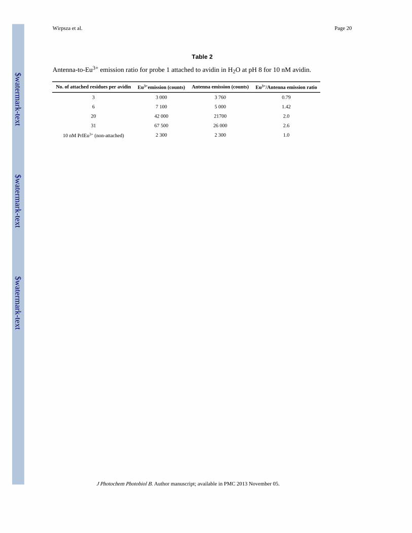
quenching comparing to emission of non-attached probes. For probe 2, increasing thenumber of attached probes resulted in further progressive quenching (Fig. 5C), while forprobe 4 the dependence of the cumulative fluorescent signal on the number of thecrosslinked probes remained linear. Attachment of Eu3+-based probe 1 also resulted in 3fold quenching, however when the number of the conjugated probes increased, a significantsuper-linear luminescence enhancement was observed (Fig. 5C). This effect can beexplained by enhancement of antenna-to-lanthanide energy transfer, which is supported bydecrease of antenna fluorescence and simultaneous increase of lanthanide emission in thecomplex (Tab. 2). One factor that reduces the brightness of the probe could be quenchingdue to the contact between the antenna-fluorophore and protein surface. This is supported bythe superior properties of the probe 4 possessing a rigid spacer between the antennafluorophore and the crosslinking group. This spacer could prevent the quenching byrestricting the fluorophore contacts with avidin.
As expected, light emission of avidin conjugates increased in heavy water (Tab. 1). Thus 1.3and 3 fold enhancement was observed for Tb3+ and Eu3+ chelates correspondingly, which isclose to enhancement factors for corresponding non-attached probes [13].
As seen from Fig. 5D, attachment of more than one BODIPY fluorophore to avidindramatically decreased the cumulative fluorescent signal due to expected FRET quenching.
3.4 Modified avidin conjugates are capable of biotin bindingExtensive modification of avidin could potentially interfere with biotin binding. To test thebinding ability of the modified protein, we titrated the conjugate with biotinylatedoligonucleotide carrying BHQ quencher. As seen from Fig. 6, incubation caused a dramaticdecrease in brightness suggesting quenching of the modified protein through binding of thebiotinylated oligonucleotide. As expected, ca. 4 fold excess of the oligo was required toachieve maximal quenching, which corresponds to saturation of all biotin binding sites.
3.5 Microscopic imaging of living cells using luminescent avidin conjugatesTo image the cells, we first treated them with acylating biotin derivative, which resulted incovalent attachment of the biotin residues to the cellular surface (Fig. 7A, B). As expected,subsequent incubation with luminescent labeled avidin conjugates resulted in the attachmentto the cells as judged by visual inspection under UV light. For microscopic imaging of thecells in time-gated mode we used Total Internal Reflection Fluorescence Microscopy(TIRFM) [16-17]. Total internal reflection is an optical phenomenon, which occurs whenlight propagating in a dense medium (such as glass) meets an interface with a less densemedium, such as water. If the light meets the interface at a small angle, some of the lightpassing through the interface is refracted and some is reflected back into the dense medium.At a certain angle all of the light is reflected. This angle is known as the critical angle, andits value depends on the refractive indices of the media (n1, n2): Θc = Sin-1(n1/n2). However,some of the energy of the beam propagates a short distance (a few hundred nanometers) intothe water, generating an evanescent wave. If this energy is not absorbed, it passes back intothe glass. However, if a fluorophore molecule is within the evanescent wave it can absorbphotons and be excited. In this way, it is possible to get fluorescence with a very lowbackground of excitation light. We used this principle in the design of the experimentalsetup for imaging of small luminescent objects (Fig. 8A). This allowed selective excitationof the surface attached objects. Repetitive laser pulses excited labeled cells and theluminescent signal collected after a short time delay allowing the decay of short-livedbackground fluorescence. Light emission images were acquired and accumulated using anICCD camera. Optical and time-gated luminescent images for bacterial and mammaliancells are shown in Fig. 8B. As expected, the images were highly contrasted.
Wirpsza et al. Page 7
J Photochem Photobiol B. Author manuscript; available in PMC 2013 November 05.
$waterm
ark-text$w
atermark-text
$waterm
ark-text
4 DISCUSSIONThis study demonstrates the fact that multiple luminescent chelates can be attached to avidinmolecule to create hypersensitive affinity probes that can be coupled to variousbiomolecules of interest. Avidin is a convenient protein for design of such probes due to itsrelatively small size (4-5 nm) and large number of exposed Lys residues to which thelanthanide chelates can be attached. Using a high concentration of reactive lanthanide labels,we were able to introduce up to 30-31 luminescent residues in a single avidin moleculeproducing highly bright conjugates. Eu3+ conjugates of probe 1 displayed fortuitousadditional signal enhancement apparently caused by proximation of the labels at the proteinsurface, which resulted in the improvement of antenna-to-lanthanide energy transfer. Thenature of this effect is not quite clear. Enhanced energy transfer could arise due toscavenging of the fraction of the antenna light (that has not been transferred to thelanthanide) by another closely positioned antenna molecule, which then transfers theabsorbed energy to the chelated lanthanide. Indeed, small overlapping of the emission andabsorption spectra of the antenna fluorophore of probe 1 is consistent with the suggestedmechanism. Also, the excited antenna could transfer the energy to the lanthanide ion of theneighboring probe. Finally, carboxylate amino acid residues on the protein surface can beinvolved in the additional coordination of the lanthanide, displacing a water molecule fromits coordination sphere, thereby enhancing the quantum yield of the metal emission [18].Terbium-based multiple label constructs displayed a significant decrease of light emissioncomparing to the sum of equivalent number of non-attached probes, which was most likelydue to the interaction of the chelate with the protein surface. Another factor of reducing thelight emission could be contact quenching resulting from the approximation of theneighboring antennae-fluorophores at high labeling density. Luminescent quenching can besuppressed by the presence of a biphenyl spacer. Generally, the rigid biphenyl group canrestrict the fluorophore contacts with the protein, and also prevent the contact quenching byinterfering with stacking interactions of the antennae.
5. CONCLUSIONSWe obtained avidin conjugates carrying multiple lanthanide chelated with detection limit in1-10 fM range as estimated by the detection sensitivity of single non-attached probes usedfor labeling. These conjugates can find wide application in biological, biophysical andbiomedical studies. They can be especially useful for imaging of single molecules,biological micro objects, and body tissues as well as the development of highly sensitiveassays in which the signal cannot be amplified (e.g. using PCR amplification technique).
AcknowledgmentsThis study was supported by NIH grant RO1 GM-307-17-21 to AM and NIH grant RO1 MN-079197 for SM andMB.
LITERATURE CITED1. Eliseeva S, Bünzli JC. Lanthanide luminescence for functional materials and bio-sciences. Chem
Soc Rev. 2010; 39:189–227. [PubMed: 20023849]
2. Hagan AK, Zuchner T. Lanthanide-based time-resolved luminescence immunoassays. Anal BioanalChem. 2011; 400:2847–2864. [PubMed: 21556751]
3. Bunzli JC. Benefiting from the unique properties of lanthanide ions. Acc Chem Res. 2006; 39:53–61. [PubMed: 16411740]
4. Dickson EF, Pollak A, Diamandis EP. Time-resolved detection of lanthanide luminescence forultrasensitive bioanalytical assays. J Photochem Photobiol B. 1995; 27:3–19. [PubMed: 7699520]
Wirpsza et al. Page 8
J Photochem Photobiol B. Author manuscript; available in PMC 2013 November 05.
$waterm
ark-text$w
atermark-text
$waterm
ark-text

5. Dickson EF, Pollak A, Diamandis EP. Ultrasensitive bioanalytical assays using time-resolvedfluorescence detection. Pharmacol Ther. 1995; 66:207–235. [PubMed: 7667396]
6. Hemmila I, Laitala V. Progress in lanthanides as luminescent probes. J Fluoresc. 2005; 15:529–42.[PubMed: 16167211]
7. Parker D. Excitement in f block: structure, dynamics and function of nine-coordinate chirallanthanide complexes in aqueous media. Chem Soc Rev. 2004; 33:156–165. [PubMed: 15026820]
8. Selvin PR. Principles and biophysical applications of lanthanide-based probes. Annu Rev BiophysBiomol Struct. 2002; 31:275–302. [PubMed: 11988471]
9. Werts MH. Making sense of lanthanide luminescence. Sci Prog. 2005; 88:101–131. [PubMed:16749431]
10. Rowley G, Henriksson T, Louie A, Nguyen P, Kramer M, Der-Balian G, Kameda N.Fluoroimmunoassays for Ferritin and IgE. Clin Chem. 1987; 33:1563.
11. Haralambidis J, Angus K, Pownall S, Duncan L, Chai M, Tregear J. The preparation of polyamide-oligonucleotide probes containing multiple non-radioactive labels. Nucl Acid Res. 1990; 18:501–505.
12. Diamandis E. Multiple labeling and time-resolvable fluorophores. Clin Chem. 1991:1486–1491.[PubMed: 1716533]
13. Krasnoperov L, Marras S, Kozlov M, Wirpsza L, Mustaev A. Luminescent probes forultrasensitive detection of nucleic acids. Bioconjugate Chem. 2010; 21:319–327.
14. Xiao M, Selvin PR. Quantum Yields of Luminescent Lanthanide Chelates and Far-Red DyesMeasured by Resonance Energy Transfer. J Am Chem Soc. 2001; 123:7067–7073. [PubMed:11459485]
15. Pillai S, Kozlov M, Marras SAE, Krasnoperov L, Mustaev A. New Cross-linking Quinoline andQuinolone Derivatives for Sensitive Fluorescent Labeling. J Fluorescence. Mar 28.2012 Epubahead of print.
16. Wazawa T, Ueda M. Total internal reflection fluorescence microscopy in single moleculenanobioscience. Adv Biochem Eng Biotechnol. 2005; 95:77–106. [PubMed: 16080266]
17. Schneckenburger H. Total internal reflection fluorescence microscopy: technical innovations andnovel applications. Curr Opin Biotechnol. 2005; 16:13–28. [PubMed: 15722010]
18. Li M, Selvin PR. Luminescent polyaminocarboxylate chelates of Terbium and Europium: Theeffect of Chelate Structure. J Am Chem Soc. 1995; 117:8132–8138.
Wirpsza et al. Page 9
J Photochem Photobiol B. Author manuscript; available in PMC 2013 November 05.
$waterm
ark-text$w
atermark-text
$waterm
ark-text

Highlights
• Luminescent avidin probes carrying multiple Eu and Tb chelates were obtained.
• Detection sensitivity of the probes was in 10-14 – 10-15 M range.
• The probes were used for labeling and time-resolved imaging of living cells.
Wirpsza et al. Page 10
J Photochem Photobiol B. Author manuscript; available in PMC 2013 November 05.
$waterm
ark-text$w
atermark-text
$waterm
ark-text
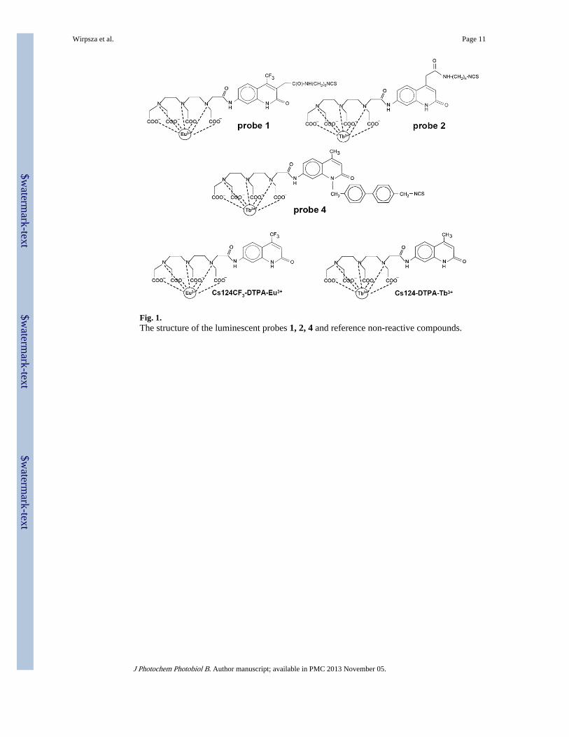
Fig. 1.The structure of the luminescent probes 1, 2, 4 and reference non-reactive compounds.
Wirpsza et al. Page 11
J Photochem Photobiol B. Author manuscript; available in PMC 2013 November 05.
$waterm
ark-text$w
atermark-text
$waterm
ark-text

Fig. 2.Synthetic route for probe 4.
Wirpsza et al. Page 12
J Photochem Photobiol B. Author manuscript; available in PMC 2013 November 05.
$waterm
ark-text$w
atermark-text
$waterm
ark-text

Fig. 3.Dependence of the number of the attached probes 1, 2, and 4 (A) and BODIPY residues (B)per avidin on the reactive fluorophores concentration in the reaction mixture.
Wirpsza et al. Page 13
J Photochem Photobiol B. Author manuscript; available in PMC 2013 November 05.
$waterm
ark-text$w
atermark-text
$waterm
ark-text
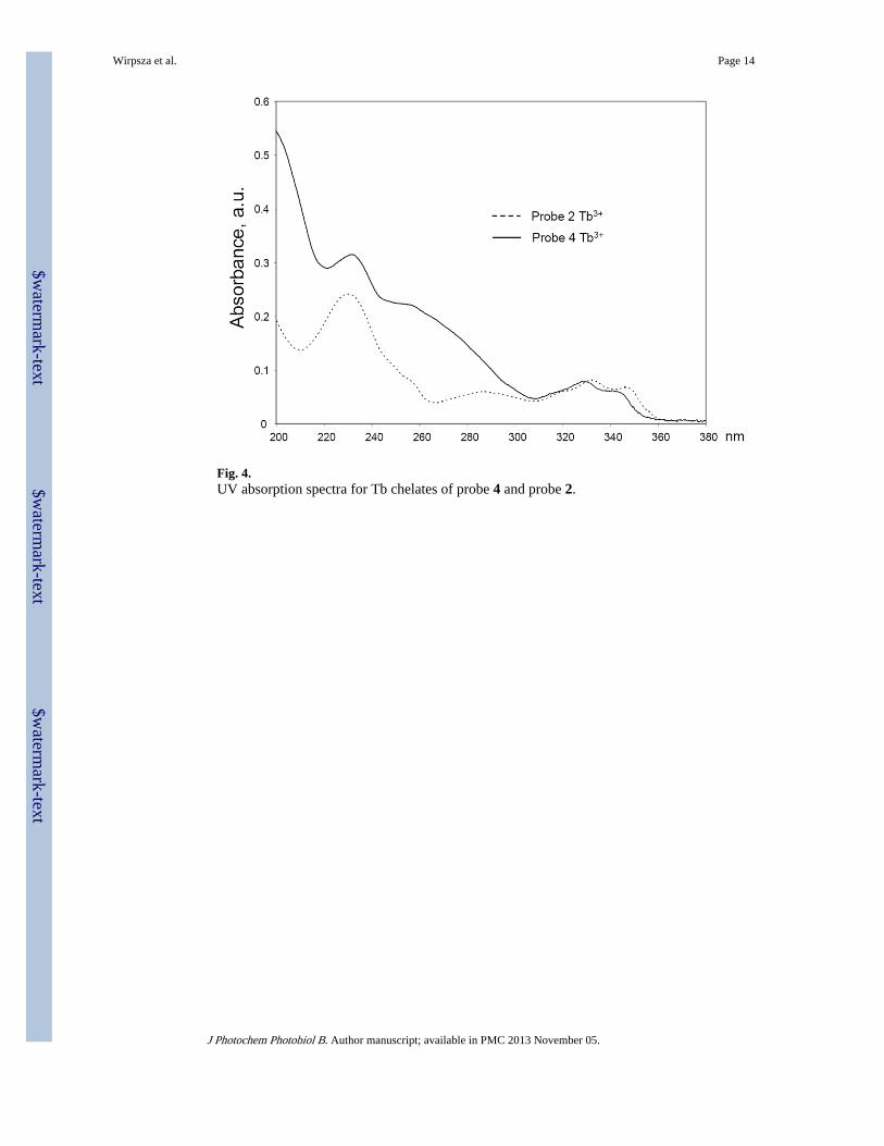
Fig. 4.UV absorption spectra for Tb chelates of probe 4 and probe 2.
Wirpsza et al. Page 14
J Photochem Photobiol B. Author manuscript; available in PMC 2013 November 05.
$waterm
ark-text$w
atermark-text
$waterm
ark-text
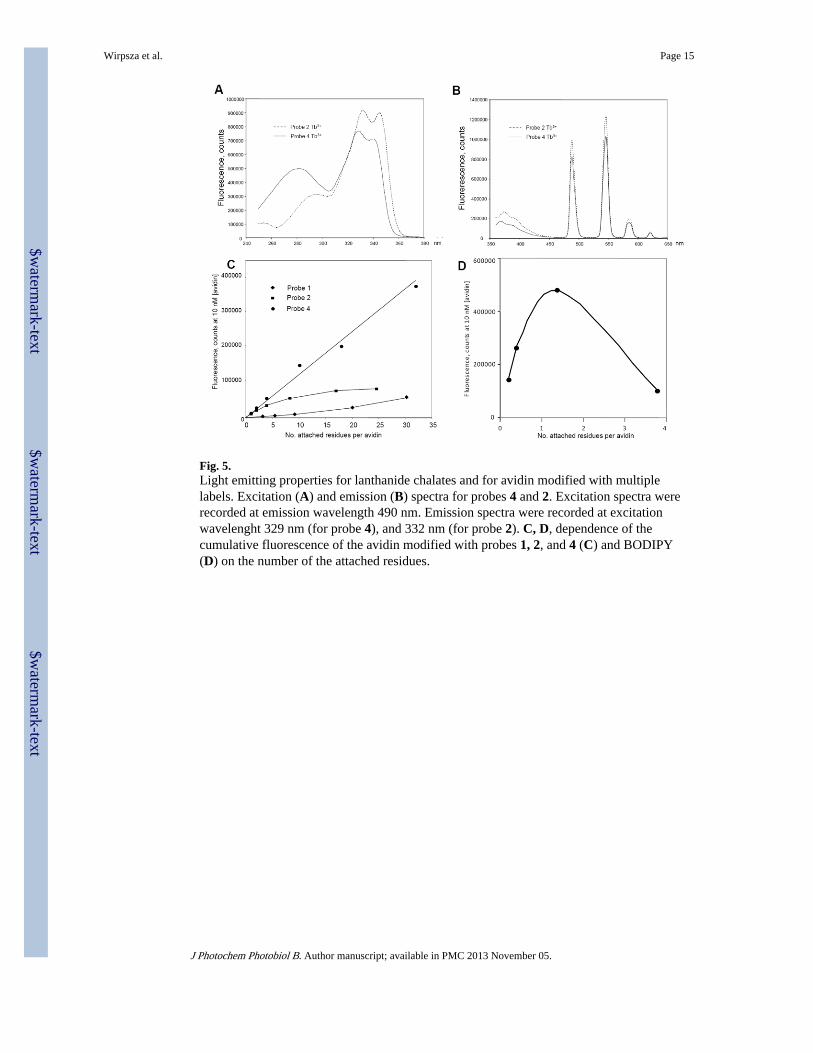
Fig. 5.Light emitting properties for lanthanide chalates and for avidin modified with multiplelabels. Excitation (A) and emission (B) spectra for probes 4 and 2. Excitation spectra wererecorded at emission wavelength 490 nm. Emission spectra were recorded at excitationwavelenght 329 nm (for probe 4), and 332 nm (for probe 2). C, D, dependence of thecumulative fluorescence of the avidin modified with probes 1, 2, and 4 (C) and BODIPY(D) on the number of the attached residues.
Wirpsza et al. Page 15
J Photochem Photobiol B. Author manuscript; available in PMC 2013 November 05.
$waterm
ark-text$w
atermark-text
$waterm
ark-text

Fig. 6.Light emission spectra of luminescent avidin conjugates in the absence (grey bars) and inthe presence (black bars) of biotinylated DNA oligo carrying a Black Hole Quencher.
Wirpsza et al. Page 16
J Photochem Photobiol B. Author manuscript; available in PMC 2013 November 05.
$waterm
ark-text$w
atermark-text
$waterm
ark-text

Fig. 7.The scheme for luminescent labeling of living cells. (A) X-ray structure of avidin displayinglysine residues to which reactive light-emitting probes can be attached. (B) The strategy forcell labeling. The cells are treated with reactive biotin derivative, followed by attachment ofthe luminescent labeled avidin conjugates to biotinylated cells.
Wirpsza et al. Page 17
J Photochem Photobiol B. Author manuscript; available in PMC 2013 November 05.
$waterm
ark-text$w
atermark-text
$waterm
ark-text
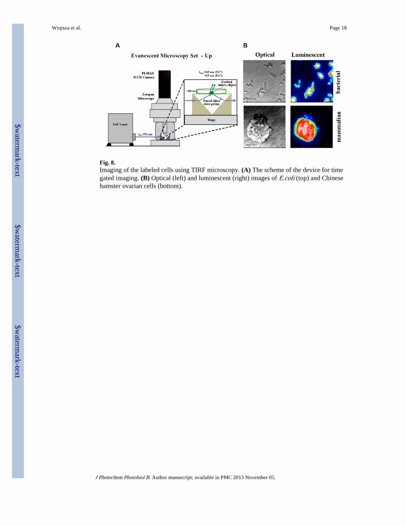
Fig. 8.Imaging of the labeled cells using TIRF microscopy. (A) The scheme of the device for timegated imaging. (B) Optical (left) and luminescent (right) images of E.coli (top) and Chinesehamster ovarian cells (bottom).
Wirpsza et al. Page 18
J Photochem Photobiol B. Author manuscript; available in PMC 2013 November 05.
$waterm
ark-text$w
atermark-text
$waterm
ark-text
$waterm
ark-text$w
atermark-text
$waterm
ark-text
Wirpsza et al. Page 19
Tabl
e 1
Fluo
resc
ence
of
avid
in c
onju
gate
s, n
on-a
ttach
ed p
robe
s, a
nd r
efer
ence
com
poun
ds in
wat
er a
nd d
eute
rium
oxi
de s
olut
ions
Pro
beN
o. r
esid
ues
per
avid
inF
luor
esce
nce
(per
10
nM a
vidi
n)F
luor
esce
nce
(per
10
nM a
vidi
n)Sp
ecif
ic f
luor
esce
nce
(cal
cula
ted
for
10 n
M f
luor
opho
re)
in H
2Oin
D2O
in H
2Oin
D2O
1
3167
500
210
000
2 25
07
000
2
2475
000
100
000
3 10
04
200
4
3037
7 00
052
0 00
012
600
17 3
00
2 (n
on-a
ttach
ed)
N/A
N/A
N/A
34 5
0042
000
4 (n
on-a
ttach
ed)
N/A
N/A
N/A
30 7
0037
000
1 (n
on-a
ttach
ed)
N/A
N/A
N/A
2 30
07
500
Ref
eren
ce c
ompo
unds
(se
e ch
art 1
)
C s
124-
DT
PA-T
b3+N
/AN
/AN
/A40
000
65 0
00
C s
124C
F3-
DT
PA-E
u3+N
/AN
/AN
/A2
500
10 0
00
J Photochem Photobiol B. Author manuscript; available in PMC 2013 November 05.
$waterm
ark-text$w
atermark-text
$waterm
ark-text
Wirpsza et al. Page 20
Table 2
Antenna-to-Eu3+ emission ratio for probe 1 attached to avidin in H2O at pH 8 for 10 nM avidin.
No. of attached residues per avidin Eu3+emission (counts) Antenna emission (counts) Eu3+/Antenna emission ratio
3 3 000 3 760 0.79
6 7 100 5 000 1.42
20 42 000 21700 2.0
31 67 500 26 000 2.6
10 nM PrIEu3+ (non-attached) 2 300 2 300 1.0
J Photochem Photobiol B. Author manuscript; available in PMC 2013 November 05.