normal white blood cell morphology - yolafoitikvth.yolasite.com/resources/blood atlas wbc_1.pdf ·...
TRANSCRIPT
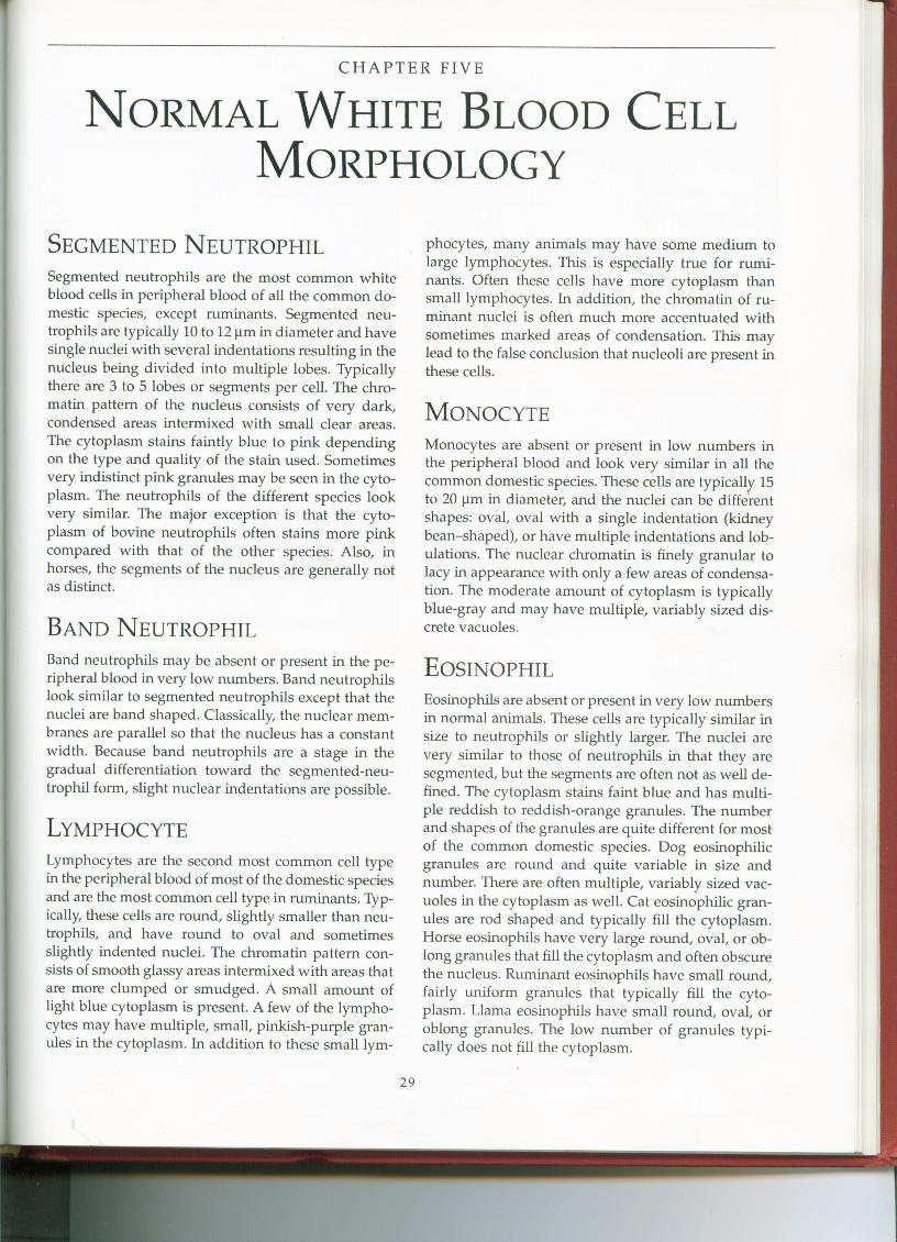
CHAPTER FIVE
NORMAL WHITE BLOOD CELLMORPHOLOGY
SEGMENTED NEUTROPHILSegmented neutrophils are the most common whiteblood cells in peripheral blood of all the common do-mestic. species, except ruminants. Segmented neu-trophils are typically 10 to 12 11min diameter and havesingle nuclei with several indentations resulting in thenucleus being divided into multiple lobes. Typicallythere are 3 to 5 lobes or segments per cell. The chro-matin pattern of the nucleus consists of very dark,condensed areas intermixed with small clear areas.The cytoplasm stains faintly blue to pink dependingon the type and quality of the stain used. Sometimesvery indistinct pink granules may be seen in the cyto-plasm. The neutrophils of the different species lookvery similar. The major exception is that the cyto-plasm of bovine neutrophils often stains more pinkcompared with that of the other species. Also, inhorses, the segments of the nucleus are generally notas distinct.
BAND NEUTROPHILBand neutrophils may be absent or present in the pe-ripheral blood in very low numbers. Band neutrophilslook similar to segmented neutrophils except that thenuclei are band shaped. Classically, the nuclear mem-branes are parallel so that the nucleus has a constantwidth. Because band neutrophils are a stage in thegradual differentiation toward the segmented-neu-trophil form, slight nuclear indentations are possible.
LYMPHOCYTELymphocytes are the second most common cell typein the peripheral blood of most of the domestic speciesand are the most common cell type in ruminants. Typ-ically, these cells are round, slightly smaller than neu-trophils, and have round to oval and sometimesslightly indented nuclei. The chromatin pattern con-sists of smooth glassy areas intermixed with areas thatare more clumped or smudged. A small amount oflight blue cytoplasm is present. A few of the lympho-cytes may have multiple, small, pinkish-purple gran-ules in the cytoplasm. In addition to these smalllym-
29
phocytes, many animals may have some medium tolarge lymphocytes. This is especially true for rumi-nants. Often these cells have more cytoplasm thansmall lymphocytes. In addition, the chromatin of ru-minant nuclei is often much more accentuated withsometimes marked areas of condensation. This maylead to the false conclusion that nucleoli are present inthese cells.
MONOCYTEMonocytes are absent or present in low numbers inthe peripheral blood and look very similar in all thecommon domestic species. These cells are typically 15to 20 11min diameter, and the nuclei can be differentshapes: oval, oval with a single indentation (kidneybean-shaped), or have multiple indentations and lob-ulations. The nuclear chromatin is finely granular tolacy in appearance with only a few areas of condensa-tion. The moderate amount of cytoplasm is typicallyblue-gray and may have multiple, variably sized dis-crete vacuoles.
EOSINOPHILEosinophils are absent or present in very low numbersin normal animals. These cells are typically' similar insize to neutrophils or slightly larger. The nuclei arevery similar to those of neutrophils in that they aresegmented, but the segments are often not as well de-fined. The cytoplasm stains faint blue and has multi-ple reddish to reddish-orange granules. The numberand shapes of the granules are quite different for mostof the common domestic species. Dog eosinophilicgranules are round and quite variable in size andnumber. There are often multiple, variably sized vac-uoles in the cytoplasm as well. Cat eosinophilic gran-ules are rod shaped and typically fill the cytoplasm.Horse eosinophils have very large round, oval, or ob-long granules that fill the cytoplasm and often obscurethe nucleus. Ruminant eosinophils have small round,fairly uniform granules that typically fill the cyto-plasm. Llama eosinophils have small round, oval, oroblong granules. The low number of granules typi-cally does not fill the cytoplasm.
30 NORMAL WHITE BLOOD CELL MORPHOLOGY
BASOPHILBasophils are rarely seen in the peripheral blood of allthe common domestic species. They are most com-monly seen in horses. Basophils are similar in size orslightly larger than neutrophils, and the cytoplasm islight purple. The nucleus is segmented but often notto the degree of the mature neutrophil. Low numbersof small, round, purple cytoplasmic granules may
sometimes be present in dog basophils. The presenceor absence of granules may be dependent on the typeof stain used. Cat basophils contain indistinct small,round, lavender granules. Both cow and horse ba-sophils have several small, well-stained purple gran-ules in the cytoplasm. Llama basophils look very sim-ilar to cow or horse basophils. Figures 5.1-5.30 showthe normal white blood cells of the common domesticspecies.
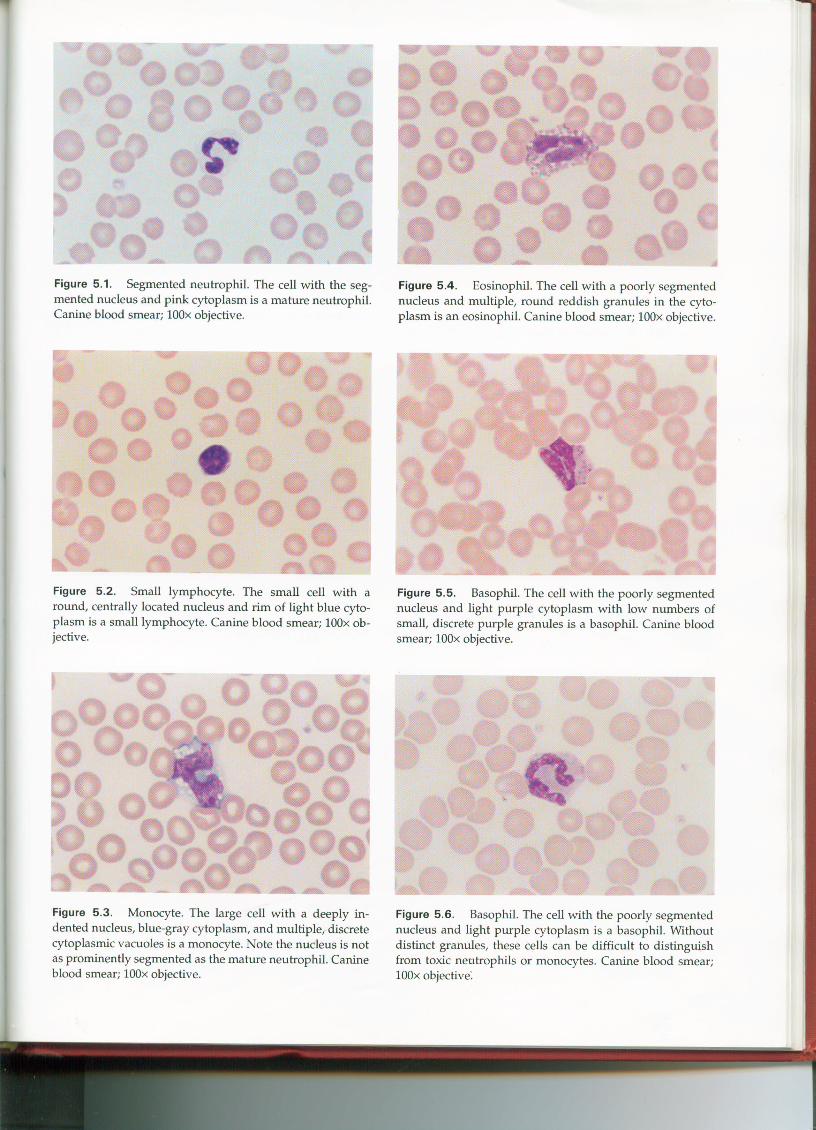
Figure 5.1. Segmented neutrophil. The cell with the seg-mented nucleus and pink cytoplasm is a mature neutrophil.Canine blood smear; IOOx objective.
Figure 5.2. Small lymphocyte. The small cell with around, centrally located nucleus and rim of light blue cyto-plasm is a small lymphocyte. Canine blood smear; lOOx ob-jective.
Figure 5.3. Monocyte. The large cell with a deeply in-dented nucleus, blue-gray cytoplasm, and multiple, discretecytoplasmic vacuoles is a monocyte. Note the nucleus is notas prominently segmented as the mature neutrophil. Canineblood smear; IOOx objective.
Figure 5.4. Eosinophil. The cell with a poorly segmentednucleus and multiple, round reddish granules in the cyto-plasm is an eosinophil. Canine blood smear; IOOx objective.
Figure 5.5. Basophil. The cell with the poorly segmentednucleus and light purple cytoplasm with low numbers ofsmall, discrete purple granules is a basophil. Canine bloodsmear; lOOx objective.
Figure 5.6. Basophil. The cell with the poorly segmentednucleus and light purple cytoplasm is a basophil. Withoutdistinct granules, these cells can be difficult to distinguishfrom toxic neutrophils or monocytes. Canine blood smear;lOOx objective:
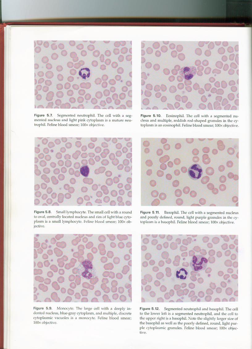
Figure 5.7. Segmented neutrophil. The cell with a seg-mented nucleus and light pink cytoplasm is a mature neu-trophil. Feline blood smear; IOOxobjective.
Figure 5.8. Small lymphocyte. The small cell with a roundto oval, centrally located nucleus and rim of light blue cyto-plasm is a small lymphocyte. Feline blood smear; IOOxob-jective.
Figure 5.9. Monocyte. The large cell with a deeply in-dented nucleus, blue-gray cytoplasm, and multiple, discretecytoplasmic vacuoles is a monocyte. Feline blood smear;IOOxobjective.
Figure 5.10. Eosinophil. The cell with a segmented nu-cleus and multiple, reddish rod-shaped granules in the cy-toplasm is an eosinophil. Feline blood smear; IOOxobjective.
Figure 5.11. Basophil. The cell with a segmented nucleusand poorly defined, round, light purple granules in the cy-toplasm is a basophil. Feline blood smear; IOOxobjective.
Figure 5.12. Segmented neutrophil and basophil. The cellto the lower left is a segmented neutrophil, and the cell tothe upper right is a basophil. Note the slightly larger size ofthe basophil as well as the poorly defined, round, light pur-ple cytoplasmic granules. Feline blood smear; lOOxobjec-tive.
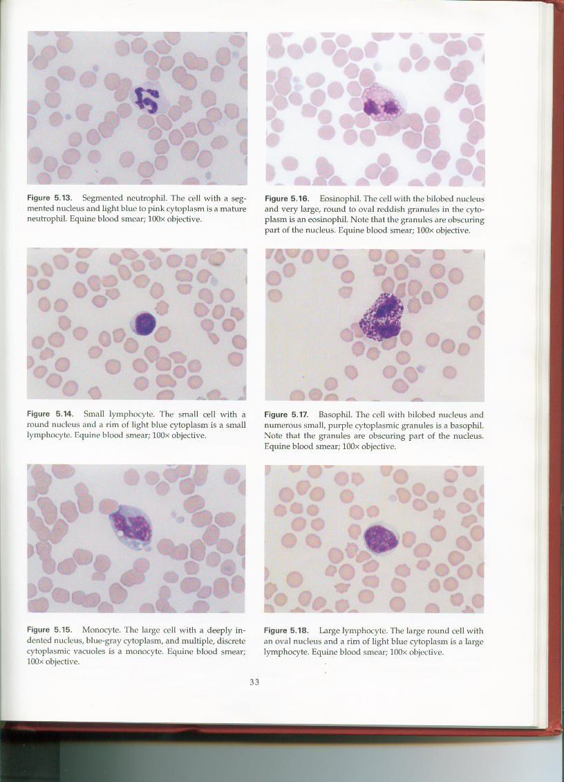
Figure 5.16. Eosinophil. The cell with the bilobed nucleusand very large, round to oval reddish granules in the cyto-plasm is an eosinophil. Note that the granules are obscuringpart of the nucleus. Equine blood smear; IOOx objective.
Figure 5.17. Basophil. The cell with bilobed nucleus andnumerous small, purple cytoplasmic granules is a basophil.Note that the granules are obscuring part of the nucleus.Equine blood smear; lOOx objective.
Figure 5.18. Large lymphocyte. The large round cell withan oval nucleus and a rim of light blue cytoplasm is a largelymphocyte. Equine blood smear; IOOx objective.
33
Figure 5.13. Segmented neutrophil. The cell with a seg-mented nucleus and light blue to pink cytoplasm is a matureneutrophil. Equine blood smear; IOOx objective.
Figure 5.15. Monocyte. The large cell with a deeply in-dented nucleus, blue-gray cytoplasm, and multiple, discretecytoplasmic vacuoles is a monocyte. Equine blood smear;lOOx objective.
Figure 5.14. Small lymphocyte. The small cell with around nucleus and a rim of light blue cytoplasm is a smalllymphocyte. Equine blood smear; IOOx objective.
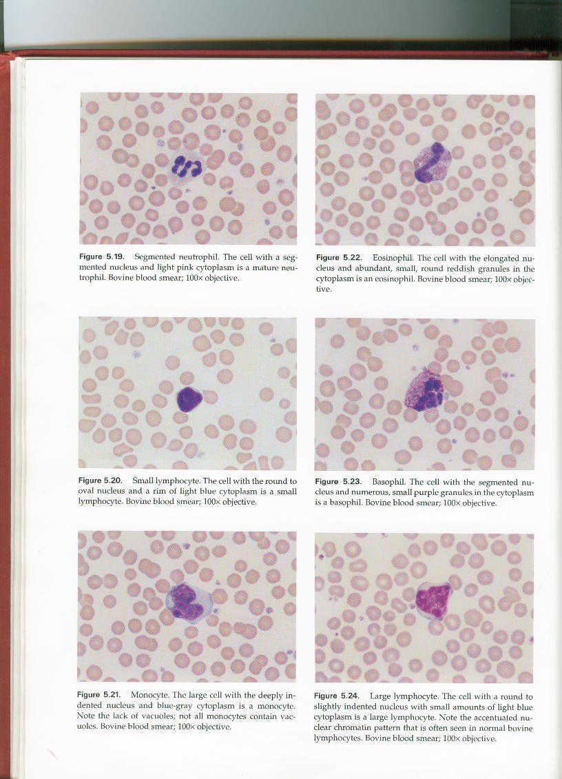
Figure 5.19. Segmented neutrophil. The cell with a seg-mented nucleus and light pink cytoplasm is a mature neu-trophil. Bovine blood smear; lOOx objective.
Figure 5.20. Small lymphocyte. The cell with the round tooval nucleus and a rim of light blue cytoplasm is a smalllymphocyte. Bovine blood smear; lOOx objective.
Figure 5.21. Monocyte. The large cell with the deeply in-dented nucleus and blue-gray cytoplasm is a monocyte.Note the lack of vacuoles; not all monocytes contain vac-uoles. Bovine blood smear; lOOx objective.
Figure 5.22. Eosinophil. The cell with the elongated nu-cleus and abundant, small, round reddish granules in thecytoplasm is an eosinophil. Bovine blood smear; lOOx objec-tive.
Figure 5.23. Basophil. The cell with the segmented nu-cleus and numerous, small purple granules in the cytoplasmis a basophil. Bovine blood smear; lOOx objective.
Figure 5.24. Large lymphocyte. The cell with a round toslightly indented nucleus with small amounts of light bluecytoplasm is a large lymphocyte. Note the accentuated nu-clear chromatin pattern that is often seen in normal bovinelymphocytes. Bovine blood smear; lOOx objective.
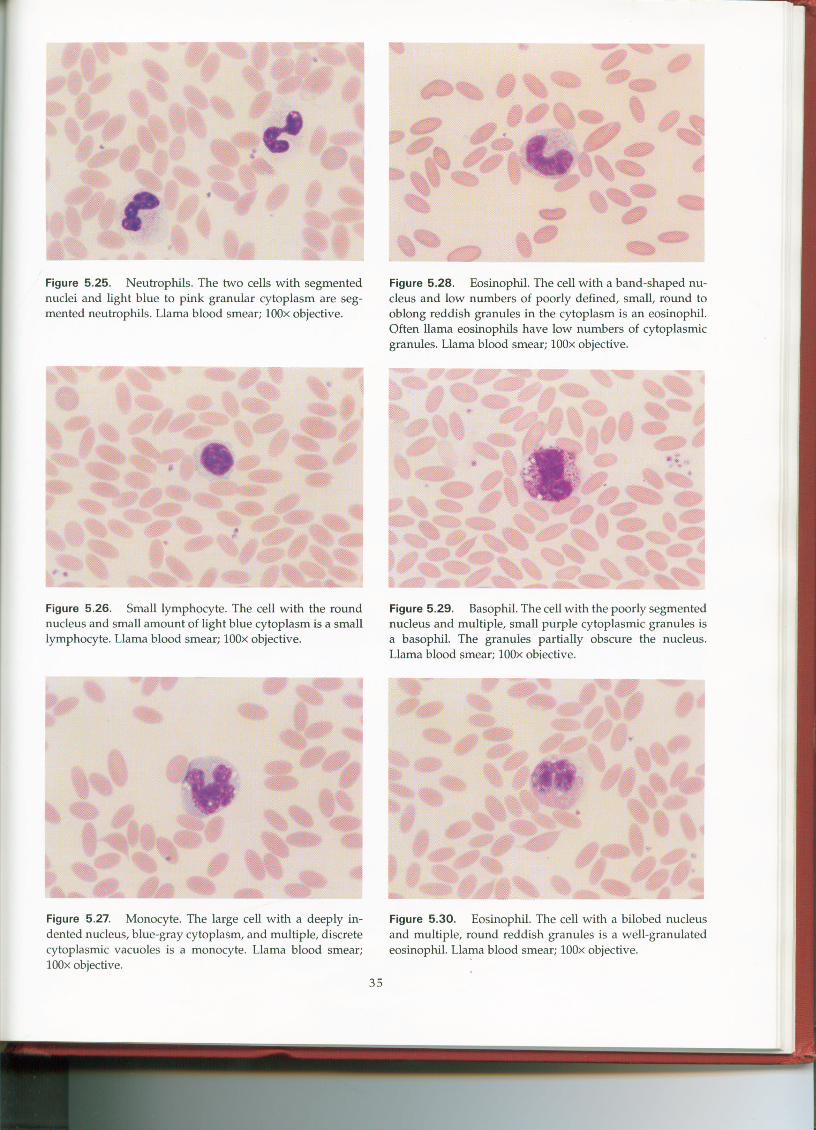
Figure 5.25. Neutrophils. The two cells with segmentednuclei and light blue to pink granular cytoplasm are seg-mented neutrophils. Llama blood smear; lOOx objective.
Figure 5.26. Small lymphocyte. The cell with the roundnucleus and small amount of light blue cytoplasm is a smalllymphocyte. Llama blood smear; lOOx objective.
Figure 5.27. Monocyte. The large cell with a deeply in-dented nucleus, blue-gray cytoplasm, and multiple, discretecytoplasmic vacuoles is a monocyte. Llama blood smear;lOOx objective.
35
Figure 5.28. Eosinophil. The cell with a band-shaped nu-cleus and low numbers of poorly defined, small, round tooblong reddish granules in the cytoplasm is an eosinophil.Often llama eosinophils have low numbers of cytoplasmicgranules. Llama blood smear; lOOx objective.
Figure 5.29. Basophil. The cell with the poorly segmentednucleus and multiple, small purple cytoplasmic granules isa basophil. The granules partially obscure the nucleus.Llama blood smear; lOOx obiective.
Figure 5.30. Eosinophil. The cell with a bilobed nucleusand multiple, round reddish granules is a well-granulatedeosinophil. Llama blood smear; lOOx objective.