red blood cell morphology in health and disease
TRANSCRIPT

Red Blood Cell Morphology in Health and Disease

Principle• Normal RBCs are minute biconcave discs. → diameter of
6-8.5 micrometers.
• Normal and diseased red cells are subject to considerable distortion during spreading of films.
• It is therefore very important to scan films carefully and select an area of minimal distortion for examination.
• Red blood cells should be examined in an area with little or no rouleaux formation. Thus, technologists always avoid thick areas where rouleaux have formed, and very thin areas where RBCs are maximally distorted.

PrincipleNotes:
• A normal RBC has a normal size (normocytic) and a normal Hb content i.e. normal color (normochromic).
• Red cells are stained by the eosin component of romanovwskydyes

Causes of Variation in RBC Morphology
• Variation in RBC morphology stems from four main reasons:
(a) Abnormal erythropoisis
(b) Inadequate Hb synthesis
(c) Damage to, or changes affecting RBCs after leaving the bone marrow
(d) Attempts of the bone marrow to compensate for anemia

Causes of Variation in RBC MorphologyTypes of Variation in RBC Morphology
The four causes mentioned above give rise to the following abnormalities of the red cells:
• Variation in Size (anisocytosis): • Macrocytosis :RBC larger than normal
• Microcytosis : RBC smaller than normal
Figure : Blood film showing Macrocytes Figure : blood film showing microcytic-hypochromic cells

• Variation in Shape (poikilocytosis)
• Tear-drop cells: cells shaped like tear drops or pears.
• Elliptocytes: they can vary in appearance from oval-shaped (Ovalocytes) to thin pencil-shaped forms (Pencil cells).
• Keratocytes: also described as helmet cells or bite cells. These cells are fragmented red blood cells that have been "scooped out" so they resemble helmets or appear as if they have been bitten. These "bites" result from either the removal of a heinz body by the phagocytic function of the spleen, or from mechanical damage.
• Schistocytes: RBC fragments.
• Echinocytes: also known as "crenated cells" and "burr cells". These cells have many tiny spicules (10-30) evenly distributed over the cell membrane. Although often confused with acanthocytes, the projections on echinocytes are much more uniform in shape and distribution in.
• Acanthocytes: also referred to as "spur cells". Spheroid RBCs with few large spiny projections. 5-10 spicules, irregular spacing and thickness (must be differentiated from echinocytes).
• Sickled cells: banana-shaped or crescent-shaped cells
• Blister cells: RBCs with vacuoles or markedly thin areas at periphery of membrane.
• Pyknocytes: irregularity contracted cells.
• Variation in Hemoglobin Content
• Hypochromasia: color is paler than normal.
• Anisochromasia : cells having a dimorphic picture.
• Target cells: Thin, hyopochromatic cell. Round area of central pigmentation. also called codocytes.
• Leptocytes : very thin cells with a colorless central part.
• Spherocytes: sphere-shaped cells with deep coloration and no central pallor.
• Stomatocyte: also called fish mouth cell. Uniconcave RBC, slitlike area of central pallor.
Other Abnormalities• Late normoblast: an immature nucleated RBC with a basophilic
nucleus.
• Polychromasia: cells with pale green or blue coloration, which is a sign of immaturity.
• Howel jolly bodies: nuclear remnants that appear as Small (1 mm), round, dense, purple bodies in RBCs.
• Basophilic stippling: Fine, medium, or coarse blue granules uniformly distributed throughout RBC.
• Pappenheimer bodies: small peripherally sited basophilic inclusions. They are composed of hemosiderin. They appear purple in color with conventional staining. Using perl's stain gives them a blue color.
• Malarial parasite: stages of different malarial species.
• Heinz bodies: oxidized and denatured Hb. Stained by supravital stains (new methylene blue, brilliant cresyl blue).


blood film showing tear-drop cellsElliptocytes. blood film showing elliptocytes.
blood film showing some helmet cells. blood film showing schistocytes (fragmented RBCs)
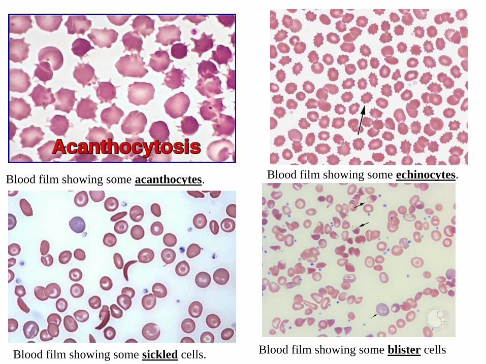
Blood film showing some echinocytes.Blood film showing some acanthocytes.
Blood film showing some sickled cells. Blood film showing some blister cells

Blood film demonstrating hypochromasiaBlood film showing a bite cell
Blood film showing target cells Blood film showing leptocytes.
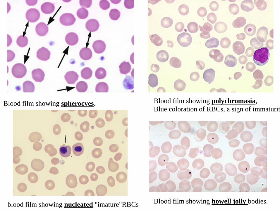
Blood film showing polychromasia,
Blue coloration of RBCs, a sign of immaturityBlood film showing spherocyes.
blood film showing nucleated "imature"RBCsBlood film showing howell jolly bodies.

Blood film showing pappenheimer bodiesBlood film showing basophilic stippling.
Blood film showing Heinz bodies.Blood film showing malarial parasite
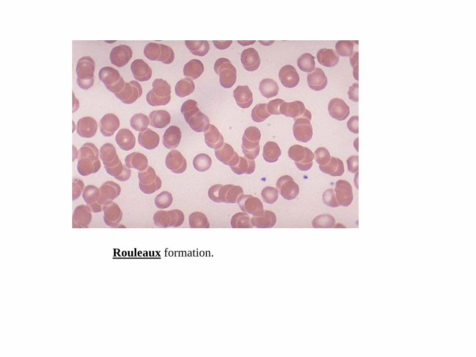
Rouleaux formation.
