ombretta masala and ram seshadri - university of...
TRANSCRIPT

27 Jun 2004 11:37 AR AR218-MR34-02.tex AR218-MR34-02.sgm LaTeX2e(2002/01/18) P1: FHD10.1146/annurev.matsci.34.052803.090949
Annu. Rev. Mater. Res. 2004. 34:41–81doi: 10.1146/annurev.matsci.34.052803.090949
Copyright c© 2004 by Annual Reviews. All rights reserved
SYNTHESIS ROUTES FOR LARGE VOLUMES OF
NANOPARTICLES
Ombretta Masala and Ram SeshadriMaterials Department and Materials Research Laboratory, University of California,Santa Barbara, California 93106; email: [email protected],[email protected]
Key Words inorganic materials, metals, semiconductors, oxides
� Abstract This review focuses on the preparation of capped nanoparticles of inor-ganic materials, classified by composition. The materials described include elementalmetals and metalloids (semiconductors), chalcogenide II–VI and IV–VI semiconduc-tors, III–V semiconductors, and oxides (both of simple- and multitransition metal).Although particular emphasis is placed on methods that yield large volumes of nanopar-ticles, recent novel methods that may not necessarily be scalable are also reviewed.The review makes apparent the richness of chemistry that has become routine to prac-titioners in the field; diverse inorganic systems with distinct chemistry require distinctmethods of preparation.
INTRODUCTION
The possibility to prepare monodisperse inorganic nanoparticles of pure materialsin the sub-20 nm size regime, associated with the capping of these particles by(typically) functionalized long-chain organic molecules, has thrown open an entirefield of research. Inorganic nanoparticles are finding myriad uses, ranging fromtraditional ones, such as coloring agents (in stained glass windows) and catalysts,to more novel ones, such as magnetic drug delivery, hypothermic cancer therapy,contrast agents in magnetic resonance imaging, magnetic and fluorescent tagsin biology, solar photovoltaics, nano bar codes, and emission control in dieselvehicles. This field is, in certain ways, reaching maturity, and to go to the nextstep, it is becoming important to develop methods of scaling up the synthesis ofthese materials.
We do not discuss the properties and uses of nanoparticles, except so far asthe properties might be a part of the process of characterization. Also, this re-view does not concern itself with the vast literature of nanophase materials, whichare monolithic materials comprising crystalline grains whose dimensions are inthe nanometer range. Such materials are typically prepared using sol-gel and re-lated techniques. This is an older, more mature area that continues to attract a
0084-6600/04/0804-0041$14.00 41

27 Jun 2004 11:37 AR AR218-MR34-02.tex AR218-MR34-02.sgm LaTeX2e(2002/01/18) P1: FHD
42 MASALA � SESHADRI
great deal of interest (1). There is growing interest in controlling the shapes ofnanophase and nanoparticulate materials, as described, for example, in the chapterby Law et al. (2) on nanotubes and nanowires and that by Kolmakov & Moskovits(3) on chemical sensing by nanostructures (see this volume). We make no attemptto describe attempts to control the shapes of nanoparticles except when they areincidental to the sysnthesis.
Au
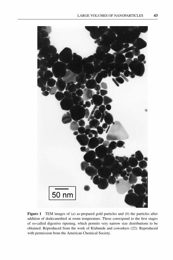
The usual synthetic route to prepare gold nanoparticles involves the reduction of asuitable metal salt (usually an halide) in solution in the presence of a stabilizer. Thefirst systematic studies of this method were by Michael Faraday who obtained col-loidal gold by reducing an aqueous solution of AuCl−4 with phosphorus (4). Sincethen, the discovery of various reducing agents and stabilizers have made possiblethe development of mild conditions that can be employed for the preparation oflarge amounts of nanoparticles of reproducible quality. Among these reductionmethods, the best known is that developed by Brust et al. (5), which involves thephase transfer of AuCl−4 from aqueous to organic solutions followed by reductionwith NaBH4 in the presence of alkanethiols to yield gold nanoparticles in the sizerange of 1–3 nm. Thiols are excellent stabilizers for capping gold nanoparticlesowing to strong Au-S bonding, which enables the isolation of the particles as solidmaterials following solvent evaporation (6). The isolated particles can be readilyredissolved in nonpolar solvents where they are indefinitely stable. Other good pro-tective agents, such as phosphanes (7), phosphines (8), amines (9), chalcogenides(10), carboxylates (11), and polymers (12, 13), have been successfully used. Thesize and morphology of the gold nanoparticles can be tuned by varying the con-centration ratio of capping agent to metal salts (14–16) and by choosing a suitablereducing agent (13). A weak reducing agent, such as citrate (17–19) or tartarate(13), will favor a slow reaction allowing particle growth over a long period to yieldfaceted and small (<1 nm) nanoparticles, whereas with relatively strong reducingagents, such as formamide (20) or o-anisidine (21), bigger and generally sphericalnanoparticles will form. Prasad et al. (22) have shown that thiol-stabilized goldcolloids obtained by reduction can be subjected to a digestive ripening processthat significantly reduces the average particle size and polydispersity. The ripen-ing process (Figure 1) involves refluxing of the particles near the boiling point ofthe solvent (usually toluene) in the presence of an excess of stabilizer and has beensuccessfully employed not only with thiols but also with other protective agents,such as amines, silanes, and phosphines (23).
Another method that is of great potential for the synthesis of gold nanoparticlesin large quantities, owing to mild conditions and relatively nontoxic reagents, isbased on the solvated metal atom dispersion (SMAD) technique. This method, firstreported by Klabunde and coworkers (24, 25), involves the vaporization of goldunder vacuum followed by deposition on the inside walls of the vacuum chambercooled to liquid nitrogen temperature simultaneously with the vapors of an organic
27 Jun 2004 11:37 AR AR218-MR34-02.tex AR218-MR34-02.sgm LaTeX2e(2002/01/18) P1: FHD
LARGE VOLUMES OF NANOPARTICLES 43
Figure 1 TEM images of (a) as-prepared gold particles and (b) the particles afteraddition of dodecanethiol at room temperature. These correspond to the first stagesof so-called digestive ripening, which permits very narrow size distributions to beobtained. Reproduced from the work of Klabunde and coworkers (22). Reproducedwith permission from the American Chemical Society.

27 Jun 2004 11:37 AR AR218-MR34-02.tex AR218-MR34-02.sgm LaTeX2e(2002/01/18) P1: FHD
44 MASALA � SESHADRI
Figure 1 (Continued)
solvent, usually toluene. There are no by-products resulting from the reduction ofthe metal salt, and the gold colloids can be easily isolated as solid materials andredispersed in nonpolar solvents. The nanoparticles are nearly spherical in shapewith sizes in the range of 1–6 nm. However, their size distribution can be furthernarrowed to 4–5 nm by the digestive-ripening procedure described earlier.
27 Jun 2004 11:37 AR AR218-MR34-02.tex AR218-MR34-02.sgm LaTeX2e(2002/01/18) P1: FHD
LARGE VOLUMES OF NANOPARTICLES 45
Ag
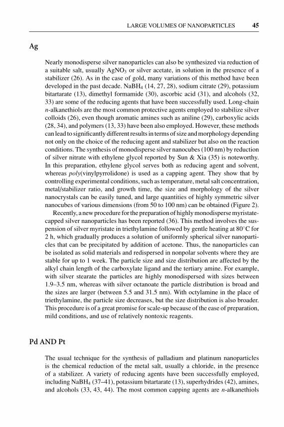
Nearly monodisperse silver nanoparticles can also be synthesized via reduction ofa suitable salt, usually AgNO3 or silver acetate, in solution in the presence of astabilizer (26). As in the case of gold, many variations of this method have beendeveloped in the past decade. NaBH4 (14, 27, 28), sodium citrate (29), potassiumbitartarate (13), dimethyl formamide (30), ascorbic acid (31), and alcohols (32,33) are some of the reducing agents that have been successfully used. Long-chainn-alkanethiols are the most common protective agents employed to stabilize silvercolloids (26), even though aromatic amines such as aniline (29), carboxylic acids(28, 34), and polymers (13, 33) have been also employed. However, these methodscan lead to significantly different results in terms of size and morphology dependingnot only on the choice of the reducing agent and stabilizer but also on the reactionconditions. The synthesis of monodisperse silver nanocubes (100 nm) by reductionof silver nitrate with ethylene glycol reported by Sun & Xia (35) is noteworthy.In this preparation, ethylene glycol serves both as reducing agent and solvent,whereas poly(vinylpyrrolidone) is used as a capping agent. They show that bycontrolling experimental conditions, such as temperature, metal salt concentration,metal/stabilizer ratio, and growth time, the size and morphology of the silvernanocrystals can be easily tuned, and large quantities of highly symmetric silvernanocubes of various dimensions (from 50 to 100 nm) can be obtained (Figure 2).
Recently, a new procedure for the preparation of highly monodisperse myristate-capped silver nanoparticles has been reported (36). This method involves the sus-pension of silver myristate in triethylamine followed by gentle heating at 80◦C for2 h, which gradually produces a solution of uniformly spherical silver nanoparti-cles that can be precipitated by addition of acetone. Thus, the nanoparticles canbe isolated as solid materials and redispersed in nonpolar solvents where they arestable for up to 1 week. The particle size and size distribution are affected by thealkyl chain length of the carboxylate ligand and the tertiary amine. For example,with silver stearate the particles are highly monodispersed with sizes between1.9–3.5 nm, whereas with silver octanoate the particle distribution is broad andthe sizes are larger (between 5.5 and 31.5 nm). With octylamine in the place oftriethylamine, the particle size decreases, but the size distribution is also broader.This procedure is of a great promise for scale-up because of the ease of preparation,mild conditions, and use of relatively nontoxic reagents.
Pd AND Pt
The usual technique for the synthesis of palladium and platinum nanoparticlesis the chemical reduction of the metal salt, usually a chloride, in the presenceof a stabilizer. A variety of reducing agents have been successfully employed,including NaBH4 (37–41), potassium bitartarate (13), superhydrides (42), amines,and alcohols (33, 43, 44). The most common capping agents are n-alkanethiols
27 Jun 2004 11:37 AR AR218-MR34-02.tex AR218-MR34-02.sgm LaTeX2e(2002/01/18) P1: FHD
46 MASALA � SESHADRI
Figure 2 (A, B) SEM and (C) TEM images of silver nanocubes. (D) X-ray diffractionpattern of silver nanocubes. The inset in (D) is a scheme of the nanocube morphology.Reproduced from Sun and Xia (35). Reproduced with permission from the AmericanAssociation for the Advancement of Science.
and polymers, but a few examples of Pt and Pd nanoparticles capped with amines(45), alkyl isocyanides (46), and cyclodextrins (47) have also been reported.
Very recently, Kim et al. (48) developed a new synthetic method for palladiumnanoparticles involving the thermal decomposition at 300◦C of a solution of themetal acetate in a surfactant. With tri-n-octylphosphine (TOP) and a mixture ofTOP/oleylamine as surfactants, highly monodisperse palladium nanocrystals ofsizes of 3, 5, and 7 nm were obtained depending on the surfactant mole ratio. Withonly oleylamine as surfactant, polydisperse nanoparticles with an average sizelarger than 10 nm were produced. The quality of the as-synthesized nanoparticles,the ease of preparation, and the fact that no further size-selective technique isrequired make this the first method that can be easily adapted for large-scaleproduction of palladium nanoparticles.
Co, Ni, Fe, AND Cu
Although significant progress has been achieved in the synthesis of monodispersenoble metal nanoparticles, synthetic routes for magnetic Fe, Co, and Ni nanoparti-cles have yet to be improved (49). Synthesis in inverse micelles and sonochemical

27 Jun 2004 11:37 AR AR218-MR34-02.tex AR218-MR34-02.sgm LaTeX2e(2002/01/18) P1: FHD
LARGE VOLUMES OF NANOPARTICLES 47
decomposition of the metal carbonyls have been reported for the preparation ofnanosized colloids of Co (50, 51), Fe (52–54), and Ni (55), but these methods yield,in most cases, polydisperse particles and are perhaps not suitable for scale up be-cause of the difficulty of reproducibility at high volumes and high concentrations.However, a few synthetic procedures that are of great promise for the synthesisof monodisperse magnetic nanoparticles have been reported. For example, Conanoparticles can be prepared with a method developed by Murray and cowork-ers, which involves the reduction of Co chloride by a superhydride in solution inthe presence of a stabilizer, usually a mixture of oleic acid and an alkylphosphine(56, 57). The average particle size can be tuned by carefully selecting the type ofalkylphosphines employed in conjunction with oleic acid because the steric hin-drance of the alkyl groups can exert a control on the rate of particle growth. Thus,bulky long-chain phosphines such as TOP will tend to inhibit the growth and favorsmall particles (2–6 nm), whereas shorter-chain and less bulky phosphines such astributylphosphine will permit growth and lead to bigger particles (7–11 nm). Theas-synthesized nanoparticles are surrounded by a robust organic shell and can bereadily dispersed in aliphatic, aromatic, and chlorinated solvents, reprecipitatedby short-chain alcohols, which further reduce the size by selective precipitation.Fatty acids other than oleic acid, such as octanoic and dodecanoic acids, can beused to cap the particles through a simple ligand exchange process even thoughacids with an alkyl chain shorter than C8 are not strong enough to stabilize theparticles. Reduction of metal acetates in solution by polyalcohols or NaBH4 in thepresence of stabilizers (oleic acid, TOP, tributylamine, and hexadecylamine havebeen employed) is also a successful route to synthesize highly monodisperse Co,Ni, and Ni-Co nanoparticles (58, 59).
A new approach for the synthesis of Co nanoparticles has been developed byDinega & Bawendi (60) based on the thermal decomposition (200◦C) of dicobal-toctacarbonyl, Co2(CO)8, in the presence of tri-n-octylphosphine oxide (TOPO)as a surfactant to yield rather polydisperse nanocrystals with an average diameterof 20 nm. One advantage of this method compared with the reduction synthesisdescribed above is the absence of by-products because CO is released and ele-mental Co is the only nonvolatile product left in the reaction vessel. Recently,variations (61–65) of this method have been reported where monodisperse Conanocrystals with tunable sizes between 3–20 nm can be obtained by using a com-bination of carboxylic acids, amines, and phosphines as surfactants, and separationof the nanocrystals from solution is achieved by magnetically induced precipita-tion rather than by addition of a polar solvent. This method can be also adapted forthe synthesis of uniform Fe nanoparticles by replacing Co2(CO)8 with Fe(CO)5 athigher temperatures (58, 66); for the synthesis of bimetallic Fe-Co (58) and Fe-Mo(67) nanoparticles by thermal decomposition of a mixture of the correspondingmetal carbonyls; for Fe-Pt (68), Co-Pt (69, 70), and Fe-Pd (70) nanoparticlesby simultaneous decomposition of, respectively, Fe(CO)5 or Co2(CO)8 and re-duction of platinum or palladium acetonate. Another organometallic precursor,Co(η3-C8H13)(η4-C8H12), has been successfully employed to prepare small Co

27 Jun 2004 11:37 AR AR218-MR34-02.tex AR218-MR34-02.sgm LaTeX2e(2002/01/18) P1: FHD
48 MASALA � SESHADRI
nanoparticles (average size 1 nm) by thermal decomposition in the presence ofpoly(vinylpyrrolidone) as a stabilizing agent (71).
Very little work has been carried out on Cu nanoparticles. Some attempts havebeen made to synthesize Cu nanoparticles by chemical reduction in solution (72,73) and reduction in reverse micelles (74); however, these methods yielded parti-cles of irregular shapes and wide size distributions. High-quality Cu nanoparticleswere recently produced via an organometallic route using the CVD precursorCu[OCH(CH3)CH2N(CH3)2] (75). When the thermal decomposition of the pre-cursor was carried out at 300◦C in hexadecylamine as a hot coordinating solvent,uniform spherical particles of 7.5 nm formed. In the presence of TOPO, the de-composition yielded bigger particles with different shapes (oval, spherical, andelongated) and sizes (8–100 nm), with the elongated ones predominating at highprecursor concentration. Thus the synthesis procedure can be controlled by tai-loring reaction parameters such as temperature, nature of the capping agent, andprecursor concentration.
Si AND Ge
Unlike II–VI and III–V semiconductors, the synthesis of group IV semiconduc-tors silicon and germanium has not been extensively studied; however, interest inthese materials has grown rapidly in the past decade owing to a number of studiessuggesting that luminescence in porous silicon and silicon nanoparticles may becaused by quantum confinement (76–78). Despite a great deal of experimental andtheoretical work, the origin of luminescence in silicon and its dependence on thenanoparticle size are still controversial. Some groups have observed clear changesin the wavelengths and intensities of luminescence with varying sizes or with mod-ification of particle surface (79–81), whereas others have reported evidence to thecontrary (82). Understanding the luminescent properties of silicon nanoparticlesis of extreme importance for the application of these materials in the electronicsindustry, in particular as components in optoelectronic devices. Belonging to thesame group, and therefore having semiconducting properties similar to silicon,germanium nanocrystals are also expected to show optical properties related toquantum confinement, and evidence of this has been provided (83–85).
Early work by Brus and coworkers (78, 81, 86–88) on the preparation of sili-con nanoparticles from the gas phase, while not important from the viewpoint ofscale-up, represents the first step toward the study of the photophysics of thesematerials. The preparation involves a three-step procedure carried out in a quartzreactor with a flowing system: First, a mixture of disilane-helium is flowed at acontrolled rate into the pyrolysis chamber at 1000◦C and 1 atm, followed by imme-diate dilution into a O2-He mixture and decrease of temperature to 200◦C–300◦Cto stop growth and aggregation. The particles are finally collected in a solutionof ethylene glycol to yield a robust colloid that is stable under air without signsof flocculation over several months. The resulting surface-oxidized nanoparticles

27 Jun 2004 11:37 AR AR218-MR34-02.tex AR218-MR34-02.sgm LaTeX2e(2002/01/18) P1: FHD
LARGE VOLUMES OF NANOPARTICLES 49
are crystalline, present weak luminescence emission at room temperature, andtheir sizes (ranging between 3 and 8 nm) can be tailored by controlling the flowrate and the initial disilane concentration because the average particle size de-creases with decreasing flow rate and decreasing concentration. The nanocrystalscan be efficiently separated by size-selective precipitation and by size-exclusionchromatography techniques, which prove to be very valuable in the study of thequantum confinement of luminescence (86, 88).
CO2-laser-induced decomposition of SiH4 in a gas flow reactor has been shownto be very useful for the synthesis of large quantities of silicon nanoparticles (89–95). In a typical reaction set-up, SiH4 in an inert gas is exposed to focused radiationfrom a pulsed CO2 laser in the reaction chamber resulting in the dissociation ofthe silane molecules by resonant laser absorption. The growth of the so-formedsilicon nuclei is abruptly stopped as soon as they leave the hot chamber, and they aresubsequently extracted from an adjacent high-vacuum chamber through a conicalnozzle. The resulting nanoparticles have sizes that can be tuned in the 3–20 nmrange by varying the silane concentration and flow rate and are characterized bystrong visible luminescence. The particles are extremely pure because the reactorwalls remain cold and therefore unreactive during the pyrolysis process. One ofthe main advantages of this method is that production of large amounts of powders(20–5000 mg/h) can be easily achieved by improving the flow reactor capabilities(89, 95).
The first example of a solution route to Si nanocrystals was developed by Heath(96) more than a decade ago and involves the reduction of SiCl4 and RSiCl3(R=H, octyl) by sodium metal in a nonpolar organic solvent at high temperatures(385◦C) and high pressures (>100 atm) to yield R-capped, hexagonal-shaped sil-icon nanoparticles. The octyl group is a more effective capping agent in terms ofsize control because nanoparticles with sizes ranging from 3 to 7 nm are formed,as opposed to single crystals ranging from 5 to 3000 nm obtained with R=H. Onthe other hand, infrared spectroscopy showed that the octyl groups are sponta-neously replaced to give nanoparticles with a surface characterized by a mixtureof Si–O, Si–H, and Si–Cl bonds. When the same reaction scheme is applied togermanium (R=phenyl), nanowires of 7–10 nm in diameter and up to 10 µ inlength are obtained (97). Reduction of germanium salts was also achieved withNaK (99) and with lithium naphthalide (98). With NaK, the resulting nanoparticlesare polydispersed (with sizes ranging between 8 and 20 nm), crystalline platelets,capped by a layer of GeO2 with traces of Ge-Cl termination on the surface, andare insoluble in most polar and nonpolar solvents. With lithium naphthalide, an-nealing with a red laser is required to obtain crystalline nanoparticles. Followingthe route discovered by Heath (96), Kauzlarich and coworkers (98–108) exploredthe synthesis of silicon and germanium nanoparticles from reactions involvingalkyl semiconductor halides or metal silicides/germanides in solution to find lessextreme conditions, higher yields, and better surface manipulation, which are nec-essary requirements for the production of nanoparticles on a large scale. Reductionof SiCl4 was successfully carried out at room temperature with sodium naphthalide

27 Jun 2004 11:37 AR AR218-MR34-02.tex AR218-MR34-02.sgm LaTeX2e(2002/01/18) P1: FHD
50 MASALA � SESHADRI
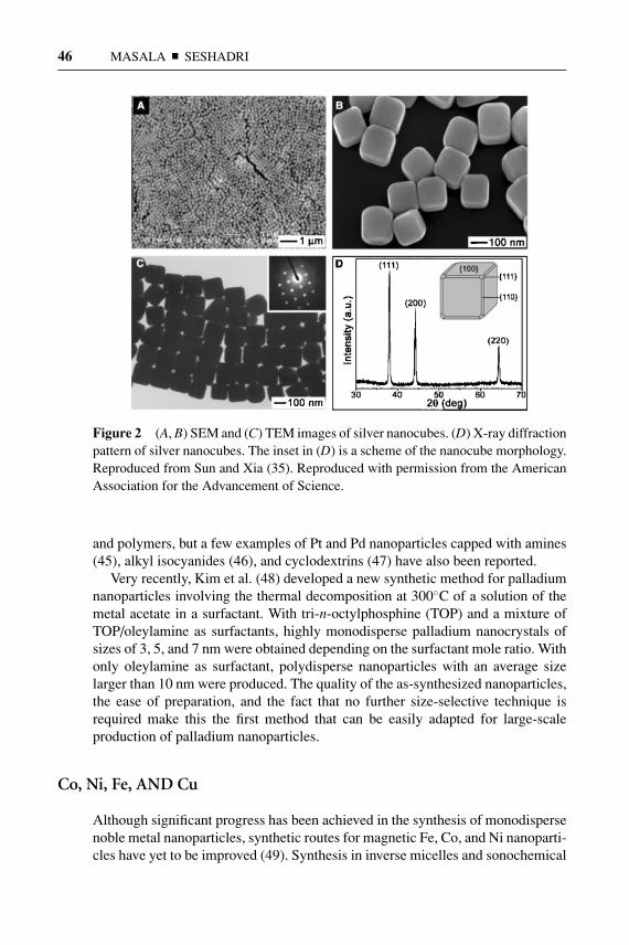
Figure 3 TEM image of alkyl-caped Si nanoparticles formed by the reduction ofSiCl4 by sodium naphthalenide in 1,2-dimethoxyethane. The surface has been cappedwith an excess of n-butyllithium. The inset shows a selected area diffraction patternof diamond-Si. Reproduced from Baldwin et al. (104). Reproduced with permissionfrom the American Chemical Society.
in 1,2-dimethoxyethane to give uniform, crystalline, halide-capped nanoparticleswhose surface can be easily modified (103, 104) (Figure 3). For example, fur-ther treatment with an excess of octanol or n-butyllithium yielded, respectively,octanoxide- and n-butyl-capped nanocrystals. The reaction of SiCl4 with metalsilicides such as KSi, Mg2Si, and NaSi has proved to be of great potential for thesynthesis of large quantities of luminescent silicon nanoparticles with a function-alized surface. It involves refluxing the metal silicide in a suitable solvent (usuallyethylene glycol dimethyl ether) under inert atmosphere in the presence of SiCl4 fol-lowed by passivation with an appropriate capping agent (100–102). Methyllithium,n-butyllithium, and several Grignard reagents have been used for the passivationstep to yield alkyl-capped nanoparticles. The resulting nanoparticles showed noevidence of surface contamination from a layer of silicon oxide (a common prob-lem in many synthetic preparations), but they presented a wide size distribution(small particles of mostly 1–5 nm were observed together with bigger particles upto 30 nm) and poor crystallinity. This method has also been employed for the syn-thesis of gram-quantities of organic-capped germanium nanoparticles (108–110).As in the case of silicon, the as-synthesized germanium nanoparticles are charac-terized by a certain degree of polydispersity, with sizes ranging from 2 to 15 nm,weak visible luminescence, but good surface stability. The formation of a layerof GeO2 over time was observed only with the 6-(tetrahydropyranyloxy)-hexylgroup as a capping agent (110). Recently, the same authors reported a variation ofthis method consisting in the oxidation of the metal silicide with Br2 followed by
27 Jun 2004 11:37 AR AR218-MR34-02.tex AR218-MR34-02.sgm LaTeX2e(2002/01/18) P1: FHD
LARGE VOLUMES OF NANOPARTICLES 51
passivation with LiAlH4, butyllithium, or a Grignard reagent to give luminescentnanoparticles with a relatively high polydispersity (1–20 nm) (105–107). Workfrom the Kauzlarich group referred to above has definitely introduced novel routesfor the synthesis of organic-capped silicon nanoparticles in solution by exploitingmoderate reaction conditions, and these routes can possibly be adopted for pro-duction on a large scale. Improvements, especially in terms of size control, highyield, and crystallinity are, however, still called for.
Another solution route to monodisperse, alkyl-passivated silicon nanoparticlesinvolves the decomposition of an organosilane precursor, such as diphenylsilane,in solvents heated and pressurized well above their critical points to temperaturesbetween 400◦C–500◦C and pressures above 80 bar (111–113). Octanol-, octene-,and octanethiol-capped silicon nanocrystals prepared by using supercritical or-ganic solvents, such as hexane and cyclohexane, have sizes that can be tuned in therange of 1.5–3.5 nm with a narrow size distribution and are very stable to surfaceoxidation upon exposure in air. They also luminesce strongly at room temperature.The process should lend itself for scale-up.
Silicon and germanium nanoparticles on a small scale can also be prepared bylaser ablation (114–119) and ultrasonic methods (120–123). In the laser ablationtechnique, the nanoparticles are generated by irradiation of a solid target of thesemiconductor metal by a focused laser beam in a reactor chamber under inertatmosphere where temperature, pressure, and residence time can be accuratelycontrolled, followed by deposition on a hard, cold substrate. The nanoparticles arehighly monodisperse, with sizes usually ranging from 1 to 5 nm, and very purebecause only the metal semiconductor and inert species are present in the system.Ultrasonic synthesis involves the electrochemical etching of a silicon wafer toform porous silicon that can be dispersed in a variety of solvents by suspensionin an ultrasonic cleaning bath for a determined period of time. Like in the case ofthe laser ablation synthesis, the main advantage of this method is the possibilityof employing high-purity semiconductor-grade substrates to avoid particle surfacecontamination by SiO2 and other impurities. The obtained nanocrystals presenta high degree of crystallinity but also irregular shapes and sizes (from a fewnanometers to several micrometers).
II–VI SEMICONDUCTORS
The synthesis and properties of II–VI semiconductor quantum dots have been ex-tensively investigated, and several reviews on current progress in this field havebeen recently published (124–127). A wide range of synthetic routes have been de-veloped for these materials, and in this section, only those methods that lend them-selves to the production of nanoparticles on a large scale will be discussed. Methodsfor IV–VI semiconductors (notably chalcogenides of Pb) are also discussed.
Some of the earliest methods for the preparation of II–VI quantum dots date backto 1984 when Brus and coworkers (128) suggested that the optical properties of the
27 Jun 2004 11:37 AR AR218-MR34-02.tex AR218-MR34-02.sgm LaTeX2e(2002/01/18) P1: FHD
52 MASALA � SESHADRI
bulk material differ significantly from those of the same material in the nanometersize. They prepared uncapped and polymer-capped CdS (129, 130) and ZnS (130)nanoparticles (in the 3–6 nm range) by an arrested precipitation technique at roomtemperature involving the slow injection of the metal salt into a solution of am-monium or sodium sulfide in a suitable solvent (acetonitrile, methanol, or water).The success of the method relies on the ability to stop the crystal growth processimmediately after nucleation begins by controlling the equilibrium between solidCdS (ZnS) and solvated metal ions in solution,
Cd(Zn)Scrystal ⇀↽ Cd2+(Zn2+)solv. + S2−solv.,
and this can be achieved by selecting an appropriate reaction temperature or anappropriate solvent. Weller and his group extended this technique to other semi-conductor materials, such as CdSe (131), HgTe (132), and (Cd,Hg)Te (133), andto a number of capping agents, such as phosphates, amines, and especially thioal-cohols (134–136). The as-synthesized thiol-capped quantum dots can be preparedon a gram scale (up to 10 g can be prepared from one synthesis reaction) as sta-ble powders that are readily redispersed in water and possess high luminescencequantum yields (up to 50%) and high photostability. The main disadvantages ofthe controlled precipitation methods are that the nanocrystals present a poor de-gree of crystallinity, with surface defects essentially owing to the low temperaturesemployed, and post-treatments such as size-selective precipitation are required toobtain narrow size distributions.
Wang et al. (137, 138) proposed a simple solution synthesis for pure quantumdots of the M chalcogenides (M = Bi, Cu, Cd, Sn, Zn; chalcogenide = S, Se) thatinvolves the reduction of S or Se in the presence of KBH4 and the correspondingmetal salt at room temperature in an appropriate solvent. The latter significantlyinfluences the quality of the final product, yielding small uniform nanoparticles (4–6 nm) in the case of ethylenediamine and a mixed metal/chalcogenide precipitatewith poor crystallinity and low yield in the case of pyridine. The role of KBH4
is probably to reduce S to S2− and Se to Se2−, which react subsequently withthe metal salt. Recently, amine-capped PbSe nanoparticles of tunable sizes andshapes were also obtained with this method (139). Like in the previous methods,the main limitation is the difficulty to achieve narrow size distributions and highcrystallinity.
Another interesting room temperature solution synthesis that can be imple-mented for large-scale production involves the application of ultrasound on chemi-cal reactions (140–142). Ultrasound irradiation results in acoustic cavitationthrough the formation, growth, and then implosive collapse of bubbles in a liquid.The collapse generates localized hot spots where high temperatures and pressuresdevelop. For example, the preparation of spherical ZnSe nanoparticles of averagesizes of 3, 4, and 5 nm has been achieved by mixing Zn(Ac)2 and selenourea inwater followed by sonication with an high-intensity ultrasonic probe under inertatmosphere for a determined period of time (142). Pb and Cu selenide nanopar-ticles can be also obtained by using the corresponding acetates. By combining

27 Jun 2004 11:37 AR AR218-MR34-02.tex AR218-MR34-02.sgm LaTeX2e(2002/01/18) P1: FHD
LARGE VOLUMES OF NANOPARTICLES 53
ultrasonic irradiation with an electrochemical pulse, CdSe (140) and PbSe (141)nanoparticles with average sizes of 4 and 12 nm, respectively, have been synthe-sized. Parameters such as temperature, sonication time, and intensity influence theparticle size, with small nanocrystals generally forming at low values. Owing tothe simple reaction set-up and moderate reaction conditions, this process could beeasily scaled up to form large quantities of small semiconductor nanoparticles.
The decomposition of molecular precursors at high temperatures in a coordi-nating solvent is, without doubt, one of the most successful routes to prepare high-quality semiconductor quantum dots. Famously, this technique was developed in1993 by Bawendi and coworkers (143) who prepared CdE nanoparticles (E = Se,S, and Te) through separate, rapid injections of a solution of (CH3)2Cd in TOP anda solution of TOP and the corresponding chalcogenide into TOPO at high tempera-tures (∼200◦C–300◦C). Bis(trimethylsilyl)- and bis(tert-butyldimethylsilyl) saltswere also employed as a source of the chalcogenide. Other than allowing particlesolubility in organic solvents, the capping agent plays a crucial role in preventingparticle aggregation and electronically passivating the semiconductor surface. TheTOPO-Cd interaction at the surface is relatively weak so that TOPO can be easilydisplaced by dissolution in other solvents such as pyridine (144). The size of thenanocrystals can be easily tuned by controlling the reaction temperature with largerparticles forming at higher temperatures. Many variations of this method have sincebeen exploited. In particular, Alivisatos and his group carried out extensive studieson the CdSe system (145–147), demonstrating that parameters such as tempera-ture, monomer concentration, and growth rate significantly influence particle sizeand morphology. Spherical quantum dots are usually favored at low monomer con-centration and slow growth rate, whereas rod-, teardrop-, and tetrapod-like shapednanoparticles are formed at increased growth rate [achieved by the addition ofa stronger coordinating solvent than TOPO like, for example, hexyl-phosphonicacid (HPA)] and at higher monomer concentrations. Multiple injections of reagentsduring material growth also control the size of these nanostructures with higher in-jection volumes favoring, for example, longer nanorods with higher aspect ratio. Asimilar approach has been also used for the preparation of ZnS and ZnSe nanoparti-cles, CdSe/ZnS, CdSe/CdS, and CdSe/ZnSe core/shell nanostructures (148–151).The atoms in the nanocrystal surface are not fully coordinated and give rise togap surface states and defects unless passivated. Passivation with an organic layergoverns the quantum dot surface chemistry and improves optical properties, suchas the emission efficiency (134). However, inorganic passivation with a semicon-ductor of higher band gap provides an excellent alternative not only to improvequantum efficiencies but also to decrease fluorescence lifetimes, and it confersgreater stability to further treatments such as the incorporation of nanoparticlesinto solid structures. Also, core/shell nanoparticles exhibit novel properties thatmake them very attractive as a new nanocrystal system on their own (152, 153). Theso-called TOPO method permits the production of highly monodisperse nanopar-ticles in quantities of hundreds of milligrams in one single experiment. Howevera great hindrance for its development on a large scale is represented by the high

27 Jun 2004 11:37 AR AR218-MR34-02.tex AR218-MR34-02.sgm LaTeX2e(2002/01/18) P1: FHD
54 MASALA � SESHADRI
temperatures employed and by the toxicity of the starting materials. In particular,alkyl metals such as (CH3)2Cd and (CH3)2Zn are pyrophoric, explosive at hightemperatures, and liberate highly toxic gases of metal oxide so that all the reactionsinvolving these chemicals must be carried out with extreme precautions under aninert atmosphere. In order to achieve milder and more versatile reaction conditions,many groups have attempted alternative synthetic routes.
Peng and coworkers have carried out remarkable studies in the effort to adaptthe TOPO method to more stable and less toxic cadmium sources (127, 154–157).A series of experiments led to the conclusion that (CH3)2Cd decomposes in purehot TOPO soon after injection, producing insoluble metallic cadmium (156). Inthe presence of TOPO and HPA, (CH3)2Cd is stabilized via the formation of aHPA-Cd complex that can be purified and employed to generate CdSe quantumdots of quality comparable to those described by Bawendi and coworkers, sug-gesting that (CH3)2Cd is not a necessary raw material and can be replaced byother cadmium salts capable of generating the same type of complex in solution.CdO (154, 157) and cadmium salts with an anion of a weak acid, such as Cd(Ac)2
and CdCO3 (155), proved to be excellent substitutes. A typical reaction involvesthe dissolution of the metal salt in a mixture of TOPO and another solvent (HPA,tetradecylphosphonic acid, fatty acids such as stearic acid and lauric acid, andamines such as n-dodecylamine have been successfully employed) at 300◦C orbelow, followed by the addition of a solution of the chalcogenide (Se, S, or Te)in TOP. Compared to the highly unstable alkylcadmium, these salts enable a slowinitial nucleation in solution with the advantages that (a) the injection temperaturecan be much lower and contained between 220◦C–300◦C; (b) both nucleation andgrowth are almost independent on injection conferring great reproducibility; and(c) this slow nucleation means that the initial injection can be completed within alonger time, and thus large amounts of stock solutions can be added to the reactionvessel, making the process more feasible for scale-up productions. Particle mor-phology can be controlled by monomer concentration, the growth temperature, andrepetitive injections of the chalcogenide solution to obtain a variety of shapes fromdots to rods and tetrapods, over a range of conditions (154, 157, 158) (Figure 4).The nanoparticles are monodisperse, with sizes ranging from of 2 to 25 nm andwith luminescence efficiencies of up to 85% (127). This method, even though atthe present time is applicable only to cadmium chalcogenides, has a great potentialfor scale-up production of high-quality quantum dots.
Another alternative to the use of alkylmetals for the synthesis of chalcogenidenanoparticles involves the use of single-source precursors, i.e., compounds con-taining both the semiconductor metal and the chalcogenide atoms already boundtogether in a single molecule. This method was reported earlier by Brennanet al. (159) for the preparation of CdSe nanoclusters in 4-ethylpyridine fromCd[Se(C2H5)]2 and then extensively investigated by O’Brien and coworkers for thesynthesis of ME (M = Cd, Zn, Pb; E = Se, S) quantum dots and related core/shellcomposites from bis(dialkyldithio-/diseleno-carbamato)metal(II) salts (160, 161).The synthesis involves the dissolution of the precursor in TOP followed by decom-position in a suitable high-boiling coordinating solvent (TOPO or 4-ethylpyridine)

27 Jun 2004 11:37 AR AR218-MR34-02.tex AR218-MR34-02.sgm LaTeX2e(2002/01/18) P1: FHD
LARGE VOLUMES OF NANOPARTICLES 55
Figure 4 TEM images of the time evolution of rice-shape CdSe nanocrystals. Thetimes are indicated. Reproduced from Peng & Peng (154). Reproduced with permissionfrom the American Chemical Society.
usually at temperatures above 200◦C. The size of the as-synthesized nanoparti-cles depends on the reaction time and temperature, precursor/capping agent ratio,and alkyl groups. For example, in the case of zinc, symmetrical or unsymmetri-cal alkyl groups lead to good quality ZnS (162) and ZnSe (163) quantum dots,whereas in the case of CdSe (164–167) and PbSe (168, 169), mainly precur-sors with unsymmetrical groups produce high-quality nanoparticles. In the caseof CdS, by varying the growth temperature and the concentration of the pre-cursor Cd[Se(C2H5)]2, a range of morphologies from monorod, birod, tripod,tetrapod to the pencil-type rod can be easily obtained (170). The synthesis ofthe precursor is a simple, two-step procedure that requires first the preparationof the (dialkyldithio-/diseleno-carbamato)dialkylammonium salt by treating CSe2
or CS2 with an excess of amine or NaOH at 0◦C, followed by reaction with anaqueous solution of cadmium or zinc chloride to yield a precipitate that is stableunder aerobic conditions (171). Other single-source precursors include nitrogen-based compounds (172–174), ethylxanthate salts (175), and molecular inorganicclusters of formula (X)4[M10Se4(SPh)16] (X = Li, (CH3)3NH). These clusters,discovered several years ago (176), have been recently employed by Cumberlandet al. (177) for the synthesis of CdSe and CdSe/ZnS nanoparticles in hexadecy-lamine as a capping agent. By selecting the decomposition temperature of theclusters, from 100◦C–300◦C, particle growth can be tuned to give monodispersenanoparticles with sizes in the 2–10 nm range. Compared to the TOPO method,the single-source precursor technique has the advantages that the syntheses forboth the precursor and quantum dots are straightforward, do not involve extremelypyrophoric materials, and permit the production of high-quality nanoparticles on agram scale.
27 Jun 2004 11:37 AR AR218-MR34-02.tex AR218-MR34-02.sgm LaTeX2e(2002/01/18) P1: FHD
56 MASALA � SESHADRI
To prepare crystalline nanoparticles, high temperatures, associated with the re-fluxing of reactants in high-boiling solvents, are often called for. There are manyproblems associated with such solvents, including possible toxicity, expense, andtheir inability to dissolve simple salts. A simple method to circumvent this problemis to use solvothermal methods, employing solvents well above their boiling pointsin enclosed vessels that support high autogenous pressures. Solvothermal (calledhydrothermal when the solvent is water) methods are widespread in their use, par-ticularly in the preparation of crystalline solids, including silicates and materialssuch as zeolites (178). An added advantage of solvothermal methods is that becausethey are carried out in closed containers, special inert conditions are not neces-sary. During the past five years, solvothermal techniques have become increas-ingly common for the synthesis of semiconductor nanoparticles. For chalcogenidenanoparticles, two preparation routes can be identified, i.e., one starting from theelements and one starting from a salt of the metal. A series of experiments withzinc and cadmium chalcogenides have shown that the solvent has a critical role inthe reaction starting from the elements (179–181). In a typical setup, powders ofmetal and chalcogenide are ground together, inserted in a autoclave filled with thesolvent up to 70% of the capacity, and then heated at a constant temperature for adetermined period of time. With pyridine (179), at 180◦C and 8 h, spherical ZnSenanoparticles with sizes between 12 and 16 nm are obtained. With water (180), at180◦C and 24 h, ZnS and CdS nanoparticles with sizes ranging from 70 to 100 nmform. The latter constitutes an important example because water is a very desir-able solvent, being harmless compared to the organic counterparts and thereforemore suitable for scale-up. With ethylenediamine (181, 182), at 120◦C and 6 h, astable complex with likely composition Zn-E-ethylenediamine precipitates in theform of an orange compound (E = S, Se). Decomposition of the complex above300◦C under nitrogen or treatment with acid solutions yields the correspondingZnE nanoparticles with a platelike morphology. Heating time is also an importantfactor in governing the particle size as shown for ZnS (179). Spherical nanoparti-cles with sizes of 3, 10, and 18 nm are obtained when the elements are heated inpyridine for 5, 10, and 24 hours, respectively.
Solvent and heating time also play an important role in the solvothermal re-actions involving metal salts. Yu et al. (183, 184) synthesized uncapped CdEnanoparticles (E = S, Se, Te) from CdC2O4, Cd(NO3)2, and CdSO4 in a rangeof solvents. Luminescent uncapped CdS, CdSe, and CdTe nanorods with diame-ters between 20–60 nm and lengths between 100 and 4800 nm are obtained fromCdC2O4 in ethylenediamine, diethylenetriamine, and triethylenetetramine at tem-peratures above 120◦C (183). Temperatures under 120◦C favor the growth of amixture of shorter nanorods and spherical particles. CdS nanorods can be ob-tained also at lower temperatures from cadmium sulfate or nitrate in polyamines;however, spherical particles with sizes ranging from 6 to 20 nm are preferredin solvents such as water, ethanol, tetrahydrofuran, butane-1,4-diol, and ethyleneglycol (184). It is interesting to note that the solvent also affects the phase of thematerials with pure hexagonal CdS prepared in ethanol or tetrahydrofuran and
27 Jun 2004 11:37 AR AR218-MR34-02.tex AR218-MR34-02.sgm LaTeX2e(2002/01/18) P1: FHD
LARGE VOLUMES OF NANOPARTICLES 57
a small amount of cubic phase present in CdS prepared in ethylenediamine orpyridine.
Recently, Seshadri and coworkers reported the first solvothermal method for thesynthesis of thiol- and TOPO-capped CdSe (185) and CdS (186) nanoparticles. Thesynthesis involves heating the autoclave containing the cadmium source (cadmiumstearate), S (Se), tetralin, and the capping agent (dodecanethiol or TOPO) in tolueneat 200◦. The driving force of the process is that tetralin is aromatized to naphthalene,a more stable compound, forming H2S (H2Se) in situ. In the presence of TOPO,monodisperse CdS nanoparticles of 4 nm are prepared with nearly 100% yield,whereas with dodecanethiol as a capping agent, CdSe nanoparticles of 3 nm indiameter are obtained. The as-synthesized nanoparticles are soluble in nonpolarsolvents such as toluene, and their size distribution can be narrowed by size-selective precipitation techniques.
III–V SEMICONDUCTORS
Synthetic studies of III–V semiconductor nanoparticles are much less advancedcompared to the II–VI analogues even though they possess a higher degree ofcovalent bonding, a less ionic lattice, and larger exciton diameters resulting inmore pronounced quantum size effects in the optical spectra, which are very de-sirable for applications in optical devices (187). The preparation routes availablefor III–V semiconductor nanocrystals are usually an adaption of the synthesesdeveloped for II–VI semiconductors. However, owing to the differences in thechemical properties, these typical procedures do not lead to the same results asin the case of the II–VI system (187–189). For example, the decomposition oforganometallic precursors at high temperatures in coordinating solvents discov-ered by Murray et al. (143) has been extended to the synthesis of ME nanocrystals(M = In, Ga; E = As, P) by several groups (190–201). This method usuallyinvolves the reaction of a metal salt, such as GaCl3 or InCl3, with the correspond-ing tris(trimethylsilyl)pnictogen, P(SiMe3)3, or As(SiMe3)3 in a suitable solventsuch as TOPO, TOP, quinoline, triglyme, and amines in the temperature range of200◦C–300◦C. To obtain high-quality nanoparticles, the growth must be carriedout over a long period of time (the process can take up to seven days), and an-nealing of the isolated particles at temperatures up to 400◦C is often necessary toimprove their crystallinity (193, 202). Even then, the as-synthesized nanocrystalspresent relatively large size distributions (with sizes usually ranging from 2 to6 nm and deviation standard of ∼20%), and further treatments, such as size-selective precipitation techniques, are required to achieve monodispersity. Thepoor quality of the nanoparticles is mainly because of the higher degree of cova-lency of the III–V semiconductor materials compared with the II–VI analogues.High covalency makes difficult the discrete nucleation step necessary to obtainmonodisperse nanocrystals (187). Recently, Battaglia & Peng (203) showed thatuniform and monodisperse InP and InAs nanoparticles can be synthesized in a

27 Jun 2004 11:37 AR AR218-MR34-02.tex AR218-MR34-02.sgm LaTeX2e(2002/01/18) P1: FHD
58 MASALA � SESHADRI
noncoordinating solvent such as octadecene in the presence of a fatty acid. Thequality of the nanocrystals depends significantly on the metal:ligand ratio andthe best results are observed only for a very narrow concentration window, i.e.,when the ratio is 1:3. In this case, luminescent spherical nanoparticles of 3 nmin diameter and narrow size distribution (∼5%) are obtained. Metal:ligand ratiosabove or below this value (1:2 or 1:4.5) result in materials with no distinguishablefeatures in the UV-vis spectra, implying a wide size distribution. The chain lengthof the acid also affects the quality of the particles, with palmitic and stearic acidspromoting ideal rates for particle nucleation and growth. Other than more controlover particle size and morphology, this synthetic variation enables the use of ahydrocarbon as a solvent in the place of the more expensive phosphorus-basedTOP or TOPO, and the process is complete within 3–4 h instead of several days.
The work of Cao & Banin (204, 205) who studied the growth of several shellstructures on a core of InAs nanoparticles is worthy of note. CdSe, GaAs, InP,ZnS, and ZnSe shells can be grown on a InAs core via a two-step procedure thatinvolves first the preparation of the InAs nanocrystals according to one of the meth-ods described above (197) and the isolation of the particles as powders. Isolation isnecessary because the size distribution is narrowed by size-selective precipitationbefore growing the shell. Shells of InP and GaAs can be grown only at high tem-peratures (240◦C) by injecting a TOP solution of the corresponding metal halideXCl3 (X = Ga, In) and pnictide E(SiMe3)3 (E = P, As) into a TOP solution ofthe InAs nanoparticles. For CdSe, ZnSe, and ZnS, the shell solution is preparedby mixing the alkyl metal with a TOP solution of Se [with S or S(SiMe3)3 in thecase of ZnS], and injection can be achieved at temperatures as low as 150◦C. Inthe case of InAs/InP and InAs/GaAs, a red shift in the photoluminescence spec-trum is observed excluding the formation of an alloy of the two materials, andthe quantum yields are significantly lowered by a factor of 8. Whereas InAs/InPnanocrystals present good solubility in organic solvents after precipitation fromthe growth solution, InAs/GaAs require annealing for several hours at 260◦C. For-mation of InAs/CdSe, InAs/ZnSe, and InAs/ZnS core/shell nanoparticles resultsin a red shift in the photoluminescence spectra as well, but in all cases an increaseof up to 20 times in the quantum yield efficiency is reported. The quantum yield ofthe core/shell nanostructures increases with the shell thickness up to a thresholdvalue beyond which further shell growth results in quenching. For ZnS and ZnSeshells, this trend can be explained considering that there is a large cell mismatchcompared to InAs, and further shell growth can lead to formation of defects at thecore-shell interface, resulting in carriers being trapped. In the case of CdSe, thereis no lattice mismatch compared to InAs, but defects may generate at the interfacebecause CdSe prefers to adopt the wurzite phase over the cubic one, which istypical for InAs (205).
A few examples of alternative syntheses that avoid the need of the volatile, toxicand sometimes pyrophoric silylated pnictide precursors are given in the literature.Green & O’Brien prepared luminescent, monodisperse (∼7 nm) InP nanoparticlesfrom the decomposition of a single-source precursor, the corresponding metal
27 Jun 2004 11:37 AR AR218-MR34-02.tex AR218-MR34-02.sgm LaTeX2e(2002/01/18) P1: FHD
LARGE VOLUMES OF NANOPARTICLES 59
diorganophosphide In(PBu2)3, at 167◦C in 4-ethylpyridine (206). Other potentialsingle-source precursors for GaAs and GaP nanoparticles have been synthesizedby Wells et al. (202, 207). The same authors prepared relatively polydisperseGaAs and GaP nanoparticles through the in situ preparation of the correspondingpnictide E(Na/K)3 (E = P, As) in toluene followed by reflux with GaCl3 solutionsin coordinating solvents such as mono- and diglyme (208). The latter method doesnot involve the use of the toxic arsines or phosphines. However, the preparation ofthe Na/K pnictides requires the handling of hazardous and pyrophoric compoundssuch as Na/K alloys and elemental phosphorus or arsenic.
The synthesis of III–V semiconductor nanoparticles in aqueous solutions ishampered by the strong hydrolysis of GaX3 and InX3 (X = Cl, Br, I) and theircorresponding organometallic compounds. Recently Gao et al. (209) reported theonly aqueous synthesis of GaP and InP nanoparticles to date, achieved undermild conditions via a solvothermal method. The reaction involves the treatmentof Ga2O3 or In2O3 with white phosphorus in the presence of I2 in alkaline waterat 120◦C–160◦C for 8 h in a sealed autoclave. The role of I2 is to induce thedismutation of phosphorus, improving the overall yield of the reaction (up to 90%for GaP):
P4 + 2I2 + 4OH− + 4H2O → 2PH3 + 2H3PO4 + 4I−.
The as-synthesized GaP and InP nanoparticles are luminescent, with a highdegree of crystallinity and with an average size of 5 and 8 nm, respectively, and goodmonodispersity. The same authors successfully employed the GaP nanocrystals inthe thermal conversion of benzene into 6-phenylfulvene (210), opening a newapplication field for these inorganic nanoparticles. Other solvothermal syntheticmethods for the preparation of InP, InAs, and GaN have been reported by Qian et al.(211–213). Spherical InP nanoparticles of average size of 15 nm are prepared byreacting In2Cl3 with Na3P in 1,2-dimethoxyethane (DME) at 180◦C (211). Withbenzene as a solvent, the nanoparticles reach 40 nm in size. The two sizes aredue to the fact that DME reacts differently from benzene, which acts only assolvent; DME is an open-chain polyether that can easily hinder the growth of thenanocrystals by forming complexes with the halide. InAs nanoparticles (averagesize 15 nm) are obtained with a similar reaction in xylene at 150◦C in the presenceof metallic zinc (212). Zinc creates a reducing environment, limiting the amountof free oxygen in the autoclave and obviating the need to perform the reaction inan inert, dry atmosphere.
OXIDES
Oxides, particularly of the transition metals, constitute a very important materialclass, displaying almost every known property and participating in a variety offunctions (214). Because of the high oxygen content of Earth’s atmosphere, ox-ides are also some of the most stable materials formed. Many transition metal
27 Jun 2004 11:37 AR AR218-MR34-02.tex AR218-MR34-02.sgm LaTeX2e(2002/01/18) P1: FHD
60 MASALA � SESHADRI
oxides (particularly those of the early transition metals, such as TiO2 and ZrO2,and the oxides of Fe) are somewhat biocompatible, or at least, not very toxic.This is perhaps the single greatest advantage of oxide materials. Although therehas been a great deal of recent work on oxide nanoparticles, the area consider-ably lags behind the state of the art of metal or chalcogenide nanoparticles, inpart because synthesis is not as well developed and because capping is less wellunderstood. Hyeon (49) has recently reviewed the synthesis of magnetic nanopar-ticles, including oxides. Rajamathi & Seshadri (215) have reviewed hydrothermaland solvothermal preparation of oxide nanoparticles, and the area itself, includingsynthesis, has been reviewed by one of us in a book chapter (216).
The pinnacle of synthetic sophistication in the preparation of functional oxidenanoparticles (the function being magnetism) has in fact been achieved by naturein magnetotactic bacteria. In 1975, Blakemore (217) demonstrated that certainbacteria found in marine marsh muds tended to rapidly navigate along a specificdirection; the local geomagnetic North or South pole, depending on whether thebacteria habit the Northern or Southern hemisphere. In addition, Blakemore usedtransmission electron microscopy to demonstrate that these bacteria containedFe-rich crystals approximately 100 nm in diameter, and Frankel et al. (218) latershowed that a magnetotactic spirillium cultured in solutions containing ferric saltsproduced uniform single crystals of spinel Fe3O4.
Microbial magnetite, as well as the ferrimagnetic sulfide biomineral greigite γ -Fe3S4 (found in magnetotactic bacteria that grow in marine, sulfidic environments),is an excellent example of the control that nature is able to exert over inorganiccrystallization. The magnetic particles are produced to be at the optimal singledomain size. Any smaller and the particles would be superparamagnetic. Anylarger and the particles would develop multiple magnetic domains, making themless effective magnets. In addition, within the magnetosome, the magnetite crystalsalign themselves in order to provide the entire bacteria a greater restoring torque,thereby producing a very effective compass. Dunin-Borkowski et al. (219) havedirectly mapped the magnetism in chains of magnetite crystals in magnetotacticbacteria using transmission electron microscopy and electron holography.
As is the case for all nanoparticles, a major concern in the preparation of oxidenanoparticles is to be able to control size and shape precisely, and further, to beable to assemble the nanoparticles in ordered arrays. Biogenic magnetite providesgreat inspiration, suggesting what laboratory preparations can aspire to and pro-viding hints on how these goals could be achieved. A UK-based company calledNanoMagneticsTM (http://nanomagnetics.co.uk) has already started the processof commercialization of biomineral-inspired magnetic nanoparticles. At the timeof writing, their website states that small magnetic particles (presumably ferrite-based) can be grown in the cavities of the protein apoferritin. The nanoparticles,along with their apoferritin coatings, are then annealed into arrays, carbonizingthe protein in the process, and resulting in putative high data density magneticstorage media. The particles grown are then assembled into planar arrays, and theprotein shells are carbonized by heating. The company website claims to have
27 Jun 2004 11:37 AR AR218-MR34-02.tex AR218-MR34-02.sgm LaTeX2e(2002/01/18) P1: FHD
LARGE VOLUMES OF NANOPARTICLES 61
achieved magnetic data storage densities of exceeding 12 Gbit in−2 in 2002 usingthis strategy.
Many transition metal ions, particularly those in high oxidation states (whichare often the more covalent ones, such as oxides of Ti4+ or Zr4+), are easilyhydrolyzed in solution according to (assuming a solvated ion)
2M3+(H2O)6 −→ M2O3 ↓ + 6H+ + 9H2O.
The water molecule is decomposed in the process. Typically, basic conditions arecalled for. However, in the preparation of oxides of amphoteric ions, such as Al(III)or Ga(III), hydrolysis can also be performed under conditions of low pH.
It is important to recognize that such hydrolysis is not possible with many ions,particularly of the electropositive elements. Alkali, alkaline earth, and rare-earthions, for example, are too stable in solution and too ionic for hydrolysis to takeplace. For example, La3+ has the equilibrium
La2O3 ↓ + 6H+ + 9H2O ⇀↽ 2La3+(H2O)6
shifted completely to the left only when the temperature is as high as 1000 K.Despite these limitations, hydrolytic routes have been the mainstay of many
preparations of nanoparticulate oxide materials, particularly magnetic oxides ofthe first-row transition metals. A large portion of the reported literature on hy-drolytic routes to oxide nanoparticles concerns aqueous systems. Nonaqueousmedia have not been greatly explored. Aprotic solvents, where hydrolysis may bemore effective, could provide promising avenues for oxide nanoparticle synthesis.
Magnetic oxide (typically ferrite spinels or γ -Fe2O3) nanoparticles, when dis-persed at high concentrations in water or oil, form ferrofluids (220). Particle sizesin these materials are usually in the 5–15 nm range—small enough that neithermagnetic nor gravitational fields should cause their precipitation. The many usesof ferrofluids in magnetic seals, bearings, dampers, etc. (220), have resulted inan extensive body of literature on the preparation and properties of dispersionsof magnetic oxide nanoparticles. Many of the preparative routes involve hydroly-sis. In many cases, the preparation simply involves raising the pH of a solution ofmetal ions taken in the correct proportion by addition of base. Sorenson et al. (221)have made detailed magnetic and Mossbauer studies of MnFe2O4 nanoparticlesprepared by precipitation at high pH followed by digestion. Cabuil and coworkers(222) have shown in the case of the preparation of CoFe2O4 and γ -Fe2O3 particlesthat changing the temperature and the nature of the base allows particles of differ-ent sizes to be prepared. In addition, they have suggested methods of dispersingthe particles in water though controlling Coulomb repulsion between the particles.The particles can be dispersed in oils by modifying their surfaces with long-chainsurfactants. Cabuil et al. (223) have also shown that size selection of particles ispossible though control of a combination of surface charge and ionic strength ofthe dispersing medium. Morales et al. (224) have made a careful study of γ -Fe2O3
nanoparticles with sizes ranging from 3 to 14 nm prepared by hydrolytic means

27 Jun 2004 11:37 AR AR218-MR34-02.tex AR218-MR34-02.sgm LaTeX2e(2002/01/18) P1: FHD
62 MASALA � SESHADRI
[and also by laser pyrolysis of Fe(CO)5 in solution]. They show that to account forthe magnetic properties of the particles, it is necessary to use a model wherein themoments on the surface of the particle are canted.
The use of polar solvents other than water to perform hydrolysis is an ex-citing prospect. Ammar et al. (225) have demonstrated that glycols (specifically1,2-propanediol) can be used as a solvent under reflux to hydrolyze a mixture ofCo(II) and Fe(III) salts to obtain equiaxed particles of CoFe2O4 with an averagediameter of 5.5 nm. A combination of Mossbauer spectroscopy, X-ray absorptionnear-edge structure (XANES), and magnetic measurements suggested that the par-ticles were well ordered both in terms of being crystalline and having all Co in thedivalent state. Rajamathi et al. have been able to capitalize on the unusual proper-ties of glycols—they are sufficiently polar, they can dissolve metal salts, and theycan support hydrolysis, and yet, they can dissolve long-chain surfactants such asamines—in the preparation of n-octylamine-capped 5 nm γ -Fe2O3 nanoparticles(226). The particles can be dissolved in toluene when a little excess amine is addedto the solvent. The particles can be precipitated through addition of a polar sol-vent such as 2-propanol and then redissolved in toluene/n-octylamine. Diethyleneglycol has also been used as a solvent by Carunto et al. (227) to prepare a numberof transition metal ferrites capped by long-chain carboxylic acids.
Pileni (228, 229) has reviewed the extensive work from her group on the use ofreverse micelles as nanoscale reaction chambers within which nanoparticles canbe prepared. In a system of water and surfactant dispersed in oil, under suitableconditions, the water forms spherical droplets of radius RW given by
RW = 3Vaq [H2O]
σ [S],
where square brackets indicate concentration, S refers to surfactant, σ is the areaper head group of the surfactant molecule, and Vaq is the volume of a watermolecule. By controlling the water and surfactant concentration, the diameter ofthe water droplet can therefore be controlled. If a nanoparticle is nucleated withinthe water sphere, its growth is limited by the size constraint of the water droplet.Pillai & Shah (230) have utilized this route to prepare high-coercivity CoFe2O4
nanoparticles by mixing two water-in-oil microemulsions, one containing metalions and the other containing base. These authors calcine the particles so theresulting material is nanophase rather than nanoparticulate. MnFe2O4 (231) andCoCrFeO4 (232) have been prepared using such reverse-micelle routes by Vestal& Zheng and characterized by using a combination of magnetization studies andpowder neutron diffraction.
Pileni has also pioneered the use of the surfactant-as-reactant approach in thepreparation of nanoparticles. For example, in the preparation of CoFe2O4 nanopar-ticles with sizes between 2 to 5 nm, instead of preparing inverse-micellar disper-sions of the Co and Fe salts, Moumen & Pileni (233–235) prepared the dodecyl-sulfonate (DS) analogs Fe(DS)2 and Co(DS)2. These were made to form micellarsolutions; to raise the pH, aqueous methylamine solution was added. Stirring for

27 Jun 2004 11:37 AR AR218-MR34-02.tex AR218-MR34-02.sgm LaTeX2e(2002/01/18) P1: FHD
LARGE VOLUMES OF NANOPARTICLES 63
two hours resulted in a magnetic precipitate. Owing to the low yield of Fe(II) toFe(III) oxidation under these conditions, an excess of Fe(DS)2 is required.
With an increase in concentration of the reactants, there is an increase in par-ticle size. Zinc-doped Co ferrite nanoparticles (236) and zinc ferrite (237) havealso been prepared using these methods. The 10-nm ferrite nanocrystal ferroflu-ids prepared using the Fe(DS)2 route form deposits with different morphologieswhen evaporated on oriented graphite substrates (238). The morphology can bestrongly influenced by applying a magnetic field during the evaporation process.Thus, magnetic properties of deposits prepared in the presence and absence of afield are quite different.
Hydrolysis can be assisted by irradiation of the reacting bath with ultrasound.In these sonochemical preparations, acoustic cavitation results in the productionof concentrated spots of extremely high temperatures. These hot spots acceleratethe rate of metal ion hydrolysis. Gedanken and coworkers (239) have preparedZnO, CuO, Co3O4, and Fe3O4 particles by subjecting solutions of the acetates toultrasound irradiation using a high-intensity horn. Particle morphologies could bealtered by using mixtures of water and dimethyl formamide instead of pure wateras the solvent. Magnetite nanorods have been prepared by these authors (240) byultrasonic irradiation Fe(II) acetate in water in the presence of β-cyclodextrin. Theauthors suggest that cyclodextrin molecules are acting as size-stabilizing agents.
Oxides, unlike the other chalcogenides (sulfides, selenides, tellurides) are notassociated with an oxide ion source in solution. However, it is possible to directlyoxidize a metal source in solution. A popular route to this is to use zero-valentcarbonyls, for example, Fe(CO)5. Decomposing these carbonyls in solvents resultsin very finely divided metal particles that are easily susceptible to oxidation. Infact, exposing them to an atmosphere of air is usually sufficient to convert theparticles to oxides.
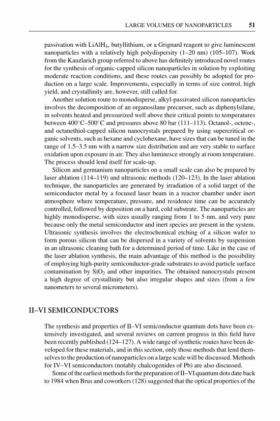
Bentzon et al. (241) have prepared Fe oxide nanoparticles by decomposingFe(CO)5 in decalin in the presence of oleic acid as the stabilizing ligand. Agingthe ferrofluid formed in air for several weeks results in a mixture of hematite andspinel phases. This is among the first reports of nanocrystal superlattice formations.Hyeon et al. (242) have prepared monodisperse γ -Fe2O3 nanoparticles in the sizerange of 4–16 nm by decomposing Fe(CO)5 complexes in octyl ether at 300◦C inthe presence of oleic acid as a capping agent (Figure 5). Oxidation of bcc-Fe to γ -Fe2O3 was achieved by the addition of the organic oxidant (CH2)2NO. The resultingcapped oxide nanoparticles can easily be redispersed in organic solvents, such ashexane or toluene. Particle size is altered by varying the ratio of Fe to the cappingagent (oleic or lauric acids). The remarkable feature of this preparation is thatthe as-prepared particles are sufficiently monodisperse that they form nanocrystalsuperlattices without the need for a size-selection process. More recently, the samegroup has extended their method of aging a metal surfactant complex followed bymild oxidation to produce Co ferrite spinel nanoparticles (243).
Wagner and coworkers have prepared yttrium oxide (244) and europium-dopedyttrium oxide nanoparticles (245) by first reducing rare-earth salts in solution using

27 Jun 2004 11:37 AR AR218-MR34-02.tex AR218-MR34-02.sgm LaTeX2e(2002/01/18) P1: FHD
64 MASALA � SESHADRI
Figure 5 TEM image of monodisperse 11 nm γ -Fe2O3 nanoparticles prepared with-out size selection. Reproduced from Hyeon et al. (242). Reproduced with permissionfrom the American Chemical Society.
alkalides (strong reducing agents that are complexes of crown ethers with alkalimetals, and where the anion is a complexed electron) and then oxidizing the rare-earth metal nanoparticle so formed in aerated water. The white powders were thenannealed in air. More examples of oxide nanoparticles prepared through directoxidation are discussed along with solvothermal methods described later in thereview.
If one starts with a precursor complex wherein the ligands bind to metal ionsthrough oxygen, it could be possible to envisage a decomposition reaction thatwould leave behind the metal oxide. For a trivalent ion, such reactions could begeneralized as
[R − O]3 − M3+ −→ M2O3 + leaving groups.
Suitable design of the R group (stable leaving groups) would ensure that thereaction proceeds in a facile manner. It should then be possible to carry out suchreactions in a suitable high-temperature solvent under solvothermal conditions,possibly in the presence of a suitable capping agent.
Rockenberger et al. (246) have described the use of cupferron complexes asprecursors to prepare transition metal oxide nanoparticles. Cupferron (N-phenyl,N-nitroso hydroxylamine) forms bidentate, univalent complexes with a numberof different transition metal ions. These complexes easily decompose to give theoxide. The authors demonstrated the preparation of γ -Fe2O3, Cu2O, and Mn3O4
27 Jun 2004 11:37 AR AR218-MR34-02.tex AR218-MR34-02.sgm LaTeX2e(2002/01/18) P1: FHD
LARGE VOLUMES OF NANOPARTICLES 65
nanoparticles prepared by injecting octylamine solutions of the corresponding cup-ferron precursors into refluxing trioctylamine. Size is controlled by the temperatureof the reaction. The particles so prepared form stable solutions in solvents such astoluene, from which they can be reprecipitated by the addition of methanol. Thiswork is quite seminal in its generality, and particularly in the manner in which itsuggests the search for suitable precursors for the preparation of oxide nanopar-ticles. Most importantly, it suggests thermolysis as an alternative to hydrolysis,which, as pointed out earlier, is simply not viable for a number of metal oxides.
An important contribution to the surface chemistry of metal oxide nanoparti-cles has been made by Rotello and coworkers (247) who have prepared γ -Fe2O3
nanoparticles by the cupferron decomposition method and compared the relativeefficacies of different long-chain surfactants as capping agents. The most stabiliz-ing capping agent (as monitored by ease of redissolution and stability in solution)was obtained by using a two-tailed surfactant (with 12-carbon tails) with the polarpart comprising a 1,3-diol.
The thermolysis of Fe(III) hydroxide caprylates in boiling tetralin under argonflow gives γ -Fe2O4 nanoparticles (248). The surfaces of the nanoparticles could bemodified by exchanging the capping caprylate groups with betaine, among otherspecies (249). Betaine-capped γ -Fe2O3 nanoparticles are reported to have highsolubility in water.
Using a combination of hydrolysis and oxidation, O’Brien et al. (250) haveshown that the treatment of a complex alkoxide containing Ba2+ and Ti4+—BaTi(O2CC7H15)[OCH(CH3)2]5, an agent for the MOCVD growth of BaTiO3—can be decomposed in diphenyl ether at 140◦C in the presence of oleic acid as acapping agent to give nanocrystalline, cubic BaTiO3. After cooling to 100◦C, 30%H2O2 is added, and crystallization is induced over a 48 h period. Changing theratio of capping agent to water and the amount of peroxide added permits the sizeof the particles to be varied. This is perhaps the only report of soluble perovskiteoxide nanoparticles and, once again, is a route of great generality and interest. Theauthors also report using such routes to prepare PbTiO3 and TiO2 nanoparticles.
In a metathesis reaction, two compounds, AB and CD, exchange species togive two new compounds AC and BD. Such routes have been explored in thepreparation of nanoparticles. Arnal et al. (251) have reported two nonhydrolyticroutes to sol-gel metal oxides, particularly of titanium. The first route involves thereaction of a metal halide with a metal alkoxide:
MCln + M(OR)n −→ 2MOn/2 + nRCl,
and the second route involves the reaction of a metal halide with an ether:
MCln + (n/2)ROR −→ MOn/2 + nRCl.
In both reactions, some of the driving force comes from the removal of thevolatile product RCl. The first of the two methods has been employed by Colvinand coworkers (252) to prepare capped TiO2 nanoparticles in refluxing heptadecane

27 Jun 2004 11:37 AR AR218-MR34-02.tex AR218-MR34-02.sgm LaTeX2e(2002/01/18) P1: FHD
66 MASALA � SESHADRI
with trioctyl phosphine oxide as the capping agent:
MCln + M(OR)n −→ 2MOn/2 + nRCl.
The particles formed clear solutions in nonpolar solvents, from which they could beprecipitated through the addition of a polar solvent such as acetone. When trioctylphosphine oxide was not taken with the starting materials, insoluble precipitateswere obtained. Recently, a similar route has been used by Hyeon and coworkersto prepare tetragonal zirconia nanoparticles (253).
Rajamathi & Seshadri (215) have reviewed the uses of solvothermal methods forthe preparation of oxide and chalcogenide nanoparticles. For oxide nanoparticles,these methods can involve hydrolysis, oxidation and thermolysis, all performed un-der hydrothermal or solvothermal conditions. Some of the more striking examplesare provided here.
An unusual reaction reported by Inoue et al. (254) is the direct oxidation of Cemetal in 2-methoxyethanol at temperatures between 200◦C and 250◦C. Most of theproduct obtained was bulk CeO2 as a yellow solid, but in addition, they obtaineda brown solution of 2 nm CeO2 nanoparticles. The CeO2 nanoparticles couldbe salted out through the addition of NaCl and redispersed into solution at will.The solutions obeyed the Beer-Lambert law for the concentration dependence ofthe optical extinction, suggesting that the nanoparticulate dispersion was a genuinesolution.
Starting with Zn powder and GaCl3 in water, Qian and coworkers (255) haveused the fact that the oxidation of Zn to Zn2+ in water is associated with theproduction of hydroxyl ions according to
Zn + 2H2O −→ Zn2+ + 2OH− + H2↑ .
The OH− ions help in the hydrolysis of Ga3+ to eventually give, in the presence ofZn2+, the spinel ZnGa2O4. The authors used a 10%–20% over-stoichiometry of Zn,and the reaction was carried out at 150◦C for 10 h. 10-nm ZnGa2O4 nanoparticleswere obtained. Excess ZnCl2 formed in the reaction could be washed away withwater.
Chemseddine & Moritz (256) have used the polycondensation of titanium alkox-ide Ti(OR)4 in the presence of tetramethylammonium hydroxide to obtain highlycrystalline anatase nanoparticles with different sizes and shapes. A general, step-wise reaction for such polycondensation could be written as
2Ti(OR)4 −→ (OR)3TiOTi(OR)3 + ROR
(OR)3TiOTi(OR)3 + Ti(OR)4 −→ (OR)3TiOTi(OR)2OTi(OR)3 + ROR,
and so on to anatase, although it might not proceed in precisely this manner inthe presence of base. The reactions were performed in two steps: First alkox-ide was added to the base at 0◦C in alcoholic solvents in a three-necked flask.The temperature was then raised to reflux. The second stage involved treating theproduct of the reflux in Ti autoclaves under a saturated vapor pressure of water

27 Jun 2004 11:37 AR AR218-MR34-02.tex AR218-MR34-02.sgm LaTeX2e(2002/01/18) P1: FHD
LARGE VOLUMES OF NANOPARTICLES 67
(2500 kPa) at temperatures between 175◦C and 200◦C for 5 h. Sufficiently monodis-perse nanocrystals were obtained so that they formed coherent superlattices asmonitored by low-angle powder X-ray diffraction. By using other hydroxides withbulkier alkyl groups (for example, tetrapropylammonium hydroxide) some controlover crystallite morphology became possible.
Wu et al. (257) have combined microemulsion and inverse-micelle techniqueswith hydrothermal techniques in the preparation of rutile and anatase TiO2 nanopar-ticles. They used the system water-cyclohexane-Triton X-100 with n-hexanol as asecond emulsifier. In the water-filled micellar pockets were Ti(OR)4 (R = butyl)acidified with HCl or HNO3. Treating the system at 120◦C–200◦C for 12–144 hgave anatase particles. When high HCl concentrations were employed, rutile rodswere obtained.
Hirano (258) has prepared spinel ZnGa2O4 nanoparticles by adjusting the pH ofZn and Ga sulphates with NH3 to different initial values (varied from 2.5 through10). The material was heat treated hydrothermally at temperatures between 150◦C
Figure 6 TEM image of 7 nm n-octylamine-capped CoFe2O4 nanoparticles preparedin solvothermal toluene. The bar is 20 nm. Reproduced from (263) with permissionfrom the Royal Society of Chemistry.

27 Jun 2004 11:37 AR AR218-MR34-02.tex AR218-MR34-02.sgm LaTeX2e(2002/01/18) P1: FHD
68 MASALA � SESHADRI
and 240◦C for times between 5 and 50 h. Particle sizes could be varied from 5 to25 nm.
Cabanas et al. (259, 260) have described the preparation of nanoparticulateCeO2-ZrO2 solid solutions under flow-hydrothermal conditions, wherein the reac-tants are taken to the final temperature very rapidly in a continuous process. Theadvantage of performing hydrothermal reactions in such a manner is, first, that alarge amount of material can be processed, permitting simple scale up. Second, thenucleation step can be made very rapid as a result of the rapid heating. This can helpseparate nucleation and growth and can thereby satisfy the famous LaMer criterion(261) for obtaining monodisperse particles. Cabanas & Poliakoff (262) have alsoprepared a number of spinel ferrite samples in this way, starting from mixtures ofdifferent Fe(II) and M(II) acetates (M = Co, Ni, Zn and Co/Ni). Most of the prepa-rations yielded a bimodal distribution of sample sizes, with the smaller samplesbeing approximately 10 nm in diameter, and the larger ones approximately 100 nm.
Recently, Thimmaiah et al. (263) have extended the thermolytic route to oxidenanoparticles devised by Rockenberger et al. (246) to solvothermal conditions.Using solvothermal toluene (typically at 220◦C for 1 h), cupferron precursors ofFe, and of Co and Fe are decomposed to obtain sub-12 nm maghemite γ -Fe2O3
nanoparticles and spinel CoFe2O4 nanoparticles. The reactions do not work inthe absence of long-chain amines (n-octylamine and n-dodecylamine), which areadded as capping agents. A complete characterization of the magnetic propertiesof these nanoparticles has been presented by the authors. This is the only report todate of capped oxide nanoparticles prepared from a solvothermal route (Figure 6).
ACKNOWLEDGMENT
This work is supported by the Petroleum Research Fund of the American ChemicalSociety under a type AC award. We also acknowledge a start-up grant from theDean, College of Engineering, UCSB, and thank Paul O’Brien for discussions andencouragement.
The Annual Review of Materials Research is online athttp://matsci.annualreviews.org
LITERATURE CITED
1. Gleiter H. 1989. Nanocrystalline materi-als. Prog. Mater. Sci. 33:223–315
2. Law M, Goldberger J, Yang P. 2004.Semiconductor nanowires and nanotubes.Annu. Rev. Mater. Res. 34:83–122
3. Kolmakov A, Moskovits M. 2004. Chem-ical sensing and catalysis by one-dimensional metal oxide nanostructures.Annu. Rev. Mater. Res. 34:151–80
4. Faraday M. 1857. Experimental rela-tions of gold (and other metals) to light.Phil. Trans. R. Soc. London 147:145–81
5. Brust M, Walker M, Bethell D, SchiffrinDJ, Whyman R. 1994. Synthesis of thiol-derivatized gold nanoparticles in a 2-phase liquid-liquid system. Chem. Com-mun. 7:801–2

27 Jun 2004 11:37 AR AR218-MR34-02.tex AR218-MR34-02.sgm LaTeX2e(2002/01/18) P1: FHD
LARGE VOLUMES OF NANOPARTICLES 69
6. Schmid G, Corain B. 2003. Nanoparticu-lated gold: syntheses, structures, electron-ics and reactivities. Eur. J. Inorg. Chem.17:3081–98
7. Schmid G, Lehnert A. 1989. The com-plexation of gold colloids. Angew. Chem.Int. Ed. Engl. 28:780–81
8. Weare WW, Reed SM, Warner MG, Hut-chinson JE. 2000. Improved synthesis ofsmall phosphine-stabilized gold nanopar-ticles. J. Am. Chem. Soc. 122:12890–91
9. Leff DV, Brandt L, Heath JR. 1996. Syn-thesis and characterization of hydropho-bic, organically-soluble gold nanocrys-tals functionalized with primary amines.Langmuir 12:4723–30
10. Brust M, Stuhr-Hansen N, Norgaard K,Christensen JB, Nielsen LK, BjornholmT. 2001. Langmuir-Blodgett films ofalkane chalcogenide (S, Se, Te) stabilizedgold nanoparticles. Nano Lett. 1:189–91
11. Yao H, Momozawa O, Hamatami T,Kimura K. 2001. Stepwise size-selectiveextraction of carboxylate-modified goldnanoparticles from an aqueous suspen-sion into toluene with tetraoctylammo-nium cations. Chem. Mater. 13:4692–97
12. Corbierre MK, Cameron NS, Sutton M,Mochrie SGJ, Lurio LB, et al. 2001.Polymer-stabilized gold nanoparticlesand their incorporation into polymer ma-trices. J. Am. Chem. Soc. 123:10411–12
13. Tan Y, Dai X, Li Y, Zhu D. 2003. Prepara-tion of gold, platinum, palladium and sil-ver nanoparticles by the reduction of theirsalts with a weak reductant-potassiumbitartrate. J. Mater. Chem. 13:1069–75
14. Heath JR, Knobler CM, Leff DV. 1997.Pressure/temperature phase diagrams andsuperlattices of organically functionalizedmetal nanocrystals monolayers: the in-fluence of particle size, size distributionand surface passivant. J. Phys. Chem. B101:189–97
15. Hostetler MJ, Wingate JE, Zhong CJ,Harris JE, Vachet RW, et al. 1998. Alka-nethiolate gold cluster molecules withcore diameters from 1.5 to 5.2 nm: core
and monolayer properties as a function ofcore size. Langmuir 14:17–30
16. Leff DV, Ohara PC, Heath JR, GelbartWM. 1995. Thermodynamic control ofgold nanocrystal size: experiment andtheory. J. Phys. Chem. 99:7036–41
17. Zhu T, Vasilev K, Kreiter M, Mittler S,Knoll W. 2003. Surface modification ofcitrate-reduced colloidal gold nanoparti-cles with 2-mercaptosuccinic acid. Lang-muir 19:9518–25
18. Frens G. 1973. Controlled nucleation forregulation of particle-size in monodis-perse gold suspensions. Nat. Phys. Sci.241:20–22
19. Turkevich J, Stevenson PC, Hillier J.1951. A study of the nucleation andgrowth processes in the synthesis of col-loidal gold. Discuss. Faraday Soc. 11:55–75
20. Han MY, Quek CH, Huang W, Chew CH,Gan LM. 1999. A simple and effectivechemical route for the preparation of uni-form nonaqueous gold colloids. Chem.Mater. 11:1144–47
21. Dai X, Tan Y, Xu J. 2002. Formation ofgold nanoparticles in the presence of o-anisidine and the dependence of the struc-ture of poly(o-anisidine) on synthetic con-ditions. Langmuir 18:9010–16
22. Prasad BLV, Stoeva SI, Sorensen CM,Klabunde KJ. 2002. Digestive ripeningof thiolate gold nanoparticles: the effectof alkyl chain length. Langmuir 18:7515–20
23. Prasad BLV, Stoeva SI, Sorensen CM,Klabunde KJ. 2003. Digestive-ripeningagents for gold nanoparticles: alternativesto thiols. Chem. Mater. 15:935–42
24. Lin S, Franklin MT, Klabunde KJ. 1986.Nonaqueous colloidal gold. Clustering ofmetal atoms in organic media. Langmuir2:259–60
25. Stoeva S, Klabunde KJ, Sorensen CM,Dragieva I. 2002. Gram-scale synthesisof monodisperse gold colloids by the sol-vated metal atom dispersion and diges-tive ripening and their organization into

27 Jun 2004 11:37 AR AR218-MR34-02.tex AR218-MR34-02.sgm LaTeX2e(2002/01/18) P1: FHD
70 MASALA � SESHADRI
two- and three-dimensional structures. J.Am. Chem. Soc. 124:2305–11
26. Brust M, Kiely CJ. 2002. Some recent ad-vances in nanostructure preparation fromgold and silver nanoparticles: a short top-ical review. Coll. Surf. A Phys. Eng. Asp.202:175–86
27. He S, Yao J, Jiang P, Shi D, Zhang H,et al. 2001. Formation of silver nanoparti-cles and self-assembled two-dimensionalordered superlattice. Langmuir 17:1571–75
28. Li X, Zhang J, Xu W, Jia H, Wang X,et al. 2003. Mercaptoacetic acid-cappedsilver nanoparticles colloid: formation,morphology and SERS activity. Langmuir19:4285–90
29. Tan Y, Li Y, Zhu D. 2003. Preparation ofsilver nanocrystals in the presence of ani-line. J. Coll. Int. Sci. 258:244–51
30. Pastoriza-Santos I, Liz-Marzan LM.1999. Formation and stabilization of silvernanoparticles through reduction by N,N-dimethylformamide. Langmuir 15:948–51
31. Sondi I, Goia DV, Matijevic E. 2003.Preparation of highly concentrated stabledispersions of uniform silver nanoparti-cles. J. Coll. Int. Sci. 260:75–81
32. Lee C, Wan C, Wang Y. 2001. Synthesisof metal nanoparticles via self-regulatedreduction by an alcohol surfactant. Adv.Funct. Mater. 11:344–47
33. Ayyappan S, Gopalan Srinivasa R, Sub-banna GN, Rao CNR. 1997. Nanoparti-cles of Ag, Au, Pd, and Cu produced byalcohol reduction of the salts. J. Mater.Res. 12:398–401
34. Wang W, Chen X, Efrima S. 1999. Silvernanoparticles capped by long chain un-saturated carboxylates. J. Phys. Chem. B103:7238–46
35. Sun Y, Xia Y. 2002. Shape-controlled syn-thesis of gold and silver nanoparticles.Science 298:2176–79
36. Yamamoto M, Nakamoto M. 2003.Novel preparation of monodisperse silvernanoparticles via amine adducts derived
from insoluble silver myristate in tertiaryalkylamine. J. Mater. Chem. 13:2064–65
37. Shen CM, Su YK, Yang HT, Yang TZ, GaoHJ. 2003. Synthesis and characterizationof n-octadecyl mercaptan-protected pal-ladium nanoparticles. Chem. Phys. Lett.373:39–45
38. Chen S, Huang K, Stearns JA. 2000. Alk-anethiolate-protected palladium nanopar-ticles. Chem. Mater. 12:540–47
39. Yee C, Scotti M, Ulman A, White H, Raf-ailovich M, Sokolov J. 1999. One-phasesynthesis of thiol-functionalized platinumnanoparticles. Langmuir 15:4314–16
40. Chen S, Kimura K. 2001. Synthesis ofthiolate-stabilized nanoparticles in pro-tolytic solvents as isolable colloids. J.Phys. Chem. B 105:5397–403
41. Perez H, Pradeau JP, Albouy PA, Perez-Olmil J. 1999. Synthesis and characteriza-tion of functionalized platinum nanopar-ticles. Chem. Mater. 11:3460–63
42. Quiros I, Yamada M, Kubo K, MizutaniJ, Kurihara M, Nishihara H. 2002. Prepa-ration of alkanethiolate-protected palla-dium nanoparticles and their size depen-dance on synthetic conditions. Langmuir18:1413–18
43. Teranishi T, Miyake M. 1998. Size con-trol of palladium nanoparticles and theircrystal structures. Chem. Mater. 10:594–600
44. Thomas PJ, Kulkarni GU, Rao CNR.2000. An investigation of two-dimen-sional arrays of thiolized Pd nanoparti-cles. J. Phys. Chem. B 104:8138–44
45. Mandal S, Selvakannan PR, Roy D,Chaudhari RV, Sastry M. 2002. A newmethod for the synthesis of hydropho-bized, catalytically active Pt nanoparti-cles. Chem. Commun. 24:3002–3
46. Horswell SL, Kiely CJ, O’Neil IA,Schiffrin DJ. 1999. Alkyl isocyanide-derivatized platinum nanoparticles. J. Am.Chem. Soc. 121(23):5573–74
47. Alvarez J, Liu J, Roman E, KaiferAE. 2000. Water-soluble platinum andpalladium nanoparticles modified with
27 Jun 2004 11:37 AR AR218-MR34-02.tex AR218-MR34-02.sgm LaTeX2e(2002/01/18) P1: FHD
LARGE VOLUMES OF NANOPARTICLES 71
thiolated β-cyclodextrin. Chem. Com-mun. 13:1151–52
48. Kim SW, Park J, Jang Y, Chung Y, HwangS, et al. 2003. Synthesis of monodis-perse palladium nanoparticles. Nano Lett.3:1289–91
49. Hyeon T. 2003. Chemical synthesis ofmagnetic nanoparticles. Chem. Commun.8:927–34
50. Petit C, Taleb A, Pileni MP. 1999. Cobaltnanosized particles organized in a 2D su-perlattice: synthesis, characterization andmagnetic properties. J. Phys. Chem. B103:1805–10
51. Lin XM, Sorensen CM, Klabunde KJ,Hadjipanayis GC. 1998. Temperature de-pendance of morphology and magneticproperties of cobalt nanoparticles pre-pared by an inverse micelle technique.Langmuir 14:7140–46
52. Wilcoxon JP, Provencio PP. 1999. Use ofsurfactant micelles to control the struc-tural phase of nanosized iron clusters. J.Phys. Chem. B 103:9809–12
53. Suslick KS, Fang M, Hyeon T. 1996.Sonochemical synthesis of iron colloids.J. Am. Chem. Soc. 118:11960–61
54. Suslick KS, Hyeon T, Fang M. 1996.Nanostructured materials generated byhigh-intensity ultrasound: sonochemicalsynthesis and catalytic studies. Chem.Mater. 8:2172–79
55. Chen DH, Hsieh CH. 2002. Synthe-sis of nickel nanoparticles in aqueouscationic surfactant solutions. J. Mater.Chem. 12:2412–15
56. Sun S, Murray CB. 1999. Synthesis ofmonodisperse cobalt nanocrystals andtheir assembly into magnetic superlat-tices. J. Appl. Phys. 85:4325–30
57. Murray CB, Sun S, Gaschler W, DoyleH, Betley TA, Kagan CR. 2001. Colloidalsynthesis of nanocrystals and nanocrystalsuperlattices. IBM J. Res. Develop. 45:47–56
58. Murray CB, Sun S, Doyle H, Betley T.2001. Monodisperse 3D transition-metal(Co, Ni, Fe) nanoparticles and their as-
sembly into nanoparticle superlattices.MRS Bull. 26:985–91
59. Hou Y, Gao S. 2003. Monodisperse nickelnanoparticles prepared from a monosur-factant system and their magnetic proper-ties. J. Mater. Chem. 13:1510–12
60. Dinega DP, Bawendi MG. 1999. Asolution-phase chemical approach to anew crystal structure of cobalt. Angew.Chem. Int. Ed. Engl. 38:1788–91
61. Giersig M, Hilgendorff M. 2002. On theroad from single, nanosized magneticclusters to multi-dimensional nanostruc-tures. Coll. Surf. 202:207–13
62. Puntes VF, Krishnan MK, Alivisatos P.2001. Synthesis, self-assembly and mag-netic behavior of a two-dimensional su-perlattice of single-crystal ε-Co nanopar-ticles. App. Phys. Lett. 78:2187–89
63. Tripp SL, Pusztay SV, Ribbe AE, Wei A.2002. Self-assembly of cobalt nanoparti-cle rings. J. Am. Chem. Soc. 124:7914–15
64. Bonnemann H, Brijoux W, BrinkmannR, Matoussevitch N, Waldofner N, et al.2003. Size-selective synthesis of air sta-ble colloidal magnetic cobalt nanoparti-cles. Inorg. Chim. Acta 350:617–24
65. Kim SW, Son SU, Lee SS, HyeonT, Chung YC. 2001. Colloidal cobaltnanoparticles: a highly active and reusablePauson-Khand catalyst. Chem. Commun.21:2212–13
66. Park SJ, Kim S, Lee S, Khim ZG, CharK, Hyeon T. 2000. Synthesis and mag-netic studies of uniform iron nanorodsand nanospheres. J. Am. Chem. Soc.122:8581–82
67. Li Y, Liu J, Wang Y, Wang ZL. 2001.Preparation of monodispersed Fe-Monanoparticles as the catalyst for CVD syn-thesis of carbon nanotubes. Chem. Mater.13:1008–14
68. Sun S, Murray CB, Weller D, FolksL, Moser A. 2000. Monodisperse FePtnanoparticles and ferromagnetic FePtnanocrystal superlattices. Science 287:1989–92

27 Jun 2004 11:37 AR AR218-MR34-02.tex AR218-MR34-02.sgm LaTeX2e(2002/01/18) P1: FHD
72 MASALA � SESHADRI
69. Sobal NS, Ebels U, Mohwald H, Gier-sig M. 2003. Synthesis of core-shell PtConanoparticles. J. Phys. Chem. B 107:7351–54
70. Chen M, Nikles DE. 2002. Synthesis ofspherical FePd and CoPt nanoparticles. J.Appl. Phys. 91:8477–79
71. Osuna J, de Caro D, Amiens C, Chau-dret B, Snoeck E, et al. 1996. Synthe-sis, characterization and magnetic prop-erties of cobalt nanoparticles from anorganometallic precursor. J. Phys. Chem.100:14571–74
72. Chen S, Sommers JM. 2001. Alkane-thiolate-protected copper nanoparticles:spectroscopy, electrochemistry and solid-state morphological evolution. J. Phys.Chem. B 105:8816–20
73. Dhas NA, Raj PC, Gedanken A. 1998.Synthesis, characterisation and propertiesof metallic copper nanoparticles. Chem.Mater. 10:1446–52
74. Lisiecki I, Billoudet F, Pileni MP. 1997.Syntheses of copper nanoparticles ingelified microemulsion and in reversemicelles. J. Mol. Liq. 72:251–61
75. Hambrock J, Becker R, Birkner A,Weib J, Fischer RA. 2002. A non-aqueous organometallic route to highlymonodisperse copper nanoparticles usingCu(OCH(Me)CH2NMe2)2. Chem. Com-mun. 1:68–69
76. Canham LT. 1990. Silicon quantum wirearray fabrication by electrochemical andchemical dissolution of wafers. Appl.Phys. Lett. 57(10):1046–48
77. King WD, Boxall DL, Lukehart CM.1997. Nanoclusters of silicon and germa-nium. J. Clust. Sci. 8(2):267–92
78. Littau KA, Szajowski PJ, Muller AJ, Kor-tan AR, Brus LE. 1993. A luminescentsilicon nanocrystal colloid via a high tem-perature aerosol reaction. J. Phys. Chem.97:1224–30
79. Ehbrecht M, Kohn B, Huisken F, LagunaMA, Paillard V. 1997. Photoluminescenceand resonant Raman spectra of siliconfilms produced by size-selected cluster
beam deposition. Phys. Rev. B 56:6958–64
80. Seraphin AA, Werwa E, KolenbranderKD. 1997. Influence of nanostructure sizeon the luminescence behavior of siliconnanoparticle thin films. J. Mater. Res.12:3386–92
81. Schuppler S, Friedman SL, Marcus MA,Adler DL, Xie YH, et al. 1994. Di-mensions of luminescent oxidized andporous silicon structures. Phys. Rev. Lett.72(16):2648–51
82. Koch F, Petrova-Koch V. 1996. Light fromSi-nanoparticle systems—a comprehen-sive review. J. Non-Cryst. Solids 198-200:840–46
83. Paine DC, Caragianis C, Kim TY, Shige-sato Y, Ishahara T. 1993. Visible pho-toluminescence from nanocrystalline Geformed by H2 reduction of Si0.6Ge0.4O2.Appl. Phys. Lett. 62(22):2842–44
84. Kanemitsu Y, Uto H, Masumoto Y, MaedaY. 1992. On the origin of visible pho-toluminescence in nanometer-size Gecrystallites. Appl. Phys. Lett. (61):2187–89
85. Venkatasubramanian R, Malta DP,Timmons ML, Hutchby JA. 1991. Visiblelight emission from quantized planarGe structures. Appl. Phys. Lett. 59(13):1603–5
86. Wilson WL, Szajowski PF, Brus LE. 1993.Quantum confinement in size-selected,surface oxidized silicon nanocrystals. Sci-ence 262:1242–44
87. Brus LE, Szajowski PF, Wilson WL,Harris TD, Schuppler S, Citrin PH. 1995.Electron spectroscopy and photophysicsof Si nanocrystals: relation to bulk c-Siand porous Si. J. Am. Chem. Soc. 117:2915–22
88. Brus LE. 1994. Luminescence of sili-con materials: chains, sheets, nanocrys-tals, nanowires, microcrystals and poroussilicon. J. Phys. Chem. 98:3575–81
89. Borsella E, Falconieri M, Botti S, MartelliS, Bignoli F, et al. 2001. Opticaland morphological characterization of Si

27 Jun 2004 11:37 AR AR218-MR34-02.tex AR218-MR34-02.sgm LaTeX2e(2002/01/18) P1: FHD
LARGE VOLUMES OF NANOPARTICLES 73
nanocrystals/silica composites preparedby sol-gel processing. Mater. Sci. Eng. B79:55–62
90. Botti S, Coppola R, Gourbilleau F, RizkR. 2000. Photoluminescence from sili-con nanoparticles synthesized by laser-induced decomposition of silane. J. Appl.Phys. 88(6):3396–401
91. Borsella E, Botti S, Cremona M, MartelliS, Montereali RM, Nesterenko A. 1997.Photoluminescence from oxidized Sinanoparticles produced by CW CO2 lasersynthesis in a continuous-flow reactor. J.Mater. Sci. Lett. 16:221–23
92. Huisken F, Kohn B, Paillard V. 1999.Structured films of light-emitting sil-icon nanoparticles produced by clus-ter beam deposition. Appl. Phys. Lett.74(25):3776–78
93. Ledoux G, Gong J, Huisken F. 2001. Ef-fect of passivation and aging on the pho-toluminescence of silicon nanoparticles.Appl. Phys. Lett. 79(24):4028–30
94. Ledoux G, Gong J, Huisken F, Guillois O,Reynaud C. 2002. Photoluminescence ofsize-separated silicon nanocrystals: con-firmation of quantum confinement. Appl.Phys. Lett. 80(25):4834–36
95. Li X, He Y, Talukdar SS, Swihart MT.2003. Process for preparing macroscopicquantities of brightly photoluminescentsilicon nanoparticles with emission span-ning the visible spectrum. Langmuir19:8490–96
96. Heath JR. 1992. A liquid-solution-phasesynthesis of crystalline silicon. Science258:1131–33
97. Heath JR, LeGoues FK. 1993. A liquidsolution synthesis of single crystal germa-nium quantum wires. Chem. Phys. Lett.208(3,4):263–68
98. Kornowski A, Giersig M, Vogel R,Chemseddine A, Weller H. 1993. Nano-meter-sized colloidal germanium parti-cles—wet-chemical synthesis, laser-ind-tuced crystallization and particle growth.Adv. Mater. 5(9):634–36
99. Heath JR, Shiang JJ, Alivisatos PA. 1994.
Germanium quantum dots: optical proper-ties and synthesis. J. Chem. Phys. 101(2):1607–15
100. Bley RA, Kauzlarich SM. 1996. A low-temperature solution phase route for thesynthesis of silicon nanoclusters. J. Am.Chem. Soc. 118:12461–62
101. Yang CS, Bley RA, Kauzlarich SM, LeeHWH, Delgado GR. 1999. Synthesis ofalkyl-terminated silicon nanoclusters bya solution route. J. Am. Chem. Soc.121:5191–95
102. Mayeri D, Phillips BL, Augustine MP,Kauzlarich SM. 2001. NMR study ofthe synthesis of alkyl-terminated siliconnanoparticles from the reaction of SiCl4
with the Zintl salt, NaSi. Chem. Mater.13:765–70
103. Baldwin RK, Pettigrew KA, Ratai E,Augustine MP, Kauzlarich SM. 2002.Solution reduction synthesis of surfacestabilized silicon nanoparticles. Chem.Commun. 17:1822–23
104. Baldwin RK, Pettigrew KA, Garno JC,Power PP, Liu GY, Kauzlarich SM.2002. Room temperature solution syn-thesis of alkyl-capped tetrahedral shapedsilicon nanocrystals. J. Am. Chem. Soc.124(7):1150–51
105. Liu Q, Kauzlarich SM. 2002. A new syn-thetic route for the synthesis of hydrogenterminated silicon nanoparticles. Mater.Sci. Eng. B 96:72–75
106. Pettigrew KA, Liu Q, Power PP, Kau-zlarich SM. 2003. A new synthetic routeto alkyl terminated silicon nanoparticles.Mat. Res. Soc. Symp. Proc. 737:F1.8.1–8.6
107. Pettigrew KA, Power PP, Kauzlarich SM.2003. Solution synthesis of alkyl- andalkyl/alkoxy-capped silicon nanoparticlesvia oxidation of Mg2Si. Chem. Mater.15:4005–11
108. Taylor BR, Kauzlarich SM, Lee HWH,Delgado GR. 1998. Solution synthesisof germanium nanocrystals demonstrat-ing quantum confinement. Chem. Mater.10:22–24

27 Jun 2004 11:37 AR AR218-MR34-02.tex AR218-MR34-02.sgm LaTeX2e(2002/01/18) P1: FHD
74 MASALA � SESHADRI
109. Taylor BR, Kauzlarich SM, DelgadoGR, Lee HWH. 1999. Solution synthesisand characterization of quantum confinedGe nanoparticles. Chem. Mater. 11:2493–500
110. Tanke RS, Kauzlarich SM, Patten TE,Pettigrew KA, Murphy DL, et al.2003. Synthesis of germanium nanoclus-ters with irreversibly attached functionalgroups: acetals, alcohols, esters and poly-mers. Chem. Mater. 15:1682–89
111. Ding Z, Quinn BM, Haram SK, Pell LE,Korgel BA, Bard AJ. 2002. Electrochem-istry and electrogenerated chemilumines-cence from silicon nanocrystal quantumdots. Science 296:1293–97
112. Holmes JD, Ziegler KJ, Doty RC, Pell LE,Johnston KP, Korgel BA. 2001. Highly lu-minescent silicon nanocrystals with dis-crete optical transitions. J. Am. Chem. Soc.123:3743–48
113. English DS, Pell LE, Yu Z, BarbaraPF, Korgel BA. 2002. Size tunable visi-ble luminescence from individual organicmonolayer stabilized silicon nanocrystalquantum dots. Nano Lett. 2(7):681–85
114. Morales AM, Lieber CM. 1998. A laserablation method for the synthesis of crys-talline semiconductor nanowires. Science279:208–11
115. Lowndes DH, Rouleau CM, Thundat TG,Duscher G, Kenik EA, Pennycook SJ.1999. Silicon and zinc telluride nanopar-ticles synthesized by low energy densitypulsed laser ablation into ambient gases.J. Mater. Res. 14(2):359–69
116. Yoshida T, Yamada Y, Orii T. 1998. Elec-troluminescence of silicon nanocrystal-lites prepared by pulsed laser ablation inreduced pressure inert gas. J. Appl. Phys.83(10):5427–32
117. Carlisle JA, Germanenko IN, PithawallaYB, El-Shall MS. 2001. Morphology,photoluminscence and electronic struc-ture in oxidized silicon nanoclusters. J.Electr. Spectr. 114–116:229–34
118. Seto T, Kawakami Y, Suzuki N, HirasawaM, Aya N. 2001. Laser synthesis of uni-
form silicon single nanodots. Nano Lett.1(6):315–18
119. Ngiam ST, Jensen KF, Kolenbrander KD.1994. Synthesis of Ge nanocrystals em-bedded in a Si host matrix. J. Appl. Phys.76(12):8201–3
120. Heinrich JL, Curtis CL, Credo GM,Kavanagh KL, Sailor MJ. 1992. Lumi-nescent colloidal silicon suspensions fromporous silicon. Science 255:66–68
121. Bley RA, Kauzlarich SM, Davis JE, LeeHWH. 1996. Characterization of siliconnanoparticles prepared from porous sili-con. Chem. Mater. 8:1881–88
122. Kim NY, Laibinis PE. 1997. Thermalderivatization of porous silicon with al-cohols. J. Am. Soc. Chem. 119:2297–98
123. Dhas NA, Raj CP, Gedanken A. 1998.Preparation of luminescent siliconnanoparticles: a novel sonochemicalapproach. Chem. Mater. 10:3278–81
124. Esteves ACC, Trindade T. 2002. Syntheticstudies on II/VI semiconductor quantumdots. Curr. Opin. Solid State Mater. Sci.6(4):347–53
125. Trindade T, O’Brien P, Pickett NL. 2001.Nanocrystalline semiconductors: synthe-sis, properties, and perspectives. Chem.Mater. 13(11):3843–58
126. O’Brien P, Pickett NL. 2004. Strategiesfor the scalable synthesis of quantum dotsand related nanodimensional materials.See Ref. 264. In press
127. Peng X. 2002. Green chemical ap-proaches toward high-quality semicon-ductor nanocrystals. Chem. Eur. J. 8(2):335–39
128. Brus LE. 1984. Electron-electron andelectron-hole interactions in small semi-conductor crystallites: the size depen-dance of the lowest excited electronicstate. J. Chem. Phys. 80(9):4403–9
129. Rossetti R, Ellison JL, Gibson JM, BrusLE. 1984. Size effects in the excited elec-tronic states of small colloidal CdS crys-tallites. J. Chem. Phys. 80(9):4464–69
130. Rossetti R, Hull R, Gibson JM, Brus LE.1985. Excited electronic states and optical
27 Jun 2004 11:37 AR AR218-MR34-02.tex AR218-MR34-02.sgm LaTeX2e(2002/01/18) P1: FHD
LARGE VOLUMES OF NANOPARTICLES 75
spectra of ZnS and CdS crystallites in the≈15 to 50 A size range: evolution frommolecular to bulk semiconducting prop-erties. J. Chem. Phys. 82(1):552–59
131. Rogach AL, Kornowski A, Gao M,Eychmuller A, Weller H. 1999. Synthe-sis and characterization of a size seriesof extremely small thiol-stabilized CdSenanocrystals. J. Phys. Chem. B 103:3065–69
132. Rogach A, Kershaw S, Burt M, HarrisonM, Kornowski A, et al. 1999. Colloidallyprepared HgTe nanocrystals with strongroom-temperature infrared luminescence.Adv. Mater. 11(7):552–55
133. Harrison MT, Kershaw SV, Burt MG,Eychmuller A, Weller H, Rogach AL.2000. Wet chemical synthesis and spec-troscopic study of CdHgTe nanocrystalswith strong near-infrared luminescence.Mater. Sci. Eng. B 69–70:355–60
134. Spanhel L, Haase M, Weller H,Henglein A. 1987. Photochemistry ofcolloidal semiconductors. Surface modifi-cation and stability of strong luminescingCdS particles. J. Am. Chem. Soc. 109:5649–55
135. Gaponik N, Talapin DV, Rogach AL,Hoppe K, Shevchenko EV, et al. 2002.Thiol-capping of CdTe nanocrystals: analternative to organometallic syntheticroutes. J. Phys. Chem. B 106:7177–85
136. Vossmeyer T, Katsikas L, Giersig M,Popovic IG, Diesner K, et al. 1994. CdSnanoclusters: synthesis, characterization,size dependent oscillator strength, tem-perature shift of the excitonic transitionenergy, and reversible absorbance shift. J.Phys. Chem. 98:7665–73
137. Wang W, Germanenko I, El-Shall MS.2002. Room-temperature synthesis andcharacterization of nanocrystalline CdS,ZnS and CdxZn1−xS. Chem. Mater. 14:3028–33
138. Wang W, Geng Y, Yan P, Liu F, Xie Y,Qian Y. 1999. A novel mild route tonanocrystalline selenides at room temper-ature. J. Am. Chem Soc. 121:4062–63
139. Lifshitz E, Bashouti M, Kloper V, KigelA, Eisen MS, Berger S. 2003. Synthe-sis and characterization of PbSe quan-tum wires, multipods, quantum rods, andcubes. Nano Lett. 3(6):857–62
140. Mastai Y, Polsky R, Koltypin Y, GedankenA, Hodes G. 1999. Pulsed sonoelec-trochemical synthesis of cadmium se-lenide nanoparticles. J. Am. Chem. Soc.121:10047–52
141. Zhu J, Aruna ST, Koltypin Y, Gedanken A.2000. A novel method for the preparationof lead selenide: pulse sonoelectrochem-ical synthesis of lead selenide nanoparti-cles. Chem. Mater. 12:143–47
142. Zhu J, Koltypin Y, Gedanken A. 2000.General sonochemical method for thepreparation of nanophasic selenides: syn-thesis of ZnSe nanoparticles. Chem.Mater. 12:73–78
143. Murray CB, Norris DJ, Bawendi MG.1993. Synthesis and characterization ofnearly monodisperse CdE (E = S, Se, Te)semiconductor nanocrystallites. J. Am.Chem. Soc. 115:8706–15
144. Alivisatos PA. 1996. Perspectives onthe physical chemistry of semiconductornanocrystals. J. Phys. Chem. 100:13226–39
145. Manna L, Scher EC, Alivisatos PA.2000. Synthesis of soluble and process-able rod-, arrow-, teardrop-, and tetrapod-shaped CdSe nanocrystals. J. Am. Chem.Soc. 122(51):12700–6
146. Peng X, Manna L, Yang W, Wickham J,Scher E, et al. 2000. Shape control of CdSenanocrystals. Nature 404(2):59–61
147. Peng X, Wickham J, Alivisatos AP. 1998.Kinetics of II–VI and III–V colloidalsemiconductor nanocrystal growth: “fo-cusing” of size distributions. J. Am. Chem.Soc. 120:5343–44
148. Hines MA, Guyot-Sionnest P. 1996. Syn-thesis and characterization of strongly lu-minescing ZnS-capped CdSe nanocrys-tals. J. Phys. Chem. 100:468–71
149. Danek M, Jensen KF, Murray CB,Bawendi MG. 1996. Synthesis of

27 Jun 2004 11:37 AR AR218-MR34-02.tex AR218-MR34-02.sgm LaTeX2e(2002/01/18) P1: FHD
76 MASALA � SESHADRI
luminescent thin film CdSe/ZnSe quan-tum dot composites using CdSe quantumdots passivated with an overlayer ofZnSe. Chem. Mater. 8:173–80
150. Dabbousi BO, Rodriguez-Viejo J, Miku-lec FV, Heine JR, Mattoussi H, et al. 1997.(CdSe)ZnS core-shell quantum dots: syn-thesis and characterization of a size seriesof highly luminescent nanocrystallites. J.Phys. Chem. B 101:9463–75
151. Peng X, Schlamp MC, Kadavanich AV,Alivisatos AP. 1997. Epitaxial growth ofhighly luminescent CdSe/CdS core/shellnanocrystals with photostability and elec-tronic accessibility. J. Am. Chem. Soc.119:7019–29
152. Rajeshwar K, de Tacconi NR, Chen-thamarakshan CR. 2001. Semiconductor-based composite materials: prepara-tion, properties, and performance. Chem.Mater. 13(9):2765–82
153. Eychmuller A. 2000. Structure and pho-tophysics of semiconductor nanocrys-tals. J. Phys. Chem. B 104(28):6514–28
154. Peng ZA, Peng X. 2001. Formationof high-quality CdTe, CdSe, and CdSnanocrystals using CdO as precursor. J.Am. Chem. Soc. 123:183–84
155. Qu L, Peng ZA, Peng X. 2001. Alter-native routes toward high quality CdSenanocrystals. Nano Lett. 1(6):333–37
156. Peng ZA, Peng X. 2001. Mechanisms ofthe shape evolution of CdSe nanocrystals.J. Am. Chem. Soc. 123:1389–95
157. Peng ZA, Peng X. 2002. Nearly mono-disperse and shape-controlled CdSenanocrystals via alternative routes: nu-cleation and growth. J. Am. Chem. Soc.124:3343–53
158. Manna L, Milliron DJ, Meisel A, ScherE, Alivisatos AP. 2003. Controlled growthof tetrapod-branched inorganic nanocrys-tals. Nature Mater. 2:382–85
159. Brennan JG, Siegrist T, Carroll PJ,Stuczynski SM, Brus LE, SteigerwaldML. 1989. The preparation of large semi-conductor clusters via the pyrolysis of a
molecular precursor. J. Am. Chem. Soc.111:4141–43
160. Pickett NL, O’Brien P. 2001. Synthe-ses of semiconductor nanoparticles usingsingle-molecular precursors. Chem. Rec.1:467–79
161. Malik MA, O’Brien P, Revaprasadu N.2002. A simple route to the synthesis ofcore/shell nanoparticles of chalcogenides.Chem. Mater. 14(5):2004–10
162. Revaprasadu N, Malik MA, O’Brien P,Wakefield G. 1999. Deposition of zincsulfide quantum dots from a single-source molecular precursor. J. Mater. Res.14(8):3237–40
163. Revaprasadu N, Malik MA, O’Brien P,Zulu MM, Wakefield G. 1998. Single-source molecular precursor for the depo-sition of zinc selenide quantum dots. J.Mater. Chem. 8(8):1885–88
164. Trindade T, O’Brien P, Zhang X. 1997.Synthesis of CdS and CdSe nanocrystal-lites using a novel single-molecule precur-sors approach. Chem. Mater. 9:523–30
165. Ludolph B, Malik MA, O’Brien P, Revap-rasadu N. 1998. Novel single moleculeprecursor routes for the direct synthesisof highly monodispersed quantum dotsof cadmium or zinc sulfide or selenide.Chem. Commun. 17:1849–50
166. Revaprasadu N, Malik MA, O’Brien P,Wakefield G. 1999. A simple route to syn-thesize nanodimensional CdSe-CdS core-shell structures from single molecular pre-cursors. Chem. Commun. 16:1573–74
167. Lazell M, O’Brien P. 1999. A novel singlesource precursor route to self capping CdSquantum dots. Chem. Commun. 20:2041–42
168. Trindade T, Monteiro OC, O’Brien P,Motevalli M. 1999. Synthesis of PbSenanocrystallites using a single-sourcemethod. The X-ray crystal structure oflead(II) diethyldiselenocarbamate. Poly-hedron 18:1171–75
169. Trindade T, O’Brien P, Zhang X, Mote-valli M. 1997. Synthesis of PbS nanocrys-tallites using a novel single molecule

27 Jun 2004 11:37 AR AR218-MR34-02.tex AR218-MR34-02.sgm LaTeX2e(2002/01/18) P1: FHD
LARGE VOLUMES OF NANOPARTICLES 77
precursors approach: X-ray single-crystalstructure of Pb(S2CNEtPri)2. J. Mater.Chem. 7(6):1011–16
170. Jun YW, Lee SM, Kang NJ, CheonJ. 2001. Controlled synthesis of multi-armed CdS nanorod architectures usingmonosurfactant system. J. Am. Chem. Soc.123:5150–51
171. Malik MA, Revaprasadu N, O’Brien P.2001. Air-stable single-source precursorsfor the synthesis of chalcogenide semi-conductor nanoparticles. Chem. Mater.13(3):913–20
172. Nair PS, Radhakrishnan T, RevaprasaduN, Kolawole GA, O’Brien P. 2002. Asingle-source route to CdS nanorods.Chem. Commun. 6:564–65
173. Crouch DJ, O’Brien P, Malik MA, Ska-bara PJ, Wright SP. 2003. A one-stepsynthesis of cadmium selenide quantumdots from a novel single source precursor.Chem. Commun. 12:1454–55
174. Nair PS, Radhakrishnan T, Revap-rasadu N, Kolawole GA, O’Brien P.2003. Cd(NH2CSNHNHCSNH2)Cl2: anew single-source precursor for the prepa-ration of CdS nanoparticles. Polyhedron22(23):3129–35
175. Nair PS, Radhakrishnan T, RevaprasaduN, Kolawole GA, O’Brien P. 2002.Cadmium ethylxanthate: a novel single-source precursor for the preparationof CdS nanoparticles. J. Mater. Chem.12(9):2722–25
176. Dance IG, Choy A, Schudder ML.1984. Syntheses, properties, and molec-ular and crystal structures of (Me4N)4
[E4M10(SPh)16] (E = S, Se; M = Zn, Cd):molecular supertetrahedral fragments ofthe cubic metal chalcogenide lattice. J.Am. Chem. Soc. 106:6285–95
177. Cumberland SL, Hanif KM, Javier A,Khitrov GA, Strouse GF, et al. 2002. In-organic clusters as single-source precur-sors for preparation of CdSe, ZnSe, andCdSe/ZnS nanomaterials. Chem. Mater.14:1576–84
178. Byrappa K, Yoshimura M. 2001. Hand-
book of Hydrothermal Technology.Norwich, New York: William AndrewPubl.
179. Li Y, Ding Y, Qian Y, Zhang Y, Yang L.1998. A solvothermal elemental reactionto produce nanocrystalline ZnSe. Inorg.Chem. 37:2844–45
180. Peng Q, Dong Y, Deng Z, Sun X,Li Y. 2001. Low-temperature elemental-direct-reaction route to II–VI semicon-ductor nanocrystalline ZnSe and CdSe.Inorg. Chem. 40:3840–41
181. Zhan JH, Yang XG, Zhang WX, WangDW, Xie Y, Qian YT. 2000. A solvother-mal route to wurzite ZnSe nanoparticles.J. Mater. Res. 15:629–32
182. Deng ZX, Wang C, Sun MX, Li DY.2002. Structure-directing coordinationtemplate effect of ethylenediamine in for-mations of ZnS and ZnSe nanocrystal-lites via solvothermal route. Inorg. Chem.41(4):869–73
183. Yu SH, Wu YS, Yang J, Han ZH, XieY, et al. 1998. A novel solventothermalsynthetic route to nanocrystalline CdE(E = S, Se, Te) and morphological con-trol. Chem. Mater. 10:2309–12
184. Yu SH, Yang J, Han ZH, Zhou Y, YangRY, et al. 1999. Controllable synthesis ofnanocrystalline CdS with different mor-phologies and particle sizes by a novelsolvothermal process. J. Mater. Chem.9:1283–87
185. Gautam UK, Rajamathi M, Meldrum F,Morgan P, Seshadri R. 2001. A solvother-mal route to capped CdSe nanoparticles.Chem. Commun. 7:629–30
186. Gautam UK, Seshadri R, Rao CNR. 2003.A solvothermal route to CdS nanocrystals.Chem. Phys. Lett. 375:560–64
187. Heath JR, Shiang JJ. 1998. Covalency insemiconductor quantum dots. Chem. Soc.Rev. 27(1):65–71
188. Green M, O’Brien P. 1999. Recent ad-vances in the preparation of semiconduc-tors as isolated nanometric particles: newroutes to quantum dots. Chem. Commun.22:2235–41

27 Jun 2004 11:37 AR AR218-MR34-02.tex AR218-MR34-02.sgm LaTeX2e(2002/01/18) P1: FHD
78 MASALA � SESHADRI
189. Green M. 2002. Solution routes to III–Vsemiconductor quantum dots. Curr. Opin.Solid State Mater. Sci. 6:355–63
190. Wells RL, Pitt CG, McPhail AT, PurdyAP, Shafieezad S, Hallock RB. 1989. Useof tris(trimethylsilyl)arsine to prepare gal-lium arsenide and indium arsenide. Chem.Mater. 1:4–6
191. Wells RL, Hallock RB, McPhail AT, PittCG, Johansen JD. 1991. Preparation ofa novel gallium arsenide single-sourceprecursor having the empirical formulaAsCl3Ga2. Chem. Mater. 3:381–82
192. Wells RL, Aubuchon SR, Kher SS,Lube MS, White PS. 1995. Synthesis ofnanocrystalline indium arsenide and in-dium phosphide from indium(III) halidesand tris(trimethylsilyl)pnicogens. Synthe-sis, characterization, and decompositionbehavior of I3InP(SiMe3)3. Chem. Mater.7:793–800
193. Olshavsky MA, Goldstein AN, AlivisatosAP. 1990. Organometallic synthesis ofGaAs crystallites exhibiting quantumconfinement. J. Am. Chem. Soc. 112:9438–39
194. Uchida H, Curtis CJ, Nozik AJ. 1991.GaAs nanocrystals prepared in quinoline.J. Phys. Chem. 95:5382–84
195. Uchida H, Curtis CJ, Kamat PV, JonesKM, Nozik AJ. 1992. Optical propertiesof GaAs nanocrystals. J. Phys. Chem.96:1156–60
196. Guzelian AA, Katari JEB, KadavanichAV, Banin U, Hamad K, et al. 1996.Synthesis of size-selected, surface-passivated InP nanocrystals. J. Phys.Chem. 100:7212–19
197. Guzelian AA, Banin U, Kadavanich AV,Peng X, Alivisatos AP. 1996. Colloidalchemical synthesis and characterizationof InAs nanocrystal quantum dots. Appl.Phys. Lett. 69(10):1432–34
198. Micic OI, Curtis CJ, Jones KM, SpragueJR, Nozik AJ. 1994. Synthesis and char-acterization of InP quantum dots. J. Phys.Chem. 98:4966–69
199. Micic OI, Sprague JR, Curtis CJ, Jones
KM, Machol JL, et al. 1995. Synthesis andcharacterization of InP, GaP, and GaInP2
quantum dots. J. Phys. Chem. 99:7754–59
200. Micic OI, Ahrenkiel SP, Nozik AJ. 2001.Synthesis of extremely small InP quan-tum dots and electronic coupling in theirdisordered solid films. Appl. Phys. Lett.78(25):4022–24
201. Malik MA, O’Brien P, Norager S, SmithJ. 2003. Gallium arsenide nanoparticles:synthesis and characterization. J. Mater.Chem. 13:2591–95
202. Wells RL, Self MF, McPhail AT,Aubuchon SR, Woudenberg RC, Jasin-ski JP. 1993. Synthesis, characteriza-tion, and thermal decomposition of[Cl2GaP(SiMe3)2]2, a potential precursorto gallium phosphide. Organometallics12:2832–34
203. Battaglia D, Peng X. 2002. Formation ofhigh quality InP and InAs nanocrystalsin a non-coordinating solvent. Nano Lett.2(9):1027–30
204. Cao YW, Banin U. 1999. Synthesis andcharacterization of InAs/InP and InAs/CdSe core/shell nanocrystals. Angew.Chem. Int. Ed. Engl. 38(24):3692–94
205. Cao YW, Banin U. 2000. Growth andproperties of semiconductor core/shellnanocrystals with InAs cores. J. Am.Chem. Soc. 122:9692–702
206. Green M, O’Brien P. 1998. A novel met-alorganic route for the direct and rapidsynthesis of monodispersed quantum dotsof indium phosphide. Chem. Commun.(22):2459–60
207. Janik JF, Wells RL, Young VG, Rhein-gold AL, Guzei IA. 1998. New pnictino-gallanes [H2GaE(SiMe3)2]3 (E = P, As)—formation, structural characterization,and thermal decomposition to affordnanocrystalline GaP and GaAs. J. Am.Chem. Soc. 120(3):532–37
208. Kher SS, Wells RL. 1994. A straight-forward, new method for the synthesisof nanocrystalline GaAs and GaP. Chem.Mater. 6:2056–62
27 Jun 2004 11:37 AR AR218-MR34-02.tex AR218-MR34-02.sgm LaTeX2e(2002/01/18) P1: FHD
LARGE VOLUMES OF NANOPARTICLES 79
209. Gao S, Lu J, Chen N, Zhao Y, Xie Y. 2002.Aqueous synthesis of III–V semiconduc-tor GaP and InP exhibiting pronouncedquantum confinement. Chem. Commun.24:3064–65
210. Gao S, Lu J, Zhan Y, Chen N, Xie Y. 2002.Thermic conversion of benzene into 6-phenylfulvene with high yield mediatedby GaP nanocrystals. Chem. Commun.23:2880–81
211. Qian YT. 1999. Solvothermal synthesisof nanocrystalline III–V semiconductors.Adv. Mater. 11(13):1101–2
212. Li DY, Duan XF, Qian YT, Yang L,Ji MR, Li CW. 1997. Solvothermal co-reduction route to the nanocrystalline III–V semiconductor InAs. J. Am. Chem. Soc.119:7869–70
213. Xie Y, Qian YT, Wang W, Zhang S,Zhang Y. 1996. A benzene-thermal syn-thetic route to nanocrystalline GaN. Sci-ence 272:1926–27
214. Rao CNR. 1989. Transition metal oxides.Annu. Rev. Phys. Chem. 40:291–326
215. Rajamathi M, Seshadri R. 2002. Oxideand chalcogenide nanoparticles from hy-drothermal/solvothermal reactions. Curr.Opin. Solid State Mater. Sci. 6:337–45
216. Seshadri R. 2004. Oxide nanoparticles.See Ref. 264. In press
217. Blakemore R. 1975. Magnetotactic bac-teria. Science 190:377–79
218. Frankel RB, Blakemore RP, Wolfe RS.1979. Magnetite in freshwater mag-netotactic bacteria. Science 203:1355–56
219. Dunin-Borkowski RE, McCartney MR,Frankel RB, Bazylinski DA, Posfai M,Buseck PR. 1998. Magnetic microstruc-ture of magnetotactic bacteria by electronholography. Science 282:1868–70
220. Cabuil V. 2000. Phase behavior of mag-netic nanoparticles dispersions in bulk andconfined geometries. Curr. Opin. ColloidInterface Sci. 5:44–48
221. Chen JP, Sorenson CM, Klabunde KJ,Hadjipanayis GC, Devlin E, Kostikas A.1996. Size-dependent magnetic proper-
ties of MnFe2O4 fine particles synthesizedby coprecipitation. Phys. Rev. B 54:9288–96
222. Lefebure S, Dubois E, Cabuil V, Neveu S,Massart R. 1998. Monodisperse magneticnanoparticles: preparation and dispersionin water and oils. J. Mater. Res. 13:2975–81
223. Massart R, Dubois E, Cabuil V, HasmonayE. 1995. Preparation and properties ofmonodisperse magnetic fluids. J. Magn.Magn. Mater. 149:1–5
224. Morales MP, Veintemillas-Verdaguer S,Montero MI, Serna CJ, Roig A, et al.1999. Surface and internal spin cantingin γ -Fe2O3 nanoparticles. Chem. Mater.11:3058–64
225. Ammar S, Helfen A, Jouini N, FievetF, Rosenman I, et al. 2001. Magneticproperties of ultrafine cobalt ferrite parti-cles synthesized by hydrolysis in a polyolmedium. J. Mater. Chem. 11:186–92
226. Rajamathi M, Ghosh M, Seshadri R. 2002.Hydrolysis and amine-capping in a glycolsolvent as a route to soluble maghemiteγ -Fe2O3 nanoparticles. Chem. Commun.10:1152–53
227. Caruntu D, Remond Y, Chou N-H, JunM-J, Caruntu G, et al. 2002. Reactivityof 3D transition metal cations in diethy-lene glycol solutions. Synthesis of tran-sition metal ferrites with the structure ofdiscrete nanoparticles complexed withlong-chain carboxylate anions. Inorg.Chem. 41:6137–46
228. Pileni MP. 1997. Reverse micelles asmicroreactors. J. Phys. Chem. 97:6961–73
229. Pileni MP. 1993. Nanosized particlesmade in colloidal assemblies. Langmuir13:3266–76
230. Pillai V, Shah DO. 1996. Synthesis ofhigh-coercivity cobalt ferrite particles us-ing water-in-oil microemulsions. J. Magn.Magn. Mater. 163:243–48
231. Liu C, Zhang ZJ. 2001. Size-dependentsuperparamagnetic properties of Mnspinel ferrite nanoparticles synthesized

27 Jun 2004 11:37 AR AR218-MR34-02.tex AR218-MR34-02.sgm LaTeX2e(2002/01/18) P1: FHD
80 MASALA � SESHADRI
from reverse micelles. Chem. Mater. 13:2092–96
232. Vestal CR, Zhang ZJ. 2002. Synthesisof CoCrFeO4 nanoparticles using mi-croemulsion methods and size-dependentstudies of their magnetic properties.Chem. Mater. 14:3817–22
233. Moumen N, Pileni MP. 1996. New synthe-ses of cobalt ferrite particles in the range2-5 nm: comparison of the magnetic prop-erties of the nanosized particles in dis-persed fluid or in powder form. Chem.Mater. 8:1128–34
234. Moumen N, Pileni MP. 1996. Control ofthe size of cobalt ferrite magnetic fluid. J.Phys. Chem. 100:1867–73
235. Moumen N, Bonville P, Pileni MP. 1996.Control of the size of cobalt ferrite mag-netic fluids: Mossbauer spectroscopy. J.Phys. Chem. 100:14410–16
236. Hochepied JF, Sainctavit Ph, Pileni MP.2001. X-ray absorption spectra and X-ray magnetic circular dichroism studiesat Fe and Co L2,3 edges of mixed cobalt-zinc ferrite nanoparticles: cationic repar-tition, magnetic structure and hysteresiscycles. J. Magn. Magn. Mater. 231:315–22
237. Hochepied JF, Bonville P, Pileni MP.2000. Nonstoichiometric zinc ferritenanocrystals: syntheses and unusual mag-netic properties. J. Phys. Chem. B 104:905–12
238. Ngo AT, Pileni MP. 2001. Assemblies offerrite nanocrystals: partial orientation ofthe easy magnetic axes. J. Phys. Chem. B105:53–58
239. Vijaya Kumar R, Diamant Y, Gedanken A.2000. Sonochemical synthesis and char-acterization of nanometer-size transitionmetal oxides from metal acetates. Chem.Mater. 12:2301–5
240. Vijaya Kumar R, Koltypin Y, Xu XN,Yeshurun Y, Gedanken A, Felner I. 2001.Fabrication of magnetite nanorods by ul-trasound irradiation. J. Appl. Phys. 89:6324–28
241. Bentzon MD, van Wonterghem J, Mørup
S, Tholen A. 1989. Ordered aggre-gates of ultrafine iron oxide particles—supercrystals. Phil. Mag. B 60:169–78
242. Hyeon T, Lee SS, Park J, Chung Y, NaHB. 2001. Synthesis of highly crystallineand monodisperse maghemite nanocrys-tallites without a size-selection process.J. Am. Chem. Soc. 123:12798–801
243. Hyeon T, Chung Y, Park J, Lee SS,Kim Y-W, Park BH. 2002. Synthesisof highly crystalline and monodispersecobalt ferrite nanocrystals. J. Phys. Chem.B 106:6831–33
244. Nelson JA, Wagner MJ. 2002. Yttriumoxide nanoparticles prepared by alkalidereduction. Chem. Mater. 14:915–17
245. Nelson JA, Brant EL, Wagner MJ. 2003.Nanocrystalline Y2O3:Eu phosphors pre-pared by alkalide reduction. Chem. Mater.15:688–93
246. Rockenberger J, Scher EC, AlivisatosAP. 1999. A new nonhydrolytic single-precursor approach to surfactant-cappednanocrystals of transition metal oxides. J.Am. Chem. Soc. 121:11595–96
247. Boal AK, Das K, Gray M, Rotello VM.2002. Monolayer exchange chemistryof γ -Fe2O3 nanoparticles. Chem. Mater.14:2628–36
248. Bourlinos AB, Simopoulos A, PetridesD. 2002. Synthesis of capped ultrafineγ -Fe2O3 particles from iron(III) hydrox-ide caprylate: a novel starting materialfor readily attainable organosols. Chem.Mater. 14:899–903
249. Bourlinos AB, Bakandritsos A, Georgak-ilas V, Petrides D. 2002. Surface modi-fication of ultrafine magnetic iron oxideparticles. Chem. Mater. 14:3226–28
250. O’Brien S, Brus L, Murray CB. 2001.Synthesis of monodisperse nanoparticlesof barium titanate: Toward a generalizedstrategy of oxide nanoparticle synthesis.J. Am. Chem. Soc. 123:12085–86
251. Arnal P, Corriu RJP, Leclercq D, MutinPH, Vioux A. 1997. A solution chemistrystudy of nonhydrolytic sol-gel routes totitania. Chem. Mater. 9:694–98

27 Jun 2004 11:37 AR AR218-MR34-02.tex AR218-MR34-02.sgm LaTeX2e(2002/01/18) P1: FHD
LARGE VOLUMES OF NANOPARTICLES 81
252. Trentler TJ, Denler TE, Bertone JF,Agrawal A, Colvin VL. 1999. Synthe-sis of TiO2 nanocrystals by nonhydrolyticsolution-based reactions. J. Am. Chem.Soc. 121:1613–14
253. Joo J, Yu T, Kim YW, Park HM, Wu F,et al. 2003. Multigram scale synthesis andcharacterization of monodisperse tetrago-nal zirconia nanocrystals. J. Am. Chem.Soc. 125:6553–57
254. Inoue M, Kimura M, Inui T. 1999. Trans-parent colloidal solution of 2 nm ceria par-ticles. Chem. Commun. 11:957–58
255. Li Y, Duan X, Liao H, Qian Y. 1998.Self-regulation synthesis of nanocrys-talline ZnGa2O4 by hydrothermal reac-tion. Chem. Mater. 10:17–18
256. Chemseddine A, Moritz T. 1999. Nano-structuring titania: control over nanocrys-tal structure, size, shape and organization.Eur. J. Inorg. Chem. 2:235–245
257. Wu M, Long J, Huang A, Luo Y, Feng S,Xu R. 1999. Microemulsion-mediated hy-drothermal synthesis and characterizationof nanosize rutile and anatase particles.Langmuir 15:8822–25
258. Hirano M. 2000. Hydrothermal synthesisand characterization of ZnGa2O4 spinelfine particles. J. Mater. Chem. 10:469–72
259. Cabanas A, Darr JA, Lester E, Poli-akoff M. 2000. A continuous and cleanone-step synthesis of nano-particulateCe1−xZrxO2 solid solutions in near-criticalwater. Chem. Commun. 11:901–2
260. Cabanas A, Darr JA, Lester E, PoliakoffM. 2001. Continuous hydrothermal syn-thesis of inorganic materials in a near-critical water flow reactor; the one-stepsynthesis of nano-particulate Ce1−xZrxO2
(x = 0–1) solid solutions. J. Mater. Chem.11:561–68
261. LaMer VK, Dinegar RH. 1950. The-ory, production and mechanism of forma-tion of monodispersed hydrosols. J. Am.Chem. Soc. 72:4847–54
262. Cabanas A, Poliakoff M. 2001. Thecontinuous hydrothermal synthesis ofnano-particulate ferrites in near criticaland supercritical water. J. Mater. Chem.11:1408–16
263. Thimmaiah S, Rajamathi M, Singh N,Bera P, Meldrum F, et al. 2001. Asolvothermal route to capped nanoparti-cles of γ -Fe2O3 and CoFe2O4. J. Mater.Chem. 11:3215–21
264. Rao CNR, Muller A, Cheetham AK, eds.2004. The Chemistry of Nanomaterials:Synthesis, Properties, and Applications.Weinheim: Wiley-VCH