open access review article occlusal position correcting ... · occlusal adjustment using bite...
TRANSCRIPT

CroniconO P E N A C C E S S EC DENTAL SCIENCE
Review Article
Occlusal Position Correcting Therapy for Temporomandibular Disorders: Review
Kengo Torii*
Visiting Professor, Department of General Dentistry, School of Life Dentistry, Nippon Dental University, Tokyo, Japan
*Corresponding Author: Kengo Torii, Visiting Professor, Department of General Dentistry, School of Life Dentistry, Nippon Dental University, Tokyo, Japan.
Citation: Kengo Torii. “Occlusal Position Correcting Therapy for Temporomandibular Disorders: Review”. EC Dental Science 17.3 (2018): 168-176.
Received: January 16, 2018; Published: February 14, 2018
AbstractOcclusal position correcting therapy entails making the habitual occlusal position consistent with the muscular contact position
(MCP). However, since the MCP is very unstable in the mouth, occlusal analysis and equilibration in the MCP should be performed on models mounted on an articulator with a bite plate-induced occlusal position wax record, which represents the MCP. Occlusal equili-bration is performed on the articulator and then in the mouth referring to the ground spots on the articulator.
Keywords: Muscular Contact Position; Habitual Occlusal Position; Bite Plate-Induced Occlusal Position
Although the effect of occlusal adjustment on temporomandibular disorders (TMDs) remains controversial, it has been reported that occlusal adjustment using bite plate-induced occlusal position as a reference position is extremely effective for the treatment of TMDs [1]. The author calls this occlusal adjustment the occlusal position correcting therapy. The bite plate-induced occlusal position (BPOP) repre-sents the muscular contact position (MCP). Brill., et al. postulated that the coincidence of the muscular and the tooth position (intercuspal position) constitutes a physiological condition, whereas the lack of coincidence of these two positions may be indicative of a pathological condition [2]. It has been reported that these two positions do not coincide in subjects with the temporomandibular joint (TMJ) clicking [3]. Similarly, it has also been reported that coincidence of the MCP and tooth position is associated with improvement in the symptoms of TMJ pain, myofascial pain, headache, tinnitus, otalgia, coxalgia and vertigo [4-7].
Occlusal position correcting therapy
Occlusal equilibration in the MCP is very difficult to obtain in the mouth, owing to extreme MCP instability. This is because of the mandible’s tendency to avoid premature contacts resulting in a shift from the MCP to the more stable intercuspal position. Therefore, occlusal analysis and equilibration in the MCP should be performed on dental models mounted on an articulator with BPOP wax record. The occlusal adjustment is then performed with referring to the ground spots on the models [8]. Therefore, an articulator with high re-producibility of the MCP is required.

169
Occlusal Position Correcting Therapy for Temporomandibular Disorders: Review
Citation: Kengo Torii. “Occlusal Position Correcting Therapy for Temporomandibular Disorders: Review”. EC Dental Science 17.3 (2018): 168-176.
Reproducibility of the mandibular movement from rest to the occlusal position
In habitual closure, wherein the interocclusal distance is less than 1 mm, the mandible closes perpendicular to the occlusal plane [9,10]. Therefore, after removing the BPOP record from models attached to an articulator, the upper model is required to move vertically downward (Figure 1, 2).
Figure 1: Upper and lower models mounted on an articulator with BPOP wax record.
Figure 2: After removing the record, upper model is moved downward until tooth contact is made.

170
Occlusal Position Correcting Therapy for Temporomandibular Disorders: Review
Citation: Kengo Torii. “Occlusal Position Correcting Therapy for Temporomandibular Disorders: Review”. EC Dental Science 17.3 (2018): 168-176.
Mandibular movement after the removal of a premature contact
After removing a premature contact, the mandible seems to rotate around the condyle [10] (Figure 3, 4). Therefore, the lower model should be located in the relationship between the condyle and the lower incisal point (Figure 5, 6).
Figure 3: After removing the incisal pin, the upper model moves in a hinge-like fashion.
Figure 4: Measurement of the distance between the con-dylar and the incisal points.

171
Occlusal Position Correcting Therapy for Temporomandibular Disorders: Review
Citation: Kengo Torii. “Occlusal Position Correcting Therapy for Temporomandibular Disorders: Review”. EC Dental Science 17.3 (2018): 168-176.
Figure 5: The lower model is located in the same relation-ship as the patient.
Figure 6: The mandible will move vertically from the rest position and then move like a hinge.
BPOP (muscular position) record
The BPOP record should not include the anterior teeth, because the bite force of the anterior teeth is weaker than that of molar teeth (strong) [11] (Figure 7).

172
Occlusal Position Correcting Therapy for Temporomandibular Disorders: Review
Citation: Kengo Torii. “Occlusal Position Correcting Therapy for Temporomandibular Disorders: Review”. EC Dental Science 17.3 (2018): 168-176.
Figure 7: BPOP wax record excludes the anterior teeth.
Exclusion of distortion with mounting plaster
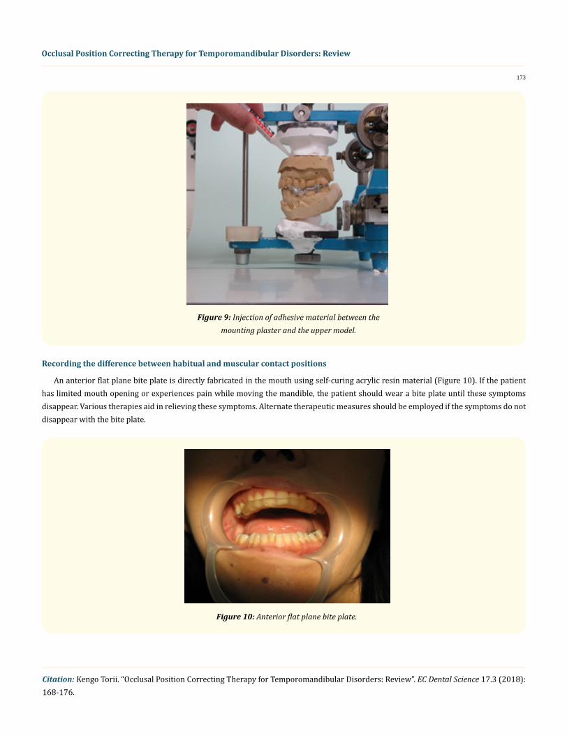
The base of the upper model should be covered with a wrapping film sheet. After the mounting plaster has set, the film is removed and then α-cyanoacrylate adhesive material is injected between the upper model and the mounting plaster (Figure 8, 9). This mounting method prevents the occurrence of dimensional change during the setting of the plaster and subsequent inaccuracy in the occlusion of the models.
Figure 8: The base of the upper model is covered with wrapping film sheet.
173
Occlusal Position Correcting Therapy for Temporomandibular Disorders: Review
Citation: Kengo Torii. “Occlusal Position Correcting Therapy for Temporomandibular Disorders: Review”. EC Dental Science 17.3 (2018): 168-176.
Figure 9: Injection of adhesive material between the mounting plaster and the upper model.
Recording the difference between habitual and muscular contact positions
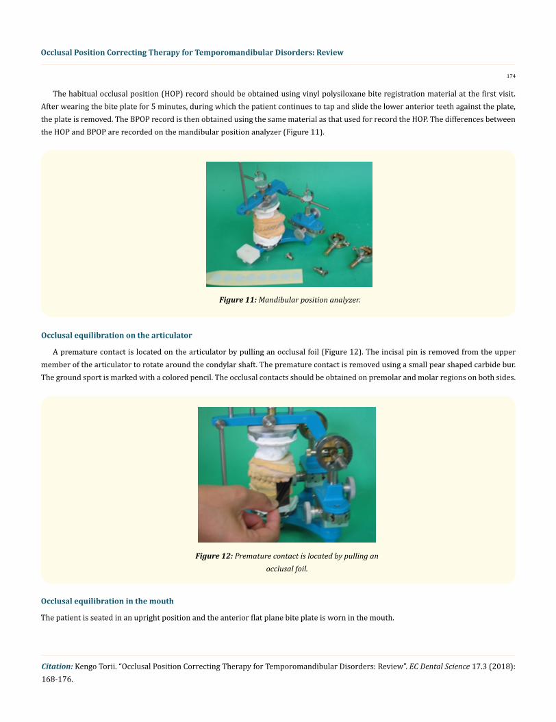
An anterior flat plane bite plate is directly fabricated in the mouth using self-curing acrylic resin material (Figure 10). If the patient has limited mouth opening or experiences pain while moving the mandible, the patient should wear a bite plate until these symptoms disappear. Various therapies aid in relieving these symptoms. Alternate therapeutic measures should be employed if the symptoms do not disappear with the bite plate.
Figure 10: Anterior flat plane bite plate.
174
Occlusal Position Correcting Therapy for Temporomandibular Disorders: Review
Citation: Kengo Torii. “Occlusal Position Correcting Therapy for Temporomandibular Disorders: Review”. EC Dental Science 17.3 (2018): 168-176.
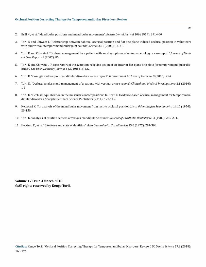
The habitual occlusal position (HOP) record should be obtained using vinyl polysiloxane bite registration material at the first visit. After wearing the bite plate for 5 minutes, during which the patient continues to tap and slide the lower anterior teeth against the plate, the plate is removed. The BPOP record is then obtained using the same material as that used for record the HOP. The differences between the HOP and BPOP are recorded on the mandibular position analyzer (Figure 11).
Figure 11: Mandibular position analyzer.
Occlusal equilibration on the articulator
A premature contact is located on the articulator by pulling an occlusal foil (Figure 12). The incisal pin is removed from the upper member of the articulator to rotate around the condylar shaft. The premature contact is removed using a small pear shaped carbide bur. The ground sport is marked with a colored pencil. The occlusal contacts should be obtained on premolar and molar regions on both sides.
Figure 12: Premature contact is located by pulling an occlusal foil.
Occlusal equilibration in the mouth
The patient is seated in an upright position and the anterior flat plane bite plate is worn in the mouth.

175
Occlusal Position Correcting Therapy for Temporomandibular Disorders: Review
Citation: Kengo Torii. “Occlusal Position Correcting Therapy for Temporomandibular Disorders: Review”. EC Dental Science 17.3 (2018): 168-176.
After the patient attains muscular conditioning by tapping and sliding the lower anterior teeth against the plate for 5 minutes, the plate is removed from the mouth and an occlusal foil is placed in the mouth with the help of a holder. The patient is then asked to close the jaw until the teeth come into contact and to hold that position. The premature contact is confirmed by laterally pulling the foil (Figure 13). The premature contact should be compared with the contact marked on the articulator. The contact which is coincident with the one located on the articulator is selected as the true premature contact and is removed. After removing two or three premature contacts, impressions of upper and lower jaws are taken along with the BPOP wax record as previously described. The upper and lower models are mounted on the articulator with the BPOP wax record for the next appointment. Occlusal adjustments are performed on the articulator and the oc-clusal equilibration is repeated in the mouth. An occlusal adjustment is completed by confirming the presence of occlusal contacts on the premolar and molar teeth on both sides in the mouth and on the articulator. After adjustment is completed, the patient may complain of cheek biting or tongue biting caused by the changed occlusal position. However, this phenomenon should cease after 3 or 5 days.
Figure 13: Premature contact is detected by pulling occlusal foils in the mouth in a similar way as in the model.
Conclusion
Occlusal analysis and equilibration in the MCP should be performed on dental models mounted on an articulator with BPOP wax re-cord. The occlusal adjustment is then performed with referring to the ground spots on the models.
Competing Interests
The author declares that he has no competing interests.
Bibliography
1. Torii K and Chiwata I. “Occlusal adjustment using the bite plate-induced occlusal position as a reference position for temporoman-dibular disorders: a pilot study”. Head and Face Medicine 6 (2010): 5.
176
Occlusal Position Correcting Therapy for Temporomandibular Disorders: Review
Citation: Kengo Torii. “Occlusal Position Correcting Therapy for Temporomandibular Disorders: Review”. EC Dental Science 17.3 (2018): 168-176.
2. Brill N., et al. “Mandibular positions and mandibular movements”. British Dental Journal 106 (1959): 391-400.
3. Torii K and Chiwata I. “Relationship between habitual occlusal position and flat bite plane-induced occlusal position in volunteers with and without temporomandibular joint sounds”. Cranio 23.1 (2005): 16-21.
4. Torii K and Chiwata I. “Occlusal management for a patient with aural symptoms of unknown etiology: a case report”. Journal of Medi-cal Case Reports 1 (2007): 85.
5. Torii K and Chiwata I. “A case report of the symptom-relieving action of an anterior flat plane bite plate for temporomandibular dis-order”. The Open Dentistry Journal 4 (2010): 218-222.
6. Torii K. “Coxalgia and temporomandibular disorders: a case report”. International Archives of Medicine 9 (2016): 294.
7. Torii K. “Occlusal analysis and management of a patient with vertigo: a case report”. Clinical and Medical Investigations 2.1 (2016): 1-3.
8. Torii K. “Occlusal equilibration in the muscular contact position”. In: Torii K. Evidence-based occlusal management for temporoman-dibular disorders. Sharjah: Bentham Science Publishers (2014): 123-149.
9. Nevakari K. “An analysis of the mandibular movement from rest to occlusal position”. Acta Odontologica Scandinavica 14.10 (1956): 20-150.
10. Torii K. “Analysis of rotation centers of various mandibular closures”. Journal of Prosthetic Dentistry 61.3 (1989): 285-291.
11. Helkimo E., et al. “Bite force and state of dentition”. Acta Odontologica Scandinavica 35.6 (1977): 297-303.
Volume 17 Issue 3 March 2018©All rights reserved by Kengo Torii.