pd-l1 ihc 22c3 pharmdx sk006 50 tests for use with ... · pd-l1 ihc 22c3 pharmdx . sk006 . 50 tests...
TRANSCRIPT

PD-L1 IHC 22C3 pharmDx SK006 50 tests for use with Autostainer Link 48
Table of Contents 1. Intended Use ......................................................................................................................................................................... 2
2. Summary and Explanation ................................................................................................................................................... 2
2.1 NSCLC ............................................................................................................................................................................ 2
2.2 Gastric or GEJ Adenocarcinoma .................................................................................................................................. 2
3. Principle of Procedure ......................................................................................................................................................... 2
4. Materials Provided................................................................................................................................................................ 4
5. Materials Required, but Not Supplied ................................................................................................................................. 4
6. Precautions ........................................................................................................................................................................... 5
7. Storage .................................................................................................................................................................................. 5
8. Specimen Preparation .......................................................................................................................................................... 5
9. Reagent Preparation ............................................................................................................................................................ 5
10. Staining Procedure on the Autostainer Link 48 Solution................................................................................................... 5
11. Quality Control ..................................................................................................................................................................... 6
12. Assay Verification ................................................................................................................................................................ 6
13. Scoring Interpretation .......................................................................................................................................................... 6
13.1 NSCLC .......................................................................................................................................................................... 6
13.2 Gastric or GEJ Adenocarcinoma ..................................................................................... Error! Bookmark not defined.
14. Slide Evaluation .................................................................................................................................................................... 9
15. Limitations ............................................................................................................................................................................ 9
15.1 General Limitations ...................................................................................................................................................... 9
16. Performance Evaluation ..................................................................................................................................................... 10
16.1 Non-Clinical Performance Evaluation: Normal and Neoplastic Tissues ............................................................... 10
16.2 Non-Clinical Performance Evaluation: NSCLC ....................................................................................................... 12
16.3 Clinical Performance Evaluation: NSCLC ............................................................................................................... 14
16.4 Non-Clinical Performance Evaluation: Gastric or GEJ Adenocarcinoma ............................................................. 18
16.5 Clinical Performance Evaluation: Gastric or GEJ Adenocarcinoma ..................................................................... 19
17. Troubleshooting ................................................................................................................................................................. 19
18. References .......................................................................................................................................................................... 19
P03951_06/SK00621-5/2017.09.10 p. 1/21
PD-L1 IHC 22C3 pharmDx SK006 50 tests for use with Autostainer Link 48 1. Intended Use For in vitro diagnostic use. PD-L1 IHC 22C3 pharmDx is a qualitative immunohistochemical assay using Monoclonal Mouse Anti-PD-L1, Clone 22C3 intended for use in the detection of PD-L1 protein in formalin-fixed, paraffin-embedded (FFPE) non-small cell lung cancer (NSCLC) and gastric or gastroesophageal junction (GEJ) adenocarcinoma tissues using EnVision FLEX visualization system on Autostainer Link 48. Non-Small Cell Lung Cancer (NSCLC) PD-L1 protein expression in NSCLC is determined by using Tumor Proportion Score (TPS), which is the percentage of viable tumor cells showing partial or complete membrane staining at any intensity. The specimen should be considered to have PD-L1 expression if TPS ≥1% and high PD-L1 expression if TPS ≥50%. PD-L1 IHC 22C3 pharmDx is indicated as an aid in identifying NSCLC patients for treatment with KEYTRUDA® (pembrolizumab). See the KEYTRUDA® product label for expression cutoff values guiding therapy in specific clinical circumstances. Gastric or Gastroesophageal Junction (GEJ) Adenocarcinoma PD-L1 protein expression in gastric or GEJ adenocarcinoma is determined by using Combined Positive Score (CPS), which is the number of PD-L1 staining cells (tumor cells, lymphocytes, macrophages) divided by the total number of viable tumor cells, multiplied by 1001. The specimen should be considered to have PD-L1 expression if CPS ≥ 1. PD-L1 IHC 22C3 pharmDx is indicated as an aid in identifying gastric or GEJ adenocarcinoma patients for treatment with KEYTRUDA® (pembrolizumab). 2. Summary and Explanation
Binding of the PD-1 ligands, PD-L1 and PD-L2, to the PD-1 receptor found on T-cells, inhibits T-cell proliferation and cytokine production. Up-regulation of PD-1 ligands occurs in some tumors and signaling through this pathway can contribute to inhibition of active T-cell immune surveillance of tumors. KEYTRUDA is a humanized monoclonal antibody that binds to the PD-1 receptor and blocks its interaction with PD-L1 and PD-L2, releasing PD-1 pathway-mediated inhibition of the immune response, including the anti-tumor immune response. In syngeneic mouse tumor models, blocking PD-1 activity resulted in decreased tumor growth (1).
2.1 NSCLC Merck Sharp & Dohme sponsored clinical study, KEYNOTE-024 (KN024), investigated the clinical validity of PD-L1 IHC 22C3 pharmDx in identifying PD-L1 expressing (TPS ≥ 50%) previously untreated NSCLC patients that may respond to KEYTRUDA treatment. Refer to ‘Clinical Performance Evaluation (NSCLC)’ section below for KN024 study details.
Merck Sharp & Dohme sponsored clinical study, KEYNOTE-010 (KN010), investigated the clinical validity of PD-L1 IHC 22C3 pharmDx in identifying PD-L1 expressing (TPS ≥ 1%) previously treated NSCLC patients that may respond to KEYTRUDA treatment. Refer to ‘Clinical Performance Evaluation (NSCLC)’ section below for KN010 study details.
2.2 Gastric or Gastroesophageal Junction (GEJ) Adenocarcinoma
Merck Sharp & Dohme sponsored clinical study, KEYNOTE-059 (KN059), investigated the clinical validity of PD-L1 IHC 22C3 pharmDx in identifying PD-L1 expressing (CPS ≥ 1) gastric or GEJ adenocarcinoma patients with at least 2 prior systemic treatments for advanced disease that may respond to KEYTRUDA treatment. Refer to ‘Clinical Performance Evaluation (Gastric or GEJ Adenocarcinoma)’ section below for KN059 study details.
3 Principle of Procedure PD-L1 IHC 22C3 pharmDx contains the optimized reagents and protocol required to complete an IHC staining procedure of FFPE specimens using Autostainer Link 48. Following incubation with the primary monoclonal antibody to PD-L1 or the Negative Control Reagent (NCR), specimens are incubated with a Linker antibody specific to the host species of the primary antibody, and then are incubated with a ready-to-use visualization reagent consisting of secondary antibody molecules and horseradish peroxidase molecules coupled to a dextran polymer backbone. The enzymatic conversion of the subsequently added chromogen results in precipitation of a visible reaction product at the site of antigen. The color of the chromogenic reaction is modified by a chromogen enhancement reagent. The specimen may then be counterstained and coverslipped. Results are interpreted using a light microscope.
1 Combined Positive Score (CPS) retains the same meaning with or without a percent (%) sign.
P03951_06/SK00621-5/2017.09.10 p. 2/21
4 Materials Provided Each kit includes 19.5 mL of PD-L1 primary antibody (approximately 3µg/mL protein concentration) and contains the reagents necessary to perform 50 tests in up to 15 individual runs. The materials listed below are sufficient for 50 tests (50 slides incubated with Primary Antibody to PD-L1 and 50 slides incubated with the corresponding Negative Control Reagent, 100 slides in total). For larger tissue sections three drop zones (3 x 150µL) per slide may be warranted. Note that this will reduce the total number of tests per kit. The kit provides materials sufficient for a maximum of 15 individual staining runs. Quantity Description 1 x 34.5 mL Peroxidase-Blocking Reagent
Buffered solution containing hydrogen peroxide, detergent and 0.015 mol/L sodium azide.
1 x 19.5 mL
Primary Antibody: Monoclonal Mouse Anti-PD-L1, Clone 22C3
Monoclonal mouse (IgG1) anti-PD-L1 in a buffered solution, containing stabilizing protein, and 0.015 mol/L sodium azide.
1 x 15 mL
Negative Control Reagent
Monoclonal mouse control IgG antibody in a buffered solution, containing stabilizing protein, and 0.015 mol/L sodium azide.
1 x 34.5 mL Mouse LINKER
Rabbit secondary antibody against mouse immunoglobulins in a buffered solution containing stabilizing protein and 0.015 mol/L sodium azide.
1 x 34.5 mL Visualization Reagent-HRP
Dextran coupled with peroxidase molecules and goat secondary antibody molecules against rabbit and mouse immunoglobulins in a buffered solution containing stabilizing protein and an antimicrobial agent.
15 x 7.2 mL DAB+ Substrate Buffer
Buffered solution, containing hydrogen peroxide and an antimicrobial agent.
1 x 5 mL DAB+ Chromogen
3,3’-diaminobenzidine tetrahydrochloride in organic solvent.
1 x 34.5 mL DAB Enhancer
Cupric sulfate in water.
6 x 30 mL EnVision FLEX Target Retrieval Solution, Low pH (50x)
Buffered solution, pH 6.1, containing detergent and an antimicrobial agent.
15 slides PD-L1 IHC 22C3 pharmDx Control Slides
Each slide contains sections of two pelleted, formalin-fixed paraffin-embedded cell lines: NCI-H226* with moderate PD-L1 protein expression and MCF-7 with negative PD-L1 protein expression.
P03951_06/SK00621-5/2017.09.10 p. 3/21

PD-L
1 IH
C 2
2C3
phar
mD
x XX
XXX
*Dr. AF Gazdar and Dr. JD Minna at NIH are acknowledged for their contribution in developing NCI-H226 (ATCC Number: CRL-5826™) (2). Note: All reagents included are formulated specifically for use with this kit. In order for the test to perform as specified, no substitutions, other than EnVision FLEX Target Retrieval Solution, Low pH (50x) (Code K8005) can be made. PD-L1IHC 22C3 pharmDx has been tailored for use with Autostainer Link 48. Please refer to the User Guides for your Autostainer Link 48 and PT Link for further information. 5 Materials Required, but Not Supplied PT Link Pre-treatment Module (Code PT100/PT101/PT200) Autostainer Link 48 (Code AS480) EnVision FLEX Wash Buffer (20x) (Code K8007) Hematoxylin (Code K8008) Distilled or deionized water (reagent-quality water) Timer Positive and negative tissues to use as process controls (see Quality Control section) Microscope slides: Dako FLEX IHC Microscope Slides (Code K8020) or Fisherbrand Superfrost Plus charged slides Coverslips Permanent mounting medium and ancillary reagents required for mounting coverslips Light microscope (4x–40x objective magnification) 6 Precautions 1. For in vitro diagnostic use. 2. For professional users. 3. This product contains sodium azide (NaN3), a chemical highly toxic in pure form. At product concentrations, though not classified
as hazardous, NaN3 may react with lead and copper plumbing to form highly explosive build-ups of metal azides. Upon disposal, flush with large volumes of water to prevent metal azide build-up in plumbing (3).
4. Primary Antibody, Negative Control Reagent, Linker, and Visualization Reagent contain material of animal origin. 5. Specimens, before and after fixation, and all materials exposed to them, should be handled as if capable of transmitting infection,
and disposed of with proper precautions (4). 6. Incubation times, temperatures, or methods other than those specified may give erroneous results. 7. Reagents have been optimally diluted. Further dilution may result in loss of antigen staining. 8. The Visualization Reagent, Liquid DAB+ chromogen and prepared DAB+ Substrate-Chromogen solution may be affected
adversely if exposed to excessive light levels. Do not store system components or perform staining in strong light, such as direct sunlight.
9. Paraffin residuals may lead to false negative results. 10. Use of reagent volumes other than recommended may result in loss of visible PD-L1 immunoreactivity. 11. Results from a small study, showed a similar dynamic range of PD-L1 expression in primary and metastatic NSCLC specimen
pairs. It is possible there may be differences in PD-L1 expression in primary tumors versus metastatic sites in the same patient. 12. Large tissue sections may require 3x150 µl of reagent. 13. As a general rule, persons under 18 years of age are not allowed to work with this product. Users must be carefully instructed in
the proper work procedures, the dangerous properties of the product and the necessary safety instructions. Please refer to Safety Data Sheet (SDS) for additional information.
14. Wear appropriate Personal Protective Equipment to avoid contact with eyes and skin. 15. Unused solution should be disposed of according to local, State and Federal regulations. 16. Safety Data Sheet available for professional users on request.
Danger DAB+ Chromogen: 1–5% biphenyl-3,3',4,4'-tetrayltetraammonium tetrachloride H350 May cause cancer. H341 Suspected of causing genetic defects. P201 Obtain special instructions before use. P202 Do not handle until all safety precautions have been read and understood. P280 Wear protective gloves. Wear eye or face protection. Wear protective clothing. P308 + P313 IF exposed or concerned: Get medical attention. P405 Store locked up. P501 Dispose of contents and container in accordance with all local, regional, national and international regulations.
Warning EnVision FLEX Target Retrieval Solution, Low pH (50x): 1-5% Citric acid H319 Causes serious eye irritation. H411 Toxic to aquatic life with long lasting effects. P280 Wear eye or face protection.
MCF-7: PD-L1 negative
NCI-H226: PD-L1 positive
P03951_06/SK00621-5/2017.09.10 p. 4/21
P273 Avoid release to the environment. P264 Wash hands thoroughly after handling. P305 + P351 + P338
IF IN EYES: Rinse cautiously with water for several minutes. Remove contact lenses, if present and easy to do. Continue rinsing.
P337 + P313 If eye irritation persists: Get medical attention. P501 Dispose of contents and container in accordance with all local, regional, national and international regulations.
7 Storage Store all components of PD-L1 IHC 22C3 pharmDx, including Control Slides, in the dark at 2-8 °C when not in use on Autostainer Link 48. Do not use the kit after the expiration date printed on the outside of the kit box. If reagents are stored under any conditions other than those specified in this package insert, they must be validated by the user. There are no obvious signs to indicate instability of this product, therefore, positive and negative controls should be run simultaneously with patient specimens.
8. Specimen Preparation Tissue specimens must be handled to preserve the tissue for IHC staining. Standard methods of tissue processing should be used for all specimens.
8.1 Paraffin-embedded Specimens Formalin-fixed, paraffin-embedded (FFPE) tissue specimens are suitable for use. Alternative fixatives have not been validated and may give erroneous results. Fixation time for 12-72 hours in 10% neutral buffered formalin (NBF) is recommended, however, a study with limited samples showed fixation times of 4-168 hours in 10% NBF did not systematically alter PD-L1 detection. Fixation times of ≤3 hours may result in variable PD-L1 detection. Specimens should be blocked into a thickness of 3 or 4 mm, fixed in formalin and dehydrated and cleared in a series of alcohols and xylene, followed by infiltration with melted paraffin. The paraffin temperature should not exceed 60 °C. NSCLC FFPE tissue blocks which are 5 years or older may result in a loss of PD-L1 immunoreactivity. Please refer to Section 15.2 (Product Specific Limitations) for gastric or GEJ adenocarcinoma specimens.
Tissue specimens should be cut into sections of 4-5 µm. After sectioning, tissues should be mounted on Dako FLEX IHC microscope slides (Code K8020), or Fisherbrand Superfrost Plus slides and then placed in a 58 ± 2 °C oven for 1 hour.
8.2 Cut Section Storage Recommendation
To preserve antigenicity, tissue sections, once mounted on slides, should be held in the dark at 2-8 °C (preferred), or at room temperature up to 25°C, and stained within 6 months of sectioning. Slide storage and handling conditions should not exceed 25°C at any point post-mounting to ensure tissue integrity and antigenicity.
9 Reagent Preparation The following reagents must be prepared prior to staining: EnVision FLEX Target Retrieval Solution, Low pH (50x) Prepare a sufficient quantity of 1x Target Retrieval Solution, Low pH by diluting Target Retrieval Solution, Low pH (50x) 1:50 using distilled or deionized water (reagent-quality water); the pH of 1x Target Retrieval Solution must be 6.1 ± 0.2. 1x Target Retrieval Solution pH below 5.9 may give erroneous results. One 30 mL bottle of Target Retrieval Solution, Low pH (50x) diluted 1:50 will provide 1.5 L of 1x reagent, sufficient to fill one PT Link tank which will treat up to 24 slides per use. Discard 1x Target Retrieval Solution after three uses and do not use after 5 days following dilution. Additional EnVision FLEX Target Retrieval Solution, Low pH (50x) if required, is available as Code K8005. EnVision FLEX Wash Buffer (20x) Prepare a sufficient quantity of Wash Buffer by diluting Wash Buffer (20x) 1:20 using distilled or deionized water (reagent-quality water) for the wash steps. Store unused 1x solution at 2-8 ºC for no more than one month. Discard buffer if cloudy in appearance. Refer to the User Guide for your Autostainer Link 48 for further information. EnVision FLEX Wash Buffer (20x) is available as Code K8007. DAB+ Substrate-Chromogen Solution This solution should be mixed thoroughly prior to use. Any precipitate developing in the solution does not affect staining quality. To prepare DAB+ Substrate-Chromogen Solution, add 1 drop of Liquid DAB+ Chromogen per mL of DAB+ Substrate Buffer and mix. Prepared Substrate-Chromogen is stable for 5 days if stored in the dark at 2-8 °C. Important Notes:
• If using an entire bottle of DAB+ Substrate Buffer, add 9 drops of DAB+ chromogen. Although the label states 7.2 mL, this is the useable volume and does not account for the “dead volume” in the bottle.
• The color of the Liquid DAB+ Chromogen in the bottle may vary from clear to lavender-brown. This will not affect the performance of this product. Dilute per the guidelines above. Addition of excess Liquid DAB+ Chromogen to the DAB+ Substrate Buffer will result in deterioration of the positive signal.
10 Staining Procedure on the Autostainer Link 48 Solution
Procedural Notes The user should read these instructions carefully and become familiar with all components and instrumentation prior to use (see Section 6, Precautions).
P03951_06/SK00621-5/2017.09.10 p. 5/21

All reagents should be equilibrated to room temperature (20-25 °C) prior to immunostaining. Likewise, all incubations should be performed at room temperature. Do not allow tissue sections to dry during the staining procedure. Dried tissue sections may display increased nonspecific staining. All of the required steps and incubation times for staining are preprogrammed in the Dako Link software. Please refer to the User Guides for Autostainer Link 48 and PT Link for further information on programming protocols and loading slides and reagents. Note: The reagents and instructions supplied in this system have been designed for optimal performance when used with the recommended reagents and materials. Further dilution of the reagents or alteration of incubation times or temperatures may give erroneous or discordant results. Staining Protocol Please select the PD-L1 IHC 22C3 pharmDx staining protocol from the options in the Dako Link drop down menu. All of the required steps and incubation times for staining are preprogrammed in the Autostainer Link 48. If the appropriate PD-L1 IHC 22C3 pharmDx protocols are not on your server, please contact your local Technical Service Representative to obtain the protocols. Step 1: Deparaffinization, Rehydration and Target Retrieval (3-in-1) Procedure For details, please refer to the PT Link User Guide. Set PT Link (Code PT100/PT101/PT200) Preheat and Cool to 65 °C. Set Heat to 97 °C for 20 minutes. ►Fill PT Link tanks with 1.5 L per tank of Target Retrieval Solution, Low pH, 1x working solution to cover the tissue sections. ►Preheat the Target Retrieval Solution to 65 °C. ►Immerse Autostainer racks containing mounted, FFPE tissue sections into the pre-heated Target Retrieval Solution, Low pH, (1x
working solution) in PT Link tank. Incubate for 20 minutes at 97 °C. ►When target retrieval incubation has been completed and the temperature has cooled to 65 °C, remove each Autostainer slide rack
with the slides from the PT Link tank and immediately place the Autostainer rack with slides into a tank (e.g., PT Link Rinse Station, Code PT109) containing diluted, room temperature Wash Buffer (Code K8007).
►Incubate slides in diluted, room temperature Wash Buffer for 5 minutes. Step 2: Staining Procedure After deparaffinization, rehydration and target retrieval (3-in-1) procedure, the Autostainer racks with slides are placed on Autostainer Link 48. The instrument will perform the staining process by applying the appropriate reagent, monitoring the incubation time and rinsing slides between reagents. The reagent times are preprogrammed in the Dako Link software. Step 3: Counterstain Slides should be counterstained for 5 minutes with Hematoxylin (Link) (Code K8008). The Hematoxylin incubation time is preprogrammed in the protocol. Step 4: Mounting Non-aqueous, permanent mounting media is required. Note: Some fading of stained slides may occur, depending on several factors including, but not limited to, counterstaining, mounting materials and methods, and slide storage conditions. To minimize fading, store slides in the dark at room temperature (20-25 °C). 11, Quality Control Reagents in PD-L1 IHC 22C3 pharmDx have been quality controlled by immunohistochemistry using the target retrieval and staining procedures outlined above. Deviations in the recommended procedures for tissue fixation, processing and embedding in the user’s laboratory may produce significant variability in results. Quality controls should be included in each staining run. These quality controls are specified in Table 2 and include: an H&E stained patient tissue specimen; lab-supplied positive and negative control tissues; and a Dako-supplied Control Cell Line Slide (5). In the USA, consult the quality control guidelines of the College of American Pathologists (CAP) Accreditation Program for Immunohistochemistry; see also CLSI Quality Assurance for Immunocytochemistry, Approved Guideline (5, 6, 7) for additional information. 12 Assay Verification Prior to initial use of a staining system in a diagnostic procedure, the user should verify the assay’s performance by testing it on a series of lab-supplied tissues with known IHC performance characteristics representing known positive and negative tissues. Refer to the quality control procedures outlined in the quality control section above. These quality control procedures should be repeated for each new antibody lot, or whenever there is a change in assay parameters. Troubleshooting options for potential problems, their causes and suggested corrective actions are outlined in Table 12 16.
13. Staining and Scoring Interpretation 13.1 NSCLC All viable tumor cells on the entire slide must be evaluated and included in the PD-L1 scoring assessment. A minimum of 100 viable tumor cells must be present in the PD-L1 stained slide for the specimen to be considered adequate for PD-L1 evaluation. Slide evaluation should be performed by a pathologist using a light microscope. For evaluation of the immunohistochemical staining and scoring, an objective of 10-40x magnification is appropriate. Any perceptible membrane staining of tumor cells should be included in the scoring. PD-L1 protein expression is determined by using Tumor Proportion Score (TPS), which is the percentage of viable tumor cells showing partial or complete membrane staining at any intensity. Score partial or complete cell membrane staining (≥ 1+) that is perceived distinct from cytoplasmic staining. Cytoplasmic staining should be considered non-specific staining and is excluded in the assessment of staining intensity. Normal cells and tumor associated
P03951_06/SK00621-5/2017.09.10 p. 6/21
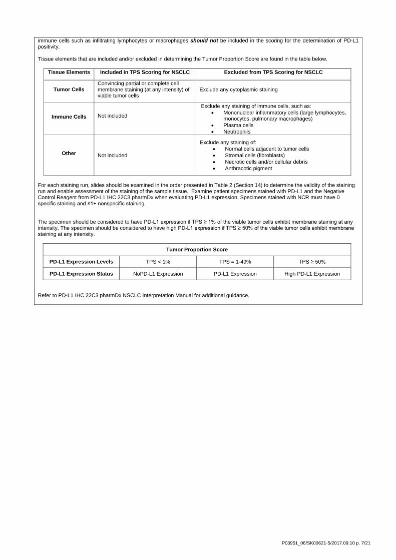
immune cells such as infiltrating lymphocytes or macrophages should not be included in the scoring for the determination of PD-L1 positivity. Tissue elements that are included and/or excluded in determining the Tumor Proportion Score are found in the table below.
Tissue Elements Included in TPS Scoring for NSCLC Excluded from TPS Scoring for NSCLC
Tumor Cells Convincing partial or complete cell membrane staining (at any intensity) of viable tumor cells
Exclude any cytoplasmic staining
Immune Cells Not included
Exclude any staining of immune cells, such as: • Mononuclear inflammatory cells (large lymphocytes,
monocytes, pulmonary macrophages) • Plasma cells • Neutrophils
Other Not included
Exclude any staining of: • Normal cells adjacent to tumor cells • Stromal cells (fibroblasts) • Necrotic cells and/or cellular debris • Anthracotic pigment
For each staining run, slides should be examined in the order presented in Table 2 (Section 14) to determine the validity of the staining run and enable assessment of the staining of the sample tissue. Examine patient specimens stained with PD-L1 and the Negative Control Reagent from PD-L1 IHC 22C3 pharmDx when evaluating PD-L1 expression. Specimens stained with NCR must have 0 specific staining and ≤1+ nonspecific staining. The specimen should be considered to have PD-L1 expression if TPS ≥ 1% of the viable tumor cells exhibit membrane staining at any intensity. The specimen should be considered to have high PD-L1 expression if TPS ≥ 50% of the viable tumor cells exhibit membrane staining at any intensity.
Tumor Proportion Score
PD-L1 Expression Levels TPS < 1% TPS = 1-49% TPS ≥ 50%
PD-L1 Expression Status NoPD-L1 Expression PD-L1 Expression High PD-L1 Expression
Refer to PD-L1 IHC 22C3 pharmDx NSCLC Interpretation Manual for additional guidance.
P03951_06/SK00621-5/2017.09.10 p. 7/21

13.2 Gastric or Gastroesophageal Junction (GEJ) Adenocarcinoma
All viable tumor cells on the entire tissue must be evaluated and included in PD-L1 expression assessment. A minimum of 100 viable tumor cells must be present in the PD-L1 stained slide (biopsy and resection) for the specimen to be considered adequate for evaluation. If patient specimens include more than one biopsy (ie. 3-5 endoscopic biopsies) on a slide, all tissues on the slide need to be evaluated to generate a single CPS for determining the PD-L1 expression level. Each biopsy should not be reported independently. PD-L1 expression in gastric or GEJ adenocarcinoma is determined by Combined Positive Score (CPS), which is the number of PD-L1 staining cells (tumor cells, lymphocytes, macrophages) divided by the total number of viable tumor cells, multiplied by 100*. Distinction of viable tumor cells, lymphocytes, and macrophages is essential for accurate denominator estimation. Although the result of the calculation can exceed 100, the maximum score is defined as CPS 100. CPS is defined as follows:
CPS = # PD-L1 staining cells (tumor cells, lymphocytes, macrophages)
Total # of viable tumor cells × 100
*Combined Positive Score (CPS) retains the same meaning with or without a percent (%) sign. Slide evaluation must be performed by a pathologist using a light microscope. For evaluation of the immunohistochemical staining, an objective of 10-20x magnification is appropriate. For determination of PD-L1 expression, an objective of 20x magnification is required. By definition, PD-L1 staining cells in gastric or GEJ adenocarcinoma are:
• Tumor cells with convincing partial or complete linear membrane staining (at any intensity) that is perceived distinct from cytoplasmic staining and
• Lymphocytes and macrophages (mononuclear inflammatory cells, MICs) within the tumor nests and/or adjacent supporting stroma with convincing membrane and/or cytoplasmic staining (at any intensity). MICs must be directly associated with the response against the tumor.
The table below provides details about which tissue elements are included in and excluded from the CPS numerator in gastric or GEJ adenocarcinoma. The CPS denominator includes all viable invasive tumor cells (PD-L1 staining and non-staining). Dysplasia, carcinoma in situ and all other cells are excluded. Table 1. CPS Numerator Inclusion/Exclusion Criteria
Tissue Elements Included in the Numerator Excluded from the Numerator
Tumor Cells Convincing partial or complete linear membrane
staining (at any intensity) of viable invasive gastric or GEJ adenocarcinoma tumor cells
• Non-staining tumor cells • Tumor cells with only cytoplasmic staining • Adenoma, dysplasia, and carcinoma in situ
Immune Cells
Membrane and/or cytoplasmic* staining (at any intensity) of mononuclear inflammatory cells (MICs) within tumor nests and adjacent supporting stroma**:
o Lymphocytes (including lymphocyte aggregates)
o Macrophages*** Only MICs directly associated with the response to the tumor are scored.
• Non-staining MICs • MICs associated with adenoma, dysplasia,
and carcinoma in situ • MICs (including lymphoid aggregates)
associated with ulcers, chronic gastritis, and other processes not associated with the tumor
• MICs associated with normal structures • Neutrophils, eosinophils and plasma cells
Other Cells • Not included • Normal cells (including ganglion cells) • Stromal cells (including fibroblasts) • Necrotic cells and/or cellular debris
*In MICs membrane and cytoplasmic staining are often indistinguishable due to high nuclear to cytoplasmic ratio. Therefore, membrane and/or cytoplasmic staining of MICs is included in the CPS numerator. **Adjacent MICs are defined as being within the same 20x field as the tumor. However, MICs that are NOT directly associated with the response to the tumor should be excluded. ***Macrophages and histiocytes are considered the same cells. For each staining run, slides should be examined in the order presented in Table 2 (Section 14) to determine the validity of the staining run and enable assessment of the staining of the sample tissue. Examine patient specimens stained with PD-L1 and the Negative Control Reagent from PD-L1 IHC 22C3 pharmDx when evaluating PD-L1 expression. Specimens stained with NCR must have 0 specific staining and ≤1+ nonspecific staining. The specimen should be considered to have PD-L1 expression if CPS ≥ 1.
Combined Positive Score
PD-L1 Expression Level CPS < 1 CPS ≥ 1
PD-L1 Expression Status No PD-L1 Expression PD-L1 Expression
P03951_06/SK00621-5/2017.09.10 p. 8/21
Refer to PD-L1 IHC 22C3 pharmDx Gastric or GEJ Interpretation Manual for additional guidance.
14 Slide Evaluation Table 2. Recommended Order of Slide Evaluation
Specimens Rationale Requirements 1. H&E (Lab-supplied)
A hematoxylin and eosin (H&E) stain of the tissue specimen is evaluated first to assess tissue histology and preservation quality.
The PD-L1 IHC 22C3 pharmDx and H&E stain should be performed on serial sections from the same paraffin block of the specimen. Tissue specimens should be intact, well preserved, and should confirm tumor indication.
2. Control Cell Line Slide (Supplied with kit)
The Control Cell Line Slide stained with the PD-L1 primary antibody from PD-L1 IHC 22C3 pharmDx should be examined to ascertain that all reagents are functioning properly. The Control Cell Line Slide contains the PD-L1-positive cell line pellet and PD-L1-negative cell line pellet.
One Control Cell Line Slide should be stained with the Primary Antibody to PD-L1 in each staining run. NCI-H226 (PD-L1-positive control cell line) acceptance criteria: • Cell membrane staining of ≥ 70% of cells at ≥ 2+ average staining
intensity. • Non-specific staining < 1+ intensity. MCF-7 (PD-L1-negative control cell line) acceptance criteria: • No specific staining. • Non-specific staining < 1+ intensity. Note that staining of a few cells in the
MCF-7 cell pellet may occasionally be observed. The following acceptance criteria are applicable: the presence of ≤ 10 total cells with distinct plasma membrane staining, or cytoplasmic staining with ≥ 1+ intensity within the boundaries of the MCF-7 cell pellet are acceptable.
If either of the Control Cell Lines does not meet these criteria, all results with the patient specimens should be considered invalid.
3. Positive Control Tissue Slides (Lab-supplied)
The Positive Control Tissue Slides stained with both PD-L1 primary antibody and Negative Control Reagent should be examined next. These slides verify that the fixation method and epitope retrieval process are effective. Known positive tissue controls should only be utilized for monitoring the correct performance of processed tissues and test reagents, NOT as an aid in formulating a specific diagnosis of patient samples.
Controls should be biopsy/surgical specimens of the same tumor indication as the patient specimen, fixed, processed and embedded as soon as possible in the same manner as the patient sample(s). Use intact specimens for interpretation of staining results as necrotic or degenerated cells often stain non-specifically. The tissues selected for use as the positive tissue controls should give weak to moderate positive staining when stained with PD-L1 to aid in detection of subtle changes in assay sensitivity. Two positive tissue control slides should be included in each staining run. Slide stained with PD-L1: Presence of brown plasma membrane staining should be observed. Non-specific staining should be ≤1+. Slide stained with Negative Control Reagent: No membrane staining. Non-specific staining should be ≤ 1+. If the positive tissue controls fail to demonstrate appropriate positive staining, results with the test specimens should be considered invalid.
4. Negative Control Tissue Slides (Lab-supplied)
The Negative Control Tissue Slides (known to be PD-L1 negative) stained with both PD-L1 primary antibody and Negative Control Reagent should be examined next to verify the specificity of the labeling of the target antigen by the primary antibody. Alternatively, negative portions of the Positive Control Tissue may serve as the Negative Control Tissue, but this should be verified by the user.
Controls should be biopsy/surgical specimens of the same tumor indication as the patient specimen, fixed, processed and embedded as soon as possible in the same manner as the patient sample(s). Two negative tissue control slides should be included in each staining run. Slide stained with PD-L1: No membrane staining in tumor cells. Non-specific staining should be ≤ 1+. Slide stained with Negative Control Reagent: No membrane staining. Non-specific staining should be ≤ 1+. If specific cell membrane staining occurs in the Negative Control Tissue Slides, results with the patient specimen should be considered invalid.
5. Tonsil Control Tissue (optional) (Lab-supplied)
Use human tonsil tissue fixed, processed and embedded in a manner similar to the patient sample(s) as an additional control material to verify sensitivity, specificity and nonspecific background staining of the assay.
Strong positive staining should be detected in portions of the crypt epithelium and weak to moderate staining of the follicular macrophages in the germinal centers. Negative staining should be observed in endothelium, fibroblasts as well as surface epithelium.
6 Patient tissue slide stained using the Negative Control Reagent
Examine patient specimens stained with the Negative Control Reagent from PD-L1 IHC 22C3 pharmDx. Negative Control Reagent is used in place of the
Absence of cell membrane staining verifies the specific labeling of the target antigen by the primary antibody. Non-specific staining should be ≤ 1+.
P03951_06/SK00621-5/2017.09.10 p. 9/21
primary antibody and aids in interpretation of specific staining at the antigen site.
7 Patient tissue slide stained using the PD-L1 primary antibody
Examine the entire slide of the patient specimens stained with the PD-L1 primary antibody from PD-L1 IHC 22C3 pharmDx last. Refer to Summary and Explanation, Limitations, and Performance Characteristics for specific information regarding PD-L1 IHC 22C3 pharmDx immunoreactivity.
Positive staining intensity should be assessed within the context of any non-specific background staining observed on the patient’s Negative Control Reagent slide in the same run. As with any immunohistochemical test, a negative result means that the antigen was not detected, not necessarily that the antigen was absent in the cells/tissue assayed. All viable tumor cells on the entire PD-L1 stained patient slide must be evaluated and included in the PD-L1 scoring assessment. A minimum of 100 viable tumor cells must be present for the specimen to be considered adequate for PD-L1 evaluation. Refer to Section 13 for scoring interpretation guidelines in PD-L1 expression.
15 Limitations
15.1 General Limitations 1. Immunohistochemistry is a multi-step diagnostic process that requires specialized training in the selection of the appropriate
reagents; tissue selection, fixation, and processing; preparation of the immunohistochemistry slide; and interpretation of the staining results.
2. Tissue staining is dependent on the handling and processing of the tissue prior to staining. Improper fixation, freezing, thawing, washing, drying, heating, sectioning, or contamination with other tissues or fluids may produce artifacts, antibody trapping, or false-negative results. Inconsistent results may be due to variations in fixation and embedding methods, or to inherent irregularities within the tissue.
3. Excessive or incomplete counterstaining may compromise proper interpretation of results. 4. The clinical interpretation of any positive staining or its absence must be evaluated within the context of clinical presentation,
morphology and other histopathological criteria. The clinical interpretation of any staining, or its absence, must be complemented by morphological studies and proper controls as well as other diagnostic tests. It is the responsibility of a qualified pathologist, who is familiar with the antibodies, reagents and methods used, to interpret the stained preparation. Staining must be performed in a certified, licensed laboratory under the supervision of a pathologist who is responsible for reviewing the stained slides and assuring the adequacy of positive and negative controls.
5. Tissues from persons infected with hepatitis B virus and containing hepatitis B surface antigen (HBsAg) may exhibit non-specific staining with horseradish peroxidase (7).
6. Reagents may demonstrate unexpected reactions in previously untested tissue types. The possibility of unexpected reactions even in tested tissue types cannot be completely eliminated due to biological variability of antigen expression in neoplasms, or other pathological tissues. Contact Dako Technical Support with documented unexpected reactions.
7. False-positive results may be seen due to non-Immunological binding of proteins or substrate reaction products. They may also be caused by pseudoperoxidase activity (erythrocytes) and endogenous peroxidase activity (cytochrome C) (7).
8. The reagents and instructions supplied in this system have been designed for optimal performance. Further dilution of the reagents or alteration of incubation times or temperatures may give erroneous or discordant results.
15.2 Product-Specific Limitations 1. False-negative results could be caused by degradation of the antigen in the tissues over time. Specimens should be stained within
six months of mounting of tissues on slides when stored in the dark at 2-8°C (preferred), or at room temperature up to 25°C. 2. For optimal and reproducible results, the PD-L1 protein requires target retrieval pre-treatment when tissues are routinely fixed
(neutral buffered formalin) and paraffin embedded. 3. Do not substitute reagents from different lot numbers of this product, or from kits of other manufacturers. The only exception is the
EnVision FLEX Target Retrieval Solution, Low pH (50x), which, if required, is available as Code K8005. 4. Stained control cell lines should be used only for validation of the staining run and should not be used to score the staining reaction
in tissue sections. 5. Use of PD-L1 IHC 22C3 pharmDx on tissues with fixatives other than formalin has not been validated. 6. Use of PD-L1 IHC 22C3 pharmDx on fine needle aspirates has not been validated. 7. Use of PD-L1 IHC 22C3 pharmDx on decalcified tissues has not been validated. 8. The clinical study in gastric or GEJ adenocarcinoma was conducted with the guidance to provide a minimum of 3 and up to 5 core
needle tissue biopsies per subject. The reliability of determining subject’s PD-L1 expression level based on testing fewer passes is unknown.” Refer to Section 13.2 for guidance on scoring interpretation.
9. If PD-L1 expression is not detected in an archival* gastric or GEJ adenocarcinoma specimen, evaluate the feasibility of obtaining an additional tumor biopsy for PD-L1 testing. (See Table 15 in Clinical Section 16.5)
*In the context of clinical trial KN059, a newly obtained biopsy was defined as a specimen obtained up to 6 weeks (42 days) prior to initiation of treatment on Day 1 (Cycle 1) with KEYTRUDA and with no additional anti-cancer treatment having been given after the specimen was obtained. Specimens that were >42 days were classified as archival.
16. Performance Evaluation 16.1 Non-Clinical Performance Evaluation: Normal and Neoplastic Tissues Normal tissues: Table 3 summarizes Monoclonal Mouse Anti-PD-L1, Clone 22C3 immunoreactivity on the recommended panel of normal tissues. Plasma membrane staining was observed on immune cells and cells of epithelial origin. Cytoplasmic staining was noted in some cell types but was not recorded as positive staining. All tissues were formalin-fixed and paraffin-embedded and stained with PD-L1 IHC 22C3 pharmDx according to the instructions in this package insert. There were no unexpected results observed in cell types
P03951_06/SK00621-5/2017.09.10 p. 10/21
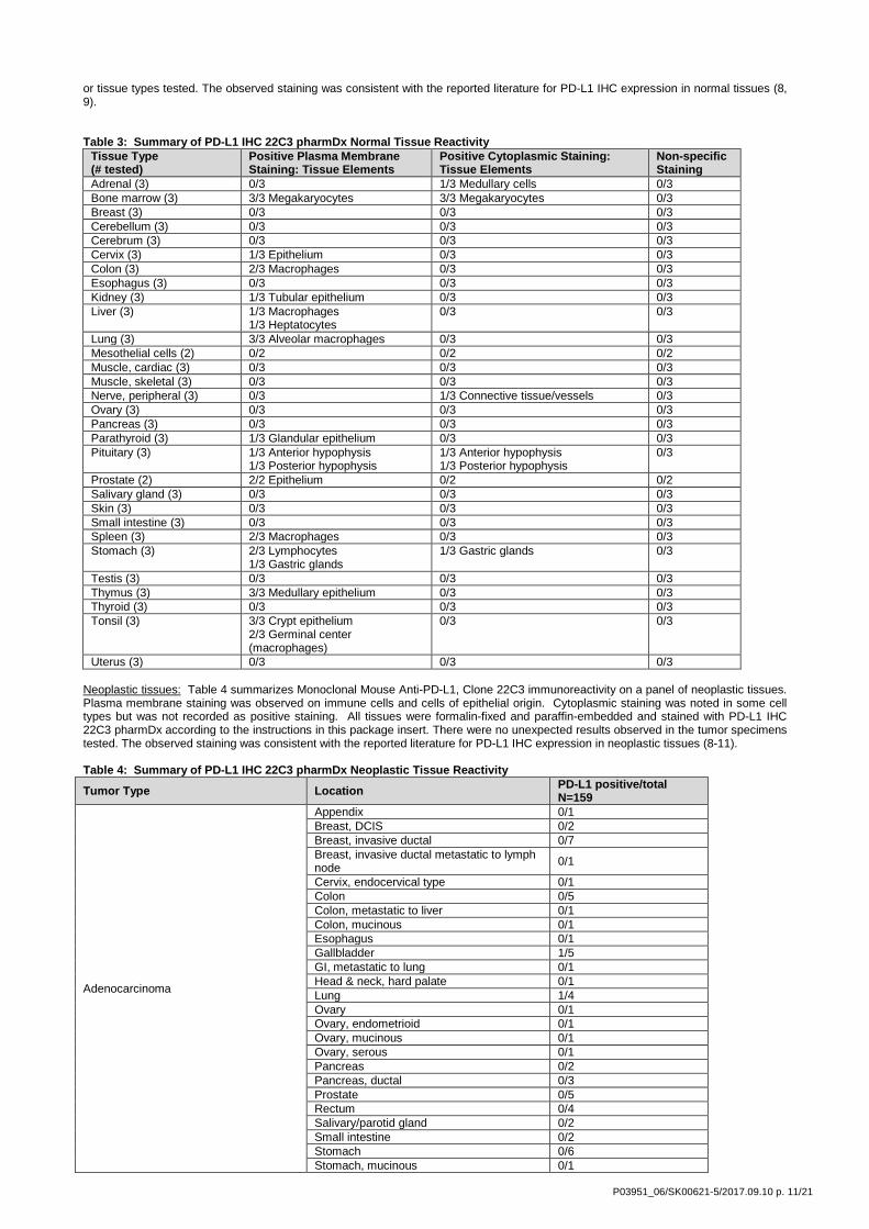
or tissue types tested. The observed staining was consistent with the reported literature for PD-L1 IHC expression in normal tissues (8, 9). Table 3: Summary of PD-L1 IHC 22C3 pharmDx Normal Tissue Reactivity
Tissue Type (# tested)
Positive Plasma Membrane Staining: Tissue Elements
Positive Cytoplasmic Staining: Tissue Elements
Non-specific Staining
Adrenal (3) 0/3 1/3 Medullary cells 0/3 Bone marrow (3) 3/3 Megakaryocytes 3/3 Megakaryocytes 0/3 Breast (3) 0/3 0/3 0/3 Cerebellum (3) 0/3 0/3 0/3 Cerebrum (3) 0/3 0/3 0/3 Cervix (3) 1/3 Epithelium 0/3 0/3 Colon (3) 2/3 Macrophages 0/3 0/3 Esophagus (3) 0/3 0/3 0/3 Kidney (3) 1/3 Tubular epithelium 0/3 0/3 Liver (3) 1/3 Macrophages
1/3 Heptatocytes 0/3 0/3
Lung (3) 3/3 Alveolar macrophages 0/3 0/3 Mesothelial cells (2) 0/2 0/2 0/2 Muscle, cardiac (3) 0/3 0/3 0/3 Muscle, skeletal (3) 0/3 0/3 0/3 Nerve, peripheral (3) 0/3 1/3 Connective tissue/vessels 0/3 Ovary (3) 0/3 0/3 0/3 Pancreas (3) 0/3 0/3 0/3 Parathyroid (3) 1/3 Glandular epithelium 0/3 0/3 Pituitary (3) 1/3 Anterior hypophysis
1/3 Posterior hypophysis 1/3 Anterior hypophysis 1/3 Posterior hypophysis
0/3
Prostate (2) 2/2 Epithelium 0/2 0/2 Salivary gland (3) 0/3 0/3 0/3 Skin (3) 0/3 0/3 0/3 Small intestine (3) 0/3 0/3 0/3 Spleen (3) 2/3 Macrophages 0/3 0/3 Stomach (3) 2/3 Lymphocytes
1/3 Gastric glands 1/3 Gastric glands 0/3
Testis (3) 0/3 0/3 0/3 Thymus (3) 3/3 Medullary epithelium 0/3 0/3 Thyroid (3) 0/3 0/3 0/3 Tonsil (3) 3/3 Crypt epithelium
2/3 Germinal center (macrophages)
0/3 0/3
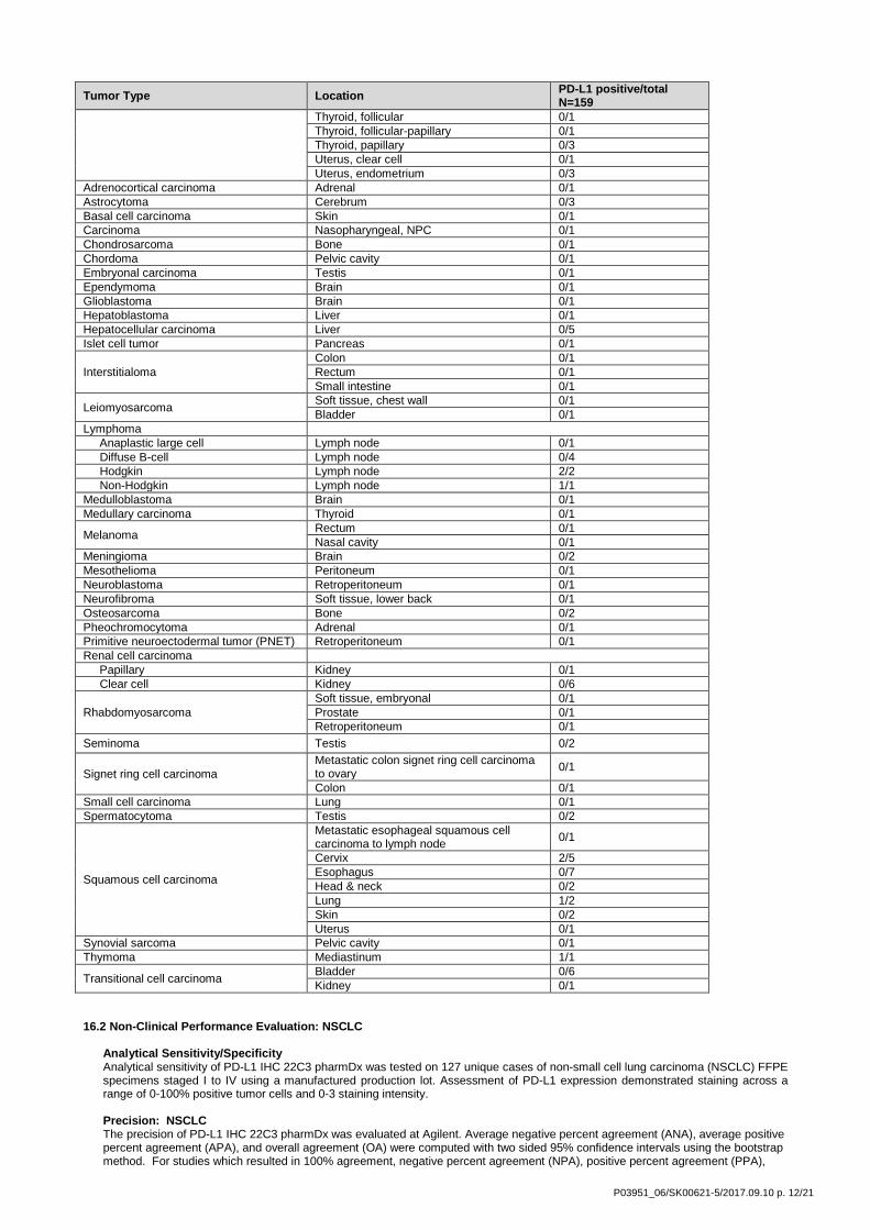
Uterus (3) 0/3 0/3 0/3 Neoplastic tissues: Table 4 summarizes Monoclonal Mouse Anti-PD-L1, Clone 22C3 immunoreactivity on a panel of neoplastic tissues. Plasma membrane staining was observed on immune cells and cells of epithelial origin. Cytoplasmic staining was noted in some cell types but was not recorded as positive staining. All tissues were formalin-fixed and paraffin-embedded and stained with PD-L1 IHC 22C3 pharmDx according to the instructions in this package insert. There were no unexpected results observed in the tumor specimens tested. The observed staining was consistent with the reported literature for PD-L1 IHC expression in neoplastic tissues (8-11). Table 4: Summary of PD-L1 IHC 22C3 pharmDx Neoplastic Tissue Reactivity
Tumor Type Location PD-L1 positive/total N=159
Adenocarcinoma
Appendix 0/1 Breast, DCIS 0/2 Breast, invasive ductal 0/7 Breast, invasive ductal metastatic to lymph node 0/1
Cervix, endocervical type 0/1 Colon 0/5 Colon, metastatic to liver 0/1 Colon, mucinous 0/1 Esophagus 0/1 Gallbladder 1/5 GI, metastatic to lung 0/1 Head & neck, hard palate 0/1 Lung 1/4 Ovary 0/1 Ovary, endometrioid 0/1 Ovary, mucinous 0/1 Ovary, serous 0/1 Pancreas 0/2 Pancreas, ductal 0/3 Prostate 0/5 Rectum 0/4 Salivary/parotid gland 0/2 Small intestine 0/2 Stomach 0/6 Stomach, mucinous 0/1
P03951_06/SK00621-5/2017.09.10 p. 11/21
Tumor Type Location PD-L1 positive/total N=159
Thyroid, follicular 0/1 Thyroid, follicular-papillary 0/1 Thyroid, papillary 0/3 Uterus, clear cell 0/1 Uterus, endometrium 0/3
Adrenocortical carcinoma Adrenal 0/1 Astrocytoma Cerebrum 0/3 Basal cell carcinoma Skin 0/1 Carcinoma Nasopharyngeal, NPC 0/1 Chondrosarcoma Bone 0/1 Chordoma Pelvic cavity 0/1 Embryonal carcinoma Testis 0/1 Ependymoma Brain 0/1 Glioblastoma Brain 0/1 Hepatoblastoma Liver 0/1 Hepatocellular carcinoma Liver 0/5 Islet cell tumor Pancreas 0/1
Interstitialoma Colon 0/1 Rectum 0/1 Small intestine 0/1
Leiomyosarcoma Soft tissue, chest wall 0/1 Bladder 0/1
Lymphoma Anaplastic large cell Lymph node 0/1 Diffuse B-cell Lymph node 0/4 Hodgkin Lymph node 2/2 Non-Hodgkin Lymph node 1/1 Medulloblastoma Brain 0/1 Medullary carcinoma Thyroid 0/1
Melanoma Rectum 0/1 Nasal cavity 0/1
Meningioma Brain 0/2 Mesothelioma Peritoneum 0/1 Neuroblastoma Retroperitoneum 0/1 Neurofibroma Soft tissue, lower back 0/1 Osteosarcoma Bone 0/2 Pheochromocytoma Adrenal 0/1 Primitive neuroectodermal tumor (PNET) Retroperitoneum 0/1 Renal cell carcinoma Papillary Kidney 0/1 Clear cell Kidney 0/6
Rhabdomyosarcoma Soft tissue, embryonal 0/1 Prostate 0/1 Retroperitoneum 0/1
Seminoma Testis 0/2
Signet ring cell carcinoma Metastatic colon signet ring cell carcinoma to ovary 0/1
Colon 0/1 Small cell carcinoma Lung 0/1 Spermatocytoma Testis 0/2
Squamous cell carcinoma
Metastatic esophageal squamous cell carcinoma to lymph node 0/1
Cervix 2/5 Esophagus 0/7 Head & neck 0/2 Lung 1/2 Skin 0/2 Uterus 0/1
Synovial sarcoma Pelvic cavity 0/1 Thymoma Mediastinum 1/1
Transitional cell carcinoma Bladder 0/6 Kidney 0/1
16.2 Non-Clinical Performance Evaluation: NSCLC
Analytical Sensitivity/Specificity Analytical sensitivity of PD-L1 IHC 22C3 pharmDx was tested on 127 unique cases of non-small cell lung carcinoma (NSCLC) FFPE specimens staged I to IV using a manufactured production lot. Assessment of PD-L1 expression demonstrated staining across a range of 0-100% positive tumor cells and 0-3 staining intensity.
Precision: NSCLC The precision of PD-L1 IHC 22C3 pharmDx was evaluated at Agilent. Average negative percent agreement (ANA), average positive percent agreement (APA), and overall agreement (OA) were computed with two sided 95% confidence intervals using the bootstrap method. For studies which resulted in 100% agreement, negative percent agreement (NPA), positive percent agreement (PPA),
P03951_06/SK00621-5/2017.09.10 p. 12/21

and overall agreement (OA) were computed with two sided 95% confidence intervals using the Wilson Score method for the TPS ≥ 1% cutoff and ≥ 50% cutoff and TPS ≥ 50% cutoff.
Table 5: Precision of PD-L1 IHC 22C3 pharmDx tested at one site (TPS ≥ 1%)
Precision Study Diagnostic Cutoff Study Design % Agreement (95% CI)
Inter-instrument
TPS ≥ 1% Each of 24 NSCLC specimens (12 PD-L1-negative and 12 PD-L1-positive) with a range of PD-L1 IHC expression was tested on each of six Autostainer Link 48 instruments.
NPA 100% (94.0-100%) PPA 100% (94.0-100%) OA 100% (96.9-100%)
Inter-operator
TPS ≥ 1% Each of 24 NSCLC specimens (12 PD-L1-negative and 12 PD-L1-positive) with a range of PD-L1 IHC expression was tested using six analysts on one Autostainer Link 48 instrument.
NPA 100% (93.9-100%) PPA 100% (94.0-100%) OA 100% (96.9-100%)
Inter-day
TPS ≥ 1% Each of 24 NSCLC specimens (12 PD-L1-negative and 12 PD-L1-positive) with a range of PD-L1 IHC expression was tested on six non-consecutive days on the Autostainer Link 48 instrument.
NPA 100% (94.0-100%) PPA 100% (94.0-100%) OA 100% (96.9-100%)
Inter-lot TPS ≥ 1% Each of 24 NSCLC specimens (13 PD-L1-negative and 11 PD-L1-positive) with a range of PD-L1 IHC expression was tested with three replicates and each of three reagent lots on the Autostainer Link 48 instrument.
ANA 98.3% (95.9-100%) APA 97.9% (94.6-100%) OA 98.1% (95.3-100%)
Intra-run (Repeatability) TPS ≥ 1% Each of 24 NSCLC specimens (12 PD-L1-negative and 12 PD-L1-positive) with a range of PD-L1 IHC expression was tested with six replicates within a run on the Autostainer Link 48 instrument.
NPA 100% (94.0-100%) PPA 100% (93.8-100%) OA 100% (96.8-100%)
NPA= Negative Percent Agreement; PPA= Positive Percent Agreement; OA=Overall Agreement ANA=Average Negative Agreement; APA=Average Positive Agreement; TPS=Tumor Proportion Score
Table 6: Precision of PD-L1 IHC 22C3 pharmDx tested at one site (TPS ≥ 50%)
Precision Study Diagnostic Cutoff Study Design % Agreement (95% CI)
Inter-instrument
TPS ≥ 50% Each of 16 NSCLC specimens (10 PD-L1-negative and 6 PD-L1-positive) with a range of PD-L1 IHC expression was tested on each of six Autostainer Link 48 instruments.
NPA 100% (92.9-100%) PPA 100% (88.6-100%) OA 100% (95.4-100%)
Inter-operator
TPS ≥ 50% Each of 16 NSCLC specimens (10 PD-L1-negative and 6 PD-L1-positive) with a range of PD-L1 IHC expression was tested using six analysts on one Autostainer Link 48 instrument.
NPA 100% (92.7-100%) PPA 100% (88.6-100%) OA 100% (95.4-100%)
Inter-day
TPS ≥ 50% Each of 16 NSCLC specimens (10 PD-L1-negative and 6 PD-L1-positive) with a range of PD-L1 IHC expression was tested on six non-consecutive days on the Autostainer Link 48 instrument.
NPA 100% (92.9-100%) PPA 100% (88.6-100%) OA 100% (95.4-100%)
Inter-lot TPS ≥ 50% Each of 16 NSCLC specimens (8 PD-L1-negative and 8 PD-L1-positive) with a range of PD-L1 IHC expression was tested with three replicates and each of three reagent lots on the Autostainer Link 48 instrument.
NPA 100% (92.6-100%) PPA 100% (92.6-100%) OA 100% (96.2-100%)
Intra-run (Repeatability) TPS ≥ 50% Each of 16 NSCLC specimens (10 PD-L1-negative and 6 PD-L1-positive) with a range of PD-L1 IHC expression was tested with six replicates within a run on the Autostainer Link 48 instrument.
NPA 100% (92.9-100%) PPA 100% (88.6-100%) OA 100% (95.4-100%)
Intra-day TPS ≥ 50% Each of 16 NSCLC specimens (10 PD-L1-negative and 6 PD-L1-positive) with a range of PD-L1 IHC expression was tested on two runs within a day, repeated over three days, on the Autostainer Link 48 instrument.
NPA 100% (88.3-100%) PPA 100% (82.4-100%) OA 100% (92.4-100%)
NPA= Negative Percent Agreement; PPA= Positive Percent Agreement; OA=Overall Agreement; TPS=Tumor Proportion Score
External Reproducibility: NSCLC The reproducibility of PD-L1 IHC 22C3 pharmDx was evaluated at three external testing sites. Average agreements were calculated since no natural reference exists in reproducibility parameters such as site and observer. Average negative percent agreement (ANA), average positive percent agreement (APA), and overall percent agreement (OA) were computed with two sided 95% confidence intervals using the bootstrap method for the TPS ≥ 1% cutoff and TPS ≥ 50% cutoff.
Table 7: Reproducibility of PD-L1 IHC 22C3 pharmDx tested at three external sites (TPS ≥ 1%)
P03951_06/SK00621-5/2017.09.10 p. 13/21
Reproducibility Study Diagnostic Cutoff Study Design % Agreement (95% CI)
Inter-site TPS ≥ 1% Each of 36 NSCLC specimens (16 PD-L1-negative and 20 PD-L1-positive) with a range of PD-L1 IHC expression was tested on five non-consecutive days. Inter-site analysis was performed between three sites on a total of 2700 pair-wise comparisons.
ANA 94.8% (90.3-98.4%) APA 95.5% (91.2-98.7%) OA 95.2% (90.8-98.6%)
Intra-site TPS ≥ 1% Each of 36 NSCLC specimens (16 PD-L1-negative and 20 PD-L1-positive) with a range of PD-L1 IHC expression was tested on five non-consecutive days at each of three study sites. Intra-site analysis was performed for three sites on a total of 1080 pair-wise comparisons.
ANA 96.2% (94.1-97.5%) APA 96.7% (95.0-97.9%) OA 96.5% (95.2-97.4%)
Inter-observer TPS ≥ 1% Scoring of 62 NSCLC specimens (28 PD-L1-negative and 34 PD-L1-positive) with a range of PD-L1 IHC expression, stained with PD-L1 IHC 22C3 pharmDx, was performed by three pathologists, one at each of three study sites, on three non-consecutive days. Inter-observer analysis was performed between three sites on a total of 1674 pair-wise comparisons.
ANA 85.8% (79.3-91.8%) APA 88.2% (82.2-93.3%) OA 87.1% (81.0-92.6%)
Intra-observer TPS ≥ 1% Scoring of 62 NSCLC specimens (28 PD-L1-negative and 34 PD-L1-positive) with a range of PD-L1 IHC expression, stained with PD-L1 IHC 22C3 pharmDx, was performed by three pathologists, one at each of three study sites, on three non-consecutive days. Intra-observer analysis was performed for three sites on a total of 558 pair-wise comparisons.
ANA 93.7% (90.0-96.1%) APA 94.8% (91.6-96.7%) OA 94.3% (92.0-95.9%)
ANA=Average Negative Agreement; APA=Average Positive Agreement; OA=Overall Agreement; TPS=Tumor Proportion Score
Table 8: Reproducibility of PD-L1 IHC 22C3 pharmDx tested at three external sites (TPS ≥ 50%)
Reproducibility Study Diagnostic Cutoff Study Design % Agreement (95% CI)
Inter-site TPS ≥ 50% Each of 36 NSCLC specimens (21 PD-L1-negative and 15 PD-L1-positive) with a range of PD-L1 IHC expression was tested on five non-consecutive days. Inter-site analysis was performed between three sites on a total of 2700 pair-wise comparisons.
ANA 90.3% (84.4-95.2%) APA 85.2% (75.6-92.9%) OA 88.3% (81.4-94.3%)
Intra-site TPS ≥ 50% Each of 36 NSCLC specimens (21 PD-L1-negative and 15 PD-L1-positive) with a range of PD-L1 IHC expression was tested on five non-consecutive days at each of three study sites. Intra-site analysis was performed for three sites on a total of 1080 pair-wise comparisons.
ANA 91.9% (88.8-94.8%) APA 87.6% (82.5-92.2%) OA 90.2% (86.3-93.7%)
Inter-observer TPS ≥ 50% Scoring of 62 NSCLC specimens (30 PD-L1-negative and 32 PD-L1-positive) with a range of PD-L1 IHC expression, stained with PD-L1 IHC 22C3 pharmDx, was performed by three pathologists, one at each of three study sites, on three non-consecutive days. Inter-observer analysis was performed between three sites on a total of 1674 pair-wise comparisons.
ANA 92.6% (87.8-96.7%) APA 92.8% (88.1-96.8%) OA 92.7% (88.1-96.8%)
Intra-observer TPS ≥ 50% Scoring of 62 NSCLC specimens (30 PD-L1-negative and 32 PD-L1-positive) with a range of PD-L1 IHC expression, stained with PD-L1 IHC 22C3 pharmDx, was performed by three pathologists, one at each of three study sites, on three non-consecutive days. Intra-observer analysis was performed for three sites on a total of 558 pair-wise comparisons.
ANA 96.4% (94.0-98.5%) APA 96.5% (94.3-98.6%) OA 96.4% (94.3-98.6%)
ANA=Average Negative Agreement; APA=Average Positive Agreement; OA=Overall Agreement; TPS=Tumor Proportion Score
16.3 Clinical Performance Evaluation: NSCLC KEYNOTE 024: Controlled trial of first-line treatment of patients with NSCLC The efficacy of KEYTRUDA was investigated in Trial 24, a randomized (1:1), open-label, multicenter, controlled trial (9). Key eligibility criteria were metastatic NSCLC, PD-L1 expression tumor proportion score (TPS) of 50% or greater by an immunohistochemistry assay using PD-L1 IHC 22C3 pharmDx, and no prior systemic treatment for metastatic NSCLC. Patients with EGFR or ALK genomic tumor aberrations; autoimmune disease that required systemic therapy within 2 years of treatment; a medical condition that required immunosuppression; or who had received more than 30 Gy of thoracic radiation within the prior 26 weeks were ineligible. Patients were randomized to receive KEYTRUDA 200 mg every 3 weeks (n=154) or investigator’s choice platinum-containing chemotherapy (n=151; including pemetrexed + carboplatin, pemetrexed + cisplatin, gemcitabine + cisplatin, gemcitabine + carboplatin, or paclitaxel +
P03951_06/SK00621-5/2017.09.10 p. 14/21
carboplatin. Non-squamous patients could receive pemetrexed maintenance). Patients were treated with KEYTRUDA until unacceptable toxicity or disease progression, or up to 35 administrations. Subsequent disease progression could be retreated for up to 1 additional year. Treatment could continue beyond disease progression if the patient was clinically stable and was considered to be deriving clinical benefit by the investigator. Assessment of tumor status was performed every 9 weeks. Patients on chemotherapy who experienced progression of disease were offered KEYTRUDA. Among the 305 patients in Trial 24, baseline characteristics were: median age 65 years (54% age 65 or older); 61% male; 82% White and 15% Asian; and 35% and 65% with an ECOG performance status 0 and 1, respectively. Disease characteristics were squamous (18%) and non-squamous (82%); M1 (99%); and brain metastases (9%). The major efficacy outcome measure was progression-free survival (PFS) as assessed by blinded independent central review (BICR) using Response Evaluation Criteria on Solid Tumors Version 1.1 (RECIST 1.1). Additional efficacy outcome measures were overall survival (OS) and objective response rate (ORR) as assessed by BICR using RECIST 1.1. Table 9 summarizes key efficacy measures for the entire intent to treat (ITT) population. Table 9: Efficacy Results in Trial 24
Endpoint KEYTRUDA 200 mg every 3 weeks n=154
Chemotherapy n=151
PFS* Number (%) of patients with event 73 (47%) 116 (77%) Hazard ratio† (95% CI) 0.50 (0.37, 0.68) --- p-Value‡ <0.001 --- Median in months (95% CI) 10.3 (6.7, NA) 6.0 (4.2, 6.2) OS Number (%) of patients with event 44 (29%) 64 (42%) Hazard ratio† (95% CI) 0.60 (0.41, 0.89) --- p-Value‡ 0.005 --- Median in months (95% CI) Not reached
(NA, NA) Not reached (9.4, NA)
Objective Response Rate* ORR % (95% CI) 45% (37, 53) 28% (21, 36) Complete response % 4% 1% Partial response % 41% 27% * Assessed by BICR using RECIST 1.1 † Hazard ratio (KEYTRUDA compared to chemotherapy) based on the stratified Cox proportional hazard model ‡ Based on stratified Log rank test NA = not available
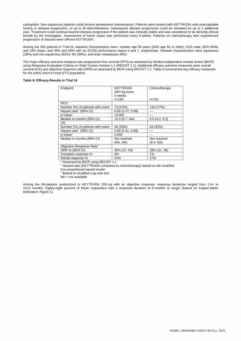
Among the 69 patients randomized to KEYTRUDA 200 mg with an objective response, response durations ranged from 1.9+ to 14.5+ months. Eighty-eight percent of these responders had a response duration of 6 months or longer (based on Kaplan-Meier estimation; Figure 1).
P03951_06/SK00621-5/2017.09.10 p. 15/21
Figure 1: Kaplan-Meier Curve for Overall Survival in Trial 24
KEYNOTE 010: Controlled trial of NSCLC patients previously treated with chemotherapy The efficacy of KEYTRUDA was investigated in Trial 10, a randomized (1:1), open-label, multicenter, controlled trial (10). Key eligibility criteria were advanced NSCLC that had progressed following platinum-containing chemotherapy, and if appropriate, targeted therapy for ALK or EGFR mutations, and PD-L1 expression tumor proportion score (TPS) of 1% or greater by a clinical trial assay (CTA) version of PD-L1 IHC 22C3 pharmDx. Forty-four and 56 percent of patients were enrolled based on testing of an archival tumor sample or a new tumor sample, respectively. Patients with autoimmune disease; a medical condition that required immunosuppression; or who had received more than 30 Gy of thoracic radiation within the prior 26 weeks were ineligible. Patients were randomized (1:1:1) to receive 2 mg/kg (n=344) or 10 mg/kg (n=346) of KEYTRUDA every 3 weeks or 75 mg/m2 of docetaxel every 3 weeks (n=343). Patients were treated with KEYTRUDA until unacceptable toxicity or disease progression that was symptomatic, was rapidly progressive, required urgent intervention, occurred with a decline in performance status, or was confirmed at 4 to 6 weeks with repeat imaging. Patients without disease progression were treated for up to 24 months or 35 administrations, whichever was longer. Subsequent disease progression could be retreated for up to 1 additional year. Assessment of tumor status was performed every 9 weeks. The primary efficacy outcome measures were OS and PFS as assessed by BICR using RECIST 1.1. Based on the CTA, a total of 1,033 NSCLC patients were randomized in the study. To evaluate the clinical utility of PD-L1 IHC 22C3 pharmDx, archived clinical study samples were retrospectively tested at a US based reference laboratory with PD-L1 IHC 22C3 pharmDx. Out of the 1,033 patients, tumor tissue from 529 patients was retrospectively tested with the PD-L1 IHC 22C3 pharmDx test. Specimens from 413 patients had PD-L1 expression (≥ 1% of viable tumor cells exhibiting membrane staining at any intensity) and samples from 94 patients did not have PD-L1 expression (< 1% of viable tumor cells exhibiting membrane staining at any intensity). Within these 413 patients with PD-L1 expression, specimens from 163 patients had high PD-L1 expression (≥ 50% of viable tumor cells exhibiting membrane staining at any intensity). The level of agreement achieved between the CTA and PD-L1 IHC 22C3 pharmDx is shown in Table 10. Table 10: CTA vs. PD-L1 IHC 22C3 pharmDx Agreement
Agreement Rates PD-L1 Cut-off
Negative Percent Agreement (95% Confidence Interval (CI))
Positive Percent Agreement (95% Confidence Interval (CI))
CTA vs. PD-L1 IHC 22C3 pharmDx TPS ≥ 1% 94.5% [91.4%-96.6%] 80.0% [76.9%-82.8%] TPS ≥ 50% 98.3% [97.1%-99.0%] 73.2% [67.9%-77.9%]
Among randomized patients having PD-L1 expression by PD-L1 IHC 22C3 pharmDx, the demographic and other baseline characteristics were well balanced between the treatment arms. The median age was 63 years (44% age 65 or older). The majority of patients were white (77%) and male (58%); baseline ECOG performance status was 0 (29%) or 1 (71%). Seventy-eight percent (78%) of patients were former/current smokers. Twenty-two percent (22%) of patients had squamous histology and 69% had non-squamous histology. The baseline and demographic characteristics were similarly well balanced across pembrolizumab and docetaxel arms in the overall clinical study. Efficacy results are summarized in Tables 11 and 12. KEYTRUDA demonstrated durable clinical benefit in NSCLC patients with PD-L1 expression (TPS ≥ 1%), which was enhanced in patients with high PD-L1 expression (TPS ≥ 50%), as determined by PD-L1 IHC 22C3 pharmDx. The magnitude of benefit was comparable to that in the overall clinical trial. The tables below summarize key efficacy measures in the overall population with PD-L1 expression (TPS ≥ 1%) and in the high PD-L1 expression (TPS ≥ 50%) subset for the overall clinical study (TPS ≥ 1% by CTA) and in the population with PD-L1 expression by PD-L1 IHC 22C3 pharmDx. The Kaplan-Meier curve for OS (TPS ≥ 1%), as determined by PD-L1 IHC 22C3 pharmDx) is shown in Figure 2. Efficacy results were similar for the 2 mg/kg and 10 mg/kg KEYTRUDA arms.
0 3 6 9 12 15 18 21
Time in Months
0
10
20
30
40
50
60
70
80
90
100
Ove
rall
Sur
viva
l (%
)
Treatment armKEYTRUDAChemotherapy
154 136 121 82 39 11 2 0
151 123 106 64 34 7 1 0
Number at RiskKEYTRUDA:
Chemotherapy:
P03951_06/SK00621-5/2017.09.10 p. 16/21
Table 11: Response to KEYTRUDA in Previously Treated NSCLC Patients: Overall Clinical Trial and Patients with PD-L1 Expression, TPS ≥ 1%, as determined by PD-L1 IHC 22C3 pharmDx
Endpoint KEYTRUDA 2 mg/kg every 3 weeks
KEYTRUDA 10 mg/kg every 3 weeks
Docetaxel 75 mg/m2 every 3 weeks
Clinical Trial PD-L1 IHC 22C3 pharmDx
Clinical Trial PD-L1 IHC 22C3 pharmDx
Clinical Trial PD-L1 IHC 22C3 pharmDx
Number of patients 344 140 346 142 343 131 OS Deaths (%) 172 (50%) 59 (42%) 156 (45%) 59 (42%) 193 (56%) 67 (51%) Hazard ratio* (95% CI)
0.71 (0.58, 0.88)
0.54 (0.37, 0.78)
0.61 (0.49, 0.75)
0.57 (0.39, 0.82) --- ---
p-Value† <0.001 <0.001 <0.001 0.00115 --- --- Median in months (95% CI)
10.4 (9.4, 11.9)
11.8 (9.6, NA)
12.7 (10.0, 17.3)
12.0 (8.7, NA)
8.5 (7.5, 9.8)
7.5 (6.3, 9.9)
PFS‡ Events (%) 266 (77%) 97 (63%) 255 (74%) 103 (73%) 257 (75%) 94 (72%) Hazard ratio* (95% CI)
0.88 (0.73, 1.04)
0.68 (0.50, 0.92)
0.79 (0.66, 0.94)
0.79 (0.59, 1.06) --- ---
p-Value† 0.068 0.00578 0.005 0.05767 --- --- Median in months (95% CI)
3.9 (3.1, 4.1)
4.9 (4.1, 6.2)
4.0 (2.6, 4.3)
4.0 (2.2, 4.6)
4.0 (3.1, 4.2)
3.8 (2.2, 4.2)
Overall response rate‡
ORR %§ (95% CI)
18% (14, 23)
24% (17, 32)
18% (15, 23)
20% (14, 28)
9% (7, 13)
5% (2, 11)
* Hazard ratio (KEYTRUDA compared to docetaxel) based on the stratified Cox proportional hazard model † Based on stratified Log rank test ‡ Assessed by BICR using RECIST 1.1 § All responses were partial responses
Table 12: Response to KEYTRUDA in Previously Treated NSCLC Patients: Overall Clinical Trial and Patients with PD-L1 High Expression, TPS ≥ 50%, as determined by PD-L1 IHC 22C3 pharmDx
Endpoint KEYTRUDA 2 mg/kg every 3 weeks
KEYTRUDA 10 mg/kg every 3 weeks
Docetaxel 75 mg/m2 every 3 weeks
Clinical Trial PD-L1 IHC 22C3 pharmDx
Clinical Trial PD-L1 IHC 22C3 pharmDx
Clinical Trial PD-L1 IHC 22C3 pharmDx
Number of patients 139 56 151 60 152 47
OS
Deaths (%) 58 (42%) 18 (32%) 60 (40%) 19 (32%) 86 (57%) 25 (53%) Hazard ratio* (95% CI)
0.54 (0.38, 0.77)
0.45 (0.24, 0.84)
0.50 (0.36, 0.70)
0.29 (0.15 0.56) --- ---
p-Value† <0.001 0.00541 <0.001 <0.001 --- --- Median in months (95% CI)
14.9 (10.4, NA)
Not reached (9.3, NA)
17.3 (11.8, NA)
Not reached (8.3, NA)
8.2 (6.4, 10.7)
7.2 (4.4, 8.3)
PFS‡
Events (%) 89 (64%) 33 (59%) 97 (64%) 34 (57%) 118 (78%) 33 (70%) Hazard ratio* (95% CI)
0.58 (0.43, 0.77)
0.47 (0.28, 0.80)
0.59 (0.45, 0.78)
0.41 (0.24, 0.70) --- ---
p-Value† <0.001 0.00221 <0.001 <0.001 --- --- Median in months (95% CI)
5.2 (4.0, 6.5)
5.9 (4.2, 9.0)
5.2 (4.1, 8.1)
4.8 (2.8, NA)
4.1 (3.6, 4.3)
3.9 (2.0, 4.3)
Overall response rate ‡
ORR %§ (95% CI)
30% (23, 39)
37% (25, 52)
29% (22, 37)
28% (18, 41)
8% (4, 13)
4% (1, 15)
P03951_06/SK00621-5/2017.09.10 p. 17/21

* Hazard ratio (KEYTRUDA compared to docetaxel) based on the stratified Cox proportional hazard model † Based on stratified Log rank test ‡ Assessed by BICR using RECIST 1.1 § All responses were partial responses
Figure 2: Kaplan-Meier Curve for Overall Survival by Treatment Arm (TPS ≥ 1% by PD-L1 IHC 22C3 pharmDx, Intent to Treat Population)
Additional robustness analyses were conducted to consider the potential impact of missing data arising from patients with PD-L1 expression (TPS ≥ 1%) by PD-L1 IHC 22C3 pharmDx, but who may have had no PD-L1 expression (TPS <1%) by the CTA. Patients with such test results are part of the intended use/ intent to diagnose (ITD)/ population of PD-L1 IHC 22C3 pharmDx; however, they were excluded from the clinical trial due to no PD-L1 expression upon CTA screening. To account for these missing data, a sensitivity analysis was conducted to understand the plausible range for the hazard ratio (HR) estimated based on PD-L1 IHC 22C3 pharmDx in the TPS ≥ 1% and TPS ≥ 50% subpopulations under an ITD framework to verify the consistency with the observed HR based on enrolment with the CTA. The HR sensitivity analysis results showed that the HR estimates are robust to any assumed attenuation of the treatment effect under the ITD framework.
16.4 Non-Clinical Performance Evaluation: Gastric or Gastroesophageal Junction (GEJ) Adenocarcinoma The following histologies were tested in the non-clinical performance evaluation of gastric or GEJ adenocarcinoma: intestinal, diffuse including signet ring cell carcinoma, and mucinous types.
Analytical Sensitivity/Specificity: Gastric or GEJ Adenocarcinoma Analytical sensitivity of PD-L1 IHC 22C3 pharmDx was tested on 100 FFPE gastric or GEJ adenocarcinoma specimens (stage I to IV) using a manufactured production lot. Assessment of PD-L1 expression demonstrated staining across a range of CPS 0-100. 60% of the specimens had PD-L1 expression, with expression defined by CPS ≥ 1. Precision: Gastric or GEJ Adenocarcinoma The precision of PD-L1 IHC 22C3 pharmDx in gastric or GEJ adenocarcinoma was evaluated at Agilent. Inter-instrument, inter-operator, inter-day, and inter-lot were tested in combined precision. Repeatability was tested in intra-run precision. Intra-observer and inter-observer precision were also assessed. Negative percent agreement (NPA), positive percent agreement (PPA), and overall agreement (OA) were computed with two sided 95% confidence intervals using the bootstrap method for the CPS ≥ 1 cutoff. For studies which resulted in 100% agreement, the two sided 95% confidence intervals were computed using the Wilson Score method.
Table 13: Precision of PD-L1 IHC 22C3 pharmDx in gastric or GEJ adenocarcinoma, tested at one site (CPS ≥ 1)
Precision Diagnostic Cutoff Study Design % Agreement (95% CI): Combined Precision (inter-instrument, inter-operator, inter-lot, inter-day,)
CPS ≥ 1 Each of 24 gastric or GEJ adenocarcinoma specimens (12 PD-L1-positive and 12 PD-L1-negative) with a range of PD-L1 expression were tested using three Autostainer Link 48 instruments, four operators, three kit lots, over three non-consecutive days.
NPA 100% (94.9-100%) PPA 95.8% (91.4-100%) OA 97.9% (92.5-100%)
Intra-run precision (Repeatability)
CPS ≥ 1 Each of 24 gastric or GEJ adenocarcinoma specimens (12 PD-L1-negative and 12 PD-L1-positive) with a range of PD-L1 IHC expression was tested with
NPA 96.9% (92.4-100%) PPA 100% (93.5-100%) OA 98.3% (92.5-100%)
P03951_06/SK00621-5/2017.09.10 p. 18/21

Precision Diagnostic Cutoff Study Design % Agreement (95% CI): five replicates within a run on the Autostainer Link 48 instrument.
Inter-observer precision
CPS ≥ 1 60 gastric or GEJ adenocarcinoma specimens (30 PD-L1-negative and 30 PD-L1-positive) with a range of PD-L1 IHC expression, stained with PD-L1 IHC 22C3 pharmDx, was scored by three pathologists over three non-consecutive days
NPA 96.0% (81.4-93.3%) PPA 96.8% (86.7-95.0%) OA 96.5% (84.6-94.3%)
]Intra-observer precision
CPS ≥ 1 60 gastric or GEJ adenocarcinoma specimens (30 PD-L1-negative and 30 PD-L1-positive) with a range of PD-L1 IHC expression, stained with PD-L1 IHC 22C3 pharmDx, was scored by three pathologists over three non-consecutive days
NPA 91.5% (87.9-95.1%) PPA 96.1% (91.2-96.5%) OA 94.1% (90.0-95.9%)
NPA= Negative Percent Agreement; PPA= Positive Percent Agreement; OA=Overall Agreement; CPS=Combined Positive Score
External Reproducibility: Gastric or GEJ Adenocarcinoma The reproducibility of PD-L1 IHC 22C3 pharmDx was evaluated at three external sites. Negative percent agreement (NPA), positive percent agreement (PPA), and overall agreement (OA) were computed with two sided 95% confidence intervals (CI) using the bootstrap method for the CPS ≥1 cutoff. In an initial study the acceptance criteria for the CI lower bound of OA and NPA for inter-observer reproducibility and CI lower bound of NPA for intra-observer reproducibility were not met. A root cause assessment indicated that one of the three observers in the study did not pass post-study proficiency testing. A second inter- and intra-observer study was conducted with three naïve observers, and the results met the acceptance criteria. Results are shown in Table 14 below. Proficiency assessment is recommended to ensure correct observer scoring interpretation.
Table 14: Reproducibility of PD-L1 IHC 22C3 pharmDx in gastric or GEJ adenocarcinoma, tested at three external sites (CPS ≥ 1)
Reproducibility Study Diagnostic Cutoff Study Design % Agreement (95% CI) Inter-site CPS ≥ 1 Each of 36 Gastric or GEJ adenocarcinoma
specimens (16 PD-L1 negative and 20 PD-L1 positive) with a range of PD-L1 IHC expression was tested on five non-consecutive days. Inter-site analysis was performed between three sites on a total of 540 pair-wise comparisons.
NPA 92.5% (86.2-97.5%) PPA 91.7% (84.7-97.7%) OA 92.0% (87.4-96.3%)
Intra-site CPS ≥ 1 Each of 36 Gastric or GEJ adenocarcinoma specimens (16 PD-L1 negative and 20 PD-L1 positive) with a range of PD-L1 IHC expression was tested on five non-consecutive days at each of three study sites. Intra-site analysis was performed for three sites on a total of 540 pair-wise comparisons.
NPA 93.1% (89.2-96.5%) PPA 98.2% (96.4-99.6%) OA 95.7% (93.7-97.6%)
Inter-observer CPS ≥ 1 Scoring of 68 Gastric or GEJ adenocarcinoma specimens (36 PD-L1- negative and 32 PD-L1-positive) with a range of PD-L1 IHC expression, stained with PD-L1 IHC 22C3 pharmDx, was performed by three pathologists, one at each of three study sites, on three non-consecutive days. Inter-observer analysis was performed between three sites on a total of 612 pair-wise comparisons.
NPA 96.6% (92.9-99.4%) PPA 96.5% (93.1-99.3%) OA 96.6% (94.0-98.7%)
Intra-observer CPS ≥ 1 Scoring of 68 Gastric or GEJ adenocarcinoma specimens (36 PD-L1-negative and 32 PD-L1-positive) with a range of PD-L1 IHC expression, stained with PD-L1 IHC 22C3 pharmDx, was performed by three pathologists, one at each of three study sites, on three non-consecutive days. Intra-observer analysis was performed for three sites on a total of 612 pair-wise comparisons.
NPA 97.2% (94.8-99.1%) PPA 97.2% (94.8-99.3%) OA 97.2% (95.3-98.9%)
NPA= Negative Percent Agreement; PPA= Positive Percent Agreement; OA=Overall Agreement; CPS=Combined Positive Score
16.5 Clinical Performance Evaluation: Gastric or Gastroesophageal Junction (GEJ) Adenocarcinoma The efficacy of KEYTRUDA was investigated in KEYNOTE 059 (KN059), a multicenter, non-randomized, open-label multi-cohort trial that enrolled 259 patients with gastric or GEJ adenocarcinoma who progressed on at least 2 prior systemic treatments for advanced disease. Previous treatment must have included a fluoropyrimidine and platinum doublet. HER2/neu positive patients must have previously received treatment with approved HER2/neu targeted therapy. Patients with active autoimmune disease or a medical condition that required immunosuppression or with clinical evidence of ascites by physical exam were ineligible. Patients received KEYTRUDA 200 mg every 3 weeks until unacceptable toxicity or disease progression that was symptomatic, rapidly progressive, required urgent intervention, occurred with a decline in performance status, or was confirmed at least 4 weeks later with repeat imaging. Patients without disease progression were treated for up to 24 months. Assessment of tumor status was
P03951_06/SK00621-5/2017.09.10 p. 19/21

performed every 6 to 9 weeks. The major efficacy outcome measures were ORR according to RECIST 1.1, as assessed by blinded independent central review, and duration of response. PD-L1 expression levelfor 259 patient tumor biopsy or resection tissue (167 archival and 90 newly obtained (refer to definition in Table 15) was determined using PD-L1 IHC 22C3 pharmDx. PD-L1 expression level for 2 samples was not evaluable. Overall, fifty eight percent (148/257) of the patients had tumors that expressed PD-L1 with a combined positive score (CPS) ≥ 1. Seventy three percent (66/90) patients whose tumors were newly obtained for PD-L1 testing and 49% (82/167) in patients whose archival tumors were tested expressed PD-L1 at CPS ≥ 1 (Table 15). Table 15: Tumor PD-L1 Expression by Specimen Type Tumor Tissue PD-L1 Expression (CPS ≥ 1) n (%) No PD-L1 Expression (CPS < 1) n (%) Overall study n=257 (%) 148 (58) 109 (42)
Archival Tissue* n=167
82 (49) 85 (51)
Newly Obtained tissue* n= 90
66 (73) 24 (27)
*In the context of clinical trial KN059, a newly obtained biopsy was defined as a specimen obtained up to 6 weeks (42 days) prior to initiation of treatment on Day 1 (Cycle 1) with KEYTRUDA and with no additional anti-cancer treatment having been given after the specimen was obtained. Specimens that were >42 days were classified as archival. Of 148 patients with PD-L1 expression at CPS ≥ 1, 143 were assessed to be either microsatellite stable (MSS) tumor status or had undetermined MSI or MMR status. The baseline characteristics of these 143 patients were: median age 64 years (47% age 65 or older); 77% male; 82% White, 11% Asian; and ECOG PS of 0 (43%) and 1 (57%). Eighty-five percent had M1 disease and 7% had M0 disease. Fifty-one percent had two and 49% had three or more prior lines of therapy in the recurrent or metastatic setting. For the 143 patients that have PD-L1 expression (CPS ≥ 1), the ORR was 13.3% (95% CI: 8.2, 20.0); 1.4% had a complete response and 11.9% had a partial response. Among the 19 responding patients, the duration of response ranged from 2.8+ to 19.4+ months, with 11 patients (58%) having responses of 6 months or longer and 5 patients (26%) having responses of 12 months or longer.
17. Troubleshooting Table 16: Troubleshooting
Problem Probable Cause Suggested Action 1. No staining of slides 1a. Programming error.
1a. Verify that the PD-L1 IHC 22C3 pharmDx program was selected for programming of slides.
1b. Lack of reaction with DAB+ Substrate-Chromogen Solution (DAB)
1b. Verify that DAB+ Substrate-Chromogen Solution was prepared properly.
1c. Sodium azide in wash buffer. 1c. Use only Dako Wash Buffer (Code K8007). 1d. Degradation of Control Slide 1d. Check kit expiration date and kit storage
conditions on outside of package. 2. Weak staining of specimen slides. 2a. Inappropriate fixation method used. 2a. Ensure that only neutral buffered formalin fixative
and approved fixation methods are used. 2b. Insufficient reagent volume applied. 2b. Check size of tissue section and reagent volume
applied. 2c. Inappropriate wash buffer used. 2c. Use only Dako Wash Buffer, Code K8007.
3. Weak staining of specimen slides or of the positive cell line on the Control Slide provided with the kit
3a. Inadequate target retrieval. 3a. Verify that the 3-in-1 pre-treatment procedure was correctly performed.
3b. Inappropriate wash buffer used. 3b. Use only Dako Wash Buffer, Code K8007 4. Excessive background staining of slides.
4a. Paraffin incompletely removed. 4a. Verify that the 3-in-1 pre-treatment procedure was correctly performed.
4b. Slides dried while loading onto the Autostainer Link 48.
4b. Ensure slides remain wet with buffer while loading and prior to initiating run.
4c. Nonspecific binding of reagents to tissue section.
4c. Check for proper fixation of the specimen and/or the presence of necrosis.
4d. Inappropriate fixation method used. 4d. Ensure that only neutral buffered formalin fixative and recommended fixation methods are used.
5. Tissue detached from slides. 5a. Use of incorrect microscope slides. 5a. Use Dako FLEX IHC Microscope Slides, (Code K8020), or Fisherbrand Superfrost Plus slides.
5b. Inadequate preparation of specimens
5b. Cut sections should be placed in a 58 ± 2 °C oven for 1 hour prior to staining.
6. Excessively strong specific staining. 6a. Inappropriate fixation method used. 6a. Ensure that only approved fixatives and fixation methods are used.
6b. Inappropriate wash buffer used. 6b. Use only Dako Wash Buffer, Code K8007. 7. Target Retrieval Solution is cloudy in appearance when heated.
7. When heated the Target Retrieval Solution turns cloudy in appearance.
7. This is normal and does not influence staining.
NOTE: If the problem cannot be attributed to any of the above causes, or if the suggested corrective action fails to resolve the problem, please call Agilent Technical Support for further assistance. Additional information on staining techniques and specimen preparation can be found in Dako Education Guide: Immunohistochemical Staining Methods (5) (available from Dako).
18 References
1. Keytruda® package insert. 2. R. Phelps, et al., Journal of Cellular Biochemistry Supplement (1996) 24:32-91.
P03951_06/SK00621-5/2017.09.10 p. 20/21
3. Department of Health, Education and Welfare, National Institutes for Occupational Safety and Health, Rockville, MD. “Procedures for the decontamination of plumbing systems containing copper and/or lead azides.” DHHS (NIOSH) Publ. No. 78-127, Current 13. August 16, 1976.
4. Clinical and Laboratory Standards Institute (formerly NCCLS). Protection of Laboratory Workers From Occupationally Acquired Infections; Approved Guideline – Third Edition. CLSI document M29-A3 [ISBN 1-56238-567-4]. Clinical and Laboratory Standards Institute, 940 West Valley Road, Suite 1400, Wayne, Pennsylvania 19087 – 1898 USA, 2000.
5. Taylor CR and Rudbeck L. Education Guide: Immunohistochemical Staining Methods. Sixth Edition. Dako, Carpinteria, California; 2013.
6. Clinical and Laboratory Standards Institute (formerly NCCCLS). Quality assurance for Immunocytochemistry; Approved guideline. CLSI document MM4-A (1-56238-396-5) CLSI, 940 West Valley Road, Suite 1400, Wayne, Pennsylvania 19087-1898 USA; 1999.
7. Omata M, Liew C-T, Ashcavai M, Peters RL. Nonimmunologic binding of horseradish peroxidase to hepatitis B s surface antigen: a possible source of error in immunohistochemistry. Am J Clin Path 1980; 73:626.
8. Okazaki T, Honjo T. PD-1 and PD-1 ligands: from discovery to clinical application. International Immunol 2007(19); 7:813. 9. Brown JA, Dorfman DM, Ma F-R, Sullivan EL, Munoz O, Wood CR, et al. Blockade of Programmed Death-1 Ligands on
Dendritic Cells Enhances T Cell Activation and Cytokine Production. J Immunol 2003;170:1257. 10. Cooper WA, Tran T, Vilain RE, Madore J, Seliger CI, Kohonen-Cornish M, et al. PD-L1 expression is a favorable prognostic
factor in early stage non-small cell carcinoma. Lung Cancer 2015; 89:181. 11. Chen B, Chapuy B, Ouyang J et al. PD-L1 Expression Is Characteristic of a Subset of Aggressive B-cell Lymphomas and
Virus-Associated Malignancies. Clin Cancer Res 2013; 19:3462-3473. 12. Reck, M, et al. Pembrolizumab versus Chemotherapy for PD-L1–Positive Non–Small-Cell Lung Cancer (KEYNOTE-024).
The New England Journal of Medicine (NJEM.org) 2016 Oct 09; 1-11. 13. Herbst RS, et al. Pembrolizumab versus docetaxel for previously treated, PD-L1-positive, advanced non-small-cell lung
cancer (KEYNOTE-010): a randomised controlled trial. The Lancet online; 2015 Dec 19. 14. Garon EB, Rizvi NA, Hui R, Leighl N, Balmanoukian AS, Eder JP, et al. Pembrolizumab for the treatment of non-small-cell
lung cancer. New Eng J Med 2015; 372:2018-28. 15. Muro K, Chung HC, Shankaran V, Geva R, Catenacci D, Gupta S, Eder JP, Golan T, Le D, Burtness B, McRee A, Lin C-C,
Pathiraja K, Lunceford J, Emancipator K, Juco J, Koshiji M, Bang Y. Pembrolizumab for patients with PD-L1–positive advanced gastric cancer (KEYNOTE-012): a multicentre, open-label, phase 1b trial. Lancet Oncol. 2016;17(6):717-726.
Edition 04/17
P03951_06/SK00621-5/2017.09.10 p. 21/21