perioperative complications of neuraxial blockade
TRANSCRIPT

The Immediate and Delayed Complications of Perioperative and Obstetric Central Neuraxial Blockade and their Management
Author: Sarah Cooper (Obstetric Anaesthetist), Louise Jobling (Obstetric Anaesthetist) Mark Stubbington (ST 7 Anaesthesia) Approved by: Anaesthetic Department Review date: October 2019 Version:1 Purpose
• To Highlight and raise awareness of common and rarer complications of central neuraxial blockade (CNB) and how they should be managed
• To provide background information to facilitate the diagnosis, assessment and management of
complications of CNB (Separate policies are available for the management of inadvertent dural puncture and post dural puncture headache and links are provided to national (AAGBI) guidance for other specific situations).
• To enable the assessing anaesthetist to better differentiate between anaesthetic and non
anaesthetic causes of neurological deficit and provide advice to patients. • To facilitate timely planning of investigations and management in cases where central pathology
(Spinal/ Epidural haematoma or abscess) is suspected. • To ensure that there is a clear pathway for the communication of findings and follow up care for
obstetric patients who have experienced a complication of central neuraxial blockade. This document is intended to be used alongside existing policies relating to obstetric anaesthesia and perioperative analgesia - Epidural Analgesia (Ref SA0198), Intrathecal Opioid Analgesia (Ref SA0103). 1. Adverse/exaggerated physiological response: High or total spinal anaesthesia, cardiac arrest and urinary retention 1.1 High or total spinal anaesthesia (TSA) Adequate surgical anaesthesia for LSCS is usually reflected by a sensory block at the 5th thoracic dermatome to light touch or the 4th thoracic dermatome to sharp prick or cold.
Relative or absolute overdose of local anaesthetic drug can result in high or total spinal anaesthesia respectively. Absolute overdose occurs when the actual dose administered exceeds the calculated dose for particular patient and route of administration. Relative overdose occurs when the pharmacological response exceeds the expected response for a particular patient and route of administration.
1

Relative overdose during single shot spinal is most likely to cause a high block and the most common causes of total spinal are the administration of an epidural dose through an unidentified intrathecal epidural catheter, a spinal after a failed epidural top up, and a full epidural top up that spreads more than expected.
Risk factors include:
• spinal after failed epidural
• positioning
• high BMI
• recently administered epidural top up
• epidural sited following accidental dural puncture or in the presence of spinal canal abnormalities
• short stature.
Clinical presentation:
• Rapid onset, development and progression of symptoms
• Initial presentation - usually cardiorespiratory features predominate
Neurological signs and symptoms play key role in diagnosis and subsequent management
2
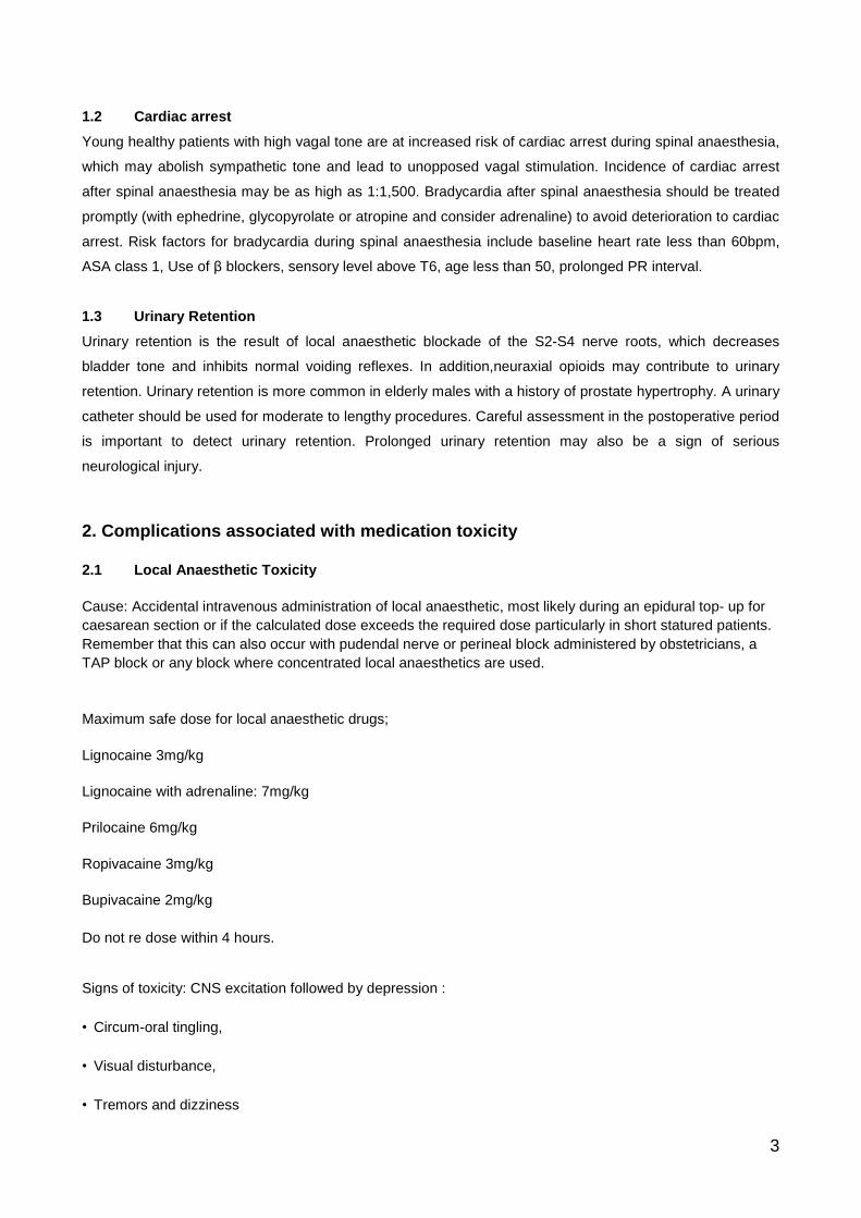
1.2 Cardiac arrest Young healthy patients with high vagal tone are at increased risk of cardiac arrest during spinal anaesthesia,
which may abolish sympathetic tone and lead to unopposed vagal stimulation. Incidence of cardiac arrest
after spinal anaesthesia may be as high as 1:1,500. Bradycardia after spinal anaesthesia should be treated
promptly (with ephedrine, glycopyrolate or atropine and consider adrenaline) to avoid deterioration to cardiac
arrest. Risk factors for bradycardia during spinal anaesthesia include baseline heart rate less than 60bpm,
ASA class 1, Use of β blockers, sensory level above T6, age less than 50, prolonged PR interval.
1.3 Urinary Retention Urinary retention is the result of local anaesthetic blockade of the S2-S4 nerve roots, which decreases
bladder tone and inhibits normal voiding reflexes. In addition,neuraxial opioids may contribute to urinary
retention. Urinary retention is more common in elderly males with a history of prostate hypertrophy. A urinary
catheter should be used for moderate to lengthy procedures. Careful assessment in the postoperative period
is important to detect urinary retention. Prolonged urinary retention may also be a sign of serious
neurological injury.
2. Complications associated with medication toxicity
2.1 Local Anaesthetic Toxicity
Cause: Accidental intravenous administration of local anaesthetic, most likely during an epidural top- up for caesarean section or if the calculated dose exceeds the required dose particularly in short statured patients. Remember that this can also occur with pudendal nerve or perineal block administered by obstetricians, a TAP block or any block where concentrated local anaesthetics are used.
Maximum safe dose for local anaesthetic drugs;
Lignocaine 3mg/kg
Lignocaine with adrenaline: 7mg/kg
Prilocaine 6mg/kg
Ropivacaine 3mg/kg
Bupivacaine 2mg/kg Do not re dose within 4 hours.
Signs of toxicity: CNS excitation followed by depression :
• Circum-oral tingling,
• Visual disturbance,
• Tremors and dizziness
3

• Convulsions
• Apnoea and unconsciousness
• Ringing in the ears / deafness
Myocardium:
• Blockade of Na+ channels in the myocardium and direct myocardial depression
• Prolonged PR and QRS intervals
• Prolonged refractory period
• Re-entrant dysrhythmias and ventricular fibrillation
Bupivacaine has a high affinity for and slow dissociation from myocardial Na+ channels, resulting in
prolonged effect and resistance to treatment. Levobupivacaine is relatively safer when compared to
bupivacaine on account of reduced affinity for myocardial Na+ channels but toxicity can still occur
The AAGBI has published a guideline for management of local anaesthetic toxicity:
4

5

6

2.2 Intravascular Injection
The risk of serious complications related to an intravascular injection, when performing a spinal anaesthetic, is low due to the small amount of local anaesthetic required to induce anaesthesia. The risk of serious complications lies with the administration of epidural or caudal anaesthesia, where relatively large amounts of local anaesthetic are administered.
Local anaesthetic agents must be stored separately from any other drugs or fluids. During preparation for the block, syringes must be labelled clearly and appropriately and kept separate from any drugs intended for intravenous injection.
During the block, the patient’s vital signs should be monitored continually according to AAGBI standards (ECG,Pulse oximetry and non invasive arterial pressure monitoring). The use of a test dose of 1mg/kg 2% lidocaine (up to a max of 3.5ml) is recommended prior to any epidural top up with bupivacaine concentrations stronger than 0.15%, (i.e stronger top ups than the prepared bags for epidural infusion).
Repeated aspiration to check that the needle or catheter has not migrated intravascularly, may be useful. Slower injection may reduce the peak plasma LA concentration and could allow earlier detection of intravascular needle placement.
Maintain communication with the patient during the procedure, to aid detection of signs of intravascular injection such as perioral tingling or tinnitus.
The National Patient Safety Agency (NPSA) issued a Safety Alert in 2009, warning that Luer connectors common to spinal, epidural and regional anaesthesia needles and i.v cannula increased the risk of wrong route injection.
BTHFT will be introducing NRfit needles, syringes, filters, catheter connectors, catheters and pump disposables in the near future and these must then be used for all neuraxial blocks in order to reduce the risk of wrong route medication errors.
2.3 Allergic reactions
Anaphylaxis to local anaesthetics is very uncommon. It is more likely to occur with esters than amides. Methylparaben or metabisulphites are preservatives that may be the cause in some cases. Preservative free preparations should be used where possible.
Where allergy to LA is suspected, patients should be managed in line with the anaphylaxis guidelines found in the red emergency folders available in all theatres.
2.4 Chemical Arachnoiditis
Inflammation of the arachnoid meningeal layer and subarachnoid space. There have been associations made with this and needle contamination with chlorhexidine. Presents variably but often with progressive symptoms of parasthesia, numbness or leg weakness. The most common symptom is pain.
Use preservative free drugs. Use low concentration chlorhexidine (0.5%) and ensure it is administered via Chloraprep applicators. Allow to dry on the skin before needle insertion. AAGBI guideline reference - https://www.aagbi.org/sites/default/files/skin%20antisepsis%20for%20central%20neuraxial%20blockade_0.pdf
7

3. Complications associated with Needle or Catheter Insertion
3.1 Subdural Injection
The subdural space is a potential space found between the dura and arachnoid mater. It contains a small amount of serous fluid and extends intracranially. Local anaesthetics can travel higher in the subdural space than in the epidural space. The small dose of local anaesthetic associated with a spinal anaesthetic may result in a failed spinal. Larger doses used in epidural analgesia, may result in Horner’s syndrome. Still larger doses (associated with epidural anaesthesia) have on rare occasions resulted in total spinal anaesthesia. Treatment is the same as with high neuraxial blockade (supportive measures such as intubation, mechanical ventilation and cardiovascular support). Detection of subdural placement of epidural catheters can be difficult as aspiration is usually negative. Be suspicious of subdural catheter placement where slow incremental dosing produces a higher and faster than expected progression of anaesthetic block.
3.2 Back Pain
Backache is common during pregnancy and often continues afterwards. There is no evidence to suggest that neuraxial anaesthesia causes permanent worsening of preexisting back pain. The incidence of back pain after neuraxial anaesthesia in the adult population is not different from that after general anaesthesia. The pain is usually mild, localised in the low back, rarely radiates to the low extremities and has a duration of only a few days. The risk factors for development of back pain include lithotomy position, multiple attempts as block placement, duration of surgery longer than 2.5 hours, body mass index greater than 32kg/m2 and a history of back pain. Acute Severe back pain along with neurological deficit after central neuraxial block may indicate central pathology that requires urgent assessment and management. See section 5.
3.3 Shearing off of catheter
Epidural catheters can be sheared while withdrawing a catheter through a tuohy needle, therefore this should be avoided.
4. Accidental Dural Puncture and Post Dural Puncture Headache
Please see separate policy: Inadvertent Dural Puncture in Obstetric Patients
5. Neurological Injury
This can be a transient or permanent complication. Fortunately, neurological injury is extremely rare with an incidence of 0.08 - 0.16%.
The following general good practice steps are recommended:
• Document pre-existing neurological deficits.
• Document conditions that may contribute to postoperative neurological deficits such as peripheral vascular disease, diabetes, intervertebral disc disease and spinal/neurological disorders.
• Perform subarachnoid techniques below L2 in adults and L3 in children. Multiple attempts increase the incidence of trauma.
• Remove and redirect the needle when encountering parasthesia. If pain is encountered during needle insertion, catheter insertion or injecting medication, immediately stop the procedure and reassess, as direct injury to a spinal nerve root may cause permanent injury.
8

• Trainees should never hesitate to seek senior help with patients who have pre-existing risk factors for neurological complications, or where neuraxial technique is predicted to be or proves to be technically challenging.
• If a patient experiences a neurological deficit after neuraxial blockade, consider epidural haematoma or abscess (see sections 5.4 and 5.5). Neurological deficits may occur related to surgical positioning, improper positioning in the postoperative period, or as a result of direct trauma related to the surgical procedure. Obstetric patients are at increased risk as described in section 5.1.
5.1 Neurological deficits in PostPartum Patients
This section deals with neurological deficits occurring below the umbilicus. The majority of neurological complications are due to compressive neuropathy as a result of prolonged labour or poor patient positioning or mode of delivery. The Obstetric Anaesthetists Association (OAA) quotes the incidence of permanent nerve damage with an epidural as about 1:24,000. The NAP 3 report on complications of all central neuraxial blockade found that the incident of permanent harm following obstetric CNB was 1in 80,000 (judged pessimistically) and 1 in 320 425 judged optimistically. Obstetric CNB appears to be associated with less frequent major complications than when it is performed for other indications. This is probably because of the relative health of the obstetric population and the short duration of catheterisation.
The temporal association between anaesthetic intervention with neuraxial block and onset of neurological symptoms often means that anaesthetists are consulted early in the presentation of a neurological complication following childbirth. When neurological complications do occur, prompt recognition and management can reduce the risk of permanent neurological deficit. Therefore, despite the majority of nerve injury being related to factors other than regional anaesthesia/analgesia, it is important to have knowledge of diagnosis, investigation and management of neurological injury.
Leg strength MUST be assessed regularly as part of routine monitoring during an epidural infusion, hourly for the first 24 hours and 4 hourly thereafter. Please see appendix : “Management of leg weakness with an epidural infusion”
5.2 Non anaesthetic causes of Postpartum Neurological Deficit Compressive Neuropathy occurs in about 1:100 births, usually caused by the foetal head compressing the lumbosacral trunk, positioning, or instrumental delivery. Ischaemic neuropathy is estimated to occur in 1:500,000 births and relates to prolonged hypotension or obstruction of the internal iliac arteries by the foetal head in prolonged labour.
Compressive Nerve Injury Nerves affected Cause Presentation
Lateral Cutaneous Nerve of the Thigh
L2-3 Compression of the nerve as it passes under the inguinal ligament
Sensory loss over the anterolateral aspect of the thigh
Lumbosacral plexus L4,5 S1-5 Compression of the plexus against sacral ala. Usually from the foetal head in the second stage.
Numbness over the lateral aspect of the thigh, lower leg and dorsum of the foot. Results in foot drop, that is almost always unilateral and on the opposite side to the fatal occiput.
9

Common Perineal Nerve L4-5, S1-2 Prolonged lithotomy position. The nerve is vulnerable to compression as it passes over the head of the fibula when patients are positioned in stirrups without due care.
Numbness over the lateral aspect of the lower leg and dorsum of the foot, foot drop. Ankle reflex intact.
Femoral Nerve L2-4 Compression of nerve against inguinal canal during forceps delivery or LSCS. Femoral neuropathy can occur bilaterally 25% of the time and is therefore often mistaken for an intraspinal lesion.
Sensory loss over anterior thigh and inner aspect of lower leg. Weak knee extension. Often presents with difficulty climbing stairs. Loss of knee jerk. Reduced or absent patellar reflex is the most reliable objective sign in femoral neuropathy.
Obturator Nerve L2-4 Compression of nerve by foetal head or forceps. Obturator neuropathy, which occurs bilaterally 25% of the time, can also be mistaken for an intraspinal lesion.
Usually unilateral sensory loss over inner thigh and weak hip adduction and rotation.
The duration of symptoms for compressive neuropathies is 6 weeks to 2 months with symptoms in almost all patients resolving in this time. In order to try and prevent such compressive nerve injuries, good general measures are to change lower extremity position frequently during a prolonged second stage of labour, avoid prolonged thigh flexion, avoid extreme thigh abduction and external rotation and minimise motor and inappropriately dense sensory block when using epidural analgesia, by using lower concentrations of local anaesthetic where possible. 5.3 Anaesthetic causes of Postpartum Neurological Deficit Neurological complications secondary to CNB are as a result of damage to the spinal cord or nerve roots. This may be due to trauma, neurotoxicity, infection, ischaemia or haematoma. Central lesions, usually associated with anaesthetic interventions, are more commonly accompanied by back pain (supplied by the posterior rami) as the anterior and posterior rami of the nerve root are affected. Nerve damage can be secondary to direct needle or injection trauma disrupting the fibres of a single nerve. This causes parasthesia, loss of sensation and muscular weakness in the distribution of the nerve. Prevention is by avoiding contact with the nerves. Direct spinal cord trauma when placing spinal, epidural or CSE is signalled by pain on needle insertion and causes prolonged motor and sensory weakness at and below the level of injury. This can be unilateral or bilateral and may be accompanied by urinary symptoms. If there is pain on needle insertion or injection of local anaesthetic, withdraw the needle. The conus medullaris usually ends at L1 but may extend to L2,3 in 10% of patients. Therefore aim for L3,4 as the highest landmark. Scanning of the back can identify the correct interspace. Cauda equina syndrome - consisting of backache, nerve root pain, saddle anaesthesia, paraplegia and sphincter dysfunction, can occur when caudal equine nerve fibres are damaged due to compression or trauma. In all cases observe strict asepsis for all CNB techniques. Exercise caution in patients with coagulopathies, bleeding disorders and who are on
10
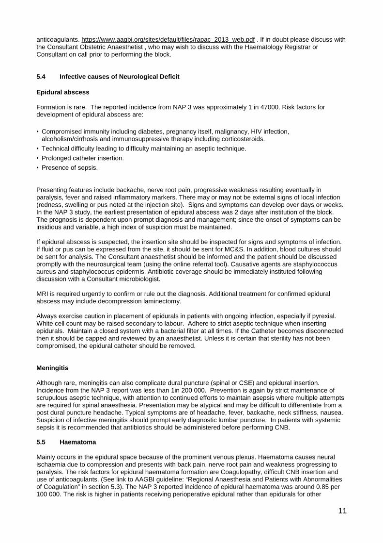
anticoagulants. https://www.aagbi.org/sites/default/files/rapac_2013_web.pdf . If in doubt please discuss with the Consultant Obstetric Anaesthetist , who may wish to discuss with the Haematology Registrar or Consultant on call prior to performing the block. 5.4 Infective causes of Neurological Deficit Epidural abscess Formation is rare. The reported incidence from NAP 3 was approximately 1 in 47000. Risk factors for development of epidural abscess are: • Compromised immunity including diabetes, pregnancy itself, malignancy, HIV infection,
alcoholism/cirrhosis and immunosuppressive therapy including corticosteroids. • Technical difficulty leading to difficulty maintaining an aseptic technique. • Prolonged catheter insertion. • Presence of sepsis. Presenting features include backache, nerve root pain, progressive weakness resulting eventually in paralysis, fever and raised inflammatory markers. There may or may not be external signs of local infection (redness, swelling or pus noted at the injection site). Signs and symptoms can develop over days or weeks. In the NAP 3 study, the earliest presentation of epidural abscess was 2 days after institution of the block. The prognosis is dependent upon prompt diagnosis and management; since the onset of symptoms can be insidious and variable, a high index of suspicion must be maintained. If epidural abscess is suspected, the insertion site should be inspected for signs and symptoms of infection. If fluid or pus can be expressed from the site, it should be sent for MC&S. In addition, blood cultures should be sent for analysis. The Consultant anaesthetist should be informed and the patient should be discussed promptly with the neurosurgical team (using the online referral tool). Causative agents are staphylococcus aureus and staphylococcus epidermis. Antibiotic coverage should be immediately instituted following discussion with a Consultant microbiologist. MRI is required urgently to confirm or rule out the diagnosis. Additional treatment for confirmed epidural abscess may include decompression laminectomy. Always exercise caution in placement of epidurals in patients with ongoing infection, especially if pyrexial. White cell count may be raised secondary to labour. Adhere to strict aseptic technique when inserting epidurals. Maintain a closed system with a bacterial filter at all times. If the Catheter becomes disconnected then it should be capped and reviewed by an anaesthetist. Unless it is certain that sterility has not been compromised, the epidural catheter should be removed. Meningitis Although rare, meningitis can also complicate dural puncture (spinal or CSE) and epidural insertion. Incidence from the NAP 3 report was less than 1in 200 000. Prevention is again by strict maintenance of scrupulous aseptic technique, with attention to continued efforts to maintain asepsis where multiple attempts are required for spinal anaesthesia. Presentation may be atypical and may be difficult to differentiate from a post dural puncture headache. Typical symptoms are of headache, fever, backache, neck stiffness, nausea. Suspicion of infective meningitis should prompt early diagnostic lumbar puncture. In patients with systemic sepsis it is recommended that antibiotics should be administered before performing CNB. 5.5 Haematoma Mainly occurs in the epidural space because of the prominent venous plexus. Haematoma causes neural ischaemia due to compression and presents with back pain, nerve root pain and weakness progressing to paralysis. The risk factors for epidural haematoma formation are Coagulopathy, difficult CNB insertion and use of anticoagulants. (See link to AAGBI guideline: “Regional Anaesthesia and Patients with Abnormalities of Coagulation” in section 5.3). The NAP 3 reported incidence of epidural haematoma was around 0.85 per 100 000. The risk is higher in patients receiving perioperative epidural rather than epidurals for other
11

indications (obstetric/chronic pain etc). Please see flow chart below for assessment and management of suspected epidural haematoma. 5.6 Recommended Technique for Avoiding Neurological Damage • Careful assessment before performing CNB is essential. • Document existing neurological deficits after careful examination. • Think carefully about anticoagulation/coagulopathy/thrombocytopenia/infection/sepsis and discuss with
consultant on call if unsure. • Aseptic technique: Handwash, hat, mask, gloves, gown, 0.5% chlorhexidine (prepared away from epidural
and spinal needles and be allowed to dry once applied to the skin. • A bacterial filter for drawing up and administering drugs for CNB • Avoid touching equipment that enters the patient • Avoid prolonged epidural catheterisation • Location of CNB - lowest palpable space. Above L3,4 should be avoided - it is well known that
anaesthetists are inaccurate in identifying the level and are often one space higher than they think. • If persistent pain in insertion of needle, placement of catheter or injection of drugs, remove needle or
discontinue drug administration. • Avoid prolonged periods of hypotension to maintain spinal cord perfusion. • Take care when positioning patients to avoid compressive neuropathy. • Inform patients of the risks and document the risks discussed. • Review all patients undergoing CNB following the procedure. • Any neurological deficit should be promptly assessed (within 30 minutes) and reviewed by a senior
anaesthetist. 5.7 Assessment and management of patient with reported neurological deficit (aimed at assessment of obstetric patients but most aspects can be applied to other perioperative patients with suspected complication of CNB). Please see appendix 1 flow chart. History • Neurological: including conditions predisposing to neuropathy e.g backache, obesity, disc disease,
diabetes, malignancy, coagulopathy, infection, previous trauma. • Labour/ mode of delivery - Instrumental delivery (type), posture during labour, use of retractors or
diathermy, period of full dilatation, injections given by obstetrician and hypotension. • Drugs - particularly anticoagulants, steroids, hypoglycaemics • Anaesthetic - type of block, degree of difficulty, possibility of inadvertent dural puncture, bloody tap, spinal
catheters, type/baricity/concentration of anaesthetic, additives, details of aseptic technique, site of injection, pain/parasthesia during procedure.
• Current symptoms: Pain, altered or absent sensation, motor deficits, sphincter dysfunction. Any RED FLAG SINISTER SYMPTOMS?
• Faecal incontinence can occur after vaginal delivery even in those patients without perineal tears (2-5%) and is very rarely due to CNB. Furthermore, misoprostol is a pro kinetic agent which can cause diarrhoea. A common cause of faecal incontinence is sphincter damage to the innervation of the sphincter during difficult vaginal delivery. In lower lumbosacral plexopathy, urinary incontinence can even accompany perianal sensory disturbance. In any case, MRI must be done on all such patients in order to rule out compression by a space occupying lesion.
Examination • Detailed neurological examination and examination of the back. • Dysfunction of lower extremities should be assessed without delay and limb or life threatening aetiologies
excluded. • Deteriorating symptoms or onset after a symptom-free interval should be treated seriously. This implies
changing pathology such as increasing compression from an enlarging mass (i.e. haematoma/abscess). Investigations
12

The presence of any sinister symptoms should prompt immediate consideration of MRI spine to exclude a central lesion (Epidural abscess or haematoma). Rapid referral to Neurosurgery at Leeds General Infirmary may be necessary for decompression - PERMANENT INJURY OCCURS BETWEEN 6 AND 12 HOURS FROM THE ONSET OF SYMPTOMS. Role of EMG (nerve conduction studies) Electrophysiological investigations can differentiate between central and peripheral nerve injury, identify the muscles affected and give a likely prognosis of neural recovery. It may be able to identify the precise lesion site. They can also produce a temporal estimate as to the timing of the injury, which is of particular note with regards to litigation. However, EMG only measures large nerve fibre changes and may take as long as 3 weeks after injury to show changes.
6. Ensuring Follow up and timely assessment of all patients after central neuraxial block: 6.1 Non obstetric Patients with Epidural Analgesia: All patients returning to the ward with epidural analgesia need to be referred to the Acute Pain Team for follow up. Routine monitoring for all patients with epidurals must be recorded hourly for the first 24 hours and at least 4 hourly thereafter: • Pain score • Conscious level • IV site inspection • Epidural catheter site inspection (daily) • Blood pressure • Respiratory rate • Oxygen saturation • Leg strength assessment (Bromage scale). Please refer to the guideline: Epidural Analgesia Ref SA0198. Appendix 2: Assessment and escalation process for suspected epidural haematoma and/or epidural abscess in patients with postoperative epidural infusions at the BRI. Appendix 3: Management of leg weakness with epidural analgesia. For help and advice during weekday office hours, please contact the Acute Pain Team (bleep 417 or via switchboard). Out of hours or if unable to contact the Acute Pain Team, contact the acute anaesthetist on call (ext 4328 or via switchboard). 6.2 Care of non obstetric patients after spinal anaesthesia: For patients who have received intrathecal opiates, bear in mind the increased risk of post operative respiratory depression. This is more likely to occur in very young or very elderly patients. Using the Pain Management Observations order, via Requests/Care plans on EPR, will ensure that these patients have the appropriate level of observation postoperatively. Specifically, they will then receive extra monitoring to detect respiratory depression; respiratory rate, conscious level/sedation and oxygen saturation. The standard care of all patients returning to the ward after spinal anaesthesia, includes: • Observations every 30 minutes for 2 hours : Blood Pressure, Heart Rate, Respiration Rate and SpO2
sedation and pain score.
Sinister Symptoms: Acute onset back pain
Radicular leg pain Urinary and anal dysfunction
Lower leg numbness and weakness
13

• If after 2 hours the observations are within safe limits, the observations can be documented hourly. If after 4 hours observations remain within safe limits, this can be reduced further to 2 hourly until 24 hours post op (or discharge if day case surgery).
• Assess the motor and sensory block and record on return to the ward and then 4 hourly up to 24 hours. In
most cases the effect of the spinal should have worn off within 4-6 hours. • Ward nursing teams should contact the Acute Pain Team or on call Anaesthetist urgently for advice if any
potential adverse events are detected: Increasing or prolonged motor block, new or increased backache, numbness or parasthesia to the legs and post-dural puncture headache.
Patients should be given verbal and ideally written information prior to discharge, regarding symptoms to be concerned about after having had spinal anaesthesia. This is particularly important for day case patients who have had spinal anaesthesia. Please see appendices 2 and 3 for suggested care pathway and patient information leaflet. 6.3 Obstetric Patients All obstetric patients who have received anaesthetic input during labour or for operative delivery, will need to be followed up by a member of the obstetric anaesthetic team, ideally the day after delivery or surgery. At the time of anaesthetic intervention, the anaesthetist involved will document the procedure on medway and add the patient to the list for follow up the next day. Details of the follow up visit should be documented using the medway Postnatal anaesthetic workflow. If there are no causes for concern at the follow up visit, the patient can be removed from the follow up list. If there are causes for concern (see above), the patient will need to be assessed as described and all findings clearly documented in the comments section on the follow up workflow. Do not remove the patient from the follow up list. Ensure that the team you hand over to at the end of your shift knows about any patients with concerns from follow up, and that they understand the next steps for management including discussions with other specialities. Ideally use the handover sheet morning and evening and ensure that any patients under our care who are not on labour ward, are included on the sheet. Patients who have had a suspected complication of CNB should have a letter sent to the G.P explaining the assessment, investigations, suspected diagnosis and management. (see appendix 4 for standard letter). Please email [email protected] giving the patient’s name, hospital number and the nature of the problem. Where needed, patients can then be offered a follow up appointment in the obstetric anaesthetic clinic. 6.4 If a potential complication of CNB is found at follow up or after being alerted by ward staff: • Any patient who is suspected of having a neurological deficit after a CNB technique, must be promptly
assessed and discussed with a senior anaesthetist as per section 5.7 and the flow chart in appendix 1. • Complete a Datix form for each patient with a likely neurological complication of CNB • Ensure that the patient’s GP receives information regarding the assessment, investigation and outcome
of a suspected complication of CNB (this can be on the discharge summary or in a separate letter). 14
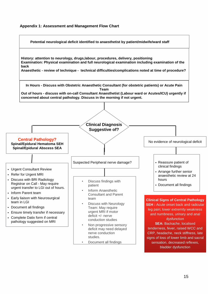
Appendix 1: Assessment and Management Flow Chart
Potential neurological deficit identified to anaesthetist by patient/midwife/ward staff
History: attention to neurology, drugs,labour, procedures, delivery, positioning Examination: Physical examination and full neurological examination including examination of the back Anaesthetic - review of technique - technical difficulties/complications noted at time of procedure?
In Hours - Discuss with Obstetric Anaesthetic Consultant (for obstetric patients) or Acute Pain Team
Out of hours - discuss with on-call Consultant Anaesthetist (Labour ward or Acutes/ICU) urgently if concerned about central pathology. Discuss in the morning if not urgent.
Clinical Diagnosis Suggestive of?
Central Pathology? Spinal/Epidural Hematoma SEH Spinal/Epidural Abscess SEA
Suspected Peripheral nerve damage?
No evidence of neurological deficit
• Urgent Consultant Review • Refer for Urgent MRI • Discuss with BRI Radiology
Registrar on Call - May require urgent transfer to LGI out of hours.
• Inform Parent team • Early liaison with Neurosurgical
team in LGI • Document all findings • Ensure timely transfer if necessary • Complete Datix form if central
pathology suggested on MRI
• Discuss findings with patient
• Inform Anaesthetic Consultant and Parent team
• Discuss with Neurology Team: May require urgent MRI if motor deficit +/- nerve conduction studies
• Non progressive sensory deficit may need delayed nerve conduction studies.
• Document all findings
• Reassure patient of clinical findings
• Arrange further senior anaesthetic review at 24 hours
• Document all findings
Clinical Signs of Central Pathology SEH : Acute onset back and radicular
leg pain; lower extremity weakness and numbness, urinary and anal
dysfunction SEA: Backache, localised
tenderness, fever, raised WCC and CRP, headache, neck stiffness, late signs of loss of lower limb and sacral
sensation, decreased reflexes, bladder dysfunction
15

Appendix 2: Example care pathway for non-obstetric patients after spinal anaesthetic
CARE PATHWAY FOR SPINAL ANAESTHETIC DAY OF OPERATION - RECOVERY ROOM Completed? Yes/No/NA
1 Dr/RN or ODP
Document observations - Blood Pressure, Heart rate, Respiratory rate, SpO2 every 5 mins until within safe limits for the patient, then every 15 minutes. Document Pain and sedation score, motor and sensory block
2 Anaesthetist
Ensure adequate IV access, Naloxone prescribed and O2 therapy in progress if required. Complete Pain Management Observations order if needed.
DAY OF OPERATION - ON RETURN TO WARD Completed? Yes/No/NA
1 RN Document the following observations every 30 minutes for 2 hours; Blood Pressure, Heart Rate, Respiration Rate, SpO2 , sedation and pain score. Assess the motor and sensory block and record on return to the ward and then 4 hourly up to 24 hours.
2 RN or Dr
If after 2 hours the observations are within safe limits (i.e NEWS 2 score low), they can be documented hourly. If after 4 hours observations remain within safe limits, this can be reduced to 2 hourly until 24 hours post op, or discharge (if day case patient).
3 RN or Dr
Monitor for adverse events and report URGENTLY to the Acute Pain Team or out of hours the On Call Anaesthetist: New/increased backache Unexpected leg weakness, numbness or pins and needles to legs Increasing/prolonged motor block (4-6 hours) Postural puncture headache (postural headache worse on standing, eased/disappears on lying down
DAY 1 POST- OP Completed? Yes/No/NA
1 RN or Dr
Continue above observations as directed until 24 hours post-op then 4 hourly until care pathway discontinued. (observations can be discontinued earlier if care pathway completion criteria met)
16

2 RN or Dr
Monitor for adverse events and report to the Acute Pain Team or out of hours the On Call anaesthetist: New / increased backache Unexpected leg weakness, numbness or pins and needles to legs. Increasing/prolonged motor block Postural puncture headache (postural headache worse on standing, eased/disappears on lying down).
CRITERIA FOR DISCONTINUING PATHWAY DATE AND TIME ACHIEVED
No new or increased backache
No unexpected weakness, numbness or pins and needles to legs
Motor function and mobility as pre-op/within limits of surgery
Able to pass urine normally (see below if catheter in situ) / able to pass urine normally once catheter removed
No postural puncture headache (postural headache worse on standing, eased/disappears when lying down).
Post epidural infusion/spinal anaesthetic information sheet given to patient?
If catheter in situ but all other criteria met, post spinal observations can be discontinued
Assess ability to pass urine once catheter removed and contact on-call anaesthetist if any concerns.
17

Appendix 3: Patient information following spinal anaesthesia Name
Hospital Number
DOB
Date Epidural Catheter/ Spinal Inserted:
You had an epidural infusion or spinal anaesthetic whilst you were in hospital. Although epidural infusions and spinal anaesthetics can provide excellent pain relief, rarely,serious complications such as a collection of pus or a blood clot around the spinal cord can arise after you have been discharged from hospital. Urgent treatment of these rare complications is necessary to prevent permanent injury. Therefore, if you experience any of the following symptoms in the next few days or weeks, go to the Accident and Emergency Department immediately. Bring this letter with you and ask to see the Anaesthetist on-call. Symptoms to be concerned about after you have had an epidural/spinal anaesthetic
• Redness, pus, tenderness or pain at the epidural /spinal site
• High temperature, neck stiffness
• Feeling generally unwell despite the fact that all seems well with the surgical wound
• New severe back pain
• New weakness, increasing numbness or new loss of sensation in your legs
• New loss of bladder or bowel control
• Continuous headache, worse on standing, not relieved by simple pain relief.
If you have any concerns about your epidural/spinal anaesthetic after you have been discharged from
hospital, telephone 01274 542200 and ask to speak to the Acute on-call Anaesthetist (via switchboard).
Following discharge from Maternity, please contact the Labour ward on extension 4515 and ask to speak to
the Obstetric Anaesthetist on duty.
Anaesthetic Department
Bradford Teaching Hospitals NHS Foundation trust.
18

Resources: Christie L, Picard J & Weinberg G. Local anaesthetic systemic toxicity. CEACCP 15(3) June 2015. 136-142. Benzon, Honorio T., Asher, Yogen G. & Hartrick, Craig T. Back Pain and Neuraxial Anesthesia. Anesthesia & Analgesia: June 2016 - Volume 122 - Issue 6 - p 2047–2058 doi: 10.1213/ANE.0000000000001270 Russell R, Dundas R, Reynolds F. Long term backache after childbirth: prospective search for causative factors. British Medical Journal 1996; 312: 1384-1388. NAP 3 - Major complications of central neuraxial block in the United Kingdom. Report and Findings, January 2009. https://www.rcoa.ac.uk/system/files/CSQ-NAP3-Full_1.pdf Spinal Anaesthesia for Day Surgery Patients; a Practical Guide. 3rd Edition. British Association of Day Surgery. 1919
19