pharmacokinetics, urinary excretion and plasma protein binding of pralidoxime in goats
TRANSCRIPT

Pp
Aa
Gb
a
ARRAA
KPGPBPU
1
ldpp3Ofe
ToT
0d
Small Ruminant Research 95 (2011) 179–183
Contents lists available at ScienceDirect
Small Ruminant Research
journa l homepage: www.e lsev ier .com/ locate /smal l rumres
harmacokinetics, urinary excretion and plasma protein binding ofralidoxime in goats
nu Rahala,b,∗, J.K. Malikb
Department of Pharmacology and Toxicology, College of Veterinary & Animal Sciences,.B. Pant University of Agriculture & Technology, Pantnagar 263145, UA, IndiaDivision of Pharmacology and Toxicology, Indian Veterinary Research Institute, Izatnagar, Bareilly 243122, UP, India
r t i c l e i n f o
rticle history:eceived 1 May 2010eceived in revised form 6 October 2010ccepted 7 October 2010vailable online 23 November 2010
eywords:ralidoximeoats
a b s t r a c t
The blood levels of cholinesterase reactivator pralidoxime (2-PAM) were determined ingoats following single dose intravenous administration @ 30 mg/kg body weight injected as6% freshly prepared solution. Blood and urine samples were collected at different predeter-mined time intervals and 2-PAM was analysed by spectrometric method with the minimumdetection of 1.0 �g ml−1. The peak plasma concentration was 49.52 ± 3.99 �g ml−1 at 2 minpost administration which rapidly declined to 15.53 ± 2.11 �g ml−1 at 10 min. Thereafter,it gradually disappeared to 1.33 ± 0.41 �g ml−1 at 1.5 h. The pharmacokinetic parameterswere determined by employing two-compartment open model. The t1/2˛, t1/2ˇ , Vdarea and
harmacokineticsloodlasmarine
ClB were calculated to be 1.68 ± 0.35 min, 21.17 ± 1.65 min, 1277.95 ± 195.67 ml/kg and41.17 ± 3.66 ml/kg/min, respectively. Approximately 52% of the total administered dose waseliminated in urine within 24 h. The plasma protein binding was estimated by equilibriumdialysis technique. The in vitro plasma protein binding of 2-PAM was 64.7%.
Based on these data, a satisfactory intravenous dosage regimen of 2-PAM in goats wouldbe 38 mg/kg body weight repeated at hourly intervals.
. Introduction
Organophosphorus pesticide poisoning is a major prob-em in the developing world where health care is oftenistant. The number of intoxications with organophos-horus pesticides (OPs) is estimated at some 3,000,000er year, and the number of deaths and casualties some
00,000 per year (Eyer, 2003; Eddleston et al., 2009).rganophosphorus compounds (OPCs) are widely usedor pest control and for the control of vector-borne dis-ases. These compounds also include the nerve poisons
∗ Corresponding author at: Department of Veterinary Pharmacology &oxicology, College of Veterinary & Animal Sciences, G.B. Pant Universityf Agriculture & Technology, Pantnagar 263145, UA, India.el.: +91 5944 233069.
E-mail address: [email protected] (A. Rahal).
921-4488/$ – see front matter © 2010 Elsevier B.V. All rights reserved.oi:10.1016/j.smallrumres.2010.10.005
© 2010 Elsevier B.V. All rights reserved.
that are among the most toxic group of chemical war-fare agents. Toxicology of organophosphates, as well asthe therapeutic approaches in acute organophosphatepoisoning, is of importance to many different groupsof health professionals. The management of OPC poi-soning is complex and includes nonspecific and specificmeasures. The dominant underlying toxicodynamic mech-anism in organophosphate poisoning is inhibition ofacetylcholinesterase (AChE). Oximes, as cholinesterasereactivators, are the only causal antidotes used againstOPCs. Pralidoxime (2-PAM), the first pyridinium oxime, isstill effective in the first 24–48 h after exposure, especiallywhen highly lipophilic organophosphates have accumu-
lated in fat and are gradually released (Mokhlesi et al.,2003).A particular problem in interpreting the beneficial roleand efficacy of oximes in clinical practice is a deficiency ofpublished data. The often cited ineffectiveness of oximes

minant Research 95 (2011) 179–183
90 min.The values of pharmacokinetic parameters are pre-
sented in Table 1. Tables 2 and 3 represent the renalexcretion and in vitro plasma protein binding of 2-PAM
Table 1Pharmacokinetic parameters (mean ± SE) of 2-PAM in plasma followingsingle dose (30 mg/kg, i.v.) administration of 2-PAM in goats (n = 6).
Kinetic parameter Unit Mean ± SE
A �g ml−1 84.58 ± 14.61B �g ml−1 19.62 ± 2.61˛ min−1 0.47 ± 0.06ˇ min−1 0.0337 ± 0.002t1/2˛ min 1.68 ± 0.35t1/2ß min 21.17 ± 1.65AUC �g min ml−1 756.17 ± 62.19AUMC �g min2 ml−1 19116.23 ± 2386.44MRT min 25.23 ± 2.55Vdarea ml kg−1 1277.95 ± 195.67Vc ml kg−1 319.33 ± 46.0Vp ml kg−1 661.83 ± 133.83Vdss ml kg−1 1037.97 ± 154.98ClB ml kg−1 min−1 41.17 ± 3.66Fc Ratio 3.48 ± 0.59T/p Ratio 0.63 ± 0.113
A = Zero time intercept of distribution slope in the two compartmentmodel, B = zero time intercept of elimination slope in the two compart-ment model, ˛ = distribution rate constant, ˇ = elimination rate constant,t1/2˛ = distribution half-life, t1/2ß = elimination half-life, ClB = clearance ofdrug, Vdarea = apparent volume of distribution, Vc = volume of central
180 A. Rahal, J.K. Malik / Small Ru
may be due to inappropriate dosing and/or generalisa-tion regarding an effective oxime concentration. Moreover,remarkable species differences in the susceptibility tooximes further require caution when animal data areextrapolated. A fundamental concept in pharmacology isthat a drug must reach specific tissues in the body in a suf-ficient concentration to exert therapeutic effects withoutcausing excessive harmful or toxic effects. Pharmacoki-netic properties of a medication need to be known sothat an appropriate amount of the drug can be admin-istered to reach target tissues and produce therapeuticresponses in a fairly predictable and timely manner. In thepresent study, blood, plasma and erythrocyte concentra-tions of pralidoxime have been investigated along with itsin vitro plasma protein binding and urinary excretion ingoats.
2. Materials and methods
2.1. Experimental animals and treatment
Six healthy goats were procured from the local market and quar-antined in the departmental animal shed for fortnight before thecommencement of experiment. During this period, animals were fedgreen fodder and water ad libitum.
Pralidoxime (obtained from Sigma Chemical Co., USA) was adminis-tered as freshly prepared 6% solution in isotonic saline into left jugularvein of animal in a single dose of 30 mg/kg body weight.
2.2. Sample collection and processing
To study the pharmacokinetics, about 5 ml of blood samples werewithdrawn from contralateral jugular vein into heparinized glass testtubes at 0, 2, 5, 10, 20, 30, 45, 60 and 90 min and 2, 3, 4, 5, 7, 10, 12 and 24 hafter the drug administration. Immediately after blood collection, packedcell volume (PCV) of the sample was assessed using a capillary. About2 ml of blood was preserved separately and rest of the blood was used toharvest plasma. Plasma was separated immediately at room temperatureafter centrifugation and stored at −20 ◦C till analysis, usually next day.
For studying urinary excretion of pralidoxime, Foley’s pediatriccatheter no.14F was passed into the urinary bladder of the animal so thatthe whole amount of urine formed by animal can be collected at any prede-termined time interval without any contamination and spillage. Followingadministration of pralidoxime, urine samples were collected at 0, 1, 2, 3, 6,12 and 24 h intervals. The total volume of urine was measured and sampleof 10 ml each were stored at −20 ◦C till further analysis.
In vitro plasma protein binding of pralidoxime was determined by theequilibrium dialysis technique as described by Kunin et al. (1959). Plasmawith known concentration of 2-PAM i.e. 2, 4, 8, 20 and 40 �g ml−1 wasdialysed (pore size, 40A) with phosphate buffer (0.2 M; pH 7.4) for 24 h at37 ◦C.
2.3. Analysis of sample
The levels of pralidoxime in blood, plasma and urine were mea-sured using spectrophotometric method of Creasey and Green (1959) asmodified by Maksimovic and Vojvodic (1969). The exact concentrationof pralidoxime in sample was calculated with standard curve, simulta-neously prepared in blood, plasma and urine of goats. The erythrocytepenetration of pralidoxime was calculated using the blood and plasmalevels and PCV values. The minimum detection level of pralidoxime by thismethod was 1.0 �g ml−1. The standard curve of 2-PAM was in straight line
between concentrations 1.0 to 40 �g ml−1. The initial pharmacokineticparameters were computed by least square technique as described bythe methods of Baggot (1977) and Gibaldi and Parrier (1982). The kineticanalysis was then done using a non-linear curve fitting programme (statisversion 3, M/s clydesoft, Glasgow, UK). The dosage regimen was computedby the method of Baggot (1977) and Johu Dein (1980).Fig. 1. Mean blood, plasma and erythrocyte concentrations and best fittedlines of regression (�g ml−1) vs. time plot of pralidoxime following singledose (30 mg/kg) i.v. administration in goats (n = 6).
3. Results
Blood, plasma and erythrocytic levels of 2-PAM atvarious time intervals after its single intravenous admin-istration (30 mg/kg) are depicted in Fig. 1. At 2 min,the plasma level was 47.59 ± 6.44 �g ml−1, which rapidlydeclined to 15.53 ± 2.11 �g ml−1 at 15 min. The therapeuticlevels (≥4 �g ml−1) was maintained for 45 min and there-after the levels slowly decreased to 1.33 ± 0.41 �g ml−1 at
compartment, Vp = volume of peripheral compartment, Vdss = volumeof distribution at steady state, AUC = total area under the concentra-tion time-curve, AUMC = total area under the first moment concentrationtime-curve, MRT = mean residence time, Fc = fraction of drug in the cen-tral compartment, T/p = Tissue plasma ratio. Values are expressed asMean ± SE.
A. Rahal, J.K. Malik / Small Ruminant
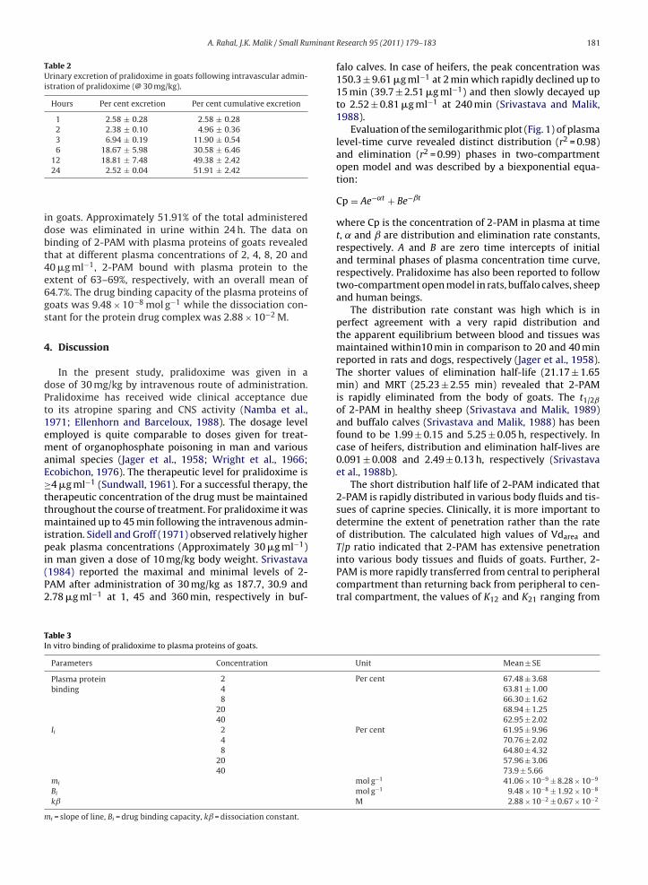
Table 2Urinary excretion of pralidoxime in goats following intravascular admin-istration of pralidoxime (@ 30 mg/kg).
Hours Per cent excretion Per cent cumulative excretion
1 2.58 ± 0.28 2.58 ± 0.282 2.38 ± 0.10 4.96 ± 0.363 6.94 ± 0.19 11.90 ± 0.546 18.67 ± 5.98 30.58 ± 6.46
idbt4e6gs
4
dPt1emaE≥ttmipi(P2
T/p ratio indicated that 2-PAM has extensive penetration
TI
m
12 18.81 ± 7.48 49.38 ± 2.4224 2.52 ± 0.04 51.91 ± 2.42
n goats. Approximately 51.91% of the total administeredose was eliminated in urine within 24 h. The data oninding of 2-PAM with plasma proteins of goats revealedhat at different plasma concentrations of 2, 4, 8, 20 and0 �g ml−1, 2-PAM bound with plasma protein to thextent of 63–69%, respectively, with an overall mean of4.7%. The drug binding capacity of the plasma proteins ofoats was 9.48 × 10⁻⁸ mol g−1 while the dissociation con-tant for the protein drug complex was 2.88 × 10⁻² M.
. Discussion
In the present study, pralidoxime was given in aose of 30 mg/kg by intravenous route of administration.ralidoxime has received wide clinical acceptance dueo its atropine sparing and CNS activity (Namba et al.,971; Ellenhorn and Barceloux, 1988). The dosage levelmployed is quite comparable to doses given for treat-ent of organophosphate poisoning in man and various
nimal species (Jager et al., 1958; Wright et al., 1966;cobichon, 1976). The therapeutic level for pralidoxime is4 �g ml−1 (Sundwall, 1961). For a successful therapy, the
herapeutic concentration of the drug must be maintainedhroughout the course of treatment. For pralidoxime it was
aintained up to 45 min following the intravenous admin-stration. Sidell and Groff (1971) observed relatively highereak plasma concentrations (Approximately 30 �g ml−1)
n man given a dose of 10 mg/kg body weight. Srivastava1984) reported the maximal and minimal levels of 2-AM after administration of 30 mg/kg as 187.7, 30.9 and.78 �g ml−1 at 1, 45 and 360 min, respectively in buf-
able 3n vitro binding of pralidoxime to plasma proteins of goats.
Parameters Concentration
Plasma proteinbinding
248
2040
Ii 248
2040
mi
Bi
kˇ
i = slope of line, Bi = drug binding capacity, kˇ = dissociation constant.
Research 95 (2011) 179–183 181
falo calves. In case of heifers, the peak concentration was150.3 ± 9.61 �g ml−1 at 2 min which rapidly declined up to15 min (39.7 ± 2.51 �g ml−1) and then slowly decayed upto 2.52 ± 0.81 �g ml−1 at 240 min (Srivastava and Malik,1988).
Evaluation of the semilogarithmic plot (Fig. 1) of plasmalevel-time curve revealed distinct distribution (r2 = 0.98)and elimination (r2 = 0.99) phases in two-compartmentopen model and was described by a biexponential equa-tion:
Cp = Ae−˛t + Be−ˇt
where Cp is the concentration of 2-PAM in plasma at timet, ˛ and ˇ are distribution and elimination rate constants,respectively. A and B are zero time intercepts of initialand terminal phases of plasma concentration time curve,respectively. Pralidoxime has also been reported to followtwo-compartment open model in rats, buffalo calves, sheepand human beings.
The distribution rate constant was high which is inperfect agreement with a very rapid distribution andthe apparent equilibrium between blood and tissues wasmaintained within10 min in comparison to 20 and 40 minreported in rats and dogs, respectively (Jager et al., 1958).The shorter values of elimination half-life (21.17 ± 1.65min) and MRT (25.23 ± 2.55 min) revealed that 2-PAMis rapidly eliminated from the body of goats. The t1/2ˇ
of 2-PAM in healthy sheep (Srivastava and Malik, 1989)and buffalo calves (Srivastava and Malik, 1988) has beenfound to be 1.99 ± 0.15 and 5.25 ± 0.05 h, respectively. Incase of heifers, distribution and elimination half-lives are0.091 ± 0.008 and 2.49 ± 0.13 h, respectively (Srivastavaet al., 1988b).
The short distribution half life of 2-PAM indicated that2-PAM is rapidly distributed in various body fluids and tis-sues of caprine species. Clinically, it is more important todetermine the extent of penetration rather than the rateof distribution. The calculated high values of Vdarea and
into various body tissues and fluids of goats. Further, 2-PAM is more rapidly transferred from central to peripheralcompartment than returning back from peripheral to cen-tral compartment, the values of K12 and K21 ranging from
Unit Mean ± SE
Per cent 67.48 ± 3.6863.81 ± 1.0066.30 ± 1.6268.94 ± 1.2562.95 ± 2.02
Per cent 61.95 ± 9.9670.76 ± 2.0264.80 ± 4.3257.96 ± 3.0673.9 ± 5.66
mol g−1 41.06 × 10⁻⁹ ± 8.28 × 10⁻⁹
mol g−1 9.48 × 10⁻⁸ ± 1.92 × 10⁻⁸
M 2.88 × 10⁻² ± 0.67 × 10⁻²

182 A. Rahal, J.K. Malik / Small Ruminant Research 95 (2011) 179–183
Table 4Intravenous priming (D′) and maintenance (Dm) doses of pralidoxime calculated on the basis of pharmacokinetic parameters at various dosage intervals tomaintain a target concentration of 4 �g ml−1.
Dosing interval(h)
Priming dose(D′ = Cpmin ×Vdarea × eˇt)(mg/kg)
Maximumplasmaconcentrationafter D′ (Cpmax)(�g ml−1)
Minimumplasmaconcentrationafter D′ (Cpmin)(�g ml−1)
Maintenancedose(Dm = Cpmin ×Vdarea × (eˇt − 1)(mg/kg)
Maximumplasmaconcentrationafter Dm
(Cpmax)
Minimumplasmaconcentrationafter Dm
(Cpmin)
1 38.6 34.8 4.61
0.251 ± 0.043 min−1 and 0.115 ± 0.013 min−1, respectively.This is further substantiated by low values of clearance.Similar to present study, high values of Vdarea and T/pratio of 2-PAM have also been reported in sheep andbuffalo calves (Srivastava and Malik, 1988, 1989). The val-ues of Vdarea and T/p ratio in sheep and buffalo calveswere 1.08 ± 0.26 l/kg and 4.39 ± 0.98, and 1.01 ± 0.05 l/kgand 5.12 ± 0.44, respectively. In man, the value of volumeof distribution was comparatively lower (0.73 ± 0.18 l/kg)(Thompson et al., 1987).
The effectiveness of an acetylcholinesterase reactiva-tor also depends on its penetration into erythrocytes, asorganophosphorus insecticides are well known to inhibitthe erythrocyte ChE to greater extent than other esterases(Srivastava et al., 1984). Accordingly, it was also thoughtimportant to calculate the extent of penetration of 2-PAMinto erythrocytes. At different blood concentrations, 2-PAMpenetrates into erythrocytes to the extent of 59–172% oftotal blood concentration. Any correlation between con-centration of 2-PAM in blood and extent of penetration intoerythrocytes could not be established though an increasein erythrocyte penetration was evident with the passageof time. Good penetration of 2-PAM into erythrocytesreflects that 2-PAM may be beneficial in the treatmentof organophosphate insecticide (OPI) poisoning in sheep(Srivastava et al., 1988a).
The results of the present urinary excretion study sug-gest that 2-PAM is mainly eliminated from the body viakidney. The rapid excretion from kidney seems to be themain contributor to its very small half-life. In buffalocalves also 52% cumulative renal excretion of 2-PAM wasreported after its intravenous administration during 24 h(Srivastava, 1984). Higher cumulative renal excretion of 2-PAM (about 78%) has been reported in heifer (Srivastavaet al., 1988b).
The plasma protein binding is an important index toassess the active fraction of the drug in blood and tis-sue compartments as only unbound or free drug is mainlyavailable at the target site for eliciting the desired pharma-cological actions as well as for the distribution and renalclearance. The results on plasma protein binding suggestedthat good percentage of 2-PAM was available in bloodstream for its therapeutic action.
Marked difference in the pharmacokinetic behaviour of
pralidoxime implies that its therapeutic dosage regimenneed to be computed by employing kinetic data establishedin the present study. A concentration of 4 �g/ml is consid-ered to be adequate therapeutic concentration (Sundwall,1961). Taking dosage intervals as 1 h, the priming and(�g ml−1) (�g ml−1)
33.5 30.2 4.0
maintenance doses of 2-PAM (Table 4) are calculated(Baggot, 1977). On the basis of the present investigation,it is concluded that the most appropriate dosage scheduleof DAM, in the treatment of OPI toxicity in goats would be38.6 mg/kg followed by 33.5 mg/kg at hourly intervals.
References
Baggot, J.D., 1977. The Basis of Veterinary Clinical Pharmacology, 1st ed.W.B. Saunders Co., Philadelphia.
Creasey, N.H., Green, A.L., 1959. 2-Hydroxyiminomethyl-N-methylpyridinium methane-sulphonate (P2S), an antidote toorganophosphorus poisoning. Its preparation, estimation andstability. J. Pharm. Pharmacol. 11, 485–490.
Ecobichon, D.J., 1976. Species differences in the reactivation oforganophosphate inhibited plasma esterases by diacetyl monoxime.Can. J. Physiol. 54, 86–93.
Eyer, P., 2003. The role of oximes in the management of organophosphoruspesticide poisoning. Toxicol. Rev. 22 (3), 165–190.
Eddleston, M., Eyer, P., Worek, F., Juszczak, E., Alder, N., Mohamed, F.,Senarathna, L., Hittarage, A., Azher, S., Jeganathan, K., Jayamanne,S., Meyer, L., Dawson, A.H., Sheriff, M.H.R., Buckley, N.A., 2009.Pralidoxime in acute organophosphorus insecticide poisoning—a ran-domised controlled trial. PLoS Med. 6 (6), e1000104.
Ellenhorn, M.J., Barceloux, D.G., 1988. Medical toxicology: Diagnosis andtreatment of human red cells. Biochem. Pharmacol. 23, 2663–2670.
Gibaldi, M., Parrier, D., 1982. Pharmacokinetics. Marcel Dekker, New York.Jager, B.V., Stagg, G.N., Green, N., Jager, L., 1958. Studies on distribution
and disappearance of pyridine-2-aldoxime methiodide (PAM) and ofdiacetylmonoxime (DAM) in man and experimental animals. Bull.Johns Hopkins Hosp. 102, 225–234.
Johu Dein, B., 1980. Clinical pharmacokinetics. J. Vet. Pharmacol. Ther. 3,4.
Kunin, C.M., Dornbus, J., Finland, M., 1959. Distribution and excretion offour tetracycline analogues in normal young man. J. Clin. Invest. 38,1950–1963.
Maksimovic, M., Vojvodic, V., 1969. Selection of the method for the deter-mination of oxime content in biological material. Arh. Hig. Rada.Toksikol. 20, 173–176.
Mokhlesi, B., Leikin, J.B., Murray, P., Corbridge, T.C., 2003. Adult Toxicol.Crit. Care. Chest 123, 897–922.
Namba, T., Nolte, C.T., Jackrel, J., Grob, D., 1971. Poisoning due toorganophosphate insecticides: acute and chronic manifestations. Am.J. Med. 50, 475–492.
Sidell, F.R., Groff, W.A., 1971. Intramuscular and intravenous administra-tion of small doses of 2-pyridinium aldoxime methochloride in man.J. Pharm. Sci. 60, 1224–1228.
Srivastava, A.K., 1984. Pharmacokinetics and Therapeutic Evaluation ofOximes in Buffalo Calves. Ph.D. Thesis, Punjab Agricultural Univ., Lud-hiana.
Srivastava, A.K., Paul, B.S., Malik, J.K., 1984. Comparative inhibition ofblood cholinesterases and carboxylesterase in buffalo calves intoxi-cated with fenitrothion. Ind. J. Pharm. 16, 60.
Srivastava, A.K., Khanikor, H.N., Malik, J.K., 1988a. An experimental studyon biodistribution of diacety1monoxime in sheep. Acta Vet. Brno 57,
13–18.Srivastava, A.K., Khanikor, H.N., Malik, J.K., 1988b. Disposition kinetics anddosage regimen of 2-formyl-1-mehtyl pyridinium oxime (2-PAM) inheifers. Acta Vet. (Hung.) 57, 13–18.
Srivastava, A.K., Malik, J.K., 1988. Pharmacokinetics of pralidoxime inBubalus bubalis. Brit. Vet. J. 144, 236–239.

minant
S
S
A. Rahal, J.K. Malik / Small Ru
rivastava, A.K., Malik, J.K., 1989. Disposition kinetics and urinary excre-tion of 2-pyridine aldoxime methiodide in sheep. Vet. Rec. 125,436–437.
undwall, A., 1961. Minimum concentration of N-methylpyridinium-2-aldoxime methane sulfonate (P2S) which reverse neuromuscularblock. Biochem. Pharmacol. 8, 413–417.
Research 95 (2011) 179–183 183
Thompson, D.R., Thompson, G.D., Greenwood, R.B., Trammel, H.L., 1987.Therapeutic dosing of pralidoxime chloride. Drug Intell. Clin. Pharm.21, 590–593.
Wright, F.C., Hunt, L.M., Palmer, J.S., 1966. The biochemical effects ofcoumaphos and three oximes on certain enzymes system and bloodprotein elements in cattle. Am. J. Vet. Res. 27, 177–185.