physiological psychology introduction: physiology psychology describes or evaluates mechanisms for...
TRANSCRIPT

Physiological Psychology Introduction: Physiology Psychology describes
or evaluates mechanisms for behavior.
Behavior in its simplest definition is movement. These movements are muscular contractions which are recognizably different, yet performed publicly which makes it easy to study behavior between species.
A species physiology must be compatible to its place in nature to secure survival. .
What are some of these
Physiological issues Identity problem- does ?
Brain=behavior? Continuity problem – what is
the relation between humans and animals?
Religious view- human are different kinds of entities
Scientific-human are part of the animal continuum

Mind and Body Problem Important Psychological Issue
The mind and body problem deals with three important philosophical views.
Idealism suggest mental monism or the absence of the physical world. If one believes this he/she is more likely to behave introvertly.
Materialism is the idea that there is no mental. Those who believe this view behave extrovertly.
Epiphenominalism suggest that the mind is a side effect of the brain and the mind holds no power.

Techniques of Studying the Brain
Lesion or Abrasion methods- cutting, severing, or destroying a part of the brain. How does it effect behavior?
Used on animals-Not on humans except after the fact
After WWI many men who had suffered brain injuries were studied.
Links:http://www.bic.mni.mcgill.ca/
Stimulation methods-extensively used- electric stimulation, electrode implants.
Stimulation to certain parts of brain have been shown to cause: aggression, submission, and extreme sexual pleasure.
Science Fiction has already explored some concerns of using stimulation to reward or punish humans.

Techniques for Studying the Brain
Chemical Stimulation- A cannula, small tube, is inserted into the brain and crystalline forms of neurotransmitters are introduced.
Recording Technique- Measures the activity of neurons. Recorder is inserted into axon. Electrode stimulates cell’s activity. Example-EEG- electroencephalogram.
Biochemical Technique- used to map out various neurotransmitter systems. Example- How levels of transmitters is linked to depression. Drug therapy can alter these levels.
Imaging Technique- New-Uses forms of energy and computers to create detailed pictures of the brain. Example-MRI (magnetism), CAT Scan (X-rays), and PET Scan (metabolic activity).

NeuronsThe Basic Unit of the Nervous System
Estimated 10-12 billion or higher!
Large number of neurons= more complex nervous system.
One Neuron can connect to as many as 75 more neurons.
Pyramidal neuron located in
Hippocampus.
Three Types of Neurons: Afferent or Sensory- run from
sense organs to central nervous system.
Efferent or motor- run from the central nervous system to the muscles.
Interneurons or multipolar- Found within the brain and are multiply connected to other neurons.
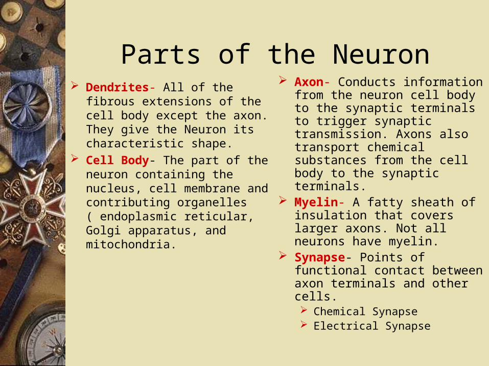
Parts of the Neuron Dendrites- All of the fibrous
extensions of the cell body except the axon. They give the Neuron its characteristic shape.
Cell Body- The part of the neuron containing the nucleus, cell membrane and contributing organelles ( endoplasmic reticular, Golgi apparatus, and mitochondria.
Axon- Conducts information from the neuron cell body to the synaptic terminals to trigger synaptic transmission. Axons also transport chemical substances from the cell body to the synaptic terminals.
Myelin- A fatty sheath of insulation that covers larger axons. Not all neurons have myelin.
Synapse- Points of functional contact between axon terminals and other cells. Chemical Synapse Electrical Synapse

The Neuron
http://faculty.washington.edu/chudler/synapse.html

Nerve ConductionWithin the Neuron Depolarization- An electrical
process -70 mill volts When the neuron is resting, not
conducting nerve impulses, it is polarized.
The cell is invaded by Na ions and the expulsion of K ions. The electric charge is gone. Depolarization occurs!
Between Neurons A chemical process When depolarization reaches
the terminal buttons, neurotransmitters are released into the synapse.
Either excite or inhibit the following neuron!
Sending neuron-presynaptic neuron
Receiving neuron- postsynaptic neuron.

Parts of the Brain:Frontal Lobe (Forebrain)
Location: serendip.brynmawr.edu/bb/kinser/

Four Types of Messengers:1. Neurotransmitters: released by terminal buttons of
neurons and detected by receptors in the membrane of another cell a short distance away.
2. Neuromodulators: released in large amounts from the terminal buttons, but diffused throughout part of the brain, affecting many neurons
3. Hormones: produced by endocrine glands, released into extracellular fluid - stimulate cell receptors on membrane surface or deep within nuclei of cells, including neurons
4. Pheromones: chemicals released into the environment through sweat, urine, or secretions of special glands. Most receptors in nose of other animals, but may also be detected in skin or other organs

Frontal Lobe Location- In the anterior most part of the brain (under
the forehead) Function:
Determines our consciousness of our environment. Determines how we initiate and respond to our environment. Daily decisions in our daily lives. Controls emotional responses and expressive language. Assigns meanings to the words we use. Involves word association. Controls memory for habits and motor activities. Emotional control center. Links:
http://www.waiting.com/brainfunction.html http://www.radiology.wisc.edu/Med_Students/neuroradiology/
fmri/sld012.htm

Parietal Lobe
Location- Near the back and top of the head (Near the back and top of the head)
Functions: Contains the location for visual attention. Contains the location for touch perception. Controls goal directed voluntary movements. Controls the manipulation of objects. Integrates different senses to allow for understanding a single
concept. If not functioning correctly epileptic behavior can occur. Links:
http://www.bcm.tmc.edu/neurol/challeng/pat31/summary.html
http://www.eqi.org.au/newsletter/glossary.html

Illustration of the Occipital Lobe
Link: Traumatic Brain Injury Research http://www.neuroskills.com/index.html?main=tbi/boccipit.shtml

Occipital Lobe
Location- Located in the most posterior (Back of the head).
Functions: Center of the visual perception center. Contains the primary visual cortex. Receives projections from the lateral geniculate nucleus of the
thalamus. Numerous visual functions. Links:
http://www.geocities.com/CapeCanaveral/Launchpad/3937/sight.htm
http://www.ruf.rice.edu/~lngbrain/cglidden/occipital.html http://www.headinjuryrehab.org/occipital_lobe.htm

Temporal Lobe
Location- at the side of the head and above the ears Functions:
Auditory sensation and perception Organization and categorization of verbal material Long term memory Personality and sexual behavior Organization of sensory input The brain has two temporal lobes, one on each side of the brain The two are interchangeable, so if one is damaged, the other is
usually able to takeover the other's duties Link:
http://www.Geocities.Com/CapeCanaveral/Launchpad/3937/temporal.Htm

Biological Foundations of PsychologyDivisions of the Brain
www.psychol.uni-giessen.De/abteil/differen/ 02abiol_skript_version.ppt

The Brain Stem(Medulla oblongata, the Midbrain, and the Pons.) Medulla Oblongata Location:
Last part of the brain before reaching the spinal cord. Continuation of the spinal cord
Function: Many cranial nerves enter and leave the brain through the Medulla. Centers for cough, gag, swallow, and vomit. Cardiac Center. Respiratory Center. Links:
http://www.waiting.com/brainfuncthree.htmlhttp://www.neuroskills.com/index.html?main=tbi/bbstem.sht
mlhttp://kidshealth.org/kid/body/brain_noSW_p4.html

Illustration of the Medulla Oblongata
http://www.brainexplorer.org/brain_atlas/Brainatlas_Midbrain.shtml
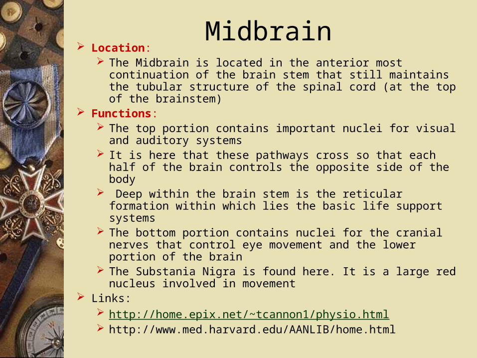
Midbrain Location:
The Midbrain is located in the anterior most continuation of the brain stem that still maintains the tubular structure of the spinal cord (at the top of the brainstem)
Functions: The top portion contains important nuclei for visual and
auditory systems It is here that these pathways cross so that each half of the brain
controls the opposite side of the body Deep within the brain stem is the reticular formation within
which lies the basic life support systems The bottom portion contains nuclei for the cranial nerves that
control eye movement and the lower portion of the brain The Substania Nigra is found here. It is a large red nucleus
involved in movement Links:
http://home.epix.net/~tcannon1/physio.html http://www.med.harvard.edu/AANLIB/home.html

Pons Location:
The Pons (meaning "bridge") lies above the medulla, and is so named because many axons cross sides within this region of the hindbrain
Functions: Arousal Assists in Controlling Autonomic Functions Relays Sensory Information Between the Cerebrum and
Cerebellum Sleep
Features of the pons are: a) basis pontis, b) middle cerebellar peduncle, and c) the superior cerebellar peduncle
All are linked to the cerebellum which sits on the posterior side of the pons. Damage to any of the structures would result in impaired coordination of movement and/or posture
Links:http://psych.athabascau.ca/html/Psych402/Biotutorials/pons www.bigchalk.com

Cerebellum Location:
Two peach-size mounds of folded tissue at the base of the brain Overlies the pons
Functions: The cerebellum ("little brain") has convolutions similar to those
of cerebral cortex, only the folds are much smaller. Like the cerebrum, the cerebellum has an outer cortex, an inner white matter, and deep nuclei below the white matter
New skills are learned by trial and error and then coded into the cerebellar memory
Coordinates movement of muscles and joints by synthesizing data from the brain stem, the spinal cord, and another brain areas such as cerebral cortex
The cerebellum fine tunes our motor activity or movement Links:
http://web.sfn.org/content/Publications/BrainBackgrounders/cerebellum.htm
http://thalamus.wustl.edu/course/cerebell.html

Thalamus Location:
The Thalamus is shaped like two footballs; each is located deep in the hemispheres of the forebrain
A large mass of gray matter deeply situated in the forebrain. There is one on either side of the midline
Functions: It relays to the cerebral cortex information received from diverse
brain regions. Sort of a requisite 'last pit stop' for information going to cortex
Axons from every sensory system (except olfaction) synapse here as the last relay site before the information reaches the cerebral cortex
Information from all sensory receptors except smell is processed in the thalamus before being sent to the cerebral cortex
Links: http://psych.athabascau.ca/html/Psych402/Biotutorials/20/
thalamus.shtml http://www.pubs.royalsoc.ac.uk/phil_bio/news/thalamus.html

Hypothalamus Location:
The hypothalamus is a midline, structure, shaped like a funnel below the thalamus
It connects to the pituitary gland Functions:
The hypothalamus has many regulating functions The autonomic nervous system, emotions and behavior, body
temperature, hunger, thirst, sleep-waking cycles Controls the release of hormones under its control: growth,
prolactin, thyroid, corticotropin, and gonadotropins Regulation of sex hormones, blood pressure, body temperature,
water balance, respiration, and food intake, while it also plays a role in regulating complex moods, such as anger, placidity, and fatigue.
Links: http://k-2.stanford.edu/InfoPackets/EndoSys.3.0.html http://www.isat.jmu.edu/users/klevicca/neuroconn/The_Brain/
Brain_Function/Limbic_System/hypothalamus.html

Hippocampus Location:
The Hippocampus is tucked out of sight on the medial side of the temporal lobe
Its shape resembles that of a 'seahorse' Functions:
Stores and processes memories Helps find memories Affects emotions The hippocampus helps to encode memories, and then helps to
find them when you want to remember something Main relay station that determines whether a new memory should
go into long-term storage or be deleted after its short-term usefulness is over
Links: http://www.morphonix.com/software/education/science/brain/
game/specimens/hippocampus.html

Illustration of Hippocampus

Basal Ganglia Location- The basal ganglia surrounds the thalamus and is
enclosed by the cerebral cortex and cerebral white matter. The name includes: caudate, putamen, nucleus accumbens, globus
pallidus, substantia nigra, subthalamic nucleus
Functions: Controls voluntary movements and establishing postures. Controls voluntary limb movement, eye movement, and cognition. Lesions in specific nuclei tend to produce characteristic deficits. One
well-known disorder is Parkinson's disease, which is the slow and steady loss of dopaminergic neurons in synapses.
Links: http://thalamus.wustl.edu/course/cerebell.html http://www-hbp.usc.edu/Projects/basal.htm http://www.sci.uidaho.edu/med532/basal.htm

Conclusion
The study of physiology has made possible for better understanding of human behavior and function, as well as, the function and behavior of other species we share our world with.