primary testicular necrotizing vasculitis clinically presented as
TRANSCRIPT
CASE REPORT Open Access
Primary testicular necrotizing vasculitis clinicallypresented as neoplasm of the testicle: a casereportAnton Maričić1, Sanja Štifter2, Maksim Valenčić1, Gordana Ðorđević2, Dean Markić1*, Josip Španjol1,Stanislav Sotošek1 and Željko Fučkar1
Abstract
We present a case of necrotizing vasculitis with the testicle as the isolated affected organ. A 25-year-old man,pretreated for epididymo-orchitis, presented with a presumed testicular neoplasm. Radical orchiectomy wasperformed and diagnosis of necrotizing vasculitis was established. In the absence of any other sign of systemicdisease, the diagnosis of isolated necrotizing vasculitis of the testis was confirmed. Two years after the operation,the patient showed no symptoms of systemic disease.
Keywords: Necrotizing vasculitis, testicular neoplasm, radical orchiectomy, ultrasound
BackgroundSymptomatic vasculitis confined to the testis withoutclinical or laboratory evidence of systemic disease is nota common finding [1-10]. It is difficult to diagnose thiscondition clinically or using noninvasive methods. Ther-apy for this condition remains controversial. Wedescribe a case with an unusual presentation simulatinga testicular neoplasm.
Case presentationA 25-year-old Caucasian man went to a general practi-tioner because of right testicular swelling and was trea-ted with oral antibiotics for presumed epididymo-orchitis. Over the next 10 days, swelling increased, thetestis became painful, body temperature increased to 38°C, and the patient was referred for urological assess-ment. The patient was admitted to the hospital for par-enteral therapy, because peroral antibiotic therapy(ciprofloxacin) was not effective.Upon physical examination, the right testicle was
enlarged and painful on palpation, and the skin of theright hemiscrotal region was red and warm. Painincreased gradually and worsened slightly with time, butthis type of pain was not typical of the presumed
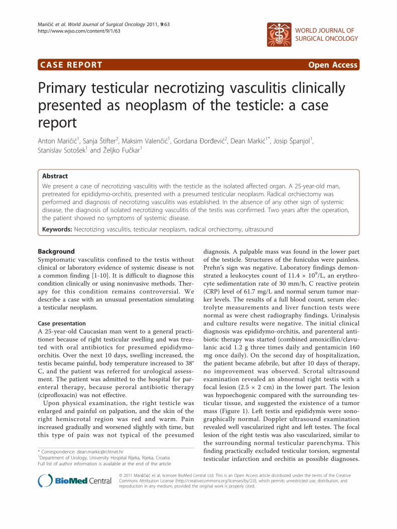
diagnosis. A palpable mass was found in the lower partof the testicle. Structures of the funiculus were painless.Prehn’s sign was negative. Laboratory findings demon-strated a leukocytes count of 11.4 × 10⁹/L, an erythro-cyte sedimentation rate of 30 mm/h, C reactive protein(CRP) level of 61.7 mg/L and normal serum tumor mar-ker levels. The results of a full blood count, serum elec-trolyte measurements and liver function tests werenormal as were chest radiography findings. Urinalysisand culture results were negative. The initial clinicaldiagnosis was epididymo-orchitis, and parenteral anti-biotic therapy was started (combined amoxicillin/clavu-lanic acid 1.2 g three times daily and gentamicin 160mg once daily). On the second day of hospitalization,the patient became afebrile, but after 10 days of therapy,no improvement was observed. Scrotal ultrasoundexamination revealed an abnormal right testis with afocal lesion (2.5 × 2 cm) in the lower part. The lesionwas hypoechogenic compared with the surrounding tes-ticular tissue, and suggested the existence of a tumormass (Figure 1). Left testis and epididymis were sono-graphically normal. Doppler ultrasound examinationrevealed well vascularized right and left testes. The focallesion of the right testis was also vascularized, similar tothe surrounding normal testicular parenchyma. Thisfinding practically excluded testicular torsion, segmentaltesticular infarction and orchitis as possible diagnoses.
* Correspondence: [email protected] of Urology, University Hospital Rijeka, Rijeka, CroatiaFull list of author information is available at the end of the article
Maričić et al. World Journal of Surgical Oncology 2011, 9:63http://www.wjso.com/content/9/1/63 WORLD JOURNAL OF
SURGICAL ONCOLOGY
© 2011 Mariččićć et al; licensee BioMed Central Ltd. This is an Open Access article distributed under the terms of the CreativeCommons Attribution License (http://creativecommons.org/licenses/by/2.0), which permits unrestricted use, distribution, andreproduction in any medium, provided the original work is properly cited.
Because ultrasound findings of the right testicle werehighly indicative of testicular neoplasm, right radicalorchiectomy was performed via an inguinal incision.Histopathological findingsThe testicle measured 4 × 3.5 × 2.5 cm and containedwell-demarcated areas of hemorrhage, 3 cm in diameter.The epididymis, investing membranes and spermaticcord appeared grossly normal. Microscopy showed thepresence of a patchy, necrotizing vasculitis affectingmedium-sized and small-sized arteries of the testicle(Figure 2). Several vessels showed fibrinoid necrosis oftheir walls and muscular layer detachment with or with-out a transmural infiltrate composed of polymorphonuc-lear leukocytes and lymphocytes. Immunofluorescencestaining for fibrin was also performed, and positivefibrin deposits were identified in arterial walls affectedwith fibrinoid necrosis (Figure 3).Follow-upThe postoperative course was uneventful. After theoperation, extensive clinical evaluation was performed toexclude other systemic diseases characterized by vasculi-tis. This included: complete blood count; erythrocytesedimentation rate; CRP; urinalysis; immunoglobulinserum level; immunological blood tests, such as rheuma-toid factor, antinuclear antibody tests and anti-neutro-phil cytoplasmic autoantibody test; complement tests;human leukocyte antigen tissue typing tests; ultrasoundof the abdomen; endoscopic examination of the ear,nose and throat; chest X-ray; and ophthalmologist
examination. All test results were normal. There was nosign of systemic disease. Two years after the diagnosis,systemic disease had not developed.
DiscussionThe most common appearance of testicular vasculitis isas part of a multiorgan or systemic disease. Involvementof the testicles is seen less frequently in Wegener’s gran-ulomatosis, Henoch-Schönlein purpura, giant cell arteri-tis, and rheumatoid arthritis, whereas testicleinvolvement is commonly associated with polyarteritisnodosa [3-5]. The microscopically observed changes arealmost identical in all vasculitis seen in other systemicdisorders. The results of postmortem studies suggestthat the testis is involved in 38-86% of cases of polyar-teritis nodosa. At the same time, less than 18% of thesecases are symptomatic, and most will show other mani-festations of polyarteritis nodosa [6,7].Isolated testicular vasculitis is not a common condi-
tion [10-13]. It is usually found in young people, as inour patient [12]. From the present literature findings, itremains unclear whether such cases represent truly iso-lated vasculitis or solely an unusual primary presenta-tion site. The pathogenesis of isolated organ vasculitis isunknown, as is why only one organ may be affected.Additionally, it is unknown whether such cases carry therisk of subsequent progression, and if so, the riskremains to be determined. It is not known whether iso-lated vasculitis has a better prognosis than does systemic
Figure 1 Ultrasound: hypoechogenic focal lesion in the lower pole of right testicle.
Maričić et al. World Journal of Surgical Oncology 2011, 9:63http://www.wjso.com/content/9/1/63
Page 2 of 5
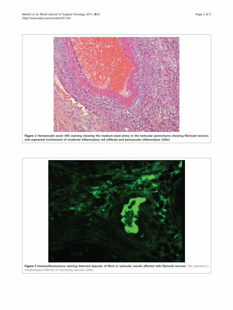
Figure 3 Immunofluorescence staining detected deposits of fibrin in testicular vessels affected with fibrinoid necrosis. This represents amorphological hallmark of necrotizing vasculitis (200x).
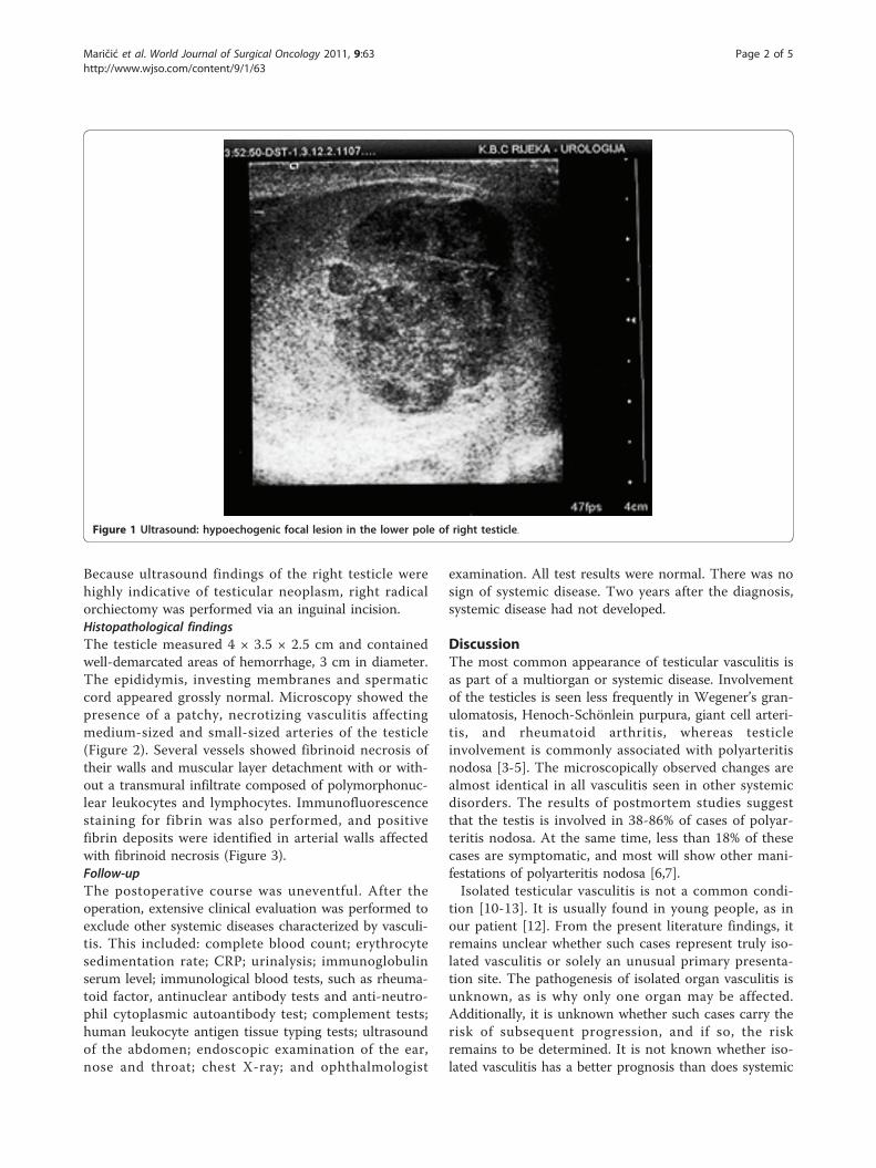
Figure 2 Hematoxylin eosin (HE) staining showing the medium-sized artery in the testicular parenchyma showing fibrinoid necrosisand segmental involvement of moderate inflammatory cell infiltrate and perivascular inflammation (200x).
Maričić et al. World Journal of Surgical Oncology 2011, 9:63http://www.wjso.com/content/9/1/63
Page 3 of 5
disease. This is important because the classic form ofpolyarteritis nodosa carries significant risks of mortalityand morbidity, even with treatment, and has a high rateof relapse [9].The conditions presenting as pain in the testicle or epi-
didymitis have been previously reported, but presentationwith clinical features suggestive of testicular neoplasm iseven more exceptional [7,10]. In the majority of reportedcases, clinical or laboratory evidence of disease in otherorgan systems on presentation was present or developedsubsequently within a short time period [5].Testicular necrotizing vasculitis is impossible to diag-
nose without tissue analysis. Our patient first presentedwith symptoms and signs in favor of inflammation(orchitis as concomitant disease cannot be excluded).However, when ultrasound with Doppler was performed,it was obvious that inflammation was not the cause ofthis lesion. Additionally, because the lesion was well vas-cularized, testicular torsion and segmental testicularinfarction were excluded. Testicular neoplasm remainedthe most probable diagnosis. After orchiectomy, histo-pathological findings were used to investigate the exis-tence of necrotizing vasculitis. Histopathologicalcharacteristics observed in necrotizing vasculitis aremainly restricted to blood vessels. Fibrinoid necrosis isthe morphological hallmark of the disease. The walls ofsmall and medium-sized testicular arteries are affected,as shown in this case report. Notably, hemorrhagicnecrosis occurs in other pathological conditions, such astesticular torsion, infarction and inflammation.Serological markers such as CRP and von Willebrand
factor are possible indicators of endothelial injury in sys-temic vasculitis but may not reflect the activity in iso-lated organ disease. Ultrasound examination may fail toshow any abnormality but can also demonstrate theexistence of a hypoechogenic mass, as in our patient[12]. Magnetic resonance imaging is a more sensitivetechnique that can demonstrate focal testicular infarc-tion, but, at present, the only “diagnostic tool” for vascu-litis is histological confirmation.To treat our patient with potentially toxic immunosup-
pressive therapy with the added risk of sterility, despitethe lack of clinical and objective laboratory evidence ofsystemic disease, presents a difficult clinical dilemma.Waterfield et al. reported on isolated testicular vasculitistreated by immunosuppressive medications. Despite ther-apy, the remaining testis became affected one year later[10]. That patient responded well to an increase inimmunosuppressive therapy. McGuirre et al. recom-mended close surveillance without additional therapy[12]. Because of his young age, we elected to performclose follow-up of our patient, instead of immunosup-pressive therapy. Two years after diagnosis, the patient isstill without symptoms of systemic disease. This is the
longest asymptomatic period in a case of testicular vascu-litis reported in the literature. In view of the high relapserate associated with polyarteritis nodosa, long-term fol-low up for these patients is essential. However, theabsence of serological markers of disease activity makesmonitoring of any future relapse quite difficult.
ConclusionPrimary testicular manifestation of necrotizing vasculitisis not a common finding. It is very important for pathol-ogists and clinicians to know that such an entity caninitially present as a testicular mass. Follow-up of thesepatients is recommended due to the risk of relapse;however, due to the rarity of the condition, the appro-priate strategies for treatment and follow-up remain tobe determined.
ConsentWritten informed consent was obtained from the patientfor publication of this Case report and any accompanyingimages. A copy of the written consent is available forreview by the Editor-in-Chief of this journal.
AbbreviationsCRP: C reactive protein.
AcknowledgementsNone
Author details1Department of Urology, University Hospital Rijeka, Rijeka, Croatia.2Department of Pathology, School of Medicine, University of Rijeka, Rijeka,Croatia.
Authors’ contributionsMA, SS and MD tracked the clinical data and drafted the manuscript. FŽ, VMand ŠJ participated in the design of the study and coordination and helpedto draft the manuscript. GÐ and SŠ provided the pathological technique.All authors read and approved the final manuscript.
Competing interestsThe authors declare that they have no competing interests.
Received: 10 March 2011 Accepted: 14 June 2011Published: 14 June 2011
References1. Raj GV, Ellington KS, Polascik TJ: Autoimmune testicular vasculitis. Urology
2003, 61:1035.2. Susanto CR, Fedder G, Looijen-Salamon MG: Acute, painful, and swollen
testicle as the presenting feature in polyarteritis nodosa. Eur J Intern Med2003, 14:441-443.
3. Huisman TK, Collins WT Jr, Voulgarakis GR: Polyarteritis nodosamasquerading as a primary testicular neoplasm; a case report andreview of the literature. J Urol 1990, 144:1236-1238.
4. Belville WD, Insalaco SJ, Dresner ML, Buck AS: Benign testis tumors. J Urol1982, 128:1198-1200.
5. Lee LM, Moloney PJ, Wong HC, Magil AB, McLoughlin MG: Testicular pain:an unusual presentation of polyarteritis nodosa. J Urol 1983,129:1243-1244.
6. Gondos B, Wong TW: Non-neoplastic diseases of the testis andepididymis. In Urological pathology. Edited by: Murphy W. Philadelphia. WBSaunders; 1989:249-313.
Maričić et al. World Journal of Surgical Oncology 2011, 9:63http://www.wjso.com/content/9/1/63
Page 4 of 5
7. Shurbaji MS, Epstein JI: Testicular vasculitis: implications for systemicdisease. Hum Pathol 1988, 19:186-189.
8. Womack C, Ansell ID: Isolated arteritis of the epididymis. J Clin Pathol1985, 38:797-800.
9. Gordon M, Luqmani RA, Adu D, Greaves I, Richards N, Michael J, Emery P,Howie AJ, Bacon PA: Relapses in patients with a systemic vasculitis. QJMed 1993, 86:779-789.
10. Warfield AT, Lee SJ, Phillips SM, Pall AA: Isolated testicular vasculitismimicking a testicular neoplasm. J Clin Pathol 1994, 47:1121-1123.
11. Atis G, Memis OF, Güngör HS, Arikan O, Saglican Y, Caskurlu T: Testicularpolyarteritis nodosa mimicking testicular neoplasm. ScientificWorldJournal2010, 10:1915-1918.
12. McGuire BB, O’Brien MF, Akhtar M, Fitzpatrick JM: Testicular vasculitismimicking a testicular neoplasm. Ir Med J 2006, 99:27-28.
13. Joudi FN, Austin JC, Vogelgesang SA, Jensen CS: Isolated testicularvasculitis presenting as a tumor-like lesion. J Urol 2004, 171:799.
doi:10.1186/1477-7819-9-63Cite this article as: Maričić et al.: Primary testicular necrotizing vasculitisclinically presented as neoplasm of the testicle: a case report. WorldJournal of Surgical Oncology 2011 9:63.
Submit your next manuscript to BioMed Centraland take full advantage of:
• Convenient online submission
• Thorough peer review
• No space constraints or color figure charges
• Immediate publication on acceptance
• Inclusion in PubMed, CAS, Scopus and Google Scholar
• Research which is freely available for redistribution
Submit your manuscript at www.biomedcentral.com/submit
Maričić et al. World Journal of Surgical Oncology 2011, 9:63http://www.wjso.com/content/9/1/63
Page 5 of 5