classification of vasculitis - columbia · pdf filevasculitis 6 polyarteritis nodosa...
TRANSCRIPT
Vasculitis 1
VasculitisEdward Dwyer, M.D.
Division of Rheumatology
VASCULITIS is a primary inflammatory disease process of the vasculature
Determinants of the Clinical Manifestations of Vasculitis:
Target organ involved
Size of vessel involved
Pathobiology of the inflammatory process of involved vasculature
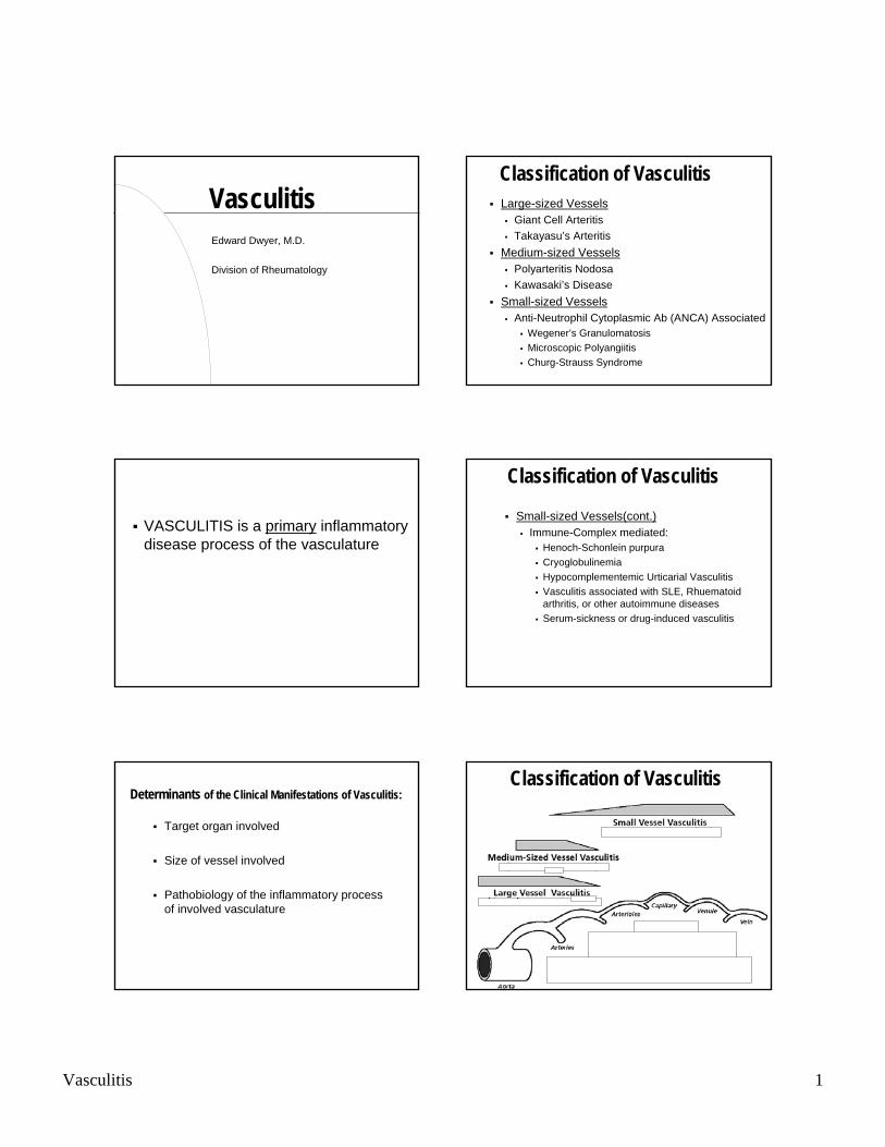
Classification of VasculitisLarge-sized Vessels
Giant Cell ArteritisTakayasu’s Arteritis
Medium-sized VesselsPolyarteritis NodosaKawasaki’s Disease
Small-sized VesselsAnti-Neutrophil Cytoplasmic Ab (ANCA) Associated
Wegener’s GranulomatosisMicroscopic PolyangiitisChurg-Strauss Syndrome
Classification of Vasculitis
Small-sized Vessels(cont.)Immune-Complex mediated:
Henoch-Schonlein purpuraCryoglobulinemiaHypocomplementemic Urticarial VasculitisVasculitis associated with SLE, Rhuematoid arthritis, or other autoimmune diseasesSerum-sickness or drug-induced vasculitis
Classification of Vasculitis
Vasculitis 2
Sequelae of Vasculitis
Stenosis and/or occlusion of involved vasculature resulting in organ ischemia or infarction
Necrosis of vessel walls resulting in aneursymal dilatation and/or thrombosis causing organ ischemia, infarction, or hemorrhage.
Diagnostic Approaches
Biopsy of involved organs
Radiographic evaluation of involved vesselsConventional AngiographyCT AngiographyMR Angiography
Serology (e.g., autoantibodies)
Giant Cell Arteritis(Temporal Arteritis)
Non-necrotizing vasculitis resulting intimalproliferation causing luminal stenosis or occlusion
Epidemiology of Giant Cell Arteritis
Age: > 50 years-old
Racial/Ethnic Background (annual Incidence)20/100,000 Northern European2/100,000 African Americans and Hispanics<1/1,000,000 Asians
Vasculature involvedThoracic aorta and major branches:
Carotid artery extra-cranial branchesTemporal arteryOccipital arteryOphthalmic arteryPosterior ciliary artery
Subclavian/axillary artery
Vasculitis 3
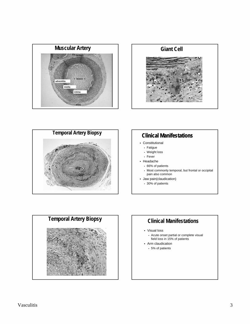
Muscular Artery
adventitia
media
intima
Temporal Artery Biopsy
Temporal Artery Biopsy
Giant Cell
Clinical ManifestationsConstitutional
FatigueWeight lossFever
Headache66% of patientsMost commonly temporal, but frontal or occipital pain also common
Jaw pain(claudication)30% of patients
Clinical Manifestations
Clinical ManifestationsVisual loss
Acute onset partial or complete visual field loss in 15% of patients
Arm claudication5% of patients

Vasculitis 4
Laboratory Abnormalities
Elevated Acute Phase ReactantsErythrocyte sedimentation rate (ESR)C-reactive proteinElevated IL-6 levels
Giant Cell Arteritis Pathogenesis
Weyand, C. M. et al. N Engl J Med 2003;349:160-169
Weyand, C. M. et al. N Engl J Med 2003;349:160-169
Giant Cell Arteritis Pathogenesis
Weyand, C. M. et al. N Engl J Med 2003;349:160-169
Giant Cell Arteritis Pathogenesis
Weyand, C. M. et al. N Engl J Med 2003;349:160-169
Giant Cell Arteritis Pathogenesis
Vasculitis 5
Optic Nerve Ischemia
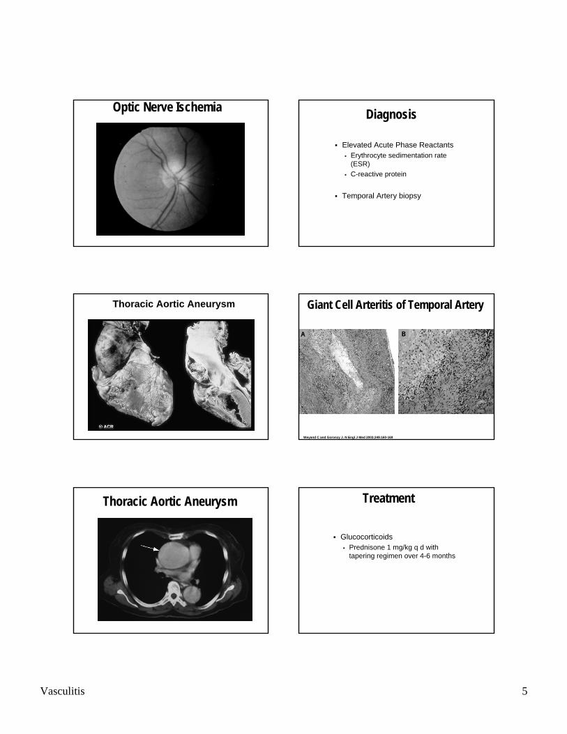
Thoracic Aortic Aneurysm
Thoracic Aortic Aneurysm
Diagnosis
Elevated Acute Phase ReactantsErythrocyte sedimentation rate (ESR)C-reactive protein
Temporal Artery biopsy
Giant Cell Arteritis of Temporal Artery
Weyand C and Goronzy J. N Engl J Med 2003;349:160-169
Treatment
GlucocorticoidsPrednisone 1 mg/kg q d with tapering regimen over 4-6 months
Vasculitis 6
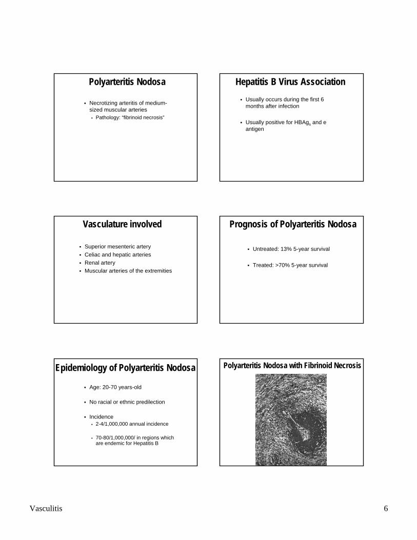
Polyarteritis Nodosa
Necrotizing arteritis of medium-sized muscular arteries
Pathology: “fibrinoid necrosis”
Vasculature involved
Superior mesenteric arteryCeliac and hepatic arteriesRenal arteryMuscular arteries of the extremities
Epidemiology of Polyarteritis Nodosa
Age: 20-70 years-old
No racial or ethnic predilection
Incidence2-4/1,000,000 annual incidence
70-80/1,000,000/ in regions which are endemic for Hepatitis B
Hepatitis B Virus AssociationUsually occurs during the first 6 months after infection
Usually positive for HBAgs and e antigen
Prognosis of Polyarteritis Nodosa
Untreated: 13% 5-year survival
Treated: >70% 5-year survival
Polyarteritis Nodosa with Fibrinoid Necrosis
Vasculitis 7
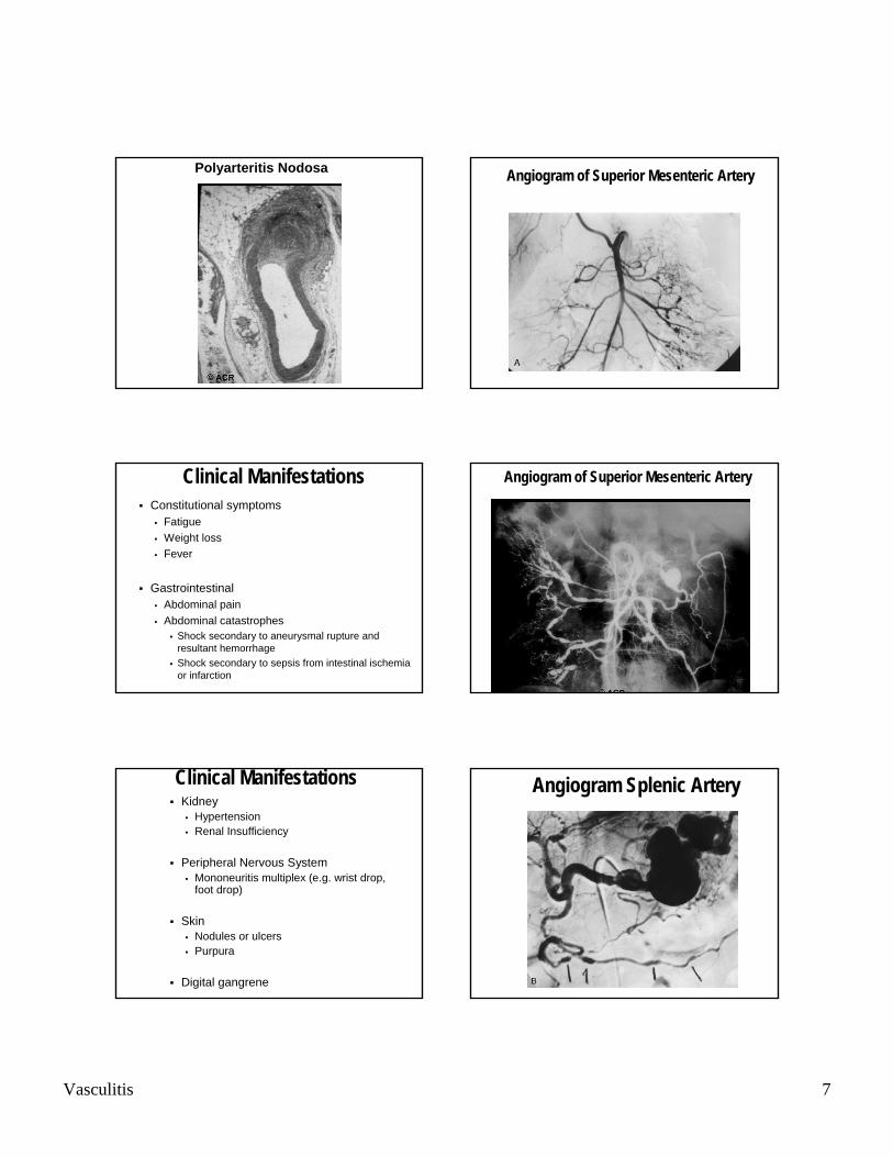
Polyarteritis Nodosa
Clinical ManifestationsConstitutional symptoms
FatigueWeight lossFever
GastrointestinalAbdominal pain Abdominal catastrophes
Shock secondary to aneurysmal rupture and resultant hemorrhageShock secondary to sepsis from intestinal ischemia or infarction
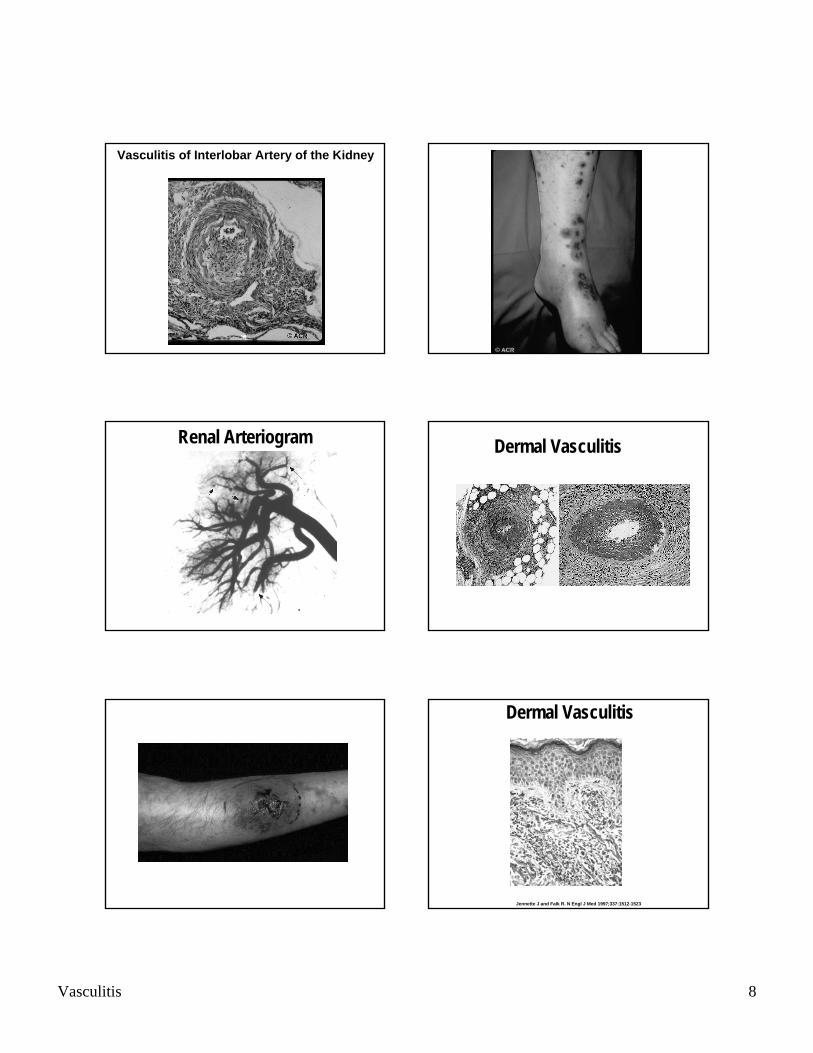
Clinical ManifestationsKidney
HypertensionRenal Insufficiency
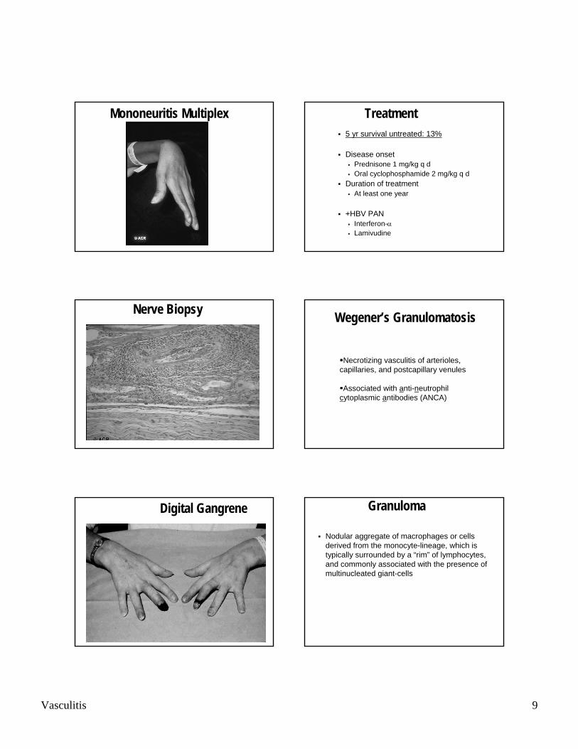
Peripheral Nervous SystemMononeuritis multiplex (e.g. wrist drop, foot drop)
SkinNodules or ulcersPurpura
Digital gangrene
Angiogram of Superior Mesenteric Artery
Angiogram of Superior Mesenteric Artery
Angiogram Splenic Artery
Vasculitis 8
Vasculitis of Interlobar Artery of the Kidney
Renal Arteriogram Dermal Vasculitis
Dermal Vasculitis
Jennette J and Falk R. N Engl J Med 1997;337:1512-1523
Vasculitis 9
Mononeuritis Multiplex
Nerve Biopsy
Digital Gangrene
Treatment5 yr survival untreated: 13%
Disease onsetPrednisone 1 mg/kg q dOral cyclophosphamide 2 mg/kg q d
Duration of treatmentAt least one year
+HBV PANInterferon-α Lamivudine
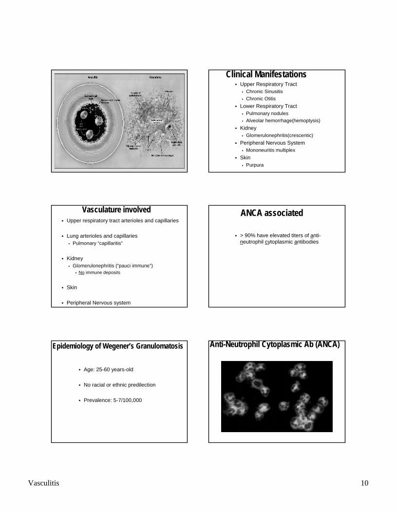
Wegener’s Granulomatosis
Necrotizing vasculitis of arterioles, capillaries, and postcapillary venules
Associated with anti-neutrophilcytoplasmic antibodies (ANCA)
Granuloma
Nodular aggregate of macrophages or cells derived from the monocyte-lineage, which is typically surrounded by a “rim” of lymphocytes, and commonly associated with the presence of multinucleated giant-cells
Vasculitis 10
Vasculature involvedUpper respiratory tract arterioles and capillaries
Lung arterioles and capillariesPulmonary “capillaritis”
KidneyGlomerulonephritis (“pauci immune”)
No immune deposits
Skin
Peripheral Nervous system
Epidemiology of Wegener’s Granulomatosis
Age: 25-60 years-old
No racial or ethnic predilection
Prevalence: 5-7/100,000
Clinical ManifestationsUpper Respiratory Tract
Chronic SinusitisChronic Otitis
Lower Respiratory TractPulmonary nodulesAlveolar hemorrhage(hemoptysis)
KidneyGlomerulonephritis(crescentic)
Peripheral Nervous SystemMononeuritis multiplex
SkinPurpura
ANCA associated
> 90% have elevated titers of anti-neutrophil cytoplasmic antibodies
Anti-Neutrophil Cytoplasmic Ab (ANCA)
Vasculitis 11
ANCA in Wegener’s Granulomatosis
Cytoplasmic reactivity (C-ANCA)Antigenic target = Proteinase 3
Serine proteinase of lysosomal granules of monocytes and azurophilic granules of neutrophils
Assay: Anti-proteinase 3 Ab titers (ELISA)
Morbidity of Wegener’s Granulomatosis
Permanent renal insufficiency- 42%End-stage renal disease- 11%Hearing loss- 35%Nasal deformities- 28%Tracheal stenosis- 13%
Mortality of Wegener’s Granulomatosis
Untreated: 10% survival at 2 years
Treated: 80% survival at 10 years
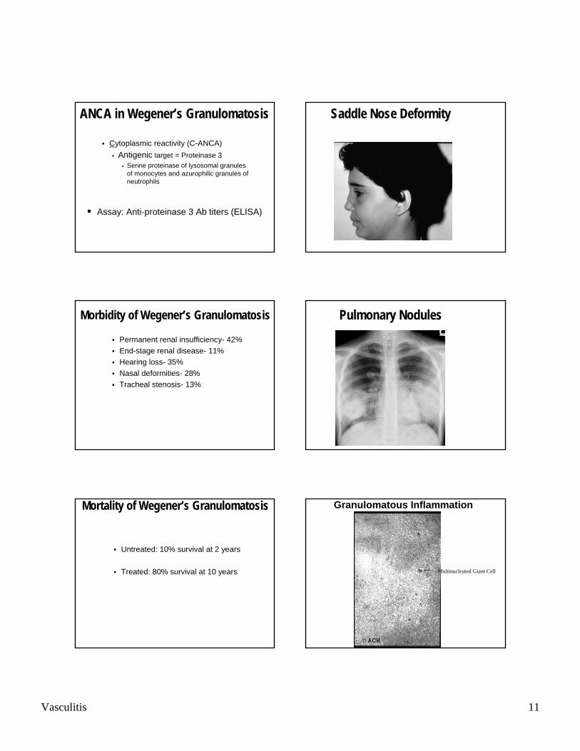
Saddle Nose Deformity
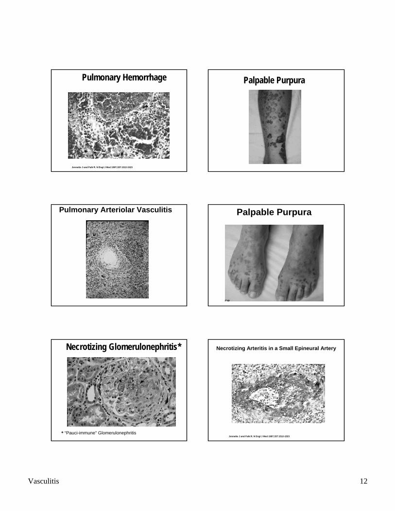
Pulmonary Nodules
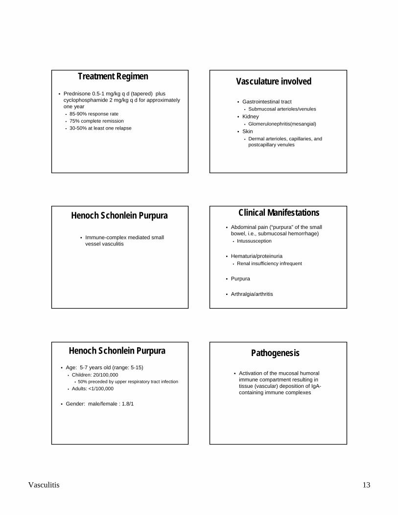
Granulomatous Inflammation
Multinucleated Giant Cell
Vasculitis 12
Pulmonary Hemorrhage
Jennette J and Falk R. N Engl J Med 1997;337:1512-1523
Pulmonary Arteriolar Vasculitis
Necrotizing Glomerulonephritis*
* “Pauci-immune” Glomerulonephritis
Palpable Purpura
Palpable Purpura
Jennette J and Falk R. N Engl J Med 1997;337:1512-1523
Necrotizing Arteritis in a Small Epineural Artery
Vasculitis 13
Treatment RegimenPrednisone 0.5-1 mg/kg q d (tapered) plus cyclophosphamide 2 mg/kg q d for approximately one year
85-90% response rate75% complete remission30-50% at least one relapse
Henoch Schonlein Purpura
Immune-complex mediated small vessel vasculitis
Henoch Schonlein PurpuraAge: 5-7 years old (range: 5-15)
Children: 20/100,00050% preceded by upper respiratory tract infection
Adults: <1/100,000
Gender: male/female : 1.8/1
Vasculature involved
Gastrointestinal tractSubmucosal arterioles/venules
KidneyGlomerulonephritis(mesangial)
SkinDermal arterioles, capillaries, and postcapillary venules
Clinical ManifestationsAbdominal pain (“purpura” of the small bowel, i.e., submucosal hemorrhage)
Intussusception
Hematuria/proteinuriaRenal insufficiency infrequent
Purpura
Arthralgia/arthritis
Pathogenesis
Activation of the mucosal humoralimmune compartment resulting in tissue (vascular) deposition of IgA-containing immune complexes

Vasculitis 14
Purpura of the Buttocks
Small Vessel Dermal Vasculitis
IgA Deposition in Dermal Vasculature
HSP Glomerulonephritis
IgA Deposition in the Mesangium
Prognosis of Henoch Schonlein Purpura
90-95% of patients exhibit spontaneous remission after 3-4 weeks, with 20-30% experiencing short-term relapses within the following 6-12 months
Vasculitis 15
Treatment
SupportiveHydrationBed restAnalgesia
Non-steroidal antiinflammatoryagents
VasculitisEdward Dwyer, M.D.
Division of Rheumatology