fatal polyarteritis presenting as gangrene*ard.bmj.com/content/annrheumdis/24/6/549.full.pdf ·...
TRANSCRIPT
Ann. rheum. Dis. (1965), 24, 549.
FATAL POLYARTERITIS PRESENTING ASASYMMETRICAL PERIPHERAL GANGRENE*
BY
M. K. KEECH AND H. E. PUROFronm the Departments of Medicine and Pathology, Wayne State University College of Medicine,
and the Detroit Receiving Hospital, Detroit, Michigan
Peripheral gangrene is one of the rarest presentingfeatures in polyarteritis nodosa. A man withparalysed right upper and left lower limbs followingpoliomyelitis in infancy was admitted to hospitalwith supraventricular tachycardia and peripheralgangrene in the non-involved extremities. In anattempt to relieve the severe foot pain following anexacerbation of the arteritis, intermittent epiduralinjections of xylocaine were given via an indwel-ling catheter. Autopsy revealed widespread vascularinvolvement with multiple infarctions of the viscera,these symptoms having been masked during life bysteroid therapy.
Case Report
A 39-year-old unmarried draughtsman was admitted inApril, 1963, during an attack of supraventricular tachy-cardia. Poliomyelitis at age 6 or 7 months had causedresidual paralysis of the right arm and left leg. 8 yearsbefore admission he had undergone surgical correctionof a deformity of the left foot. He had been admitted tohospital on three occasions for delirium tremens, hadoccasional bouts of epistaxis, and was subiect to depres-sion and suicidal tendencies. He had lost 22 lb. inweight during the past year. His mother had died oftuberculosis, his father was alive with hypertension, andan older brother had diabetes and "mental illness".The present illness began 10 weeks before admission
when he noticed stiffness of both knees followed byparaesthesiae in both legs and feet with difficulty in walkingdue to loss of sensation in the feet. This was accom-panied by loss of appetite, general weakness, and pain,tingling and numbness of the fingertips of the left hand.This loss of manual dexterity led to his discharge fromhis job because he could no longer perform his duties asa draughtsman. "Black and blue spots" developed 2weeks before admission on the toes of the right foot, oneweek before admission over the sacral area, and 3 days
* Supported by U.S. Public Health Service Training Grant TI AM5141, and the Michigan Chapter of the Arthritis and RheumatismFoundation.
before admission over the distal segments of the firstsecond, and third fingers of the left hand; 2 days beforeadmission the left testicle became tender and swollen.
Examination.-He was a thin, anxious, white male witha rapid pulse. An electrocardiogram showed supraventri-cular tachycardia of 180-190. This did not respond tocarotid massage, Valsalva's manoeuvre, gagging, eyeballpressure or vasoxyl, so he was admitted to the ward,given aramine, and slowly digitalized.
Physical Examination.-Blood pressure 122/80; tem-perature 1010 F.; lungs clear.No cardiomegaly, murmurs, or friction rubs were
found. The right testicle measured I x 2 cm. and nottender, and the left 4 x 4 cm. and very tender.
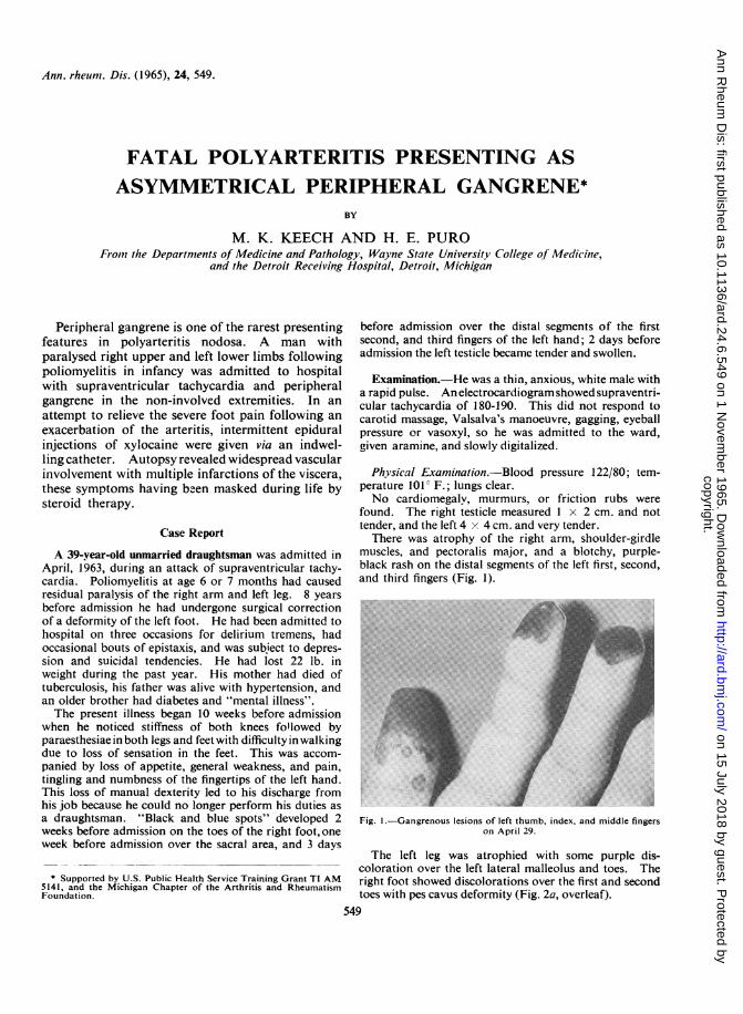
There was atrophy of the right arm, shoulder-girdlemuscles, and pectoralis major, and a blotchy, purple-black rash on the distal segments of the left first, second,and third fingers (Fig. 1).
r ... ..w ...Fig. I.-Gangrenous lesions of left thumb, index, and middle fingers
on April 29.
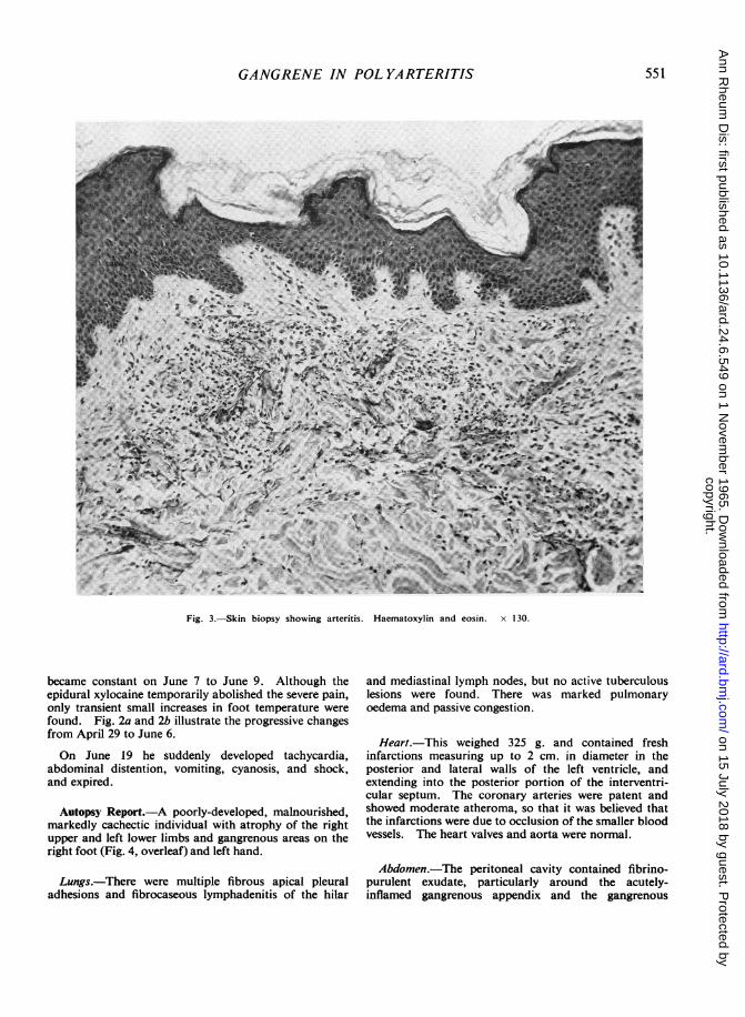
The left leg was atrophied with some purple dis-coloration over the left lateral malleolus and toes. Theright foot showed discolorations over the first and secondtoes with pes cavus deformity (Fig. 2a, overleaf).
549
copyright. on 15 July 2018 by guest. P
rotected byhttp://ard.bm
j.com/
Ann R
heum D
is: first published as 10.1136/ard.24.6.549 on 1 Novem
ber 1965. Dow
nloaded from

ANNALS OF THE RHEUMATIC DISEASES
Fig. 2 (a).-Gangrenous lesions of right first and second toes on April 29. Note pes cavus deformity. (h).-Extensionof gangrene to mid-tarsal level by June 6.
Pedal pulses were palpable and full bilaterally. Therewas weakness of muscle power in the right arm and leftleg with loss of deep tendon reflexes. Vibration senseand touch were intact.
Laboratory InvestigationsBLOOD.-Haemoglobin 9 7 g.; white blood count
11,700; polymorphs 69 per cent.; lymphocytes 18 percent.; eosinophils 10 per cent.; monocytes 3 per cent.;platelets 659,000/ c.mm.; reticulocytes 2 * 3 per cent.;erythrocyte sedimentation rate 98 mm. (Westergren).Coombs, Kline, Kahn, cryoglobulins, FBS, SGOT,and serum creatinine all normal Five estimations BUNnormal. Three lupus erythematosus preparations nega-tive. The RA Hyland latex test was positive.
URINE.-Albuminuria, 12-18 white blood cells and10-12 red blood cells.
SERUM ELECTROPHORESIS.-Total protein 6 5 g. percent.; albumin 4-13; globulin a, = 0-10, OC2 - 0 31,= 0 62, y = 1 34.
ELECTROCARDIOGRAM showed anomalous excitation(Wolf-Parkinson-White).
RADIOLOGY.-Chest x-ray revealed diminished musclemass over right chest and osteoporosis of the right arm.Intravenous pyelogram and upper gastrointestinal tractseries both normal.
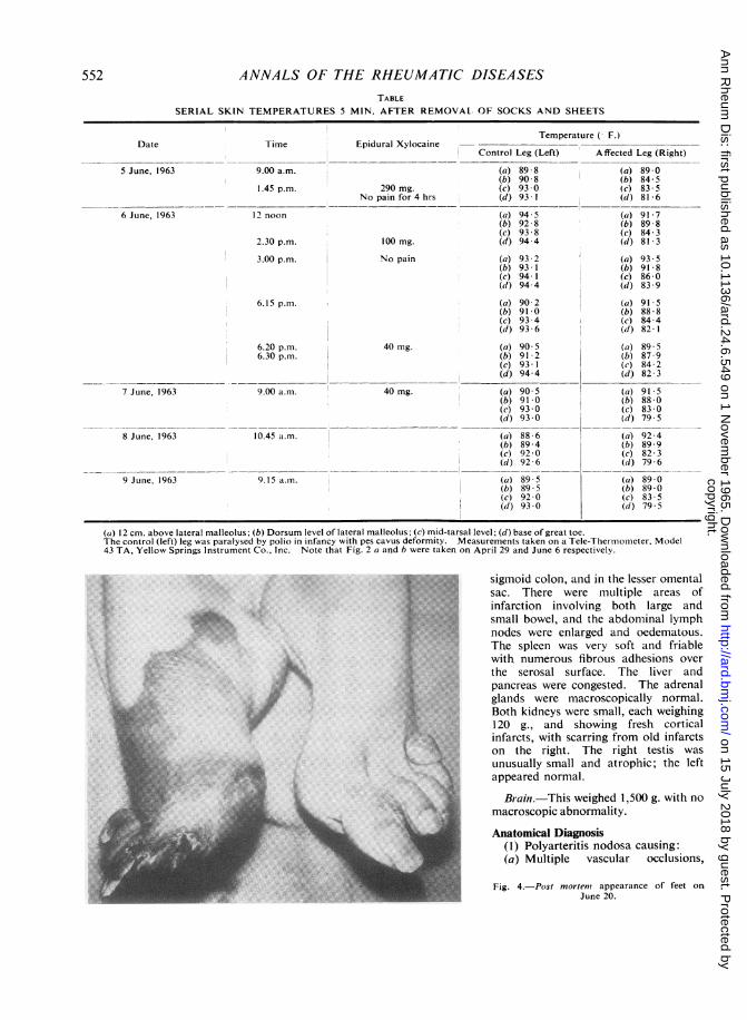
BIOPSIES.-Skin from the buttock showed a small,necrotic ulcer. Skin over left elbow contained narrowed
and obliterated arterioles with thickened, oedematouswalls infiltrated with large numbers of polymorpho-nuclear leucocytes and a few eosinophils and lympho-cytes. The specimen of gastrocnemius muscle did notcontain any large vessels, and the small vessels werenormal.
Histological Diagnosis.-Acute vasculitis, compatiblewith polyarteritis nodcsa (Fig. 3, opposite).
Course.-The patient was treated with prednisolone,50 mg. daily, ascorbic acid 300 mg. thiamine 300 mg.,priscoline 75 mg., and digoxin. The gangrenous areasremained dry and uninfected and began to separate.However, he had an exacerbation of the arteritis onJune 4 with the developmenit of further gangrenous lesionson the right foot (Fig. 2b) and left hand. Prednisolonewas increased to 80 mg. daily. Owing to the severe footpain, an epidural block with 1 per cent. xylocaine wastried on June 5. The pain was relieved for 4 hours sothe catheter was left in and the skin temperatures of bothlegs and feet recorded following further injections (Table,overleaf). The four readings on the control limb did notvary significantly. The first (lower leg) readings werealmost always the same on both sides. At ankle levelthe temperature was 2-6° lower on the affected side. Atmid-tarsal level the readings were 8-10O lower on the right.At the base of the great toe the temperature was 10-13°lower on the affected side on June 5 and 6, and 13° loweron June 7 and 9. The highest reading for the affected greattoe (83 9° F.) was taken half an hour after 100 mg.epidural xylocaine. The lowest reading (79.50 F.)
550
copyright. on 15 July 2018 by guest. P
rotected byhttp://ard.bm
j.com/
Ann R
heum D
is: first published as 10.1136/ard.24.6.549 on 1 Novem
ber 1965. Dow
nloaded from
GANGRENE IN POLYARTERITIS
~~~~ A . .~~~~~~~~~~~~~~~~~.
ftI~~~~~-AjP
Fig. 3.-Skin biopsy showing arteritis. Haematoxylin and eosin. x 130.
became constant on June 7 to June 9. Although theepidural xylocaine temporarily abolished the severe pain,only transient small increases in foot temperature werefound. Fig. 2a and 2b illustrate the progressive changesfrom April 29 to June 6.
On June 19 he suddenly developed tachycardia,abdominal distention, vomiting, cyanosis, and shock,and expired.
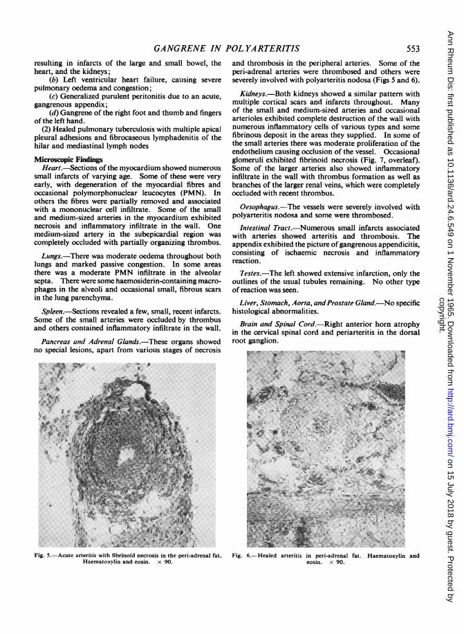
Autopsy Report.-A poorly-developed, malnourished,markedly cachectic individual with atrophy of the rightupper and left lower limbs and gangrenous areas on theright foot (Fig. 4, overleaf) and left hand.
Lungs.-There were multiple fibrous apical pleural
adhesions and fibrocaseous lymphadenitis of the hilar
and mediastinal lymph nodes, but no active tuberculouslesions were found. There was marked pulmonaryoedema and passive congestion.
Heart.-This weighed 325 g. and contained freshinfarctions measuring up to 2 cm. in diameter in theposterior and lateral walls of the left ventricle, andextending into the posterior portion of the interventri-cular septum. The coronary arteries were patent andshowed moderate atheroma, so that it was believed thatthe infarctions were due to occlusion of the smaller bloodvessels. The heart valves and aorta were normal.
Abdomen.-The peritoneal cavity contained fibrino-purulent exudate, particularly around the acutely-inflamed gangrenous appendix and the gangrenous
551
copyright. on 15 July 2018 by guest. P
rotected byhttp://ard.bm
j.com/
Ann R
heum D
is: first published as 10.1136/ard.24.6.549 on 1 Novem
ber 1965. Dow
nloaded from
ANNALS OF THE RHEUMATIC DISEASESTABLE
SERIAL SKIN TEMPERATURES 5 MIN. AFTER REMOVAL OF SOCKS AND SHEETS
Temperature (- F.)Date Time Epidural Xylocaine
Control Leg (Left) Affected Leg (Right)
5 June, 1963 9.00 a.m. (a) 89 8 (a) 89 0(b) 90- 8 (b) 84- 5
1.45 p.m. 290 mg. (c) 93 0 (c) 83 5No pain for 4 hrs (d) 93.1 (d) 81 6
6 June, 1963 12 noon (a) 94-5 (a) 91-7(b) 92 8 (b) 89 8(c) 93 8 (c) 84- 3
2.30 p.m. 100 mg. (d) 94.4 (d) 81-3
3.00 p.m. No pain (a) 93 2 (a) 93-5(b) 93-1 (b) 91 8(c) 94*1 (c) 86- 0(d) 94*4 (d) 83*9
6.15 p.m. (a) 90-2 (a) 91 5(b) 91-0 (b) 88-8(c) 93 4 (c) 84-4(d) 93 6 (d) 82-t
6.20 p.m. 40 mg. (a) 90-5 (a) 89 56.30 p.m. (b) 91-2 (b) 87-9
(c) 93*1 (c) 84 2(d) 94*4 (d) 82 3
7 June, 1963 9.00 a.m. 40 mg. (a) 90 5 (a) 915(b) 91 0 (b) 88 *0(c) 93 0 (c) 83 *0(d) 93 0 (d) 79 5
8 June, 1963 10.45 a.m. (a) 88-6 (a) 92-4(b) 89-4 (b) 89 9(c) 92 0 (c) 82-3(d) 92-6 (d) 79 6
9 June, 1963 9.15 a.m. (a) 89 5 (a) 89-0(b) 89 5 (b) 89-0(c) 92-0 (c) 83 5(d) 93*0 (d) 79.5
(a) 12 cm. above lateral malleolus; (b) Dorsum level of lateral malleolus- (c) mid-tarsal level; (d) base of great toe.The control (left) leg was paralysed by polio in infancy with pes cavus deformity. Measurements taken on a Tele-Thermometer, Model43 TA, Yellow Springs Instrument Co., Inc. Note that Fig. 2 a and b were taken on April 29 and June 6 respectively.
*..S ...,..,..,:.....................
::.:: .: ::: :.. ...:0 g... ... :.0 'i ' SX
...
.X ...i,§:.:e.0@
:e
,& (::'.-' t
:: ,>Es; ..
:. ..6 sV ..
*K ::e:X. :
*#; -
..\..
sigmoid colon, and in the lesser omentalsac. There were multiple areas ofinfarction involving both large andsmall bowel, and the abdominal lymphnodes were enlarged and oedematous.The spleen was very soft and friablewith numerous fibrous adhesions over
the serosal surface. The liver andpancreas were congested. The adrenalglands were macroscopically normal.Both kidneys were small, each weighing120 g., and showing fresh corticalinfarcts, with scarring from old infarctson the right. The right testis was
unusually small and atrophic; the leftappeared normal.
Brain.-This weighed 1,500 g. with no
macroscopic abnormality.
Anatomical Diagnosis(1) Polyarteritis nodosa causing:(a) Multiple vascular occlusions,
Fig. 4.-Post mortens appearance of feet on
June 20.
552
A;
A".1.1
1.w
copyright. on 15 July 2018 by guest. P
rotected byhttp://ard.bm
j.com/
Ann R
heum D
is: first published as 10.1136/ard.24.6.549 on 1 Novem
ber 1965. Dow
nloaded from
GANGRENE IN POL YARTERITISresulting in infarcts of the large and small bowel, theheart, and the kidneys;
(b) Left ventricular heart failure, causing severepulmonary oedema and congestion;
(c) Generalized purulent peritonitis due to an acute,gangrenous appendix;
(d) Gangrene of the right foot and thumb and fingersof the left hand.
(2) Healed pulmonary tuberculosis with multiple apicalpleural adhesions and fibrocaseous lymphadenitis of thehilar and mediastinal lymph nodes
Microscopic FindingsHeart.-Sections of the myocardium showed numerous
small infarcts of varying age. Some of these were veryearly, with degeneration of the myocardial fibres andoccasional polymorphonuclear leucocytes (PMN). Inothers the fibres were partially removed and associatedwith a mononuclear cell infiltrate. Some of the smalland medium-sized arteries in the myocardium exhibitednecrosis and inflammatory infiltrate in the wall. Onemedium-sized artery in the subepicardial region wascompletely occluded with partially organizing thrombus.
Lungs.-There was moderate oedema throughout bothlungs and marked passive congestion. In some areasthere was a moderate PMN infiltrate in the alveolarsepta. There were some haemosiderin-containing macro-phages in the alveoli and occasional small, fibrous scarsin the lung parenchyma.
Spleen.-Sections revealed a few, small, recent infarcts.Some of the small arteries were occluded by thrombusand others contained inflammatory infiltrate in the wall.
Pancreas and Adrenal Glands.-These organs showedno special lesions, apart from various stages of necrosis
4; .
A.N,.4f.,* ~~~~~..A\
A
Fig. 5.-Acute arteritis with fibrinoid necrosis in the peri-adrenal fat.Haematoxylin and eosin. x 90.
and thrombosis in the peripheral arteries. Some of theperi-adrenal arteries were thrombosed and others wereseverely involved with polyarteritis nodosa (Figs 5 and 6).
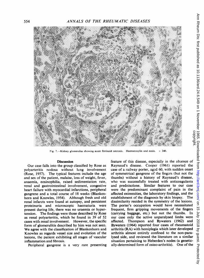
Kidneys.-Both kidneys showed a similar pattern withmultiple cortical scars and infarcts throughout. Manyof the small and medium-sized arteries and occasionalarterioles exhibited complete destruction of the wall withnumerous inflammatory cells of various types and somefibrinous deposit in the areas they supplied. In some ofthe small arteries there was moderate proliferation of theendothelium causing occlusion of the vessel. Occasionalglomeruli exhibited fibrinoid necrosis (Fig. 7, overleaf).Some of the larger arteries also showed inflammatoryinfiltrate in the wall with thrombus formation as well asbranches of the larger renal veins, which were completelyoccluded with recent thrombus.
Oesophagus.-The vessels were severely involved withpolyarteritis nodosa and some were thrombosed.
Intestinal Tract.-Numerous small infarcts associatedwith arteries showed arteritis and thrombosis. Theappendix exhibited the picture ofgangrenous appendicitis,consisting of ischaemic necrosis and inflammatoryreaction.
Testes.-The left showed extensive infarction, only theoutlines of the usual tubules remaining. No other typeof reaction was seen.
Liver, Stomach, Aorta, andProstate Gland.-No specifichistological abnormalities.
Brain and Spinal Cord.-Right anterior horn atrophyin the cervical spinal cord and periarteritis in the dorsalroot ganglion.
... ~
Fig. 6.-Healed arteritis in peri-adrenal fat. Haematoxylin andeosin. x 90.
553
copyright. on 15 July 2018 by guest. P
rotected byhttp://ard.bm
j.com/
Ann R
heum D
is: first published as 10.1136/ard.24.6.549 on 1 Novem
ber 1965. Dow
nloaded from
ANNALS OF THE RHEUMATIC DISEASES
AI
ns5,~~~~~~~~f,i+5A $ ,>R%h>^ x2f@1
(.N
4W
Fig. 7.-Kidney glomerulus showing acute fibrinoid necrosis. Haematoxylin and eosin. x 240.
DiscussionOur case falls into the group classified by Rose as
polyarteritis nodosa without lung involvement(Rose, 1957). The typical features include the age
and sex of the patient, malaise, loss of weight, fever,anaemia, eosinophilia, raised sedimentation rate,renal and gastrointestinal involvement, congestiveheart failure with myocardial infarctions, peripheralgangrene and a total course of 18 weeks (Blanken-horn and Knowles, 1954). Although fresh and oldrenal infarcts were found at autopsy, and persistentproteinuria and microscopic haematuria were
present during life, there was no uraemia or hyper-tension. The findings were those described by Roseas renal polyarteritis, which he found in 39 of 52cases with renal involvement. However, the specificform of glomerulitis described by him was not seen.
We agree with the classification of Blankenhorn andKnowles as regards vessel size and evolution of thelesions, the patient exhibiting all stages of vascularinflammation and fibrosis.
Peripheral gangrene is a very rare presenting
feature of this disease, especially in the absence ofRaynaud's disease. Cooper (1961) reported thecase of a railway porter, aged 60, with sudden onsetof symmetrical gangrene of the fingers (but not thethumbs) without a history of Raynaud's disease,who was successfully treated with anticoagulantsand prednisolone. Similar features to our casewere the predominant complaint of pain in theaffected extremities, the laboratory findings, and theestablishment of the diagnosis by skin biopsy. Thedissimilarity resided in the symmetry of the lesions.The porter's occupation would have necessitatedfrequent, firm gripping movements of the fingers(carrying baggage, etc.) but not the thumbs. Inour case only the active unparalysed limbs wereaffected. Thompson and Bywaters (1962) andBywaters (1964) reported four cases of rheumatoidarthritis (RA) with hemiplegia which later developedarthritis almost entirely confined to the non-para-lysed side, and reviewed the literature on a similarsituation pertaining to Heberden's nodes (a genetic-ally-determined form of osteo-arthritis). One of the
554
iIMS
copyright. on 15 July 2018 by guest. P
rotected byhttp://ard.bm
j.com/
Ann R
heum D
is: first published as 10.1136/ard.24.6.549 on 1 Novem
ber 1965. Dow
nloaded from
GANGRENE IN POLYARTERITIS
four had radiological erosion in the active wrist butonly osteoporosis of disuse on the inactive side.Brachial arteriograms demonstrated blocks andimpaired pulp filling in both hands, but the vesselswere of smaller calibre on the paralysed side; thisindicated that a circulating agent, possibly therheumatoid factor (RF), was partly responsible forthe vascular changes. In a series of arteriograms onpatients with rheumatoid arthritis and other dis-orders, three cases of polyarteritis nodosa showed acharacteristic picture distinct from RA (Laws, Lillie,and Scott, 1963). The digital arteries were occludeddistal to the palmar arch, their normal pattern beingreplaced by a network of irregular, tortuous, ab-normal arteries. None of these patients hadreceived steroid therapy. The abundance of theseabnormal small vessels may explain the rarity ofperipheral gangrene in polyarteritis.A high correlation of rheumatoid arteritis with
nodules and a positive test for RF was found in aseries of 35 cases by Bywaters and Scott (1963).Nodules were only found on the unaffected andactive side in patients with hemiplegia; "this iscomparable with the well-known presence of elbownodules on the opposite side to an affected shoulder".Stecher (1955) reported a patient with poliomyelitiswho later developed Heberden's nodes in the normalbut not in the paralysed hand. He states that incases of cerebral palsy or peripheral nerve damageosteo-arthritis failed to develop in the area suppliedby the affected nerve. This suggests that activemovement is important in the genesis of jointchanges (rheumatoid and osteo-arthritis) as well asin that of nodule formation. Sokoloff, McCluskey,and Bunim (1953) demonstrated that an arteritissimilar to that found in the muscle and synovialmembrane of rheumatoid patients is also the prob-able initial lesion in their early rheumatoid nodules.
Thus, it is possible that muscle activity of theextremities (in our case the fine co-ordinated move-ments of a one-handed draughtsman and fullweight-bearing on the normal foot and leg) mayfavour the development of vasculitis in a pre-disposed subject.The presence of RF in the blood may determine
the vascular changes in rheumatoid arthritis (By-waters, 1964; Epstein, 1957; Kunkel, 1960). Inexperimental animals, intimal localization of RF hasbeen demonstrated (Hess and Ziff, 1960). RF wasidentified in experimentally-induced vascular lesionsin rats given human serum that contained high titresof RF (Baum, Stastny, and Ziff, 1962). Macro-globulins may be dissociated in vitro into smallerglobulin fragments by the action of sulphydryl
reducing agents on the disulphide bonds (Deutschand Morton, 1957). A fall in circulating macro-globulin levels has been reported in patients withWaldenstrom's macroglobulinaemia and cold agglu-tinin haemolytic anaemia after the administration ofthe thiol compound, penicillamine (Ritzman, Cole-man, and Levin, 1960,1961; Bloch, Prasad, Anastasi,and Briggs, 1960). RF is a macroglobulin withphysical and chemical properties similar to Walden-strom's macroglobulin. Jaffe (1964) reportedclinical and serological improvement in a patientwith rheumatoid arthritis and arteritis manifestedby leg ulcers and gangrene. It is noteworthy thatthe lowest serum sulphhydryl cocnentrations werefound in rheumatoid arthritis complicated by ne-crotizing arteritis, in active systemic lupus erythe-matosus (SLE) and polyarteritis nodosa (Lorber,Pearson, Meredith, and Gantz-Mandell, 1964).These levels corresponded to the values found inWaldenstrom's macroglobulinaemia. These lowlevels are presumed to be due to the dehydrogena-tion of the sulphhydryl groups leading to theformation of disulphide bonds that link the proteinsub-units together to make the abnormal macro-globulin (Lorber and others, 1964). RF may beresponsible for the vasculitis that occurs in somecases of connective tissue disease (Kunkel, 1960),either because of its physical properties (increasedviscosity, cryo-precipitability, etc.) or because of itspeculiar affinity for autologous tissues. RF has alsobeen demonstrated in the synovial membranes,lymph nodes, skin, and other tissues of patients withrheumatoid arthritis (McCormick, 1963; Mellors,Nowoslawski, and Korngold, 1961; Mellors, Nowo-slawski, Korngold, and Sengson, 1961; Ball, 1962;Bland and Clark, 1962; Rodnan, Eisenbeis, andCreighton, 1962), that is, at sites where lymphocytesand plasma cells are located. A careful immuno-cytochemical study of a patient with polyarteritisnodosa and a positive latex-fixation test revealedthat cells in the spleen with the morphologicalcharacteristics of plasma cells, contained a 19Smacroglobulin. Positive staining of these cells withaggregated fluorescein-labelled human gamma glo-bulin suggested that they contained RF (Paronettoand Strauss, 1962). Our patient had a positivelatex-fixation test and a gamma globulin at the upperlimits of normal on serum protein electrophoresis(1 .34 g. per cent.). Thus, in the light of theevidence presented, it would appear reasonable to givepenicillamine a therapeutic trial in cases of polyarte-ritis with RF in an effort to reduce the likelihood offurther exacerbations of the vasculitis. Prolongedtreatment at 2 g. daily in order to achieve a fall incirculating RF would be indicated, as emphasized
555
copyright. on 15 July 2018 by guest. P
rotected byhttp://ard.bm
j.com/
Ann R
heum D
is: first published as 10.1136/ard.24.6.549 on 1 Novem
ber 1965. Dow
nloaded from
ANNALS OF THE RHEUMATIC DISEASES
by Jaffe (1964) in the management of rheumatoidarteritis
Various drugs have been related to the onset ofvasculitis in polyarteritis, SLE, and thromboticthrombocytopenic purpura, mainly the sulphon-amides, penicillin, hydralazine, and anticonvulsants(Glaser, 1963; Symmers, 1962). Also, strepto-coccal infection may precede polyarteritis nodosa.Our patient gave no history of preceding infectionand no known therapeutic antigen was administeredin the hospital before the exacerbation of thearteritis on June 4, while he was taking 50 mg.prednisolone daily. The only substance that couldpossibly be incriminated was the local applicationof bacitracin ointment for the infected skin biopsysite over the left elbow.
Corticosteroid therapy is the recognized (and only)form of treatment at the present time for severepolyarteritis. However, the healing process in thevessels may produce further ischaemia and infarctionof tissue despite satisfactory generalized responses(Glaser, 1963; Symmers and Litchfield, 1952). Along-term study of the effect of cortisone in poly-arteritis showed that, although the treated group wassuperior to the untreated group at the end of oneyear, after 3 years this advantage had disappeared(M.R.C., 1957, 1960). Thus, cortisone may prolonglife but does not produce a cure. It is well estab-lished that sudden corticosteroid withdrawal mayproduce an acute arteritis, for example in rheuma-toid arthritis, SLE, or scleroderma (Symmers, 1962).The apparent increase in rheumatoid arteritis sincethe introduction of corticosteroids has led us toquestion the role of these drugs in the pathogenesisof vasculitis (Adler, Norcross, and Lockie, 1962;Fisher, 1957). However, sixteen of the 35 cases ofrheumatoid arteritis reported by Bywaters and Scott(1963) had not had steroid therapy, and three otherseries were quoted in which the majority had notreceived steroids. The association with steroidtherapy thus appeared to be secondary, since themore severe cases were given steroids. Fatal myo-cardial infarction following steroid administrationto a patient with polyarteritis has been reported byNachman (1961), who noted the occasional para-doxical adverse effects of steroids. However, aspointed out by Smyth (1964), "most would agreethat steroid dosage should be lowered when a para-doxical adverse reaction is encountered . (but)the final answer is not yet known".
In our patient the exacerbation of the arteritis wastaken as an indication to increase the dose of pred-nisolone. We may have been at fault in this respect.Autopsy revealed old and fresh myocardial infarcts,the older lesions presumably having occurred 2
months before death during the first bout of tachy-cardia. In a study of 66 autopsies on polyarteritisnodosa, it was found that 41 had myocardial infarctsmost of which were silent (Holsinger, Osmundson,and Edwards, 1962). In another series of thirtyautopsies, 25 showed glomerular changes, and renalbiopsy was suggested as the best method of estab-lishing the diagnosis during life (Patalano andSommers, 1961). Our previous experience wouldsupport this suggestion, as the kidney appears to beone of the most constantly and diffusely involvedorgans, whereas in the other tissues the lesions aremore scattered.
SummaryA 39-year-old white male, with paralysed right
upper and left lower limbs due to poliomyelitis ininfancy, presented with a 10-week history of sys-temic symptoms and paraesthesiae of the left handand both legs and feet, followed by peripheralgangrene in the unparalysed extremities and supra-ventricular tachycardia. He had been dischargedfrom his job as a draughtsman owing to loss ofdexterity in his left (unparalysed) hand. While onsteroid therapy, a further exacerbation of thearteritis occurred and he expired during another boutof tachycardia, 18 weeks after the onset. Autopsyrevealed widespread vessel involvement with multipleinfarctions of the heart, intestine and kidneys, anacutely inflamed, gangrenous appendix and general-ized peritonitis. During life the abdominal symp-toms were masked by the steroid therapy. Possiblereasons for the localization of the gangrene to theunparalysed extremities are discussed.The literature on the rationale and use of sulph-
hydryl-reducing agents in diseases characterized byabnormal macroglobulins is reviewed.
We wish to thank Dr. P. C. Martineau for access to hisbiopsy and autopsy reports and for Figure 4, and theDepartment of Photography, headed by Mr. C. Pickard,for the illustrations.
REFERENCES
Adler, R. H., Norcross, B. M., and Lockie, L. M. (1962).J. Amer. med. Ass., 180, 922.
Ball, J. (1962). Ann. rheum. Dis., 21, 272.Baum, J., Stastny, P., and Ziff, M. (1962). Arthr. andl
Rheum., 5, 101.Bland, J. H., and Clark, L. G. (1962). Ibid., 5, 102.Blankenhorn, M. A., and Knowles, H. C. (1954). Anti.
intern. Med., 41, 887.Bloch, H. S., Prasad, A., Anastasi, A., and Briggs, D. R.
(1960). J. Lab. clin. Med., 56, 212.Bywaters, E. G. L. (1964). Canad. med. Ass., J., 91, 606.- and Scott, J. T. (1963). J. chron. Dis., 16, 905.
556
copyright. on 15 July 2018 by guest. P
rotected byhttp://ard.bm
j.com/
Ann R
heum D
is: first published as 10.1136/ard.24.6.549 on 1 Novem
ber 1965. Dow
nloaded from
GANGRENE IN POLYARTERITIS
Cooper, A. S. (1961). Guy's Hosp. Rep., 110, 110.Deutsch, H. F., and Morton, J. I. (1957). Science, 125,
600.Epstein, W. (1957). In "Serological Reactions of
Rheumatoid Arthritis", ed. R. W. Lamont-Havers, p. 22. Arthritis and RheumatismFoundation, New York.
Fisher, E. R. (1957). Amer. J. clin. Path., 27, 191.Glaser, G. H. (1963). Med. Clin. N. Amer., 47, 1475.Hess, E. V., and Ziff, M. (1960). Quoted by Odone,
D. T., Wilson, A. F., and Engleman, E. P. in Bull.rheum. Dis., 11, 229.
Holsinger, D. R., Osmundson, P. J., and Edwards, J. E.(1962). Circulation, 25, 610.
Jaffe, I. A. (1964). Ann. intern. Med., 61, 556.Kunkel, H. G. (1960). "Macroglobulins and High
Molecular Weight Antibodies", in "The PlasmaProteins", ed. F. W. Putnam, vol. 1, pp. 299, 302,305. Academic Press, Inc., New York.
Laws, J. W., Lillie, J. G., and Scott, J. T. (1963). Brit.J. Radiol., 36, 477.
Lorber, A., Pearson, C. M., Meredith, W. L., and Gantz-Mandell, L. E. (1964). Ann. intern. Med., 61, 423.
McCormick, J. N. (1963). Ann. rheum. Dis., 22, 1.Medical Research Council (1957). Brit. med. J., 1, 608.
(1960). Ibid., 1, 1399.Mellors, R. C., Nowoslawski, A., and Korngold, L.
(1961). Amer. J. Poth., 39, 533.and Sengson, B. L. (1961). J. exp.
Med., 113, 475.Nachman, R. L. (1961). Amer. J. Cardiol., 7, 288.Paronetto, F., and Strauss, L. (1962). Ann. intern. Med.,
56, 289.Patalano, V. J., and Sommers, S. C. (1961). Arch. Path.,
72, 1.Ritzmann, S. E., Coleman, S. L., and Levin, W. C. (1960).
J. clin. Invest., 39, 1330.and Levin, W. C. (1961). J. Lab. clin. Med., 57,718.
Rodnan, C. P., Eisenbeis, C. H. Jr., and Creighton, A. S.(1962). Arthr. and Rheum., 5, 316.
Rose, G. A. (1957). Brit. med. J., 2, 1148.Smyth, C. J., and others (1964). Ann. intern. Med.,
Suppl. 6, pp. 44.Sokoloff, L., McCluskey, R. T., and Bunim, J. J. (1953).
Arch. Path., 55, 475.Stecher, R. M. (1955). Ann. rheum. Dis., 14, 1.Symmers, W. St. C. (1962). Proc. roy. Soc. Med., 55, 20.
and Litchfield, J. A. (1952). Lancet, 2, 1193.Thompson, M., and Bywaters, E. G. L. (1962). Ann.
rheum. Dis., 21, 370.
Polyarterite fatale se presentant comme gangreneperipherique asymetrique.-Description clinique et
anatomo-pathologique d'un cas
RESUMEUn homme de race blanche, age de 39 ans, ayant
des membres superieur droit et inferieur gauche paralysespar la poliomyelite depuis l'enfance, s'est presente avecdes sympt6mes generaux et des paresthesies a la maingauche et aux deux jambes et pieds d'une duree de 10semaines. Cela fut suivi d'une gangrene peripherique desmembres non paralyses et d'une tachycardie supra-ventriculaire. On l'avait renvoye de son travail dedessinateur parcequ'il avait perdu l'habilite de sa maingauche (non paralysee). Pendant la therapie steroide il yeut une autre exacerbation de l'arterite et il mourut aubout d'une attaque de tachycardie 18 semaines apres ledebut de la maladie. L'autopsie revela des lesionsvasculaires etendues, de nombreux infarctus du coeur, del'intestin et des reins, l'appendice tres inflamme etgangreneux et une peritonite generalisee. Avant lamort les sympt6mes abdominaux etaient masquespar la therapie st6roide. On discute les raisons possiblesde la localisation de la gangrene aux membres nonparalyses.On discute la litterature et la raison de l'emploi des
agents qui reduisent le sulphydryl dans les maladiescaracterisees par des macroglobulines anormales.
Poliarteritis fatal presentAndose como gangrena perifericaasimetrica.-Relato clinico y anatomo-patol gico de un
caso
SUMAIUOUn hombre de raza blanca de 39 afios, con el brazo
derecho y la pierna izquierda paralizados por polio-mielitis desde su infancia, se present6 con sintomas gener-ales y parestesias en la mano izquierda y en ambos piesy piernas desde 10 semanas. Esto fue seguido de unagangrena periferica de las extremidades non-paralizadasy de una taquicardia supraventricular. El pacientehabia sido despedido de su trabajo de delineante por haberperdido la destreza de su mano izquierda (non-paralizada)Durante la terapia esteroide una exacerbaci6n mas de laarteritis y de la taquicardia se produjo y el enfermofalleci6 18 semanas despues del comienzo de la enferme-dad. La necropsia revelo lesiones vasculares extensas,infartos multiples del coraz6n, del intestino y de losriflones, el apendice muy inflamado y gangrenoso y unaperitonitis generalizada. Durante la vida la terapiaesteroide encubria los sintomas abdominales. Se dis-cuten las razones posibles de la localizaci6n de la gangrenaen las extremidades non-paralizadas.
Se pasa revista a la literatura acerca del empleo de losagentes de reducci6n del sulfidril en enfermedadescaracterizadas por macroglobulinas anormales.
557
copyright. on 15 July 2018 by guest. P
rotected byhttp://ard.bm
j.com/
Ann R
heum D
is: first published as 10.1136/ard.24.6.549 on 1 Novem
ber 1965. Dow
nloaded from