case report open access cutaneous polyarteritis nodosa ... · case report open access cutaneous...
TRANSCRIPT

Haviv et al. Pediatric Rheumatology 2014, 12:46http://www.ped-rheum.com/content/12/1/46
CASE REPORT Open Access
Cutaneous polyarteritis nodosa successfullytreated with topical diflucortolone valerate – acase report & review of the literatureRuby Haviv1*, Maya Capua1,2, Jacob Amir1 and Liora Harel1*
Abstract
Cutaneous Polyarteritis Nodosa (cPAN) was first described in 1931. cPAN is considered a rare disease, its trueincidence is unknown. The age of onset is diverse. Most studies have shown no significant gender predominance.cPAN presents with distinct skin findings, such as a maculopapular rash, subcutaneous nodules, livedoid vasculitis,panniculitis, ischemic finger lesions, or erythematous patchy rash.Etiology is unclear. It is still believed to be an immune complex-mediated disease, although a possible mechanismrecently proposed relates a familial form of the disease to impaired activity of Adenosine Deaminase 2. cPAN mayreflect an underlying disease, infection or medical treatment.There is no consensus as to initial treatment, dosage and length of treatment. Patients with constitutionalsymptoms, visceral involvement, a more severe course of the disease, or high acute phase reactants, were treatedmainly with systemic corticosteroids and/or cytotoxic agents for varying durations. However, persistence ofcutaneous lesions has been documented.We describe a 14 year old male suffering from persistent cPAN, with no constitutional symptoms or involvementof internal organs. The patient was treated with a local corticosteroid-based ointment during exacerbations, untilcomplete remission. Although reported in only one study, treatment with topical corticosteroid compound mayresult in significant improvement or complete regression of skin lesions in cPAN patients.
Keywords: Cutaneous polyarteritis nodosa, Periarteritis, CPAN, Topical treatment, Corticosteroid, Diflucortolonevalerate
BackgroundThe first description of limited cutaneous polyarteritisnodosa (cPAN) was published by Lindberg in 1931, de-scribing skin findings, and also extra-cutaneous findings,such as fever, malaise, myalgia, arthralgia and neuropathy(unlike systemic PAN, in which the cutaneous findings areonly secondary to internal organs involvement, mainlykidney, heart & liver) [1].cPAN is rare; its true incidence is unknown. It is esti-
mated that one third of children diagnosed with systemicPAN (sPAN), actually have cPAN [2,3], but in practice,rheumatologists may treat more cPAN patients than sPANpatients.
* Correspondence: [email protected]; [email protected] of Pediatrics C, Schneider Children’s Medical Center of Israel,Tel Aviv University, Sackler School of Medicine, Petach Tikvah, IsraelFull list of author information is available at the end of the article
© 2014 Haviv et al.; licensee BioMed Central LCommons Attribution License (http://creativecreproduction in any medium, provided the orDedication waiver (http://creativecommons.orunless otherwise stated.
Age of onset ranges from the neonatal and infantileperiod [4,5], up to age 81 [6]. Most studies do not revealany significant gender predominance [1]. A male to femaleratio of 1:1.7 was found in a large study of 79 cases [6].cPAN presents with distinct skin findings, such as a
maculopapular rash, subcutaneous nodules, livedoidvasculitis, panniculitis, ischemic finger lesions, or ery-thematous patchy rash. In a study of juvenile polyarteritis,all patients with cPAN were diagnosed with necrotizingarterial inflammation found on biopsy [3].The etiology of cPAN is unknown. It is, most probably,
an immune complex-mediated disease, with some evidenceof serum IgM anti-phosphatidylserine-prothrombin anti-bodies in patients’ sera, and deposition of C3 within vesselwalls, as shown by direct immunofluorescence techniques[7]. Recently, loss-of-function mutations, in the gene(CECR1) encoding Adenosine Deaminase 2, were found
td. This is an Open Access article distributed under the terms of the Creativeommons.org/licenses/by/4.0), which permits unrestricted use, distribution, andiginal work is properly credited. The Creative Commons Public Domaing/publicdomain/zero/1.0/) applies to the data made available in this article,
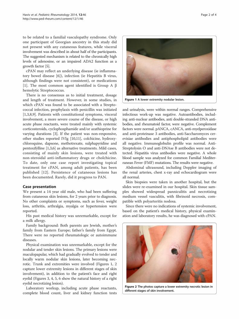
Figure 1 A lower extremity nodular lesion.
Figure 2 The photos capture a lower extremity necrotic lesion indifferent stages of skin involvement.
Haviv et al. Pediatric Rheumatology 2014, 12:46 Page 2 of 4http://www.ped-rheum.com/content/12/1/46
to be related to a familial vasculopathy syndrome. Onlyone participant of Georgian ancestry in this study didnot present with any cutaneous features, while visceralinvolvement was described in about half of the participants.The suggested mechanism is related to the chronically highlevels of adenosine, or an impaired ADA2 function as agrowth factor [5].cPAN may reflect an underlying disease (ie inflamma-
tory bowel disease [6]), infection (ie Hepatitis B virus,although findings were not consistent), or medications[1]. The most common agent identified is Group A βhemolytic Streptococcus.There is no consensus as to initial treatment, dosage
and length of treatment. However, in some studies, inwhich cPAN was found to be associated with a Strepto-coccal infection, prophylaxis with penicillin was initiated[1,3,8,9]. Patients with constitutional symptoms, visceralinvolvement, a more severe course of the disease, or highacute phase reactants, were treated mainly with systemiccorticosteroids, cyclophosphamide and/or azathioprine forvarying durations [3]. If the patient was non-responsive,other studies reported IVIg [10,11], colchicine, hydroxy-chloroquine, dapsone, methotrexate, sulphapyridine andpentoxifylline [1,3,6] as alternative treatments. Mild cases,consisting of mainly skin lesions, were treated withnon-steroidal anti-inflammatory drugs or cholchicine.To date, only one case report investigating topicaltreatment for cPAN, among adult patients, has beenpublished [12]. Persistence of cutaneous lesions hasbeen documented. Rarely, did it progress to PAN.
Case presentationWe present a 14 year old male, who had been sufferingfrom cutaneous skin lesions, for 2 years prior to diagnosis.No other complaints or symptoms, such as fever, weightloss, arthritis, arthralgia, myalgia or hypertension werereported.His past medical history was unremarkable, except for
a milk allergy.Family background: Both parents are Jewish, mother’s
family from Eastern Europe; father’s family from Egypt.There were no reported rheumatologic or autoimmunediseases.Physical examination was unremarkable, except for the
nodular and tender skin lesions. The primary lesions weremaculopapular, which had gradually evolved to tender andlocally warm nodular skin lesions, later becoming nec-rotic. Trunk and extremities were involved (Figures 1, 2capture lower extremity lesions in different stages of skininvolvement), in addition to the patient’s face and righteyelid (Figures 3, 4, 5, 6 show the natural history of a righteyelid necrotizing lesion).Laboratory workup, including acute phase reactants,
complete blood count, liver and kidney function tests
and urinalysis, were within normal ranges. Comprehensiveinfectious work-up was negative. Autoantibodies, includ-ing anti-nuclear antibodies, anti double-stranded DNA anti-bodies, and rheumatoid factor, were negative. Complementfactors were normal. pANCA, cANCA, anti-myelperoxidaseand anti-proteinase 3 antibodies, anti-Saccharomyces cer-evisiae antibodies and antiphospholipid antibodies wereall negative. Immunoglobulin profile was normal. Anti-Streptolysin O and anti-DNAse B antibodies were not de-tected. Hepatitis virus antibodies were negative. A wholeblood sample was analyzed for common Familial Mediter-ranean Fever (FMF) mutations. The results were negative.Abdominal ultrasound, including Doppler imaging of
the renal arteries, chest x-ray and echocardiogram wereall normal.Skin biopsies were taken in another hospital, but the
slides were re-examined in our hospital. Skin tissue sam-ples showed widespread panniculitis and necrotizingmedium vessel vasculitis, with fibrinoid necrosis, com-patible with polyarteritis nodosa.Since there were no indications of systemic involvement,
based on the patient’s medical history, physical examin-ation and laboratory results, he was diagnosed with cPAN.

Figure 3 The natural history of a right eyelid necrotizing lesion –early stage.
Figure 5 The natural history of a right eyelid necrotizinglesion – necrosis.
Haviv et al. Pediatric Rheumatology 2014, 12:46 Page 3 of 4http://www.ped-rheum.com/content/12/1/46
Treatment with oral prednisone was recommended.Mainly concerned about the side effects of systemicsteroidal treatment, the parents refused to any sys-temic treatment (including other oral agents, such ascolchicine), relying on the boy’s good appearance andwell-being. The patient was thus treated with topicaldiflucortolone valerate during exacerbations, three times aday, for two to three weeks at a time, until completeremission. Both facial and trunk lesions were treated,without any recurrence of a specific treated lesionreported during follow-up visits. If a lesion was not treatedwithin days of its appearance, it would have become nec-rotic. No adverse reactions were reported, no extensiveskin atrophy was marked, and no cataract was diagnosedupon follow-up. A lesion became infected only once (byStaphylococcus aureus and Pseudomonas aeruginosa), andwas successfully treated with oral antibiotics.
Figure 4 The natural history of a right eyelid necrotizinglesion – progression.
ConclusionsCurrently, there is no consensus as to initial treatment,dosage and length of treatment in cPAN patients. Wedescribe herein a 14 year old male, diagnosed with cPAN.Due to parental refusal to any systemic treatment, he wastreated with a topical corticosteroid-based compoundduring exacerbations, applying the compound threetimes a day, for two to three weeks at a time. Treatmentwas very effective, resulting in a complete remission,without any recurrence or adverse effect documentedduring follow-up. If the compound had been appliedafter a few days delay, the lesion would have becomenecrotic. Topical corticosteroid compounds may be ef-ficient in treating isolated skin lesions among cPANpatients, without mucosal or systemic signs of involve-ment. This is the first published case utilizing an ef-fective topical treatment among juvenile patients. Aprospective, multi-center study should be conducted,to evaluate different available treatments available, andweigh the different approaches in the light of the long-termadverse effects.
Figure 6 The natural history of a right eyelid necrotizinglesion – healing.

Haviv et al. Pediatric Rheumatology 2014, 12:46 Page 4 of 4http://www.ped-rheum.com/content/12/1/46
ConsentWritten informed consent was obtained from the patient’sparent for the publication of this report and any accom-panying images.
Competing interestsAll authors declare that they have no competing interests.
Authors’ contributionsRH reviewed the literature and drafted the manuscript. MC reviewed theliterature. JA treated the patient, took some of the photos and participatedin reviewing the manuscript. LH treated the patient, gathered informationduring follow-up visits, had the parents' consent for the publication of thisreport and all accompanying images, took some of the photos and participatedin reviewing the manuscript. All authors read and approved the finalmanuscript.
AcknowledgementsThe authors thank Mrs. Phyllis Curchack Kornspan for her editorial services.
Author details1Department of Pediatrics C, Schneider Children’s Medical Center of Israel,Tel Aviv University, Sackler School of Medicine, Petach Tikvah, Israel.2Department of Pediatrics Children's Hospital at, Montefiore, New-York City,NY, USA.
Received: 10 February 2014 Accepted: 16 September 2014Published: 10 October 2014
References1. Morgan AJ, Schwartz RA: Cutaneous polyarteritis nodosa: a
comprehensive review. Int J Dermatol 2010, 49(7):750–756.2. Cassidy JT, Petty RE, Laxer RM, Lindsley CB: Textbook of Pediatric
Rheumatology. 6th edition. Philadelphia, PA: Saunders; 2011.3. Ozen S, Anton J, Arisoy N, Bakkaloglu A, Besbas N, Brogan P, García-Consuegra
J, Dolezalova P, Dressler F, Duzova A, Ferriani VP, Hilário MO, Ibáñez-Rubio M,Kasapcopur O, Kuis W, Lehman TJ, Nemcova D, Nielsen S, Oliveira SK, SchiklerK, Sztajnbok F, Terreri MT, Zulian F, Woo P: Juvenile polyarteritis: results of amulticenter survey of 110 children. J Pediatr 2004, 145(4):517–522.
4. Stone MS, Olson RR, Weismann DN, Giller RH, Goeken JA: Cutaneousvasculitis in a newborn of a mother with cutaneous polyarteritis nodosa.J Am Acad Dermatol 1993, 28(1):101–105.
5. Navon Elkan P, Pierce SB, Segel R, Walsh T, Barash J, Padeh S, Zlotogorski A,Berkun Y, Press JJ, Mukamel M, Voth I, Hashkes PJ, Harel L, Hoffer V, Ling E,Yalcinkaya F, Kasapcopur O, Lee MK, Klevit RE, Renbaum P, Weinberg-ShukronA, Sener EF, Schormair B, Zeligson S, Marek-Yagel D, Strom TM, Shohat M,Singer A, Rubinow A, Pras E, Winkelmann J, Tekin M, Anikster Y, King MC,Levy-Lahad E: Mutant Adenosine Deaminase 2 in a Polyarteritis NodosaVasculopathy. N Engl J Med 2014, 370(10):921–931.
6. Daoud MS, Hutton KP, Gibson LE: Cutaneous periarteritis nodosa: aclinicopathological study of 79 cases. Br J Dermatol 1997, 136(5):706–713.
7. Kawakami T, Yamazaki M, Mizoguchi M, Soma Y: High titer of anti-phosphatidylserine-prothrombin complex antibodies in patientswith cutaneous polyarteritis nodosa. Arthritis Care Res 2007,57(8):1507–1513.
8. David J, Ansell BM, Woo P: Polyarteritis nodosa associated withStreptococcus. Arch Dis Child 1993, 69(6):685–688.
9. Fink CW: The Role of the Streptococcus in Poststreptococcal RecativeArthritis and Childhood Polyarteritis Nodosa. J Rheumatol Suppl 1991,29:14–20.
10. Marie I, Miranda S, Girszyn N, Soubrane JC, Vandhuick T, Levesque H:Intravenous immunoglobulins as treatment of severe cutaneouspolyarteritis nodosa. Intern Med J 2012, 42(4):459–462.
11. Uziel Y, Silverman ED: Intravenous Immunoglobulin Therapy in a Childwith Cutaneous Polyarteritis Nodosa. Clin Exp Rheumatol 1998,16(2):187–189.
12. Rogaslki C, Sticherling M: Panarteritis cutanea benigna – an entity limited tothe skin or cutaneous presentation of a systemic necrotizing vasculitis ?Report of seven cases and review of the literature. Int J Dermatol 2007,46(8):817–821.
doi:10.1186/1546-0096-12-46Cite this article as: Haviv et al.: Cutaneous polyarteritis nodosasuccessfully treated with topical diflucortolone valerate – a case report& review of the literature. Pediatric Rheumatology 2014 12:46.
Submit your next manuscript to BioMed Centraland take full advantage of:
• Convenient online submission
• Thorough peer review
• No space constraints or color figure charges
• Immediate publication on acceptance
• Inclusion in PubMed, CAS, Scopus and Google Scholar
• Research which is freely available for redistribution
Submit your manuscript at www.biomedcentral.com/submit