polyarteritis nodosa*t - british journal of...
TRANSCRIPT
Brit. J. Ophthal. (1968) 52, 141
POLYARTERITIS NODOSA*tWITH AN UNUSUAL OCULAR PRESENTATION
BY
G. M. MACLURENorth Riding Infirmary, Middlesbrough, Teesside
POLYARTERITIS nodosa (periarteritis nodosa), first described by Kussmaul and Maier (1866),is a relatively rare disease, but considerable attention has been paid to it in recent years.
Duke-Elder (1962), describing the condition, wrote: "this is a disseminated disease ofobscure cause characterized by necrosing obliterative lesions of small arteries and arterioleswhich appear nodular owing to the formation of aneurysms or granulomatous prolifera-tion". Harvey (1963) described the lesions as segmental in their distribution andinvolving arteries throughout most of the body so that the resulting clinical picture was oneof polymorphic manifestations which might seem unrelated. In its typical form, poly-arteritis nodosa is a well-defined entity, but is not always typical (Anderson, 1961).
This case of polyarteritis nodosa with an unusual ocular presentation is reported in fulldetail because it demonstrates the difficulty in diagnosis of the underlying condition withsuch a presentation. Harbert and McPherson (1947) described a similar case, but thelacrimal gland was not involved. Cogan (1955) reviewed and reported cases of corneo-scleritis or scleritis associated with polyarteritis nodosa, and Moore and Sevel (1966) haverecently reviewed cases associated with either polyarteritis nodosa or Wegener's granulo-matosis, in none of which the lacrimal gland appeared to be affected. Dacryoadenopathydoes not appear to have been reported as a presenting symptom in polyarteritis nodosa.
Case ReportA man aged 60 years first attended the Nottingham Eye Hospital on December 18, 1964, complainingof a swelling of the outer part of the right upper eyelid of 4 weeks' duration, and a red sore righteye for about 6 weeks. There was also a, 5-week history of an upper respiratory tract infectionwhich had been slow to settle with tablets oxytetracycline 250 mg. four times a day given by thegeneral practitioner. The patient was also attending the E.N.T. Department for "indrawn ear-drums", and was treated for eustachian catarrh with some residual bilateral deafness.
History.-There had been recurrent attacks of bronchitis in 1921, 1922, 1924 (Jan.), 1924 (Nov.), 1925,1929, 1930, and 1947, for which the usual treatment appears to have been Mist. Expect. In 1953, the patienthad been treated with aspirin and liniment for rheumatoid arthritis of the wrists and fingers. There wereupper respiratory tract infections in 1954, 1960, 1963, and Nov. 1964, but only for the last attack hadsystemic antibiotics been given. At no time were sulphonamides recorded as having been given.On July 31, 1954, the blood pressure was recorded as 165/85.In March, 1964, there had been a complaint of pain in the legs when walking, but no abnormality or
underlying cause was found.
* Received for publication January 10, 1967.t Address for reprints: North Riding Infirmary, Middlesbrough, Teesside.
141
on 6 May 2018 by guest. P
rotected by copyright.http://bjo.bm
j.com/
Br J O
phthalmol: first published as 10.1136/bjo.52.2.141 on 1 F
ebruary 1968. Dow
nloaded from

Examination.-The visual acuity was 6/9 in each eye. A slight swelling was present in the regionof the right lacrimal gland. There was a mild generalized conjunctivitis, with a small area ofnodular episcleritis and superficial punctate keratitis in the right eye. The left eye was white andquiet. The intra-ocular pressures, fundi, and discs were normal.
Treatment.-Gutt. IDU 2-hourly and gutt. scopolamine 0-1 per cent. three times a day to theright eye. Tabs. oxytetracycline 250 mg. four times a day were continued, and tabs. Becosymfour times a day were added.
Out-patient Progress.-On December 29, 1964, the keratitis had improved but the swelling in the rightlacrimal gland region had increased. This was diagnosed as a dacryoadenitis not sensitive to oxytetra-cycline, which was, therefore, changed to chlortetracycline capsules, 250 mg. four times a day. After 2weeks the swelling had resolved and the capsules and Becosym were discontinued. There was still somesuperficial punctate keratitis, and the drops were changed to oculentum Terramycin with polymyxin B.X rays of the skull, frontal sinuses, and antra showed no abnormality and there was no apical infection of
the teeth. Chest x ray showed no focal lung lesion, but there were some chronic bronchitis changes.On January 26, 1965, the nodular episcleritis, which had almost settled previously, had become more
marked, but the keratitis was much improved.
Hospital Admission.-On February 12, 1965, 2 months after his first attendance, he complainedfor the first time of malaise with some nausea and vomiting. His right eye had again become pain-ful and red, and the swelling of the lacrimal gland was much larger. The left eye was still white andquiet. He was admitted to the Eye Hospital for further investigation, and treatment was startedwith tabs. erythromycin 250 mg. four times a day.
Examination.-A general medical examination failed to show any clinical evidence of a neoplasm, andthe chest was normal. Two teeth showed caries, but there was no infection or ulceration in the mouth.There was a mild pyrexia of 99.40 F. and the blood pressure was 160/70. The fundi were normal.
Laboratory Investigations.-Hb 12A4 g./100 ml. (84 per cent.); white blood count 10,000/c.mm. (neutrophils6,100; eosinophils 900; lymphocytes 2,100; monocytes 400). Wassermann reaction and Kahn test negative.Erythrocyte sedimentation rate 110 mm. in 1 hour (Westergren). Total serum proteins 7-2 g./100 ml., withdecreased albumin (3-1 g.) and increased globulin (4 1 g.). Three specimens of stool, taken on successivedays, were all negative for occult blood. Urine showed only a trace of albumin.
Hospital Progress.-A week later, the pain, nausea, and vomiting had all settled, and the patientfelt much better. His temperature, which had been between 99-2 and 99.40 F. during the week,was normal on the 8th day. The lacrimal gland swelling was still present.
After another week, the-erythrocyte sedimentation rate had risen to 125 mm. in 1 hour and the whiteblood count was 9,000 (eosinophilia 630 per c.mm.). Rouleau formation in the blood raised the possibilityof myelomatosis, but this was not confirn}ed. A bone marrow examination showed no evidence of leu-kaemia. X ray of the dorsal lumbar spine showed no metastases, but there was narrowing of some of thediscs and general osteo-arthritis. X rays of the hands and pelvis showed no abnormality.
After a further week, he complained of some indigestion and the erythromycin was discontinued. Theerythrocyte sedimentation rate had risen to 154 mm. and the white blood count to 12,500 per c.mm. (eosino-philia 500 per c.mm.). Total serum proteins 7 0 g./100 ml. (albumin 2-8 g., globulin 4-2 g.). Hb was only7-1 g./100 ml. (49 per cent.) and a transfusion of two pints of blood was therefore given.
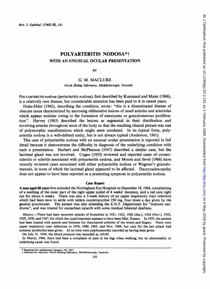
Biopsy.-A specimen from the right lacrimal gland region was taken on March 12, 1965, and sentto Prof. Norman Ashton, Department of Pathology, Institute of Ophthalmology, London, whoreported as follows:
Sections of the biopsy taken from the region of the right lacrimal gland showed no glandular tissue. Thespecimen consisted of a mass of fibro-fatty tissue containing within it numerous blood vessels showingintense subacute perivascular inflammatory reaction in which eosinophils were a predominant feature. Afew giant cells were present (Figs 1 and 2, opposite).
G. M. MACLURE142
on 6 May 2018 by guest. P
rotected by copyright.http://bjo.bm
j.com/
Br J O
phthalmol: first published as 10.1136/bjo.52.2.141 on 1 F
ebruary 1968. Dow
nloaded from
POL YARTERITIS NODOSA
~~~~~~~~~. 7~~~~~ --
FIG. 1.-Biopsy from region of lacrimal gland, FIG. 2.-High-power view of Fig. 1. The bloodshowing fibro-fatty tissue containing numerous vessels show intense subacute perivascular inflamm-blood vessels. x 73. atory reaction. A few giant cells are present
(eosinophils are a prominent feature although notdemonstrated in the black and white picture).
x115.There was no evidence of a neoplasm, and the histological picture was highly suggestive of periarteritis
nodosa or some other form of nodular vasculitis, which would fit in well with the patient's clinical condition.Further Progress.-A few days later the patient complained of nausea and abdominal pain, and,
within a few hours, passed slightly blood-stained urine. The following day, the haematuria wasmore marked.The next day (4 weeks after admission) he complained of severe abdominal pain, and had gross
haematuria, albuminuria, occult blood in the faeces, a slight haemoptysis, and an epistaxis. Bloodurea 560 mg./100 ml.; Hb 10.1 g./100 ml. (69 per cent.); white blood count 14,000 per c.mm.(eosinophilia 280 per c.mm.). The patient was transferred to a medical ward.
Progress in Medical Ward.-His general condition was noted to be poor, and he was uraemic, butthe only abnormal finding was the nodular episcleritis. The keratitis had cleared, and there wasno lacrimal gland swelling following the biopsy.Blood pressure 160/90; erythrocyte sedimentation rate 132 mm. in 1 hour. The serum potassium was
raised (6*9 mEq./litre). He was started on restricted fluids. Next day (March 20, 1965) the blood ureahad risen to 600 mg./100 ml., and the urine output was diminished, with gross haematuria.On March 21, 1965, the blood urea was 640 mg./100 ml. and the serum potassium 6-8 mEq./litre. The
patient was now anuric. The blood urea and setum potassium readings were 700 and 840 mg./100 ml. and7-4 and 8 -2 mEq./litre on the following two days.
Termination.-On March 24, 1965, the patient lapsed into coma, and he died the next day, 6weeks after admission to hospital, 14 weeks after he was first seen, and 5 months after the onset ofsymptoms.
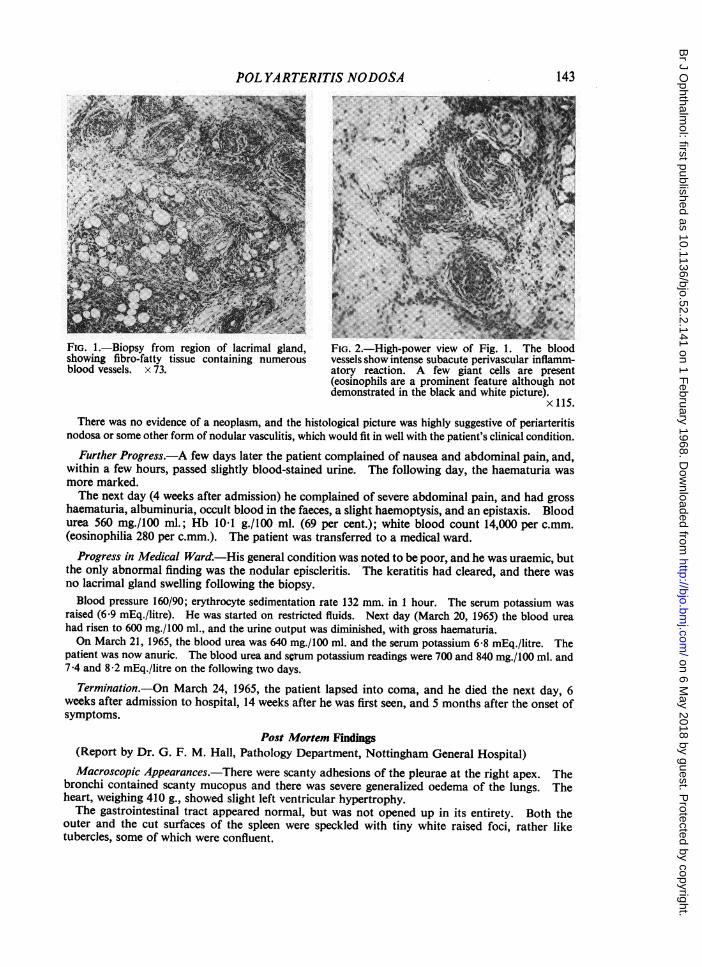
Post Mortem Findings(Report by Dr. G. F. M. Hall, Pathology Department, Nottingham General Hospital)Macroscopic Appearances.-There were scanty adhesions of the pleurae at the right apex. The
bronchi contained scanty mucopus and there was severe generalized oedema of the lungs. Theheart, weighing 410 g., showed slight left ventricular hypertrophy.The gastrointestinal tract appeared normal, but was not opened up in its entirety. Both the
outer and the cut surfaces of the spleen were speckled with tiny white raised foci, rather liketubercles, some of which were confluent.
143
on 6 May 2018 by guest. P
rotected by copyright.http://bjo.bm
j.com/
Br J O
phthalmol: first published as 10.1136/bjo.52.2.141 on 1 F
ebruary 1968. Dow
nloaded from
144 G. M. MACLURE
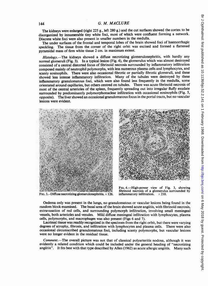
The kidneys were enlarged (right 225 g., left 280 g.) and the cut surfaces showed the cortex to bedisorganized by innumerable tiny white foci, most of which were confluent forming a network.Discrete white foci were also present in smaller numbers in the medulla.The under surfaces of the frontal and temporal lobes of the brain showed foci of haemorrhagic
speckling. The tissue from the corner of the right orbit was excised and formed a flattenedpyramidal mass of firm white tissue 2 cm. in maximum extent.
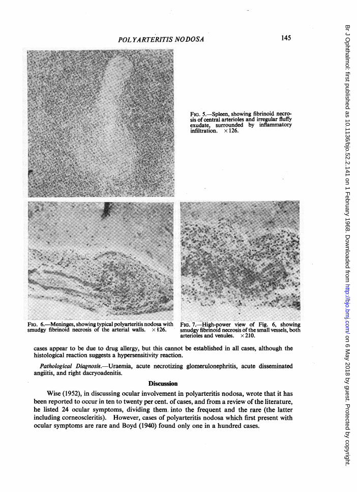
Histology.-The kidneys showed a diffuse necrotizing glomerulonephritis, with hardly anynormal glomeruli (Fig. 3). In a typical lesion (Fig. 4), the glomerulus which was almost destroyedconsisted of a central distorted focus of fibrinoid necrosis surrounded by inflammatory infiltrationcomposed mainly of neutrophil polymorphs, with less numerous plasma cells and lymphocytes, andscanty eosinophils. There were also occasional fibrotic or partially fibrotic glomeruli, and theseshowed less intense inflammatory infiltration. Many of the tubules were destroyed by theseinflammatory granulomatous foci, which were also found less frequently in the medulla, someorientated around capillaries, but others centred on tubules. There was acute fibrinoid necrosis ofmost of the central arterioles of the spleen, frequently spreading out into irregular fluffy exudatesurrounded by predominantly polymorphonuclear infiltration with occasional eosinophils (Fig. 5,opposite). The liver showed an occasional granulomatous focus in the portal tracts, but no vascularlesions were evident.
4,--4,- '-_.'=~~q~
~~~~~~AP
4..~~~04':7*
FIG.~~ X3.Dfueertzngoeuoehitis x 12. ifamaoyinitaio.x20
I.~~~~~~~~~~~~~~~~~~A 4
40~~~~~~~~~~~~~~~~~~~~~~~~~~~~~~~~4
, It
'4 4 UP~'Z.
44~ ~ ~ A&,
'. FIG. 4.-High-power view of Fig. 3, showing~~~ 'i~~~~~~ fibrinoid necrosis of a glomerulus surrounded by
FIG. 3.-Diffuse necrotizing glomerulonephritis. x 126. inflammatory infiltration. x 210.
Oedema only was present in the lungs, no granulomatous or vascular lesions being found in therandom block examined. The basal area of the brain showed acute angiitis, with fibrinoid necrosis,extravasation of red cells, and surrounding polymorph infiltration, involving small meningealvessels, both arterioles and venules. Mild diffuse meningeal infiltration with lymphocytes, plasmacells, polymorphs, and macrophages was also present (Figs 6 and 7).
Lacrimal tissue was readily recognized in the specimen from the right orbit, but there were varyingdegrees of atrophy, fibrosis, and infiltration with lymphocytes and plasma cells. There were alsooccasional circumscribed granulomatous foci, including scanty polymorphs, but vascular lesionswere no longer evident in the residual tissue.
Comment.-The overall picture was not that of classical polyarteritis nodosa, although it wasevidently a related condition which could be included under the general heading of "necrotizingangiitis". It fits best with that type described by Allen (1962) as acute allergic angiitis. Many such
on 6 May 2018 by guest. P
rotected by copyright.http://bjo.bm
j.com/
Br J O
phthalmol: first published as 10.1136/bjo.52.2.141 on 1 F
ebruary 1968. Dow
nloaded from
POL YARTERITIS NODOSA 145
i.-Spleen, showing fibrinoid necro-central arterioles and irregular fluffyte, surrounded by inflammatoryation. x 126.
*Y '4 .; ~~~~~~~~~~~~~~~~~~~~~~~~~~~~~~~.
. .........
PP." W;. .::.....:.,:.
FIG. 6.-Meninges, showing typical polyarteritis nodosa with FIG. 7.-High-power view of Fig. 6, showingsmudgy fibrinoid necrosis of the arterial walls. X 126. smudgy fibrinoid necrosis of the small vessels, both
arterioles and venules. x 210.
cases appear to be due to drug allergy, but this cannot be established in all cases, although thehistological reaction suggests a hypersensitivity reaction.
Pathological Diagnosis.-Uraemia, acute necrotizing glomerulonephritis, acute disseminatedangiitis, and right dacryoadenitis.
DiscussionWise (1952), in discussing ocular involvement in polyarteritis nodosa, wrote that it has
been reported to occur in ten to twenty per cent. of cases, and from a review of the literature,he listed 24 ocular symptoms, dividing them, into the frequent and the rare (the latterincluding corneoscleritis). However, cases of polyarteritis nodosa which first present withocular symptoms are rare and Boyd (1940) found only one in a hundred cases.
on 6 May 2018 by guest. P
rotected by copyright.http://bjo.bm
j.com/
Br J O
phthalmol: first published as 10.1136/bjo.52.2.141 on 1 F
ebruary 1968. Dow
nloaded from
According to Duke-Elder (1965a), the collagen diseases all involve the lacrimal gland andare commonly associated with the development of keratoconjunctivitis sicca in varyingdegrees. This is well demonstrated by the biopsy and post mortem specimens of thelacrimal gland, showing varying degrees of atrophy and fibrosis. He was also of theopinion (Duke-Elder, 1965b) that a hyperaemic conjunctivitis may be the only complication.The corneal complications, however, are more common and serious. These may be limitedto the cornea itself as a superficial oedema with scattered punctate epithelial lesions, butmore usually the sclera is involved as well.The precise aetiology of polyarteritis nodosa is still unknown, but various factors have
been suggested. Gruber (1925) stated that this disease might represent a reaction evokedby various infections, allergic or toxic agents. This theory received support from cases ofserum sickness (Clark and Kaplan, 1937; Rich, 1942) and from numerous animalexperiments, such as those of Rich and Gregory (1943). Arkin (1930) believed a specificinfection to be present, probably caused by a filterable virus.Rose and Spencer (1957) have presented evidence that abnormal immune reactions to
respiratory infections may be the basis for polyarteritis nodosa, and Harvey (1963) saidthat many patients who acquire this disease are already suffering from a chronic or recentacUte respiratory infection. In the present case an upper respiratory tract infectiondeveloped almost simultaneously with the eye symptoms, and there was a long history ofeleven similar attacks and attacks of bronchitis going back to 1921. It seems possible,therefore, that all these attacks were a factor in the aetiology.
Hollenhorst and Henderson (1951) are of the opinion that polyarteritis nodosa is alwayspart of a hypersensitivity reaction, characterized by an abnormal response of the mesen-chymal derivatives to an antigen. Goldberger (1959) believed the mechanism to be anantigen-antibody reaction damaging the vessel wall and also the kidney. The arteritis soproduced leads to a decreased circulating blood volume, which, with kidney injury, leads toaldosterone secretion and so to a further intra-arterial oedema and necrosis and, finally, tothe establishment of persistent polyarteritis.Zeek (1952), using the generic term necrotizing angiitis, recognized five groups which she
believed were morphologically and clinically distinct, but admitted that cases occurredwhich could not be rigidly classified. Her two groups relevant to this case are periarteritisnodosa and hypersensitivity angiitis. Allen (1962), describing the latter condition, wrotethat focal necrotizing glomerulonephritis was a frequent and often fatal accompaniment.The condition was found in this case and is well demonstrated in Figs 3 and 4. The spleenshows acute fibrinoid necrosis with surrounding polymorphonuclear inflammatory infiltra-tion with occasional eosinophils (Fig. 5) but, unlike Allen's description, there was no
evidence of any necrotizing angiitis in the lungs.The onset of polyarteritis nodosahas frequently been observed after the administration
of numerous therapeutic agents, such as thiouracil (Gibson and Quinlan, 1945), iodine(Rich, 1945), phenylbutazone (Hodge and Lawrence, 1957), sulphonamides (French, 1946;Lichtenstein and Fox, 1946), penicillin (Waugh, 1952), chloramphenicol (Rose and Spencer,1957), and streptomycin (Edge, Fazlullah and Ward, 1955). A hypersensitivity reaction, inthe form of classical polyarteritis nodosa or of generalized hypersensitivity angiitis, does notappear to have been described after the administration of tetracyclines. Indeed, Symmers(1962) concluded that "the role of drug allergy in the aetiology of any case of any of the'collagen diseases' is far from having been established".
146 G. M. MACLURE
on 6 May 2018 by guest. P
rotected by copyright.http://bjo.bm
j.com/
Br J O
phthalmol: first published as 10.1136/bjo.52.2.141 on 1 F
ebruary 1968. Dow
nloaded from
POL YARTERITIS NODOSA
The tetracyclines are known to have certain toxic side-reactions (Goodman and Gilman,1965) and one of these, an adult-type acquired Fanconi syndrome, has been reported byZimmerman and Werther (1964) who also discussed several cases reported by other authors,and more recently by Fulop and Drapkin (1965). These cases have usually followed theingestion of "outdated" batches of tetracycline which have been exposed to conditions ofheat and moisture during storage. The presence of citric acid in the formulation allows thedevelopment of a high pH and the formation of anhydrotetracycline and epianhydrotetra-cycline which are toxic degradation products. To exclude such toxic effects in this case, acareful check was made of all the tetracyclines used, and none contained citric acid(Haydock, 1965, 1966).
Lepper, Wolfe, Zimmerman, Caldwell, Spies, and Dowling (1951) first observed that thetetracyclines might damage the liver, and Sborov and Sutherland (1951) reported thatpatients receiving large oral doses of tetracyclines developed clinical evidence of hepaticdysfunction. Temporary deterioration in renal function has been reported in patients witha pre-existing significant renal impairment causing retention of the drug (Shils, 1962, 1963).Ma6lek, Zdstava, Zik, Kocvara, and Kolc (1963), using a fluorescence technique, foundthat a tetracycline complex formed in pathologically altered tissues which had quite differ-ent properties and remained in them for days or weeks. Thus, in this patient, the underlyingpathology would cause retention in the tissues and the blood, giving rise to much higherblood levels and probably further tissue damage.
Allen (1962) concluded that "all of the clinical as well as the morphological features ofhypersensitivity angiitis, allergic granulomatous angiitis, and periarteritis nodosa may occurin an individual case as if dependent on the concentration of the allergen and the reactivityof the host".The tetracyclines given for the original symptoms, which, in retrospect, were manifesta-
tions of the polyarteritis nodosa, are unlikely to have caused the disease. However, it isprobable that a hypersensitivity reaction and toxic reactions due to greater retention of thetetracyclines in the tissues were factors in accelerating the course of the disease.
SummaryA case of polyarteritis nodosa, which presented with the unusual ocular complication of
dacryoadenopathy, is described in detail. The aetiology is discussed, particular referencebeing made to the tetracyclines and their possible role in lesions of this type.
Pathological support for the diagnosis is given by a report of the general post mortemfindings and the biopsy, and is demonstrated in the photomicrographs.
I am deeply indebted to Mr. J. Horton Young for permission to report this case under his care at theNottingham Eye Hospital, and for many helpful suggestions. I should like to thank Mr. H. Fraser, Mr.L. P. Jameson Evans, and Dr. D. R. Barry for their help and comments.
I am especially grateful to Prof. Norman Ashton for the biopsy report, photomicrographs, and comments,Dr. G. F. M. Hall for the post mortem report, and Dr. R. J. Twort for permission to use the medical caserecords.
REFERENCESALLEN, A. C. (1962). "The Kidney; Medical and Surgical Diseases", 2nd ed., p. 609. Churchill, London.ANDERSON, W. A. D. (1961). "Pathology", 4th ed., p. 419. Mosby, St. Louis.ARKIN, A. (1930). Amer. J. Path., 6, 401.BoYD, L. J. (1940). Bull. N. Y. med. Coll., 3, 32.CLARK, E., and KAPLAN, B. I. (1937). Arch. Path., 24, 458.COGAN, D. G. (1955). Trans. Amer. ophthal. Soc., 53, 321.
147
on 6 May 2018 by guest. P
rotected by copyright.http://bjo.bm
j.com/
Br J O
phthalmol: first published as 10.1136/bjo.52.2.141 on 1 F
ebruary 1968. Dow
nloaded from
148 G. M. MACLURE
DUKE-ELDER, S. (1962). "System of Ophthalmology", vol. 7, p 198. Kimpton, London.,(1965a). Idem, vol. 8, pt. 1, p. 135.(1965b). Idem, vol. 8, pt. 2, p. 1101.
EDGE, J. R., FAZLULLAH, S., and WARD, J. (1955). Lancet, 1, 1153.FRENCH, A. J. (1946). Amer. J. Path., 22, 679.FuLOp, M., and DRAPKIN, A. (1965). New Engl. J. Med., 272, 986.GBSON, P. C., and QUINLAN, J. T. (1945). Lancet, 2, 108.GOLDBERGER, E. (1959). Amer. J. Cardiol., 3, 656.GOODMAN, L. S., and GILMAN, A. (1965). "The Pharmacological Basis of Therapeutics", 3rd ed., p. 1248.
Macmillan, New York; Collier-Macmillan, London.GRUBER, G. B. (1925). , Virchows Arch. path. anat., 258, 441,HARBERT, F., and MCPHERSON, S. D. (1947). Amer. J. Ophthal., 30, 727.HARVEY, A. MCGEHEE (1963). In "Cecil-Loeb Textbook of Medicine", 11th ed., ed. P. B. Beeson and W.
McDermott, p. 481. Saunders, Philadelphia.HAYDOCK, J. (1965, 1966). Ministry of Health-personal communications.HODGE, P. R., and LAWRENCE, J. R. (1957). Med. J. Aust., 1, 640.HOLLENHORST, R. W., and HENDERSON, J. W. (1951). Amer. J. med. Sci., 221, 211.KussMAuL, A., and MABR, R. (1866). Dtsch. Arch. klin. Med., 1, 484.LEPPER, M. H., WOLFE, C. K., ZIMMERMAN, H. J., CALDWELL, E. R., SPIES, H. W., and DOWLING, H. F.
(1951). Arch. intern. Med., 88, 271.LICHTENSTEIN, L., and Fox, L. J. (1946). Amer. J. Path., 22, 665.MALEK, P., ZASTAVA, V., ZAK, F., KOtVARA, S. V., and KOLC, J. (1963). J. Urol. (Baltimore), 89, 784.MOORE, J. G., and SEVEL, D. (1966). Brit. J. Ophthal., 50, 651.RICH, A. R. (1942). Bull. Johns Hopk. Hosp., 71, 123.
(1945). Ibid., 77, 43.and GREGORY, J. E. (1943). Ibid., 72, 65.
ROSE, G. A., and SPENCER, H. (1957). Quart. J. Med., 26, 43.SBoRov, V. M., and SUTHERLAND, D. A. (1951). Gastroenterology, 18, 598.SHILS, M. E. (1962). Clin. Pharm. and Therap., 3, 321.
(1963). Ann. intern. Med., 58, 389.SYMMERS, W. ST. C. (1962). Proc. roy. Soc. Med., 55, 20.WAUGH, D. (1952). Amer. J. Path., 28, 437.WISE, G. N. (1952). A.M.A. Arch. Ophthal., 48, 1.ZEEK, P. M. (1952). Amer. J. clin. Path., 22, 777.ZIMMERMAN, M. J., and WERTHER, J. L. (1964). J. Mt Sinai Hosp., 31, 38.
on 6 May 2018 by guest. P
rotected by copyright.http://bjo.bm
j.com/
Br J O
phthalmol: first published as 10.1136/bjo.52.2.141 on 1 F
ebruary 1968. Dow
nloaded from