four cases of polyarteritis nodosa presenting initially … · onset of pain and pitting edema in...
TRANSCRIPT

48
Received:May 30, 2016, Revised:July 31, 2016, Accepted:August 9, 2016
Corresponding to:Byoong Yong Choi, Department of Internal Medicine, Seoul Medical Center, 156 Sinnae-ro, Jungnang-gu, Seoul 02053, Korea. E-mail:[email protected]
pISSN: 2093-940X, eISSN: 2233-4718Copyright ⓒ 2017 by The Korean College of Rheumatology. All rights reserved.This is a Open Access article, which permits unrestricted non-commerical use, distribution, and reproduction in any medium, provided the original work is properly cited.
Case ReportJournal of Rheumatic Diseases Vol. 24, No. 1, February, 2017https://doi.org/10.4078/jrd.2017.24.1.48
Four Cases of Polyarteritis Nodosa Presenting Initially as Pain and Pitting Edema in Both Lower Extremities
Hyun Suk Lee1, Jun Ho Lee1, Yong Seok Lim1, Eui Chang Kim1, Hyun Mi Kwon2, Seong-He Park3, Byoong Yong Choi11Department of Internal Medicine, Seoul Medical Center, Departments of 2Internal Medicine and 3Pathology, Seoul National University Hospital, Seoul, Korea
Polyarteritis nodosa (PAN) has a broad spectrum of clinical presentation, since it affects small and medium-sized muscular ar-teries with microaneurysm formation, aneurysmal rupture with hemorrhage, thrombosis, and, consequently, organ ischemia or infarction. Although skeletal muscle involvement is well documented in patients with PAN, it can mimic more common dis-eases, and cause confusion and delays in diagnosis. PAN muscular involvement may have limited or early systemic forms with a benign course and excellent clinical response to corticosteroid therapy. Herein, we describe the clinical course and outcome of four unusual cases of PAN manifested by acute onset of pain and pitting edema in both lower extremities; in addition, we reviewed the relevant literature. (J Rheum Dis 2017;24:48-54)
Key Words. Polyarteritis nodosa, Systemic vasculitis, Edema, Musculoskeletal pain
INTRODUCTION
Polyarteritis nodosa (PAN) is a rare necrotizing vasculi-tis that can be progressive and fatal, and has a broad spec-trum of clinical presentations that can affect any organs involving small or medium sized arteries. Although skel-etal muscle involvement is well documented in patients with PAN, there is little reported on the clinical features and course of muscular PAN in Korea. Notably, its initial symptoms include pitting peripheral edema or lower limb pain that could be commonly confused with soft tissue in-fection or peripheral vascular diseases. Herein, we de-scribed four unusual cases of PAN manifested by acute onset of pain and pitting edema in both lower extremities, and reported their clinical course and outcome with liter-ature review.
CASE REPORT
Case 1A 37-year-old female without previous medical prob-
lems, presented with one week history of febrile sense, chilling, arthralgia (both knee and ankle), pain and swel-ling of both lower legs, and heating sensation of the skin. She initially developed pain and swelling in the left ankle, which was followed by involvement of the right lower limb in 3 days. Although muscle power was well pre-served, she was unable to walk by herself due to severe pain that was non-responsive to non-steroidal anti-in-flammatory drugs and opioids. On review of systems, she reported diarrhea and abdominal pain in right lower quadrant (RLQ) after meals. On physical exam, the abdo-men was soft but tender in RLQ area. Her calves were ten-der to touch and swollen. No skin rash was noted. However, there was warmth and erythema in the affected area (Figure 1A).Laboratory studies revealed the following: leukocyte

Muscular Polyarteritis Nodosa
www.jrd.or.kr 49
Figure 1. (A) Erythematous swel-ling in the right lower leg and (B)computed tomographic scan showed intramuscular central low density and peripheral en-hancement, and superficial fas-cial thickening with subcuta-neous fat edema (arrows) and (C)whole body positron emission tomography scan showed mar-kedly increased fluorodeoxy-glucose uptakes in bilateral lower leg muscles.
count, 14,000/μL with 76% neutrophils; hemoglobin level, 14.1 g/dL; erythrocyte sedimentation rate (ESR), 90 mm/h (0∼30); C-reactive protein (CRP) level 17.7 mg/dL (0∼0.4); blood urea nitrogen (BUN), 8 mg/dL, creatinine, 0.5 mg/dL, creatinine phosphokinase (CPK), 1,264 U/L (30∼188); aldolase, 17.8 U/L (<7.6); and an-giotensin converting enzyme, 52 U/L (20∼70). Liver function test and urine sediment were normal. Plasma D-dimer level was 1.5 μg/mL (<0.5). Hepatitis B and C serology, anti-nuclear antibody (ANA) and anti-neu-trophil cytoplasmic antibody (ANCA) were all negative. Her colonoscopic examination revealed nonspecific ulcer on the ileocecal valve. A computed tomographic (CT) ar-teriography of lower extremity demonstrated multifocal intramuscular lesions with central low density and pe-ripheral enhancement, and superficial fascial thickening with subcutaneous fat edema suggestive of an in-flammatory process involving distal muscle and fascia with central myonecrosis (Figure 1B). A whole body posi-tron emission tomography (PET) scan showed markedly increased fluorodeoxyglucose (FDG) uptakes in bilateral gastrocnemius, peroneus, soleus, and flexor hallucis lon-gus muscles also suggestive of active inflammation (Figure 1C). Multiple biopsy specimens were taken from right gastrocnemius muscle. Multiple focal necrosis in the muscle and adipose tissue, and infiltration of vessel
walls by neutrophils were seen histologically, suggestive of systemic necrotizing vasculitis. The patient was diag-nosed with PAN limited to the calf muscles. Edematous lesions in both lower legs and feet disappeared within 48 hours of high-dose prednisolone administration (1 mg/kg/d). ESR, CRP and CPK levels were restored to normal at 10∼14 days and her calf pain was gradually im-proved with the treatment.
Case 2A 66-year-old male was admitted with a four-week his-
tory of progressively increasing pain and swelling of both lower limbs. He was unable to walk or stand on his own due to aching pain in bilateral thigh and calf muscles. Electromyography and lumbar CT scan indicated spinal stenosis at L5-S1 level, but his pain worsened with leg swelling despite epidural block. On physical exam, he had erythematous skin rash over both lower limbs, which was non-palpable and non-blanching purpura. Despite prox-imal muscle weakness, he was able to move against mod-erate resistance. Laboratory exam showed leukocytosis (32,800/μL), elevated ESR (104 mm/h) and CRP (16.5 mg/dL). However, CPK and aldolase levels were within normal range. Magnetic resonance imaging (MRI) scan of the thigh with gadolinium enhancement disclosed diffuse edematous change and enhancement in the thigh mus-

Hyun Suk Lee et al.
50 J Rheum Dis Vol. 24, No. 1, February, 2017
Figure 2. (A) Axial T2-weightedmagnetic resonance imaging re-vealed diffuse edematous changeand enhancement in the right thigh muscles (arrows) and (B, C) infiltration of vessel walls by neutrophils were seen histolo-gically (H&E stain).
cles, especially (Figure 2A). Biopsy specimen of the vastus lateralis showed necrotizing vasculitis compatible with PAN (Figure 2B and 2C). Oral prednisolone (1 mg/kg/d) alone was started with dramatic clinical response, but ad-ditional methotrexate (MTX, 12.5 mg/wk) was required 4 months later to prevent flare-up.
Case 3A 68-year-old healthy male presented with fever and a
two-week history of painful swelling in both lower legs. He underwent lumbar CT scan to exclude neurogenic claudication, which revealed severe spinal stenosis at L3∼L4 and L4∼L5 levels. He had a unilateral epididy-mo-orchitis in the 4 weeks before admission and received antibiotics but showed no improvement. On physical ex-am, a faint erythematous macular rash covered his lower legs (Figure 3A). His bilateral calves were swollen but pit-ting edema was not observed. Motor power of extremities
was well preserved. Laboratory test results were as fol-lows: leukocyte count 39,700/μL, ESR, 120 mm/h, CRP, 22.3 mg/dL, serum albumin, 1.9 g/dL (3.4∼5.2 g/dL). CPK and D-dimer levels were normal. Results from blood and urine cultures, rheumatoid factor (RF), ANA and ANCA were all negative. MRI scan of the lower leg dem-onstrated multifocal ill-defined signal abnormality in the muscles and subcutaneous layers suggestive of a muscu-lar inflammatory process (Figure 3B and 3C). Histology of surgical biopsies of the right gastrocnemius muscle and of its fascia showed fibrinoid necrosis and leukocyte infiltration of the medium-sized vessel walls that were compatible with PAN. He was administered 60 mg/d pre-dnisolone (1 mg/kg/d) with a complete resolution of pain and swelling in both calves in 4 weeks. His corticosteroids were tapered over a few months, but MTX (12.5 mg/wk) was added after 7 months of the treatment due to a flare of calf pain and elevated ESR and CRP.

Muscular Polyarteritis Nodosa
www.jrd.or.kr 51
Figure 3. (A) Erythematous ma-cular rash on the left lower ex-tremity and (B, C) axial T2-wei-ghted magnetic resonance ima-ging in the right lower leg showed multifocal ill-defined signal abnormality in the mus-cles and subcutaneous layers (arrows).
Figure 4. Orbital computed tomographic scan revealed swel-ling and increased attenuation (arrow) of left periorbital soft tissue.
Case 4A 76-year-old male experienced myalgia, fever and sore
throat 3 weeks before admission to the hospital. He de-veloped left orbital swelling with ptosis despite a course of oral antibiotics. His orbital CT scan revealed swelling and increased attenuation of left periorbital soft tissue
suggestive of an inflammatory or infectious process in left orbital cavity (Figure 4). His symptoms progressed to in-clude night sweats, weight loss, painful swelling in right scrotum and purpuric skin rash in both lower limbs. He also noticed pain in both calves and became unable to rise from a chair or walk. His calves were tender to touch and swollen. There was moderately decreased strength in both lower legs secondary to calf pain. Laboratory exam showed a hemoglobin level (10.2 g/dL), leukocyte count (14,600/μL), ESR (101 mm/h), CRP (26.69 mg/dL), BUN (19 mg/dL), creatinine (0.85 mg/dL), and normal CPK and aldolase. RF, ANA and ANCA were all negative. MRI scan of left calf revealed heterogeneous edema in the muscles and its fascia, more prominent at soleus and flex-or hallucis longus. Diagnostic biopsy specimens from both gastrocnemius muscles demonstrated necrotizing vasculitis in the small and medium-sized vessels. Thereafter, he was started on oral prednisolone at 50 mg/day (1 mg/kg/d). He had a good response to cortico-steroid therapy, but required additional MTX (12.5 mg/week) for disease control. He developed productive cough and odynophagia that persisted 3 or 4 weeks after the treatments. Sputum examination revealed Pneumo-cystis jirovecii and gastroendoscopy disclosed candida esophagitis. After oral bactrim and fluconazole co-treat-
Hyun Suk Lee et al.
52 J Rheum Dis Vol. 24, No. 1, February, 2017
Table 1. Summary of clinical presentations in our case series
Variable Case 1 Case 2 Case 3 Case 4
Age (yr) 37 66 68 76Sex Female Male Male MaleChief complaint Calf pain Thigh pain Calf pain with fever Calf pain with feverOnset 1 week ago 4 weeks ago 4 weeks ago 1 week agoAssociated symptoms Leg edema,
arthralgiaLeg edema,
lower leg weaknessLeg edema,
scrotal swellingLeft orbital swelling,
scrotal swellingCPK Elevated Normal Normal NormalESR/CRP Increased Increased Increased IncreasedHBV serology Negative Negative Negative NegativeFFS 0 0 0 0Biopsy site Gastroc-nemius Vastus lateralis Gastroc-nemius Gastroc-nemiusEffective treatment Prednisolone
(1 mg/kg/d)Prednisolone (1 mg/kg/d)Additional methotrexate
(12.5 mg/wk)
Prednisolone (1 mg/kg/d)Additional methotrexate
(12.5 mg/wk)
Prednisolone (1 mg/kg/d)Additional methotrexate
(12.5 mg/wk)
CPK: creatinine phosphokinase, ESR: erythrocyte sedimentation rate, CRP: C-reactive protein, HBV: hepatitis B virus, FFS: five-factor score.
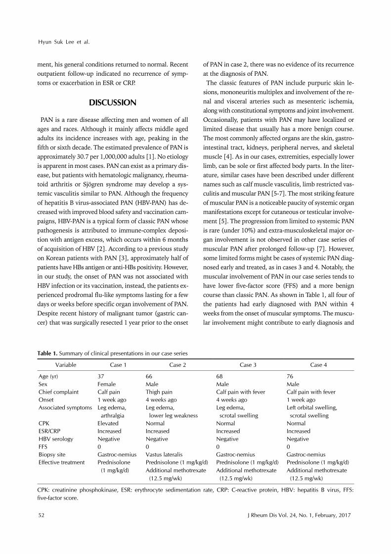
ment, his general conditions returned to normal. Recent outpatient follow-up indicated no recurrence of symp-toms or exacerbation in ESR or CRP.
DISCUSSION
PAN is a rare disease affecting men and women of all ages and races. Although it mainly affects middle aged adults its incidence increases with age, peaking in the fifth or sixth decade. The estimated prevalence of PAN is approximately 30.7 per 1,000,000 adults [1]. No etiology is apparent in most cases. PAN can exist as a primary dis-ease, but patients with hematologic malignancy, rheuma-toid arthritis or Sjögren syndrome may develop a sys-temic vasculitis similar to PAN. Although the frequency of hepatitis B virus-associated PAN (HBV-PAN) has de-creased with improved blood safety and vaccination cam-paigns, HBV-PAN is a typical form of classic PAN whose pathogenesis is attributed to immune-complex deposi-tion with antigen excess, which occurs within 6 months of acquisition of HBV [2]. According to a previous study on Korean patients with PAN [3], approximately half of patients have HBs antigen or anti-HBs positivity. However, in our study, the onset of PAN was not associated with HBV infection or its vaccination, instead, the patients ex-perienced prodromal flu-like symptoms lasting for a few days or weeks before specific organ involvement of PAN. Despite recent history of malignant tumor (gastric can-cer) that was surgically resected 1 year prior to the onset
of PAN in case 2, there was no evidence of its recurrence at the diagnosis of PAN. The classic features of PAN include purpuric skin le-
sions, mononeuritis multiplex and involvement of the re-nal and visceral arteries such as mesenteric ischemia, along with constitutional symptoms and joint involvement. Occasionally, patients with PAN may have localized or limited disease that usually has a more benign course. The most commonly affected organs are the skin, gastro-intestinal tract, kidneys, peripheral nerves, and skeletal muscle [4]. As in our cases, extremities, especially lower limb, can be sole or first affected body parts. In the liter-ature, similar cases have been described under different names such as calf muscle vasculitis, limb restricted vas-culitis and muscular PAN [5-7]. The most striking feature of muscular PAN is a noticeable paucity of systemic organ manifestations except for cutaneous or testicular involve-ment [5]. The progression from limited to systemic PAN is rare (under 10%) and extra-musculoskeletal major or-gan involvement is not observed in other case series of muscular PAN after prolonged follow-up [7]. However, some limited forms might be cases of systemic PAN diag-nosed early and treated, as in cases 3 and 4. Notably, the muscular involvement of PAN in our case series tends to have lower five-factor score (FFS) and a more benign course than classic PAN. As shown in Table 1, all four of the patients had early diagnosed with PAN within 4 weeks from the onset of muscular symptoms. The muscu-lar involvement might contribute to early diagnosis and
Muscular Polyarteritis Nodosa
www.jrd.or.kr 53
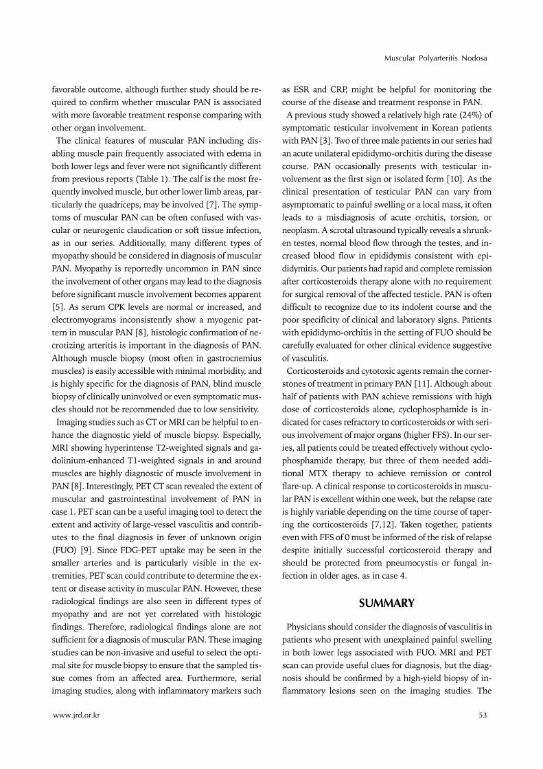
favorable outcome, although further study should be re-quired to confirm whether muscular PAN is associated with more favorable treatment response comparing with other organ involvement. The clinical features of muscular PAN including dis-
abling muscle pain frequently associated with edema in both lower legs and fever were not significantly different from previous reports (Table 1). The calf is the most fre-quently involved muscle, but other lower limb areas, par-ticularly the quadriceps, may be involved [7]. The symp-toms of muscular PAN can be often confused with vas-cular or neurogenic claudication or soft tissue infection, as in our series. Additionally, many different types of myopathy should be considered in diagnosis of muscular PAN. Myopathy is reportedly uncommon in PAN since the involvement of other organs may lead to the diagnosis before significant muscle involvement becomes apparent [5]. As serum CPK levels are normal or increased, and electromyograms inconsistently show a myogenic pat-tern in muscular PAN [8], histologic confirmation of ne-crotizing arteritis is important in the diagnosis of PAN. Although muscle biopsy (most often in gastrocnemius muscles) is easily accessible with minimal morbidity, and is highly specific for the diagnosis of PAN, blind muscle biopsy of clinically uninvolved or even symptomatic mus-cles should not be recommended due to low sensitivity.Imaging studies such as CT or MRI can be helpful to en-
hance the diagnostic yield of muscle biopsy. Especially, MRI showing hyperintense T2-weighted signals and ga-dolinium-enhanced T1-weighted signals in and around muscles are highly diagnostic of muscle involvement in PAN [8]. Interestingly, PET CT scan revealed the extent of muscular and gastrointestinal involvement of PAN in case 1. PET scan can be a useful imaging tool to detect the extent and activity of large-vessel vasculitis and contrib-utes to the final diagnosis in fever of unknown origin (FUO) [9]. Since FDG-PET uptake may be seen in the smaller arteries and is particularly visible in the ex-tremities, PET scan could contribute to determine the ex-tent or disease activity in muscular PAN. However, these radiological findings are also seen in different types of myopathy and are not yet correlated with histologic findings. Therefore, radiological findings alone are not sufficient for a diagnosis of muscular PAN. These imaging studies can be non-invasive and useful to select the opti-mal site for muscle biopsy to ensure that the sampled tis-sue comes from an affected area. Furthermore, serial imaging studies, along with inflammatory markers such
as ESR and CRP, might be helpful for monitoring the course of the disease and treatment response in PAN.A previous study showed a relatively high rate (24%) of
symptomatic testicular involvement in Korean patients with PAN [3]. Two of three male patients in our series had an acute unilateral epididymo-orchitis during the disease course. PAN occasionally presents with testicular in-volvement as the first sign or isolated form [10]. As the clinical presentation of testicular PAN can vary from asymptomatic to painful swelling or a local mass, it often leads to a misdiagnosis of acute orchitis, torsion, or neoplasm. A scrotal ultrasound typically reveals a shrunk-en testes, normal blood flow through the testes, and in-creased blood flow in epididymis consistent with epi-didymitis. Our patients had rapid and complete remission after corticosteroids therapy alone with no requirement for surgical removal of the affected testicle. PAN is often difficult to recognize due to its indolent course and the poor specificity of clinical and laboratory signs. Patients with epididymo-orchitis in the setting of FUO should be carefully evaluated for other clinical evidence suggestive of vasculitis.Corticosteroids and cytotoxic agents remain the corner-
stones of treatment in primary PAN [11]. Although about half of patients with PAN achieve remissions with high dose of corticosteroids alone, cyclophosphamide is in-dicated for cases refractory to corticosteroids or with seri-ous involvement of major organs (higher FFS). In our ser-ies, all patients could be treated effectively without cyclo-phosphamide therapy, but three of them needed addi-tional MTX therapy to achieve remission or control flare-up. A clinical response to corticosteroids in muscu-lar PAN is excellent within one week, but the relapse rate is highly variable depending on the time course of taper-ing the corticosteroids [7,12]. Taken together, patients even with FFS of 0 must be informed of the risk of relapse despite initially successful corticosteroid therapy and should be protected from pneumocystis or fungal in-fection in older ages, as in case 4.
SUMMARY
Physicians should consider the diagnosis of vasculitis in patients who present with unexplained painful swelling in both lower legs associated with FUO. MRI and PET scan can provide useful clues for diagnosis, but the diag-nosis should be confirmed by a high-yield biopsy of in-flammatory lesions seen on the imaging studies. The

Hyun Suk Lee et al.
54 J Rheum Dis Vol. 24, No. 1, February, 2017
muscular involvement of PAN may be limited forms or early systemic forms with benign course and excellent clinical response with corticosteroid therapy. However, effective treatment strategy includes careful monitoring due to high relapse rate and potential adverse events.
CONFLICT OF INTEREST
No potential conflict of interest relevant to this article was reported.
REFERENCES
1. Mahr A, Guillevin L, Poissonnet M, Aymé S. Prevalences of polyarteritis nodosa, microscopic polyangiitis, Wegener's granulomatosis, and Churg-Strauss syndrome in a French urban multiethnic population in 2000: a capture-recapture estimate. Arthritis Rheum 2004;51:92-9.
2. Guillevin L, Mahr A, Callard P, Godmer P, Pagnoux C, Leray E; French Vasculitis Study Group, et al. Hepatitis B virus-as-sociated polyarteritis nodosa: clinical characteristics, out-come, and impact of treatment in 115 patients. Medicine (Baltimore) 2005;84:313-22.
3. Bae YD, Choi HJ, Lee JC, Park JJ, Lee YJ, Lee EB, et al. Clinical features of polyarteritis nodosa in Korea. J Korean Med Sci 2006;21:591-5.
4. Pagnoux C, Seror R, Henegar C, Mahr A, Cohen P, Le Guern V, et al. Clinical features and outcomes in 348 patients with polyarteritis nodosa: a systematic retrospective study of pa-tients diagnosed between 1963 and 2005 and entered into
the French Vasculitis Study Group Database. Arthritis Rheum 2010;62:616-26.
5. Plumley SG, Rubio R, Alasfar S, Jasin HE. Polyarteritis no-dosa presenting as polymyositis. Semin Arthritis Rheum 2002;31:377-83.
6. Kamimura T, Hatakeyama M, Torigoe K, Nara H, Kaneko N, Satou H, et al. Muscular polyarteritis nodosa as a cause of fe-ver of undetermined origin: a case report and review of the literature. Rheumatol Int 2005;25:394-7.
7. Khellaf M, Hamidou M, Pagnoux C, Michel M, Brisseau JM, Chevallier X, et al. Vasculitis restricted to the lower limbs: a clinical and histopathological study. Ann Rheum Dis 2007;66:554-6.
8. Gallien S, Mahr A, Réty F, Kambouchner M, Lhote F, Cohen P, et al. Magnetic resonance imaging of skeletal muscle in-volvement in limb restricted vasculitis. Ann Rheum Dis 2002;61:1107-9.
9. Kim YJ, Kim SI, Hong KW, Kang MW. Diagnostic value of 18F-FDG PET/CT in patients with fever of unknown origin. Intern Med J 2012;42:834-7.
10. Lee LM, Moloney PJ, Wong HC, Magil AB, McLoughlin MG. Testicular pain: an unusual presentation of polyarteritis nodosa. J Urol 1983;129:1243-4.
11. Guillevin L, Cohen P, Mahr A, Arène JP, Mouthon L, Puéchal X, et al. Treatment of polyarteritis nodosa and microscopic polyangiitis with poor prognosis factors: a prospective trial comparing glucocorticoids and six or twelve cyclopho-sphamide pulses in sixty-five patients. Arthritis Rheum 2003;49:93-100.
12. Ribi C, Cohen P, Pagnoux C, Mahr A, Arène JP, Puéchal X, et al. Treatment of polyarteritis nodosa and microscopic pol-yangiitis without poor-prognosis factors: A prospective randomized study of one hundred twenty-four patients. Arthritis Rheum 2010;62:1186-97.