protection of the human heart with ischemic preconditioning during cardiac surgery: role of...
TRANSCRIPT

Protection of the human heart with ischemicpreconditioning during cardiac surgery: Role ofcardiopulmonary bypassSudip Ghosh, MDManuel Galinanes, MD, PhD, FRCS
Objective: Studies on the effects of ischemic preconditioning in the human hearthave yielded conflicting results and therefore remain controversial. This studyinvestigated whether ischemic preconditioning was able to protect against myocar-dial tissue damage in patients undergoing coronary artery surgery with cardiopul-monary bypass and on the beating heart.
Methods: A total of 120 patients were studied and divided into 3 groups: group I:cardiopulmonary bypass with intermittent crossclamp fibrillation; group II: cardiopul-monary bypass with cardioplegic arrest using cold blood cardioplegia; group III: surgeryon the beating heart. In each group (n � 40), patients were randomly subdivided (n �20/subgroup) into control and preconditioning groups (1 cycle of 5 minutes of isch-emia/5 minutes reperfusion before intervention). Ischemic preconditioning was inducedby clamping the ascending aorta in groups I and II or by clamping the coronary arteryin group III. Serial venous blood levels of troponin T were analyzed before surgery andat 1, 4, 8, 24, and 48 hours after termination of ischemia. In addition, in vitro studiesusing right atrial specimens obtained before the institution of cardiopulmonary bypass,and then again 10 minutes after initiation of bypass, were performed. The specimenswere equilibrated for 30 minutes before being allocated to 1 of the following 2 groups(n � 6 per group): (1) ischemia alone (90 minutes of ischemia followed by 120 minutesof reoxygenation) or (2) preconditioning with 5 minutes of ischemia and 5 minutes ofreoxygenation before the long ischemic insult. Creatine kinase leakage (U/g wet weight)and 3-(4,5-dimethylthiazol-2-yl)-2,5-diphenyltetrazolium bromide reduction (mmol/l pergram wet weight), an index of cell viability, were assessed at the end of the experiment.
Results: There were no perioperative myocardial infarctions or deaths in any of thegroups studied. The total release of troponin T was similar in groups I and II (patientsundergoing surgery with cardiopulmonary bypass) and in the release profile; they wereunaffected by ischemic preconditioning. In contrast, the total troponin T release for thefirst 48 hours was significantly reduced by ischemic preconditioning in group III(patients undergoing surgery without cardiopulmonary bypass) from 3.1 � 0.1 to 2.1 �0.2 ng · h · mL. Furthermore, the release profile that peaked at 8 hours in the controlgroup shifted to the left at 1 hour. In the in vitro studies, the atrial muscles obtainedbefore cardiopulmonary bypass were protected by ischemic preconditioning (crea-tine kinase � 2.6 � 0.2 and 3-[4,5-dimethylthiazol-2-yl]-2,5-diphenyltetrazoliumbromide reduction � 152 � 24 vs creatine kinase � 5.4 � 0.6 and 3-[4,5-dimethylthiazol-2-yl]-2,5-diphenyltetrazolium bromide reduction � 87 � 16 incontrols; P � .05); however, the muscles obtained 10 minutes after initiation ofcardiopulmonary bypass were already protected (creatine kinase � 0.8 � 0.1 and3-[4,5-dimethylthiazol-2-yl]-2,5-diphenyltetrazolium bromide reduction � 316 �38), and ischemic preconditioning did not result in further improvements.
Conclusions: Ischemic preconditioning is protective in patients undergoing coronaryartery surgery on the beating heart without the use of cardiopulmonary bypass, but itoffers no additional benefit when associated with bypass regardless of the mode of cardio-protection used, because cardiopulmonary bypass per se induces preconditioning.
From the Department of Integrative HumanCardiovascular Physiology and CardiacSurgery, University of Leicester, GlenfieldHospital, Leicester, United Kingdom.
This study was supported by a personalcontribution from Professor Manuel Gali-nanes.
Received for publication April 15, 2002;accepted for publication July 15, 2002.
Address for reprints: Professor M. Galinanes,Department of Integrative Human Cardiovas-cular Physiology and Cardiac Surgery, Uni-versity of Leicester, Glenfield Hospital, Le-icester LE3 9QP, United Kingdom (E-mail:sudip.ghoshglenfield-tr.trent.nhs.uk).
J Thorac Cardiovasc Surg 2003;126:133-42
Copyright © 2003 by The American Asso-ciation for Thoracic Surgery
0022-5223/2003 $30.00 � 0
doi:10.1016/S0022-5223(02)73293-5
Ghosh and Galinanes Cardiopulmonary Support and Physiology
The Journal of Thoracic and Cardiovascular Surgery ● Volume 126, Number 1 133
CSP

Myocardial injury, manifested as tran-sient cardiac contractile dysfunction(“stunning”) and myocardial necro-sis, is the most frequent complicationduring heart surgery.1 Ischemic pre-conditioning (IP), a powerful form of
endogenous protection against stunning and infarction, hasbeen demonstrated in a variety of animal species2-4 and invitro experiments involving isolated human cardiomyo-cytes5 and human atrial trabeculae.6,7 For logistic and eth-ical reasons, no clinical study can meet the strict conditionsof experimental studies on preconditioning with infarct sizeas the end point. As a result, human in vivo studies haveproduced conflicting results, and the role of IP in humansremains controversial.
Yellon and coworkers8 were the first to report that IPprotects the human heart in the setting of cardiac surgery byuse of the conservation of myocardial adenosine triphos-phate (ATP) content as the major end point. However,Perrault and colleagues9 reported that in the presence ofcardioplegic arrest, there was no difference in the release ofbiochemical markers (CK-MB) between the preconditionedand control groups. The lack of additional protection con-ferred by IP was further confirmed by the absence of dif-ference in the postarrest myocardial levels of ribonucleicmessengers coding for cardioprotective heat shock proteinsbetween the 2 groups. Similar negative results were reportedby Kaukoranta,10 Di Salvo,11 and their colleagues, whowent on to report that in the presence of hypothermia, nobeneficial effect of preconditioning was observed in similarpatients undergoing surgery with intermittent fibrillationtechniques.
The apparent discrepancy between results obtained withnoncardioplegic and cardioplegic techniques could be rec-onciled if one takes into account a possible hypothesis:Preconditioning and its salutary effects are only observed insituations of unprotected ischemia. With this in mind, thisstudy (1) investigates whether IP with 5 minutes of ischemiafollowed by 5 minutes of reperfusion is protective in pa-tients undergoing coronary artery bypass graft surgery withcardiopulmonary bypass (CPB) using cardioplegia and ven-tricular fibrillation techniques and in patients undergoingcoronary artery bypass graft surgery on the beating heartwithout CPB and (2) elucidates the underlying cause of anyprotection.
MethodsIn Vivo Studies
Patient selection and study groups. A total of 120 patientswith stable angina who underwent operations performed by asingle surgeon (M.G.) were entered in the study. They weredivided into 2 groups: those undergoing surgery with CPB (ie,on-pump) and exhibiting 3-vessel coronary disease (n � 80) andthose undergoing surgery on the beating heart without CPB (ie,
off-pump) and with single- or double-vessel coronary artery dis-ease (n � 40). Patients in the CPB group were randomized by useof a computer-generated random table to undergo surgery (1) withCPB and intermittent crossclamp fibrillation � IP (n � 20 in eachsubgroup) or (2) with cold blood cardioplegia � IP (n � 20 in eachsubgroup). Similarly, patients undergoing surgery on the beatingheart without CPB were randomized to receive or not receive IP (n� 20 in each subgroup). IP was induced by clamping the ascend-ing aorta for 5 minutes at 37°C when CPB was used or byoccluding the coronary artery to be grafted for the same period andthen reperfusing for 5 minutes before the bypass graft was per-formed. We have demonstrated that this IP protocol is the mostprotective in the human myocardium.7 Patients with low ejectionfractions (�30%), unstable angina, recent myocardial infarctions(�1 month), additional cardiac diseases, severe noncardiac dis-eases, or diabetes, and who were receiving medication that in-cluded ATP-dependent potassium channel (KATP) openers wereexcluded. The study was approved by the local ethics committee,and all patients gave written informed consent to participate in thestudy. Preoperative characteristics of the patients are highlighted inTable 1.
Operative procedure. Anesthetic technique was standardizedfor all patients. Anesthesia was induced by fentanyl and main-tained with enflurane in all patients. All operations were performedthrough a median sternotomy and with full heparinization (3mg/kg intravenously). CPB was conducted with nonpulsatile per-fusion flow (2.2-2.4 L · min�1 · m�2) with ascending aorticcannulation and 2-staged venous cannulation and moderate sys-temic hypothermia (32°C). When cardiac arrest was achieved withcold blood (6-10°C) cardioplegia, 1000 mL of the solution(composition in mmol/L: 16 MgCl2 6H20; 2 CaCl2; 20 KCl; 147NaCl; 1.0 procaine HCl, pH 7.40) was mixed with blood fromthe pump in a ratio of 1:1 and injected into the aortic rootimmediately after aortic crossclamping to obtain a myocardialtemperature of 12°C to 15°C. An additional dose of 500 mL ofcardioplegic solution was injected after 30 minutes of ischemia.In the group in which intermittent aortic crossclamp fibrillationwas used, reperfusion was performed for 3 minutes betweeneach period. One single distal coronary artery anastomosis wasperformed during each ischemic period. Proximal anastomosiswas completed on the beating heart with an aortic partialocclusion clamp in all groups.
In the off-pump group, coronary bypass grafting was performedon the beating heart using the Octopus myocardial stabilizationdevice (Medtronic Inc., Minneapolis, Minn). The suction cupswere placed on the epicardial surface on either side of the artery tobe grafted, with a suction pressure of no more than 600 mm Hg.Small coronary clamps were applied to the proximal and distalsites of the anastomosis with just enough pressure to occludecoronary flow and therefore allow grafting in a bloodless field. Theclamps were released when the anastomosis was complete. Theproximal anastomosis was performed with a partial occlusionaortic clamp.
IP was applied before the first dose of cardioplegic solution orimmediately before the aortic cross-application or the coronaryartery occlusion. The use of a Khuri Tissue pH Analyzer (VascularTech, Boston, Mass) showed that the pH of the myocardium
Cardiopulmonary Support and Physiology Ghosh and Galinanes
134 The Journal of Thoracic and Cardiovascular Surgery ● July 2003
CSP

decreased from greater than 7.3 to less than 7.0 at the end of the5-minute IP insult in all cases.
Assessment of myocardial injury. Serial venous blood sam-ples were collected before induction of anesthesia and at 1, 4, 8,24, and 48 hours after termination of ischemia in every patient forthe assessment of troponin T (TnT). This was measured by use ofthe commercially available enzyme-linked immunoabsorbent as-say kit TnT (Boehringer Mannheim, Mannheim, Germany). Thelower detection limit of the assay was 0.05 ng/mL, and concen-trations greater than the discriminator value of 0.1 ng/mL wereconsidered elevated.
Hemodynamic measurements. Heart rate, mean pulmonaryartery pressure, pulmonary capillary wedge pressure, and cardiacoutput were monitored with a pulmonary artery flotation catheter.Derived cardiovascular variables, including cardiac index and sys-temic and pulmonary vascular resistance, were calculated by stan-dard formulas. Hemodynamic data were collected at 4 time points:baseline (just after induction of anesthesia) and 1, 3, and 24 hoursafter the termination of ischemia. Changes in derived variableswere calculated and compared.
In Vitro StudiesSpecimens of human right atrium appendage were obtained frompatients undergoing elective coronary artery surgery with CPB.Samples were obtained before the institution of CPB and again10 minutes after the initiation of CPB. Two 6-0 Prolene (Ethi-con Inc, Somerville, NJ) encircling sutures were placed in theepicardial layer of the appendage once the pericardium wasopened. During the entire period of harvesting, the atrium wasexposed to the systemic circulation. The prebypass sample wasthen harvested, and a 2-stage venous cannula was inserted in thefree wall of the right atrium 3 cm away from the appendage.CPB was then instituted, and after 10 minutes, a further sampleof appendage was harvested. In each case, specimens werequickly immersed in cold (4°C) Krebs-Henseleit-HEPES me-dium, which comprised (in micromoles per liter) NaCl, 118;KCl, 4.8;, NaHCO3, 27.2; KH2PO4, 1; MgCl2, 1.2; CaCl2, 1.25;glucose, 10; and HEPES, 20. The medium was pre-bubbled with95% O2/5% CO2 to attain a PO2 of 25 to 30 kPa and pH 7.4. Theatrial appendage was immediately sliced freehand with Swann-Morton skin graft blades (Swann-Morton Ltd, Sheffield, UnitedKingdom) to a thickness of 0.3 to 0.5 mm and a weight of 5 to
10 mg each, as previously described.12 In brief, the tissue wasplaced with its epicardial surface face down on filter paper fixedto a rectangular glass base (5 � 25 cm). A ground glass slide(2.5 � 7.5 cm) was then pressed against the tissue, and theblade was drawn between the slide and tissue. The slicingapparatus and the tissue were kept wet at all times with mediumthat was stored on ice (4°C-10°C). The specimens were equil-ibrated for 30 minutes before being randomly allocated to 1 of thefollowing 2 groups (n � 6 each from different patients/group): (1)ischemia alone (90 minutes of ischemia followed by 120 minutesof reperfusion) or (2) IP with 5 minutes of ischemia and 5 minutesof reperfusion before 90 minutes of ischemia followed by 120minutes of reperfusion. For the induction of simulated ischemia,the medium was bubbled with 95% N2/5%CO2 (pH 6.80-7.00),and D-glucose was removed and substituted with D-2-deoxyglu-cose (10 mmol/L). The slices were maintained at 37°C duringthe entire ischemic period. Monitoring of PO2 with an oxygendetector electrode (Oxylite; Oxford, United Kingdom) revealedthat the PO2 in the medium was 0 kPa. Tissue pH was notmeasured in the atrial sections. At the end of the ischemicperiod, the nonoxygenated medium was removed, and the sliceswere rinsed with oxygenated medium (O2/CO2, 19:1) and in-cubated in 5 mL of oxygenated medium containing 10 mmol/Lglucose at 37°C for a further 120 minutes.
Tissue injury was determined by measuring the leakage ofcreatine kinase (CK) into the incubation medium during the120-minute reperfusion period. This was assayed by a kineticultraviolet method based on the formation of nicotinamideadenine dinucleotide (Sigma Catalog No. 1340-K), and theresults were expressed as U/g wet weight. Tissue viability wasassessed by the reduction of 3-(4,5-dimethylthiazol-2-yl)-2,5diphenyltetrazolium bromide (MTT) to blue formazan productat the end of the experimental time. In this assay, the yellowMTT is reduced to a blue formazan product by the mitochondriaof viable tissue.
The tissue was loaded into a Falcon conical tube (15 mL)(Becton Dickinson Labware, Franklin Lakes, NJ) to which 2 mL ofphosphate buffer solution (0.05 mol/L) containing MTT (1.25mg/mL, 3 mmol/L at final concentration) was added and thenincubated for 30 minutes at 37°C. After this, the tissue washomogenized in 2 mL of dimethyl sulfoxide (Homogenizer Ultra-Turrax T25, dispersing tool G8, IKA-Labortechnic, Staufen, Ger-
TABLE 1. Patient characteristics and perioperative data
Variable
With CPB Without CPB
Intermittent crossclamp Cold blood cardioplegia
Control PCControl PC Control PC
Number 20 20 20 20 20 20Age (yr � SD) 66.9 � 5.4 63.7 � 4.2 61.4 � 9.2 63.8 � 5.9 61.2 � 2.7 59.2 � 6.4LVEF (% � SD) 60.2 � 5.5 61.8 � 4.1 62.1 � 5.7 63.7 � 4.9 59.7 � 9.2 65.1 � 6.4No. of grafts/patient 3.8 � 1.5 3.6 � 0.8 3.7 � 1.2 4.1 � 0.7 1.15 � 0.6 1.2 � 0.5Ischemic time/graft
(min � SD)16.1 � 2.5 15.4 � 3.1 17.2 � 4.9 14.7 � 5.7 17.8 � 7.9 16.9 � 4.7
CPB time (min � SD) 66.4 � 12.7 70.4 � 11.4 71.4 � 13.4 69.8 � 6.7
PC, Preconditioning; LVEF, left ventricular ejection fraction (expressed as a percentage � standard deviation); CPB, cardiopulmonary bypass.
Ghosh and Galinanes Cardiopulmonary Support and Physiology
The Journal of Thoracic and Cardiovascular Surgery ● Volume 126, Number 1 135
CSP
many) at 9500 rpm for 1 minute. The homogenate was thencentrifuged at 1000g for 10 minutes, and 0.2 mL of the supernatantwas dispensed into a 98-well flat-bottom microtiter plate (NuncBrand Products, Denmark). After this, the absorbance was mea-sured on a plate reader (Benchmark; Bio-Rad Laboratories, Her-cules, Calif) at 550 nm, and the results were expressed as micro-moles per liter per milligram wet weight.
Statistical AnalysisStatistical analyses were performed using the SPSS 9.0 statisticalpackage program (SPSS Inc, Chicago, Ill) to investigate the effi-cacy of IP in the various groups of patients. Patients undergoingsurgery without CPB demonstrated less severe coronary arterydisease than those undergoing surgery with CPB (Table 2), andbecause of this, comparison between patients with and withoutCPB was not performed. A nonparametric test (Mann-Whitney U)was performed for non-Gaussian distribution of data. An unpairedStudent t test was used for continuous data (2-tailed), and the �2
test for categoric data was used to compare variables between thegroups. Repeated-measures analysis of variance was used to testthe repeated observation variables postoperatively. The area underthe curve was calculated by the method of Matthews and col-leagues.13 Data were presented as mean � SD.
ResultsIn Vivo StudiesThere were no operative deaths (first 30 postoperative days)or perioperative myocardial infarctions in any of the studygroups. Table 1 shows that the patient characteristics andperioperative data were similar within each study group, andthat there was no difference between patients who weretreated with or without IP.
Plasma Troponin TFigure 1 (A and B) shows that the profile of TnT releasein plasma was identical in patients undergoing coronarysurgery with CPB whether they were protected withintermittent fibrillation or cold blood cardioplegia. Thus,there was a significant increase in plasma TnT by 1 hourafter termination of ischemia that peaked at 4 hours, withmean values still remaining elevated at 48 hours.Notably, preconditioning did not alter this profile in bothgroups, indicating that preconditioning conveys nobenefit to patients undergoing coronary surgery withCPB.
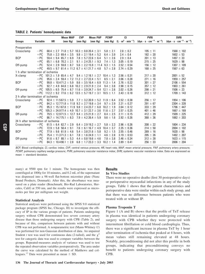
TABLE 2. Patients’ hemodynamic data
Groups Variable HRMean MAP
(mm Hg)CVP
(mm Hg)Mean PAP
(mm Hg)PCWP
(mm Hg)CI
(L · m2 · min�1)PVRI
(dyn · s · cm�5 · m�2)SVRI
(dyn · s · cm�5 · m�2)
PreoperativeCrossclamp �PC 68.4 � 2.7 71.8 � 5.7 10.3 � 0.8 20.4 � 3.1 5.8 � 2.1 2.8 � 0.2 195 � 11 1568 � 102
�PC 75.8 � 2.3 69.4 � 3.5 9.8 � 2.1 19.4 � 5.2 6.4 � 3.9 2.4 � 0.4 182 � 20 1602 � 53BCP �PC 59.4 � 9.4 65.7 � 6.9 7.8 � 5.7 22.6 � 6.4 5.1 � 2.6 2.9 � 0.4 201 � 13 1497 � 33
�PC 65.1 � 6.8 70.2 � 2.1 9.1 � 2.4 25.1 � 9.3 7.4 � 1.3 3.05 � 0.19 215 � 25 1629 � 98Off-pump �PC 52.4 � 2.9 59.8 � 8.7 5.6 � 2.2 15.9 � 11.4 9.3 � 1.5 2.52 � 0.54 156 � 12 1307 � 109
�PC 49.5 � 9.7 61.4 � 11.2 6.7 � 4.1 21.7 � 4.8 5.7 � 2.8 2.74 � 0.29 168 � 25 1567 � 631 h after termination of ischemiaX-clamp �PC 101.3 � 1.9 65.4 � 4.7 9.4 � 1.2 18.1 � 2.7 10.4 � 1.3 2.56 � 0.31 217 � 20 2001 � 52
�PC 95.6 � 2.4 59.4 � 7.2 11.2 � 2.7 22.4 � 5.1 8.5 � 2.1 2.06 � 0.28 271 � 16 1959 � 257BCP �PC 99.8 � 10.471.2 � 5.9 8.6 � 3.5 16.4 � 6.9 11.3 � 1.4 2.76 � 0.22 301 � 27 2106 � 554
�PC 92.7 � 4.9 64.8 � 6.8 10.2 � 2.9 21.9 � 2.4 9.4 � 3.8 2.98 � 0.15 357 � 12 1903 � 261Off-pump �PC 105.5 � 6.5 75.4 � 8.7 11.6 � 3.9 24.7 � 9.4 12.1 � 2.6 2.02 � 0.26 298 � 21 1506 � 23
�PC 112.2 � 9.2 77.6 � 6.2 13.5 � 5.7 19.7 � 3.1 10.5 � 1.1 2.43 � 0.18 312 � 12 1709 � 1433 h after termination of ischemiaCrossclamp �PC 92.4 � 11.567.5 � 5.9 7.7 � 3.2 20.9 � 5.2 11.9 � 6.4 2.52 � 0.38 256 � 17 1954 � 106
�PC 84.2 � 12.771.8 � 11.8 9.2 � 2.7 19.8 � 3.4 9.7 � 2.9 2.21 � 0.27 291 � 67 2304 � 328BCP �PC 95.3 � 15.167.8 � 11.9 9.8 � 2.4 23.7 � 6.8 10.2 � 1.9 2.64 � 0.12 333 � 29 1796 � 447
�PC 105.3 � 24.977.4 � 4.9 10.7 � 3.1 22.7 � 3.6 11.6 � 2.7 2.57 � 0.25 401 � 46 1667 � 169Off-pump �PC 79.5 � 12.971.5 � 12.7 9.4 � 2.5 21.9 � 2.7 8.4 � 2.5 2.11 � 0.15 412 � 29 1509 � 225
�PC 86.7 � 14.775.1 � 8.3 7.9 � 4.2 26.4 � 5.9 9.6 � 1.6 2.92 � 0.26 368 � 36 1882 � 20324 h after termination of ischemiaCrossclamp �PC 61.5 � 6.4 62.7 � 2.9 6.4 � 2.9 19.2 � 2.7 5.8 � 2.3 2.96 � 0.26 208 � 22 1804 � 234
�PC 72.9 � 5.8 58.4 � 9.1 7.6 � 6.1 16.7 � 4.9 10.4 � 3.7 2.35 � 0.36 214 � 50 2004 � 102BCP �PC 77.9 � 9.6 61.9 � 4.6 5.4 � 3.8 21.6 � 5.8 9.2 � 1.5 2.55 � 0.46 289 � 16 1620 � 98
�PC 75.4 � 11.371.3 � 6.1 7.8 � 1.6 24.9 � 1.1 4.6 � 2.8 3.19 � 0.53 295 � 26 1492 � 207Off-pump �PC 91.6 � 8.7 88.1 � 5.3 4.4 � 0.8 19.6 � 4.6 7.8 � 3.8 2.46 � 0.34 312 � 28 1623 � 115
�PC 84.3 � 10.490.1 � 2.8 6.8 � 1.7 23.3 � 3.3 10.2 � 1.4 2.89 � 0.41 256 � 33 2006 � 204
BCP, Blood cardioplegia; CI, cardiac index; CVP, central venous pressure; HR, heart rate; MAP, mean arterial pressure; PAP, pulmonary artery pressure;PCWP, pulmonary capillary wedge pressure; PVRI, pulmonary vascular resistance index; SVRI, systemic vascular resistance index. Data are expressed asmean � standard deviation.
Cardiopulmonary Support and Physiology Ghosh and Galinanes
136 The Journal of Thoracic and Cardiovascular Surgery ● July 2003
CSP

In contrast, as shown in Figure 1C, the profiles of plasmaTnT were different in patients with or without precondition-ing and off-pump surgery. Peak release occurred by 8 hoursafter termination of ischemia in the control group. Peakrelease occurred by 1 hour in the preconditioned group witha sharp decrease during the next 3 hours and with signifi-cantly lower mean plasma TnT values than in the controlgroup. The significance of the accelerated rate of enzymeleakage in the preconditioning group is not entirely clear,but it could be the result of changes within the biophysicalproperties induced by IP (thus allowing a greater transientleakage of intracellular enzymes) or of greater cumulativeischemic time in the preconditioned group compared withthe control group. TnT levels were similar in both groupsonly by the end of 48 hours.
Figure 2 shows that the cumulative plasma release ofTnT (ie, area under the curve) was similar in the patientsundergoing surgery with CPB with no significant effect ofpreconditioning. It also shows that TnT release was lower inpatients undergoing surgery without CPB, and that precon-
ditioning in this group significantly reduced the total TnTrelease by 33% when compared with the control group (2.1� 0.1 vs 3.1 � 2 ng · h�1 · mL�1 P � .05).
Hemodynamic DataTable 2 shows that the mean systemic arterial pressures,heart rate, mean pulmonary artery pressures, systemic andpulmonary vascular resistance, and cardiac indices fluctu-ated within normal ranges after the operation in both thecontrol and preconditioned groups and in the groups withand without CPB.
In Vitro StudiesFigure 3 (A and B) shows the results of the CK leakage andMTT reduction of the atrial slices obtained before bypassand 10 minutes after initiation of CPB. The results demon-strate that the increase in CK leakage and the decrease inMTT reduction caused by ischemia and reoxygenation inthe atrial muscles obtained before the institution of bypasswere significantly improved in the slices obtained 10 min-
Figure 1A. Time course of release of plasma cardiac troponin T (TnT) concentrations in patients undergoingcoronary artery bypass grafting with CPB and aortic crossclamping. In each group, patients were randomlysubdivided into control and preconditioning groups (n � 20/group). Data are expressed as mean � SD (*P < .05vs control group).
Ghosh and Galinanes Cardiopulmonary Support and Physiology
The Journal of Thoracic and Cardiovascular Surgery ● Volume 126, Number 1 137
CSP

utes after the initiation of bypass, and that this level ofprotection was identical to that of preconditioning. Thus,muscles that were obtained 10 minutes after the initiation ofbypass were already preconditioned, and the application ofIP did not result in additional benefit compared with IPalone.
DiscussionThe present study shows that the human heart is precondi-tioned by the institution of CPB, and that the use of IP incombination with other protective interventions such ascardioplegia does not result in additional protection. It alsoclearly demonstrates that the human heart can be protected
Figure 1. Cont’d. Time course of release of plasma cardiac TnT concentrations in patients undergoing coronaryartery bypass grafting with CPB and cold blood cardioplegia (B) and on the beating heart without CPB (C). In eachgroup, patients were randomly subdivided into control and preconditioning groups (n � 20/group). Data areexpressed as mean � SD (*P < .05 vs control group).
Cardiopulmonary Support and Physiology Ghosh and Galinanes
138 The Journal of Thoracic and Cardiovascular Surgery ● July 2003
CSP

by IP when patients undergo surgery without the use ofCPB. These findings have obvious important clinical impli-cations, and they warrant further discussion.
Preconditioning of the Human HeartExperimental findings on IP cannot be directly extrapolatedto humans because the mechanisms may be different fromother animal species. For both logistic and ethical reasons,no clinical study can meet the strict conditions of experi-mental studies on preconditioning in which infarct size isthe primary end point; instead, surrogate end points have tobe used. As a result, the demonstration of this phenomenonin the setting of cardiac surgery has been controversial.Yellon and colleagues8 were the first to examine the effectof two 3-minute ischemic episodes, in which each wasfollowed by 2-minute reperfusion on myocardial high-en-ergy phosphate content, in patients undergoing coronaryartery bypass graft surgery with CPB. They claimed that thehuman myocardium showed the typical biochemical fea-tures of preconditioning observed by Murry and col-leagues14 in their classic canine model of IP and thus couldbe preconditioned. There have been recent studies15,16 alsohighlighting the potential benefits of preconditioning in thecardiac surgery setting. However, Perrault and associates9
failed to show a beneficial effect of IP when this wasinduced with 3-minute aortic crossclamping followed by2-minute reperfusion before the administration of warmblood cardioplegia. Similar findings have been reported byother investigators10,17 questioning the ability of precondi-
tioning to protect the human heart. The dispute on whetherpreconditioning confers cardioprotection during cardiac sur-gery is further fueled by a more recent study by Alkhulafiand colleagues.18 By use of a protocol identical to the oneused in their first study,8 Alkhulafi and coworkers showed areduction of TnT release at 72 hours in patients exposed topreconditioning, but not at 24 or 48 hours. Notably, thesame authors reported an absence of protection in myocar-dial high-energy phosphates (in contrast with their firststudy). These opposing results are puzzling and contrastwith the overwhelming evidence that preconditioning iscardioprotective during coronary angioplasty19 and in vitroexperimental conditions using atrial trabeculae6,7 or isolatedmyocytes.20 Our finding that CPB can act as a precondi-tioning stimulus in humans is supported by another study insheep21 and sheds light on the previously mentioned con-troversy.
During cardiac surgery, there may be preoperative andintraoperative factors such as opioid agonists22 and anes-thetic agents23 that may mimic the protection of precondi-tioning. These include the use of opioid agonists, aprotinin,and, notably, CPB. It has been reported that inhalationalanesthetics can induce cardioprotection, and that this effectdiffers with the agent used.24,25 In the present study, enflu-rane was used. Although enflurane is less effective thanother inhalational anesthetics,24 and IP was shown to becardioprotective in the off-pump group, it may not be pos-sible to completely rule out some protective effect caused
Figure 2. Area under the curve of the plasma release cardiac TnT in patients undergoing coronary artery bypassgrafting with CPB and aortic crossclamping, CPB and cold blood cardioplegia, and on the beating heart withoutCPB (*P < .05 vs corresponding control group).
Ghosh and Galinanes Cardiopulmonary Support and Physiology
The Journal of Thoracic and Cardiovascular Surgery ● Volume 126, Number 1 139
CSP

by this agent. Hypothermia is another cardioprotective fac-tor26 that may influence the cardioprotection of precondi-tioning. Recently, Takeshima and colleagues27 also demon-strated that preconditioning was not protective with deephypothermia. However, moderate hypothermia alone, asused in the present studies, does not inhibit the precondi-tioning response.
Mechanism of Preconditioning by CardiopulmonaryBypassAlthough the precise mechanism of IP still remains unclear,recent investigations have clearly identified a number offactors that are essential to achieve protection. CPB inducesa systemic inflammatory reaction, and it is possible thatsome elements of this reaction may be responsible for the
observed protection. Yamashita and coworkers28 recentlyreported that interleukin 1 and tumor necrosis factor � (theproduction of which is increased by CPB) cause an eleva-tion in tissue manganese-superoxide dismutase, which wasdemonstrated when brief sublethal ischemia or anoxic in-sults were induced.29 However, this thesis is unlikely, be-cause the production of cytokines is a late event in responseto CPB that requires more than 10 minutes.
Recently, our laboratory showed that the generation offree radical species occurs soon after the institution ofCPB.30 Therefore, it is possible to speculate that free radi-cals are the primary cause of cardioprotection by CPB. Therelationship between free radicals and preconditioning wasfirst suggested by Richard and colleagues,31 who showedthat administration of oxygen free radical scavengers during
Figure 3. CK leakage (A) during the 120-minute reoxygenation period and MTT reduction (B) at the end of thereoxygenation period after 90 minutes of normothermic global ischemia with or without simulated IP. Data areexpressed as mean � SD of 6 experiments (*P < .05 vs corresponding group without preconditioning).
Cardiopulmonary Support and Physiology Ghosh and Galinanes
140 The Journal of Thoracic and Cardiovascular Surgery ● July 2003
CSP

the first reperfusion period could block the beneficial effectof preconditioning on infarct size in dogs. They thereforeproposed that the generation of low amounts of free radicalsduring the short ischemic episode is not sufficient to causecell necrosis but enough to modify cellular activity andinduce preconditioning. More recently, Pain and col-leagues32 demonstrated that the opening of mitochondrialKATP channels triggers protection through the generation offree radicals that activate protein kinase C, an obligatorystep in the signal transduction mechanism of IP. One of thepotential limitations with our in vitro studies is the use ofatrial myocardium as opposed to ventricular myocardium,and therefore any extrapolation must be conducted withcaution; however, Speechly-Dick and coworkers33 sug-gested that preconditioning exerts identical protection inboth tissues. Undoubtedly, KATP channels are present inboth atrium and ventricle,34 although their density in bothtissues is unknown.
The induction of CPB affects the body hemodynamicsthat may provoke a number of tissue responses. Thus, theloss of atrial and ventricular filling may stimulate a sympa-thetic-receptor–mediated release of local catecholamines,whereas the interruption of pulsatile systolic and diastolicblood flow to the adrenal glands may stimulate a systemiccatecholamine release. Therefore, an altered adrenergicstate may also be partially responsible for CPB-associatedpreconditioning in human myocardium. Several investiga-tors,35,36 including the current authors,37 have observed thatnorepinephrine or phenylephrine triggers preconditioning,and that this protection is prevented by adrenergic blockade.Similarly, Thornton and colleagues38 demonstrated thattyramine, an agent that causes the release of endogenouscatecholamines, reduced infarct size in rabbits when givenbefore a sustained period of ischemia. Certainly, more stud-ies are required to elucidate the mechanism of cardiopro-tection effected by CPB.
Clinical ImplicationsCardiac surgical practice is rapidly evolving, and an increas-ing number of surgeons are adopting surgery on the beatingheart, without the use of CPB, in their practice. Cardioplegicsolutions cannot be used in this situation, and the demon-stration that interventions such as IP are as effective canhave important clinical implications. However, it should berecognized that the clinical application of IP may still bedifficult and cumbersome, particularly if minimally invasiveapproaches are used. Because of this, the pharmacologicmanipulation of the signal transduction cascade of precon-ditioning may seem to be a more appropriate alternative. Inthis regard, several investigators, including the current au-thors, are endeavoring to fully elucidate the mechanism ofpreconditioning in humans to make this intervention a clin-ical reality.
CPB is known to induce a systemic inflammatory reac-tion that is believed to be responsible for increased morbid-ity. The present studies demonstrate that CPB can alsotrigger preconditioning and be cardioprotective.
References
1. Breisblatt W, Stein W, Wolfe CJ, et al. Acute myocardial dysfunctionand recovery: a common occurrence after coronary bypass surgery.J Am Coll Cardiol. 1990;15:1261-9.
2. Schott RJ, Rohmann S, Braun ER, Schaper W. Ischemic precondition-ing reduces infarct size in swine myocardium. Circ Res. 1990;66:1133-42.
3. Miller DL, Van Winckle DM. Ischemic preconditioning limits infarctsize following regional ischemia-reperfusion in in situ mouse hearts.Cardiovasc Res. 1999;42:680-4.
4. Goto M, Liu Y, Yang X-M, Ardell JL, Cohen MV, Downey JM. Roleof bradykinin in protection of ischemic preconditioning in rabbithearts. Circ Res. 1995;77:611-21.
5. Ikonomidis JS, Tumiati LC, Weisel RD, et al. Preconditioning humanventricular cardiomyocytes with brief periods of simulated ischemia.Cardiovasc Res. 1994;28:1285-91.
6. Speechly-Dick ME, Grover GJ, Yellon DM. Does ischemic precondi-tioning in the human involve PKC and the ATP-dependent K� chan-nel? Circ Res. 1995;77:1020-35.
7. Ghosh S, Standen NB, Galinanes M. Preconditioning the humanmyocardium by simulated ischemia. Studies on the early and delayedprotection. Cardiovasc Res. 2000;45:339-50.
8. Yellon DM, Alkhulaifi AM, Pugsley WB. Preconditioning the humanmyocardium. Lancet. 1993;342:276-7.
9. Perrault LP, Menasche P, Bel A, et al. Ischemic preconditioning incardiac surgery: a word of caution. J Thorac Cardiovasc Surg. 1996;112:1378-86.
10. Kaukoranta P, Lepojavi MPK, Ylitalo KV, et al. Normothermic ret-rograde blood cardioplegia with or without preceding ischemic pre-conditioning. Ann Thorac Surg. 1997;63:1268-74.
11. Di Salvo C, Hemming A, Jenkins D. Can human myocardium bepreconditioned with ischaemia under hypothermic conditions? Pro-ceedings of the Ninth Annual Meeting of the European Association forCardiothoracic Surgery. 1995:324 [Abstract].
12. Zhang JG, Ghosh S, Ockleford CD, Galinanes M. Characterization ofan in vitro model to study the short and prolonged effects of myocar-dial ischaemia and reperfusion in human myocardium. Clin Sci. 2000;99:443-53.
13. Matthews JNS, Altman DG, Campbell DJ, et al. Analysis of serialmeasurements in medical research. BMJ. 1990;300:230-5.
14. Murry CE, Jennings RB, Reimer KA. Preconditioning with ischemia:a delay of lethal injury in ischemic myocardium. Circulation. 1986;74:1124-36.
15. Wu ZK, Tarkka MR, Pehkonen E, et al. Ischaemic preconditioning hasa beneficial effect on left ventricular haemodynamic function aftercoronary artery bypass grafting operation. Scand Cardiovasc J. 2000;34:247-53.
16. Szmagala P, Morawski W, Krejca M, et al. Evaluation of peri-opera-tive myocardial tissue damage in ischemically preconditioned humanheart during aortocoronary bypass surgery. J Cardiovasc Surg. 1998;39:791-5.
17. Cremer J, Steinhoff G, Karck M, et al. Ischemic preconditioning priorto myocardial protection with cold cardioplegia in coronary surgery.Eur J Cardiothorac Surg. 1997;12:753-8.
18. Alkhulafi AM, Yellon DM, Pugsley WB. Preconditioning the humanheart during aorto-coronary bypass surgery. Eur J Cardiothorac Surg.1994;8:270-6.
19. Deutsch E, Berger M, Kussmaul WG, et al. Adaptation to ischemiaduring percutaneous transluminal coronary angioplasty: clinical, hae-modynamic and metabolic features. Circulation. 1990;82:2044-51.
20. Cleveland JC Jr, Wollmering MM, Meldrum DR, et al. Ischemicpreconditioning in human and rat ventricle. Am J Physiol. 1996;271:H1786-94.
Ghosh and Galinanes Cardiopulmonary Support and Physiology
The Journal of Thoracic and Cardiovascular Surgery ● Volume 126, Number 1 141
CSP

21. Burns PG, Krukenkamp IB, Caldarone CA, et al. Does cardiopulmo-nary bypass alone elicit myoprotective preconditioning? Circulation.1995;92(Suppl II):II447-51.
22. Schultz JEL, Rose E, Yao Z, Gross GJ. Evidence for the involvementof opioid receptors in ischemic preconditioning in rat hearts. Am JPhysiol. 1995;268:H2157-61.
23. Cope DK, Impastato WK, Cohen MV, Downey JM. Volatile anesthet-ics protect the ischemic rabbit myocardium from infarction. Anesthe-siology. 1997;86:699-709.
24. Schlack W, Preckel B, Stunneck D, Thamer V. Effects of halothane,enflurane, isoflurane, sevoflurane and desflurane on myocardial reper-fusion injury in the isolated rat heart. Br J Anaesth. 1998;81:913-19.
25. Preckel B, Schlack W, Confere T, Obal D, Barthel H, Thamer V.Effects of enflurane, isoflurane, sevoflurane and desflurane on reper-fusion injury after regional myocardial ischaemia in the rabbit heart invivo. Br J Anaesth. 1998;81:905-12.
26. Hale SL, Kloner RA. Ischemic preconditioning and myocardial hypo-thermia in rabbits with prolonged coronary artery occlusion. Am JPhysiol. 1999;276:H2029-34.
27. Takeshima S, Vaage J, Lowbeer C, Valen G. Does hypothermia orhyperkalemia influence the preconditioning response? Scand Cardio-vasc J. 1999;33:79-87.
28. Yamashita N, Shiro H, Kinya O, et al. Involvement of cytokines in themechanism of whole-body hyperthermia-induced cardioprotection.Circulation. 2000;102:452-57.
29. Yamashita N, Nishida M, Hoshida S, et al. Induction of manganesesuperoxide dismutase in rat cardiac myocytes increases tolerance tohypoxia 24 hours after preconditioning. J Clin Invest. 1994;94:2193-9.
30. Matata B, Galinanes M. Cardiopulmonary bypass exacerbates oxida-tive stress but does not increase proinflammatory cytokine release in
patients with diabetes compared to patients without diabetes: regula-tory effects of exogenous nitric oxide. J Thorac Cardiovasc Surg.2000;120:1-11.
31. Richard VJ, Murry CE, Jennings RB, Reimer KA. Oxygen-derivedfree radicals and postischemic myocardial reperfusion: therapeuticimplications. Fundam Clin Pharmacol. 1990;4:85-103.
32. Pain T, Yang XM, Critz SD, et al. Opening of mitochondrial K(ATP)channels triggers the preconditioned state by generating free radicals.Circ Res. 2000;87:460-6.
33. Speechly-Dick ME, Grover GJ, Yellon DM. Does ischemic precondi-tioning in the human involve PKC and the ATP-dependent K� chan-nel. Circ Res. 1995;77:1020-35.
34. Heidbuchel H, Vereecke J, Carmeliet E. Three different potassiumchannels in human atrium: contribution to the basal potassium con-ductance. Circ Res. 1990;66:1277-86.
35. Banerjee A, Locke-Winter C, Rogers KB, et al. Preconditioningagainst myocardial dysfunction after ischemia and reperfusion by analpha-adrenergic mechanism. Circ Res. 1993;73:656-70.
36. Cohen MV, Yang XM, Liu GS, Heusch G, Downey JM. Acetylcho-line, bradykinin, opioids, and phenylephrine, but not adenosine, triggerpreconditioning by generating free radicals and opening mitochondrialK(ATP) channels. Circ Res. 2001;89:273-8.
37. Loubani M, Galinanes M. Alpha-1 adrenoreceptors during simulatedischemia and reoxygenation of the human myocardium: effect of thedose and time of administration. J Thorac Cardiovasc Surg. 2001;122:103-12.
38. Thornton JD, Daly JF, Cohen MV, Yang X-M, Downey JM. Cat-echolamines can induce adenosine-receptor mediated protection of themyocardium but do not participate in ischemic preconditioning in therabbit. Circ Res. 1991;73:649-55.
Cardiopulmonary Support and Physiology Ghosh and Galinanes
142 The Journal of Thoracic and Cardiovascular Surgery ● July 2003
CSP